Dosimetric Analysis Report
|
|
|
- Robert Quinn
- 5 years ago
- Views:
Transcription

1 RT-safe 48, Artotinis str , Athens Greece Dosimetric Analysis Report SAMPLE, for demonstration purposes only Date of report: Date of irradiation: Irradiation system: Treatment Planning System: Number of target volumes (PTVs): 1 Number of Organs-at-risk: 1 Institution: Short description of procedure: A 3D-printer is used to construct a hollow phantom that duplicates the selected patient anatomical geometry using the patients' planning-ct DICOM images. The hollow phantom is subsequently filled with a polymer gel dosimeter. The phantom then is irradiated using the specific patient s irradiation protocol. This process is followed by a magnetic resonance imaging (MRI-T2 maps) scan of the irradiated phantom, which provides a complete set of high spatial resolution 3D-dose distribution measurements. These MR-images (T2-maps that include the measured dose distributions information) are then co-registered with the real patient planning -CT images and the corresponding DICOM-RT Structure set and treatment planning system (TPS) calculated Dose data. The dosimetric analysis report consists of a detailed 3D qualitative and quantitative comparison between TPS-calculations and corresponding patient specific derived measurements of the 3D-dose cloud, the spatial location of dose delivery and DVHs.


 blended with TPS (calculated dose) MRI 100% - RTDOSE")
2 PART I: Qualitative comparison Image registration between post-irradiation MRI and planning RTDose TPS data with structures of the Gel phantom. This is to demonstrate the coincidence of each treated target to its planned location. MRI (measured dose) blended with TPS (calculated dose) MRI 100% - RTDOSE TPS 0% MRI 50% - RTDOSE TPS 50% MRI 0% - RTDOSE TPS 100% (Brightness and contrast adjusted so that only high dose areas are depicted)
 MRI 100% -")


3 MRI (measured dose) blended with TPS (calculated dose) MRI 100% - RTDOSE TPS 0% MRI 50% - RTDOSE TPS 50% MRI 0% - RTDOSE TPS 100% (Brightness and contrast adjusted so that also low dose areas are depicted)

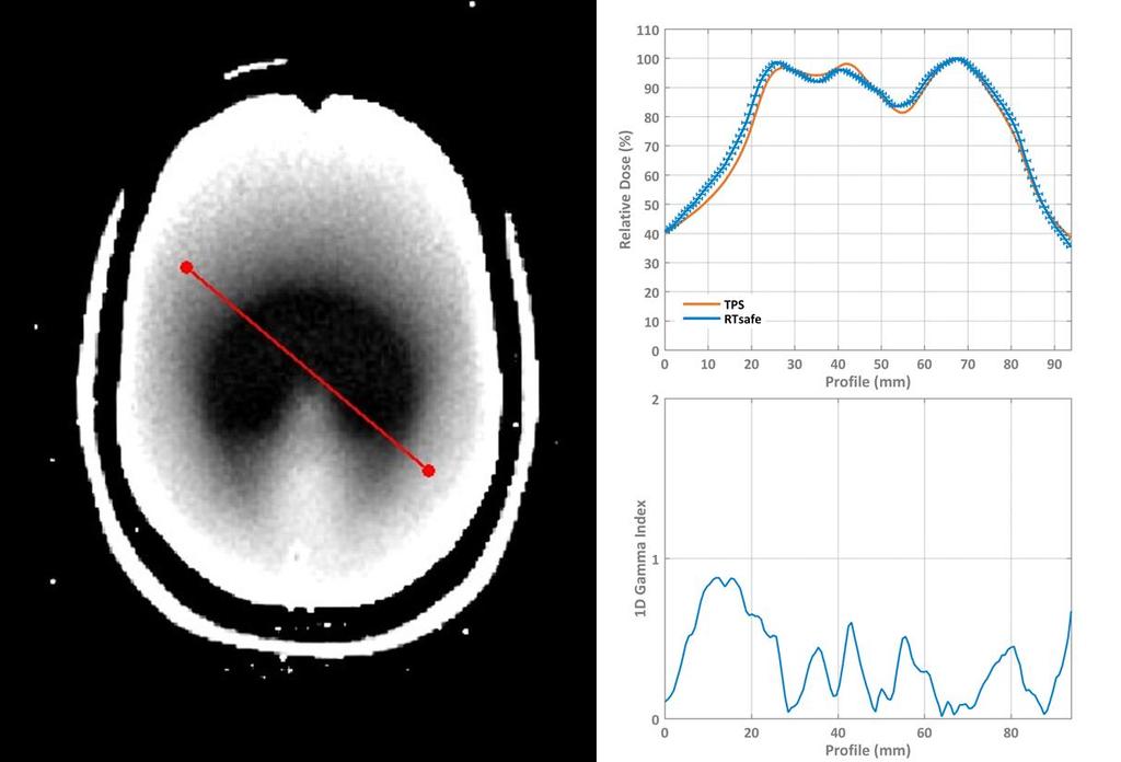
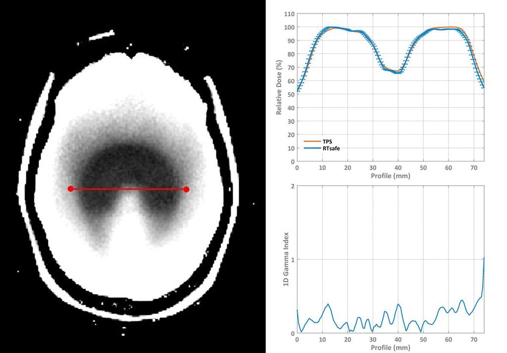
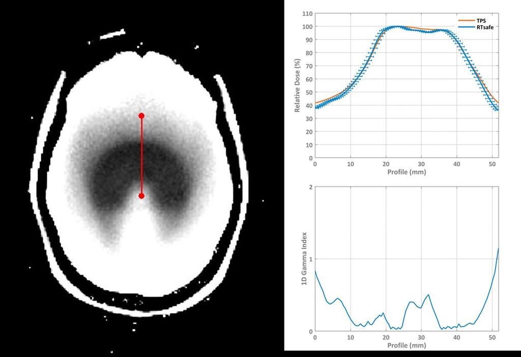
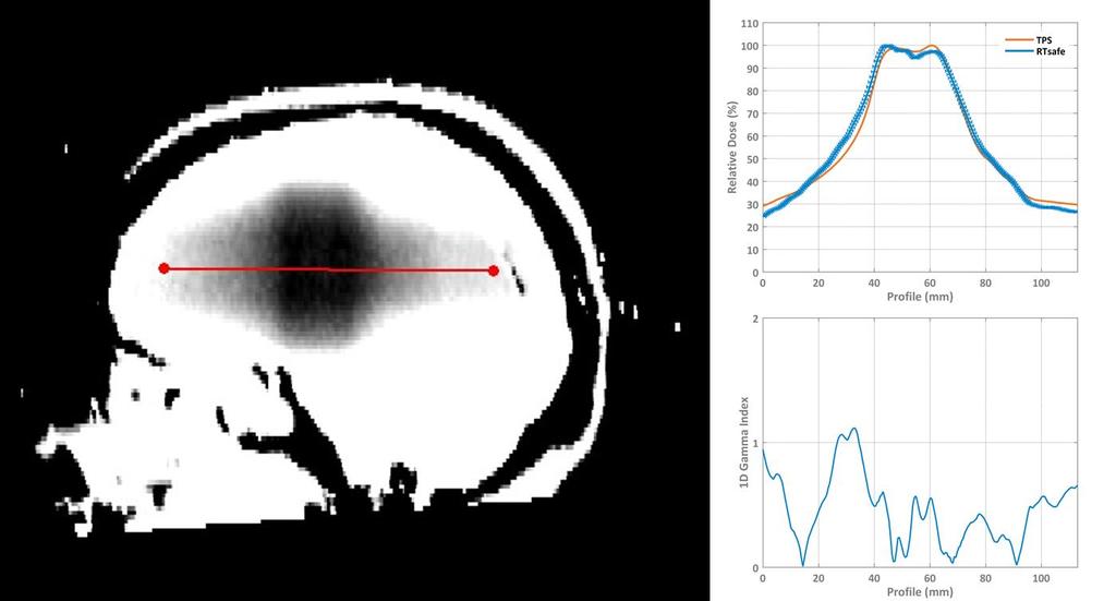
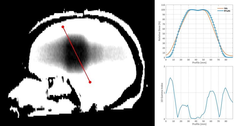
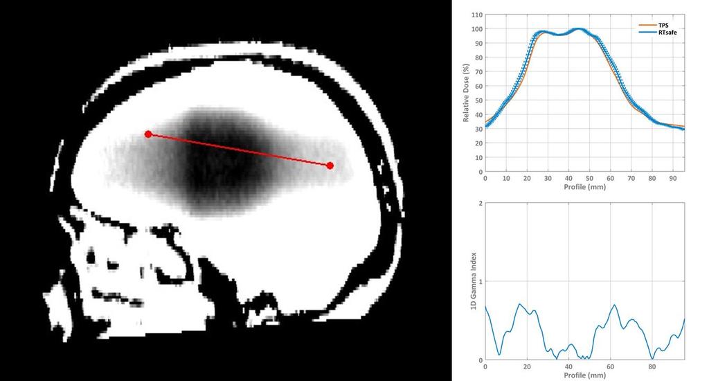
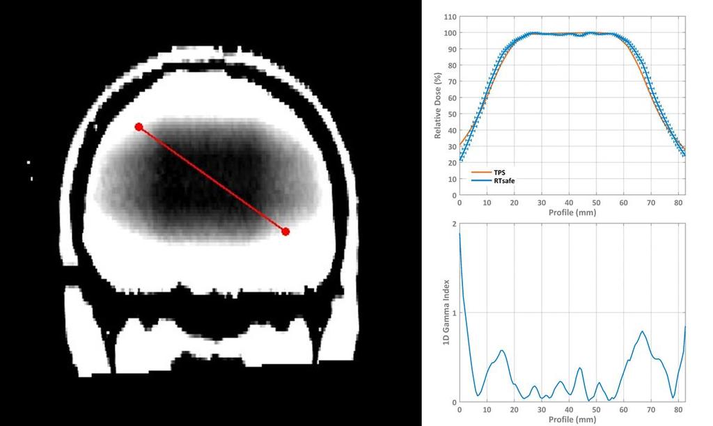
4 PART II: Profiles comparison Indicatively, a number of relative dose profiles for both the measured and TPS-calculated datasets are presented in the following figures. In order to quantitatively assess agreement between the two datasets, 1D gamma index calculations are also included. Passing criteria were 2 mm distance-to-agreement and 5% dose difference. Error bars correspond to ± 1mm spatial uncertainty.
5
6
7
8
9

10 PART III: 2D Gamma Index comparison For selected central axial slice of the irradiated phantom, 2D gamma index calculations are presented in the following figures. Again, passing criteria were 2 mm distance-to-agreement and 5% dose difference. However, a dose threshold of 2% has been applied to exclude


11 corresponding voxels from the gamma index calculations. Isodose lines are also plotted to assist comparison.

 of each structure.")
12 PART IV: DVH comparison Comparison between planned and measured relative dose distributions is presented in the following figures, in terms of cumulative Dose Volume Histograms (DVHs) for the PTV and OAR. All dose distributions were normalized to the corresponding D 50% metric (i.e., the minimum dose received by at least the 50% of the volume) of each structure.
13 PART V: DVH metrics comparison Using the aforementioned normalization (100% corresponds to D 50%), metrics derived from the above DVHs are given in the following table. Mean (%) D95 (%) Structure TPS Meas. TPS Meas. PTV Brain Stem
Evaluation of 3D Gamma index calculation implemented in two commercial dosimetry systems
 University of Wollongong Research Online Faculty of Engineering and Information Sciences - Papers: Part A Faculty of Engineering and Information Sciences 2015 Evaluation of 3D Gamma index calculation implemented
IMRT and VMAT Patient Specific QA Using 2D and 3D Detector Arrays
 IMRT and VMAT Patient Specific QA Using 2D and 3D Detector Arrays Sotiri Stathakis Outline Why IMRT/VMAT QA AAPM TG218 UPDATE Tolerance Limits and Methodologies for IMRT Verification QA Common sources
VALIDATION OF DIR. Raj Varadhan, PhD, DABMP Minneapolis Radiation Oncology
 VALIDATION OF DIR Raj Varadhan, PhD, DABMP Minneapolis Radiation Oncology Overview Basics: Registration Framework, Theory Discuss Validation techniques Using Synthetic CT data & Phantoms What metrics to
PCRT 3D. Scalable Architecture System. User-Friendly. Traceable. Continuos Development
 PCRT 3D The PCRT3D is a versatile 3D radiation treatment planning system featuring the most accurate algorithm calculations, the latest techniques in virtual simulation and the most advanced radiotherapy
Lucy Phantom MR Grid Evaluation
 Lucy Phantom MR Grid Evaluation Anil Sethi, PhD Loyola University Medical Center, Maywood, IL 60153 November 2015 I. Introduction: The MR distortion grid, used as an insert with Lucy 3D QA phantom, is
TG 132: Use of Image Registration and Fusion in RT
 TG 132: Use of Image Registration and Fusion in RT Kristy K Brock, PhD, DABR, FAAPM Associate Professor Department of Radiation Oncology, University of Michigan Chair, AAPM TG 132: Image Registration and
Using Pinnacle 16 Deformable Image registration in a re-treat scenario
 Introduction Using Pinnacle 16 Deformable Image registration in a re-treat scenario This short Hands On exercise will introduce how the Deformable Image Registration (DIR) tools in Pinnacle can be used
Coverage based treatment planning to accommodate organ deformable motions and contouring uncertainties for prostate treatment. Huijun Xu, Ph.D.
 Coverage based treatment planning to accommodate organ deformable motions and contouring uncertainties for prostate treatment Huijun Xu, Ph.D. Acknowledgement and Disclosure Dr. Jeffrey Siebers Dr. DJ
Three-dimensional dosimetry of TomoTherapy by MRI-based polymer gel technique
 JOURNAL OF APPLIED CLINICAL MEDICAL PHYSICS, VOLUME 12, NUMBER 1, WINTER 2011 Three-dimensional dosimetry of TomoTherapy by MRI-based polymer gel technique Yoichi Watanabe, 1a N. Gopishankar 2 Department
A dedicated tool for PET scanner simulations using FLUKA
 A dedicated tool for PET scanner simulations using FLUKA P. G. Ortega FLUKA meeting June 2013 1 Need for in-vivo treatment monitoring Particles: The good thing is that they stop... Tumour Normal tissue/organ
NA-MIC National Alliance for Medical Image Computing SlicerRT Extension
 NA-MIC SlicerRT Extension Csaba Pinter 1, Andras Lasso 1, Kevin Wang 2 11 Laboratory for Percutaneous Surgery, Queen s University, Canada 2 University Health Network, Toronto, ON, Canada pinter@cs.queensu.ca
Michael Speiser, Ph.D.
 IMPROVED CT-BASED VOXEL PHANTOM GENERATION FOR MCNP MONTE CARLO Michael Speiser, Ph.D. Department of Radiation Oncology UT Southwestern Medical Center Dallas, TX September 1 st, 2012 CMPWG Workshop Medical
ATC Conformance Statement Overview
 Advanced-Technology QA Consortium DICOM Workshop: ATC Conformance Statement Overview Walter R. Bosch, D.Sc. ATC DICOM Conformance Statement Overview DICOM Part 10 File Set Reader Application (ITC) Defines
SlicerRT Image-guided radiation therapy research toolkit for 3D Slicer
 SlicerRT Image-guided radiation therapy research toolkit for 3D Slicer Csaba Pinter 1, Andras Lasso 1, An Wang 2, David Jaffray 2, and Gabor Fichtinger 1 1Laboratory for Percutaneous Surgery, Queen s University,
Good Morning! Thank you for joining us
 Good Morning! Thank you for joining us Deformable Registration, Contour Propagation and Dose Mapping: 101 and 201 Marc Kessler, PhD, FAAPM The University of Michigan Conflict of Interest I receive direct
Design and performance characteristics of a Cone Beam CT system for Leksell Gamma Knife Icon
 Design and performance characteristics of a Cone Beam CT system for Leksell Gamma Knife Icon WHITE PAPER Introduction Introducing an image guidance system based on Cone Beam CT (CBCT) and a mask immobilization
THE WIRELESS PHANTOM PERFORM ACCURATE PATIENT QA IN LESS TIME THAN EVER!
 THE WIRELESS PHANTOM PERFORM ACCURATE PATIENT QA IN LESS TIME THAN EVER! Confidence in complex treatments Modern radiation therapy uses complex plans with techniques such as IMRT, VMAT and Tomotherapy.
CHAPTER 9 INFLUENCE OF SMOOTHING ALGORITHMS IN MONTE CARLO DOSE CALCULATIONS OF CYBERKNIFE TREATMENT PLANS: A LUNG PHANTOM STUDY
 148 CHAPTER 9 INFLUENCE OF SMOOTHING ALGORITHMS IN MONTE CARLO DOSE CALCULATIONS OF CYBERKNIFE TREATMENT PLANS: A LUNG PHANTOM STUDY 9.1 INTRODUCTION 9.1.1 Dose Calculation Algorithms Dose calculation
MapCHECK 2 & 3DVH. The Gold Standard for 2D Arrays
 MapCHECK 2 & 3DVH The Gold Standard for 2D Arrays Your Most Valuable QA and Dosimetry Tools THE GOLD STANDARD FOR 2D ARRAYS The MapCHECK 2 is the world s most selected independent 2D measurement array.
Position accuracy analysis of the stereotactic reference defined by the CBCT on Leksell Gamma Knife Icon
 Position accuracy analysis of the stereotactic reference defined by the CBCT on Leksell Gamma Knife Icon WHITE PAPER Introduction An image guidance system based on Cone Beam CT (CBCT) is included in Leksell
MapCHECK 2 & 3DVH The Gold Standard for 2D Arrays
 MapCHECK 2 & 3DVH The Gold Standard for 2D Arrays Your Most Valuable QA and Dosimetry Tools THE GOLD STANDARD FOR 2D ARRAYS The MapCHECK 2 is the world s most selected independent 2D measurement array.
Feasibility of 3D Printed Patient specific Phantoms for IMRT QA and Other Dosimetric Special Procedures
 Feasibility of 3D Printed Patient specific Phantoms for IMRT QA and Other Dosimetric Special Procedures ehler 046@umn.edu Eric Ehler, PhD Assistant Professor Department of Radiation Oncology What is 3D
Overview of Proposed TG-132 Recommendations
 Overview of Proposed TG-132 Recommendations Kristy K Brock, Ph.D., DABR Associate Professor Department of Radiation Oncology, University of Michigan Chair, AAPM TG 132: Image Registration and Fusion Conflict
7/31/2011. Learning Objective. Video Positioning. 3D Surface Imaging by VisionRT
 CLINICAL COMMISSIONING AND ACCEPTANCE TESTING OF A 3D SURFACE MATCHING SYSTEM Hania Al-Hallaq, Ph.D. Assistant Professor Radiation Oncology The University of Chicago Learning Objective Describe acceptance
A Study of Medical Image Analysis System
 Indian Journal of Science and Technology, Vol 8(25), DOI: 10.17485/ijst/2015/v8i25/80492, October 2015 ISSN (Print) : 0974-6846 ISSN (Online) : 0974-5645 A Study of Medical Image Analysis System Kim Tae-Eun
Tutorial. on SlicerRT and 3D Slicer modules
 Tutorial on SlicerRT and 3D Slicer modules Agenda Acquire and build SlicerRT (on an existing Slicer) Use python console in Slicer Create extension and module skeletons Use extension template Use scripted
Fractional labelmaps for computing accurate dose volume histograms
 Fractional labelmaps for computing accurate dose volume histograms Kyle Sunderland, Csaba Pinter, Andras Lasso, Gabor Fichtinger Laboratory for Percutaneous Surgery, School of Computing, Queen s University,
CONTOURING ACCURACY. What Have We Learned? And Where Do We Go From Here? BEN NELMS, PH.D. AUGUST 15, 2016
 CONTOURING ACCURACY What Have We Learned? And Where Do We Go From Here? BEN NELMS, PH.D. AUGUST 15, 2016 FIRST THINGS FIRST Happy Medical Dosimetrist s Week! OUTLINE 1. Objectives 2. The importance of
UvA-DARE (Digital Academic Repository) Motion compensation for 4D PET/CT Kruis, M.F. Link to publication
 Motion compensation for 4D PET/CT Kruis, M.F. Link to publication") UvA-DARE (Digital Academic Repository) Motion compensation for 4D PET/CT Kruis, M.F. Link to publication Citation for published version (APA): Kruis, M. F. (2014). Motion compensation for 4D PET/CT General
UvA-DARE (Digital Academic Repository) Motion compensation for 4D PET/CT Kruis, M.F. Link to publication Citation for published version (APA): Kruis, M. F. (2014). Motion compensation for 4D PET/CT General
Chapter 3 Set Redundancy in Magnetic Resonance Brain Images
 16 Chapter 3 Set Redundancy in Magnetic Resonance Brain Images 3.1 MRI (magnetic resonance imaging) MRI is a technique of measuring physical structure within the human anatomy. Our proposed research focuses
Use of MRI in Radiotherapy: Technical Consideration
 Use of MRI in Radiotherapy: Technical Consideration Yanle Hu, PhD Department of Radiation Oncology, Mayo Clinic Arizona 04/07/2018 2015 MFMER slide-1 Conflict of Interest: None 2015 MFMER slide-2 Objectives
IMRT site-specific procedure: Prostate (CHHiP)
") IMRT site-specific procedure: Prostate (CHHiP) Scope: To provide site specific instructions for the planning of CHHIP IMRT patients Responsibilities: Radiotherapy Physicists, HPC Registered Therapy Radiographers
IMRT site-specific procedure: Prostate (CHHiP) Scope: To provide site specific instructions for the planning of CHHIP IMRT patients Responsibilities: Radiotherapy Physicists, HPC Registered Therapy Radiographers
3DVH : SUN NUCLEAR On The Accuracy Of The corporation Planned Dose Perturbation Algorithm Your Most Valuable QA and Dosimetry Tools *Patent Pending
 3DVH : On The Accuracy Of The Planned Dose Perturbation Algorithm SUN NUCLEAR corporation Your Most Valuable QA and Dosimetry Tools *Patent Pending introduction State-of-the-art IMRT QA of static gantry
Raising the Bar in IMRT QA
 MapCHECK 2TM Raising the Bar in IMRT QA The leader in quick and precise measurement of modulated radiotherapy beams Benefits Proven solution for film-less rotational delivery and IMRT QA - More than 1500
Walk Through of CERR Capabilities. CERR: Introduction. Outline. Getting CERR: Control Panel. Documentation Support Community
 Walk Through of CERR Capabilities Aditya P. Apte, Ph.D. Department of Medical Physics Memorial Sloan Kettering Cancer Center New York aptea@mskcc.org AAPM 2015, July 15, 2015 CERR: Computational Environment
4 Measurement. and Analysis. 4.1 Overview and Underlying Principles 4-1
 Measurement and Analysis.1 Overview and Underlying Principles.1.1 Introductory Remarks The physics and setup for film dosimetry have been described in the previous chapters. The measurement setup for IMRT
TEPZZ Z754_7A_T EP A1 (19) (11) EP A1 (12) EUROPEAN PATENT APPLICATION. (51) Int Cl.: A61N 5/10 ( )
 (11) EP A1 (12) EUROPEAN PATENT APPLICATION. (51) Int Cl.: A61N 5/10 ( )") (19) TEPZZ Z74_7A_T (11) EP 3 07 417 A1 (12) EUROPEAN PATENT APPLICATION (43) Date of publication: 0..16 Bulletin 16/ (1) Int Cl.: A61N / (06.01) (21) Application number: 16163147.8 (22) Date of filing:
(19) TEPZZ Z74_7A_T (11) EP 3 07 417 A1 (12) EUROPEAN PATENT APPLICATION (43) Date of publication: 0..16 Bulletin 16/ (1) Int Cl.: A61N / (06.01) (21) Application number: 16163147.8 (22) Date of filing:
Use of Deformable Image Registration in Radiation Therapy. Colin Sims, M.Sc. Accuray Incorporated 1
 Use of Deformable Image Registration in Radiation Therapy Colin Sims, M.Sc. Accuray Incorporated 1 Overview of Deformable Image Registration (DIR) Algorithms that can deform one dataset to another have
Radiotherapy Plan Competition TomoTherapy Planning System. Dmytro Synchuk. Ukrainian Center of TomoTherapy, Kirovograd, Ukraine
 Radiotherapy Plan Competition 2016 TomoTherapy Planning System Dmytro Synchuk Ukrainian Center of TomoTherapy, Kirovograd, Ukraine Beam Geometry 6MV fan beam 3 jaw options 1.0, 2.5 and 5 cm 64 leaves binary
Virtual Phantoms for IGRT QA
 TM Virtual Phantoms for IGRT QA Why ImSimQA? ImSimQA was developed to overcome the limitations of physical phantoms for testing modern medical imaging and radiation therapy software systems, when there
COMPARISON OF DOSE CALCULATION ALGORITHMS FOR LEKSELL GAMMA KNIFE PERFEXION USING MONTE CARLO VOXEL PHANTOMS
 COMPARISON OF DOSE CALCULATION ALGORITHMS FOR LEKSELL GAMMA KNIFE PERFEXION USING MONTE CARLO VOXEL PHANTOMS Jan Pipek 1, Josef Novotný Jr. 1,2,3, Josef Novotný 1, Petra Kozubíková 1 1 Faculty of Nuclear
SlicerRT radiation therapy extension for 3D Slicer DICOM aspects
 SlicerRT radiation therapy extension for 3D Slicer DICOM aspects Andras Lasso, Csaba Pinter Laboratory for Percutaneous Surgery, Queen s University, Canada Commercial treatment planning systems (TPS) Expensive
Secondary 3D Dose QA Fully Automated using MOSAIQ's IQ Engine. MOSAIQ User Meeting May Antwerp
 Secondary 3D Dose QA Fully Automated using MOSAIQ's IQ Engine MOSAIQ User Meeting May 31 2013 - Antwerp Contents Project goal and collaboration Secondary 3D Dose QA project justification Secondary 3D Dose
Validation of GEANT4 for Accurate Modeling of 111 In SPECT Acquisition
 Validation of GEANT4 for Accurate Modeling of 111 In SPECT Acquisition Bernd Schweizer, Andreas Goedicke Philips Technology Research Laboratories, Aachen, Germany bernd.schweizer@philips.com Abstract.
Turbo-BrainVoyager. Setup guide
 Turbo-BrainVoyager Setup guide Turbo-BrainVoyager (TBV) is a highly optimized software package for real-time analysis and advanced visualization of functional and structural magnetic resonance imaging
Image-based Monte Carlo calculations for dosimetry
 Image-based Monte Carlo calculations for dosimetry Irène Buvat Imagerie et Modélisation en Neurobiologie et Cancérologie UMR 8165 CNRS Universités Paris 7 et Paris 11 Orsay, France buvat@imnc.in2p3.fr
The University of Chicago. Center for EPR Imaging in Vivo Physiology. Image Registration. Boris Epel
 The University of Chicago Center for EPR Imaging in Vivo Physiology Image Registration Boris Epel Imaging Methods are Complimentary CT MRI EPRI High resolution anatomic images Quantitative Poor soft tissue
A Generation Methodology for Numerical Phantoms with Statistically Relevant Variability of Geometric and Physical Properties
 A Generation Methodology for Numerical Phantoms with Statistically Relevant Variability of Geometric and Physical Properties Steven Dolly 1, Eric Ehler 1, Yang Lou 2, Mark Anastasio 2, Hua Li 2 (1) University
Help Guide. mm Copyright Mirada Medical Ltd, Mirada Medical RTx 1
 Help Guide mm3237-1.6-1 Copyright Mirada Medical Ltd, 2000-2014. Mirada Medical RTx 1 Contents Help Guide... 1 Contents... 2 Introduction to RTx... 4 Regulatory Statement... 6 Notes... 15 Data Supported...
MR-guided radiotherapy: Vision, status and research at the UMC Utrecht. Dipl. Ing. Dr. Markus Glitzner
 MR-guided radiotherapy: Vision, status and research at the UMC Utrecht Dipl. Ing. Dr. Markus Glitzner About myself Training Medizintechnik TU Graz PhD UMC Utrecht Clinical work Software implementation
An Integrated Visual Analysis System for Fusing MR Spectroscopy and Multi-Modal Radiology Imaging
 An Integrated Visual Analysis System for Fusing MR Spectroscopy and Multi-Modal Radiology Imaging Miguel Nunes, Benjamin Rowland, Matthias Schlachter, Soléakhéna Ken, Kresimir Matkovic, Anne Laprie and
3D Slicer Overview. Andras Lasso, PhD PerkLab, Queen s University
 3D Slicer Overview Andras Lasso, PhD PerkLab, Queen s University Right tool for the job Technological prototype Research tool Clinical tool Can it be done? Jalopnik.com Innovative, not robust, usually
Association between pathology and texture features of multi parametric MRI of the prostate
 Association between pathology and texture features of multi parametric MRI of the prostate 1,2 Peter Kuess, 3 D. Nilsson, 1,2 P. Andrzejewski, 2,4 P. Georg, 1 J. Knoth, 5 M. Susani, 3 J. Trygg, 2,6 T.
Commissioning of a 3D image-based treatment planning system for high-dose-rate brachytherapy of cervical cancer
 JOURNAL OF APPLIED CLINICAL MEDICAL PHYSICS, VOLUME 17, NUMBER 2, 2016 Commissioning of a 3D image-based treatment planning system for high-dose-rate brachytherapy of cervical cancer Yongbok Kim, 1 Joseph
Methodological progress in image registration for ventilation estimation, segmentation propagation and multi-modal fusion
 Methodological progress in image registration for ventilation estimation, segmentation propagation and multi-modal fusion Mattias P. Heinrich Julia A. Schnabel, Mark Jenkinson, Sir Michael Brady 2 Clinical
n o r d i c B r a i n E x Tutorial DSC Module
 m a k i n g f u n c t i o n a l M R I e a s y n o r d i c B r a i n E x Tutorial DSC Module Please note that this tutorial is for the latest released nordicbrainex. If you are using an older version please
ADVANCING CANCER TREATMENT
 The RayPlan treatment planning system makes proven, innovative RayStation technology accessible to clinics that need a cost-effective and streamlined solution. Fast, efficient and straightforward to use,
Advanced Targeting Using Image Deformation. Justin Keister, MS DABR Aurora Health Care Kenosha, WI
 Advanced Targeting Using Image Deformation Justin Keister, MS DABR Aurora Health Care Kenosha, WI History of Targeting The advance of IMRT and CT simulation has changed how targets are identified in radiation
Slide 1. Technical Aspects of Quality Control in Magnetic Resonance Imaging. Slide 2. Annual Compliance Testing. of MRI Systems.
 Slide 1 Technical Aspects of Quality Control in Magnetic Resonance Imaging Slide 2 Compliance Testing of MRI Systems, Ph.D. Department of Radiology Henry Ford Hospital, Detroit, MI Slide 3 Compliance Testing
Is deformable image registration a solved problem?
 Is deformable image registration a solved problem? Marcel van Herk On behalf of the imaging group of the RT department of NKI/AVL Amsterdam, the Netherlands DIR 1 Image registration Find translation.deformation
8/3/2017. Contour Assessment for Quality Assurance and Data Mining. Objective. Outline. Tom Purdie, PhD, MCCPM
 Contour Assessment for Quality Assurance and Data Mining Tom Purdie, PhD, MCCPM Objective Understand the state-of-the-art in contour assessment for quality assurance including data mining-based techniques
Dosimetry Simulations with the UF-B Series Phantoms using the PENTRAN-MP Code System
 Dosimetry Simulations with the UF-B Series Phantoms using the PENTRAN-MP Code System A. Al-Basheer, M. Ghita, G. Sjoden, W. Bolch, C. Lee, and the ALRADS Group Computational Medical Physics Team Nuclear
Image Co-Registration II: TG132 Quality Assurance for Image Registration. Image Co-Registration II: TG132 Quality Assurance for Image Registration
 Image Co-Registration II: TG132 Quality Assurance for Image Registration Preliminary Recommendations from TG 132* Kristy Brock, Sasa Mutic, Todd McNutt, Hua Li, and Marc Kessler *Recommendations are NOT
Mathematical methods and simulations tools useful in medical radiation physics
 Mathematical methods and simulations tools useful in medical radiation physics Michael Ljungberg, professor Department of Medical Radiation Physics Lund University SE-221 85 Lund, Sweden Major topic 1:
Magnetic Resonance Imaging Velocity. Information. Joe Lee. April 4, 2000
 Locating Arteriovenous Malformations using Magnetic Resonance Imaging Velocity Information Joe Lee April 4, 2000 1 Introduction An arteriovenous malformation (AVM) is a congenital vascular defect where
Classification of Subject Motion for Improved Reconstruction of Dynamic Magnetic Resonance Imaging
 1 CS 9 Final Project Classification of Subject Motion for Improved Reconstruction of Dynamic Magnetic Resonance Imaging Feiyu Chen Department of Electrical Engineering ABSTRACT Subject motion is a significant
ASTRO Integrating the Healthcare Enterprise. IHE-Radiation Oncology Technical Framework Volume 2 - Transactions
 ASTRO Integrating the Healthcare Enterprise IHE-Radiation Oncology Technical Framework Volume 2 - Transactions 2008 November 13, 2008 Copyright 2006-2008: ACC/HIMSS/RSNA/ASTRO Contents 1 Preface to Volume
Kevin O Donnell Senior R&D Manager Toshiba Medical Research Institute Co-Chair, DICOM Standards Committee Past Chair, IHE Radiology Planning
 1 Kevin O Donnell Senior R&D Manager Toshiba Medical Research Institute Co-Chair, DICOM Standards Committee Past Chair, IHE Radiology Planning Committee Standards & Tools 2 Learning Objectives 1) DICOM
Skull Segmentation of MR images based on texture features for attenuation correction in PET/MR
 Skull Segmentation of MR images based on texture features for attenuation correction in PET/MR CHAIBI HASSEN, NOURINE RACHID ITIO Laboratory, Oran University Algeriachaibih@yahoo.fr, nourine@yahoo.com
iplan RT Image Advanced Contouring Workstation - Driving Physician Collaboration
 iplan RT Image Advanced Contouring Workstation - Driving Physician Collaboration The iplan Contouring Workstation offers unique and innovative capabilities for faster contouring and consistent segmentation
Transitioning from pencil beam to Monte Carlo for electron dose calculations
 Transitioning from pencil beam to Monte Carlo for electron dose calculations Jessie Huang-Vredevoogd (jyhuang4@wisc.edu) University of Wisconsin NCC AAPM October 12, 2019 1 Topics to cover Background RayStation
ADVANCING CANCER TREATMENT
 3 ADVANCING CANCER TREATMENT SUPPORTING CLINICS WORLDWIDE RaySearch is advancing cancer treatment through pioneering software. We believe software has un limited potential, and that it is now the driving
High dynamic range magnetic resonance flow imaging in the abdomen
 High dynamic range magnetic resonance flow imaging in the abdomen Christopher M. Sandino EE 367 Project Proposal 1 Motivation Time-resolved, volumetric phase-contrast magnetic resonance imaging (also known
C a t p h a n / T h e P h a n t o m L a b o r a t o r y
 C a t p h a n 5 0 0 / 6 0 0 T h e P h a n t o m L a b o r a t o r y C a t p h a n 5 0 0 / 6 0 0 Internationally recognized for measuring the maximum obtainable performance of axial, spiral and multi-slice
Geant4 in Brachytherapy
 Geant4 in Brachytherapy 1. 2. 3. 4. 5. Brachytherapy: Brief Overview Clinical applications Basic research Ultrafast & biology applications Issues for the work group 1 Brachytherapy: Overview Brachy: Greek
IHE Radiation Oncology Technical Framework Supplement. Treatment Delivery Workflow (TDW) Draft for Public Comment
 Draft for Public Comment") Integrating the Healthcare Enterprise IHE Radiation Oncology Technical Framework Supplement Treatment Delivery Workflow (TDW) Draft for Public Comment Date: January 29, 2010 Author: David Murray Email:
Integrating the Healthcare Enterprise IHE Radiation Oncology Technical Framework Supplement Treatment Delivery Workflow (TDW) Draft for Public Comment Date: January 29, 2010 Author: David Murray Email:
OnDemand3D Fusion Technology
 CYBERMED INC., ONDEMAND3D TECHNOLOGY INC. OnDemand3D Fusion Technology White Paper December 2009 USA Republic of Korea www.ondemand3d.com Introduction OnDemand3D TM Fusion is registration technology to
A fluence convolution method to account for respiratory motion in three-dimensional dose calculations of the liver: A Monte Carlo study
 A fluence convolution method to account for respiratory motion in three-dimensional dose calculations of the liver: A Monte Carlo study Indrin J. Chetty, a) Mihaela Rosu, Neelam Tyagi, Lon H. Marsh, Daniel
TARDIS Working Practice Document No: 035
 TARDIS Working Practice Document No: 035 Uploading CT or MR Images All stroke patients should have a baseline scan taken before randomisation. Any other CT scans or MRIs, taken up until completion of the
Monaco VMAT. The Next Generation in IMRT/VMAT Planning. Paulo Mathias Customer Support TPS Application
 Monaco VMAT The Next Generation in IMRT/VMAT Planning Paulo Mathias Customer Support TPS Application 11.05.2011 Background What is Monaco? Advanced IMRT/VMAT treatment planning system from Elekta Software
ViewRay System Commissioning
 ViewRay System Commissioning Kyle R. Padgett, PhD, DABR Assistant Professor University of Miami Viewray Team: Matt Studenski, PhD Chet Ford, PhD Yidong Yang, PhD Nesrin Dogan, PhD Medical Physics Residents:
Commissioning of MR-only simulation for radiotherapy planning
 Radiation Oncology Ingenia MR-RT MR-only sim Commissioning of MR-only simulation for radiotherapy planning Gerald Schubert, Teuvo Vaara, Matti Lindström, Reko Kemppainen, and Marieke van Grootel-Rensen
How would, or how does, the patient position (chin extended) affect your beam arrangement?
 affect your beam arrangement?") 1 Megan Sullivan Clinical Practicum II Parotid Lab July 29, 2016 PLAN 1: IPSILATERAL WEDGE PAIR TECHNIQUE The ipsilateral wedge pair technique consisted of an anterior oblique field at 45 degrees and a
1 Megan Sullivan Clinical Practicum II Parotid Lab July 29, 2016 PLAN 1: IPSILATERAL WEDGE PAIR TECHNIQUE The ipsilateral wedge pair technique consisted of an anterior oblique field at 45 degrees and a
Creating a Knowledge Based Model using RapidPlan TM : The Henry Ford Experience
 DVH Estimates Creating a Knowledge Based Model using RapidPlan TM : The Henry Ford Experience Karen Chin Snyder, MS, DABR AAMD Region V Meeting October 4, 2014 Disclosures The Department of Radiation Oncology
1. Learn to incorporate QA for surface imaging
 Hania Al-Hallaq, Ph.D. Assistant Professor Radiation Oncology The University of Chicago ***No disclosures*** 1. Learn to incorporate QA for surface imaging into current QA procedures for IGRT. 2. Understand
A software tool for the quantitative evaluation of 3D dose calculation algorithms
 A software tool for the quantitative evaluation of 3D dose calculation algorithms William B. Harms, Sr., Daniel A. Low, John W. Wong, a) and James A. Purdy Washington University School of Medicine, Mallinckrodt
SIGMI Meeting ~Image Fusion~ Computer Graphics and Visualization Lab Image System Lab
 SIGMI Meeting ~Image Fusion~ Computer Graphics and Visualization Lab Image System Lab Introduction Medical Imaging and Application CGV 3D Organ Modeling Model-based Simulation Model-based Quantification
System Messages for MRIdian 4.1
 L 0063 System Messages for MRIdian 4.1 April 2016 SY00001 System found auto recover data on startup during system startup check. System shutdown when while plan was open. The system found plan recovery
Qalitätssicherung an Multileafkollimatoren. Dr. Lutz Müller Würzburg jan 2004
 Qalitätssicherung an Multileafkollimatoren Dr. Lutz Müller Würzburg jan 2004 IMRT Verification - present Target Volume Constraints Inverse Planning Algorithm Fluence Map Leaf Sequencer Leaf & Gantry sequence
Reconstruction in CT and relation to other imaging modalities
 Reconstruction in CT and relation to other imaging modalities Jørgen Arendt Jensen November 1, 2017 Center for Fast Ultrasound Imaging, Build 349 Department of Electrical Engineering Center for Fast Ultrasound
3DVH FAQs. What is PDP questions
 3DVH FAQs What is PDP questions 1. Explain the PDP in layman terms. How does PDP work? a. Very simply, PDP uses measured diode data, and compares it to the expected treatment plan data. The differences
Prototype of Silver Corpus Merging Framework
 www.visceral.eu Prototype of Silver Corpus Merging Framework Deliverable number D3.3 Dissemination level Public Delivery data 30.4.2014 Status Authors Final Markus Krenn, Allan Hanbury, Georg Langs This
REAL-TIME ADAPTIVITY IN HEAD-AND-NECK AND LUNG CANCER RADIOTHERAPY IN A GPU ENVIRONMENT
 REAL-TIME ADAPTIVITY IN HEAD-AND-NECK AND LUNG CANCER RADIOTHERAPY IN A GPU ENVIRONMENT Anand P Santhanam Assistant Professor, Department of Radiation Oncology OUTLINE Adaptive radiotherapy for head and
INTRODUCTION TO MEDICAL IMAGING- 3D LOCALIZATION LAB MANUAL 1. Modifications for P551 Fall 2013 Medical Physics Laboratory
 INTRODUCTION TO MEDICAL IMAGING- 3D LOCALIZATION LAB MANUAL 1 Modifications for P551 Fall 2013 Medical Physics Laboratory Introduction Following the introductory lab 0, this lab exercise the student through
PrecisePLAN 2.15 Export
 Elekta Limited. DICOM Conformance Statement for PrecisePLAN 2.15 Export (DICOM Release 2.15) 2006 Elekta Limited. All rights reserved. DRC-173-0031-01a DICOM Conformance Statement - PrecisePLAN 2.15 Export
IMSURE QA SOFTWARE FAST, PRECISE QA SOFTWARE
 QA SOFTWARE FAST, PRECISE Software for accurate and independent verification of monitor units, dose, and overall validity of standard, IMRT, VMAT, SRS and brachytherapy plans no film, no phantoms, no linac
Analysis of RapidArc optimization strategies using objective function values and dose-volume histograms
 JOURNAL OF APPLIED CLINICAL MEDICAL PHYSICS, VOLUME 11, NUMBER 1, WINTER 2010 Analysis of RapidArc optimization strategies using objective function values and dose-volume histograms Mike Oliver, a Isabelle
Whole Body MRI Intensity Standardization
 Whole Body MRI Intensity Standardization Florian Jäger 1, László Nyúl 1, Bernd Frericks 2, Frank Wacker 2 and Joachim Hornegger 1 1 Institute of Pattern Recognition, University of Erlangen, {jaeger,nyul,hornegger}@informatik.uni-erlangen.de
Dynalog data tool for IMRT plan verification
 Dynalog data tool for IMRT plan verification Poster No.: R-0051 Congress: 2014 CSM Type: Scientific Exhibit Authors: V. Sashin; FOOTSCRAY/AU Keywords: Computer applications, Radiation physics, Experimental,
ATTENTION! The whole content of the lecture with all the animations can be dowloaded from:
 ATTENTION! The whole content of the lecture with all the animations can be dowloaded from: http://www.onko.szote.uszeged.hu/letoltes/radioter_phys_basis/radiother_phys_basis.zip Size > 300 M!!! The dowloaded.zip