Dynamic Contrast enhanced MRA
|
|
|
- Matilda Thompson
- 5 years ago
- Views:
Transcription

1 Dynamic Contrast enhanced MRA Speaker: Yung-Chieh Chang Date : Department of Radiology, Taichung Veterans General Hospital, Taichung, Taiwan 1
2 Outline Basic and advanced principles of Diffusion Weighted Images (DWI) Echo Planar Imaging (EPI) 2. Diffusion weighted imaging (DWI) Basic and advanced principles of Diffusion Weighted Images (DWI) 2 1. Diffusion-weighted whole-body imaging with background body signal suppression (DWIBS) 2. Intravoxel Incoherent Motion (IVIM) Dynamic Contrast enhanced MRA Preliminary conclusions and discussion
3 Conventional CEMRA Arterial phase Venous phase
4 Conventional CEMRA
5 Conventional CEMRA 6 5 artery vein acquisition order time All 3 sections acquired in 10 seconds
6 kx Conventional CEMRA ( 3 sections) C3 C6 C9 C12 B2 B5 B8 B11 ky A1 A4 A7 A10 B2 B5 B8 B11 C3 C6 C9 C12 : pre-contrast : post-contrast ky C6 B5 A4 C12 B11 A10 B5 B11 C6 C12
7 Suppose the image of arteries and veins arteries vein arteries vein


8 Conventional CEMRA artery vein C6 C9 C12 B5 B8 B11 A4 A7 A B5 B8 B11 C6 C9 C Arterial phase Venous phase


9 Fill in the k-space center different timing too early exactly too late From left to right: early to late scanning Technique patient s ( Heart Rate & Blood pressure )

10 Suppose the image of arteries and veins arteries vein arteries vein


11 Dynamic Contrast enhanced MRA artery vein B5 C7 B9 3 A4 A6 A8 2 1 B C7 B9 F1 F2 F3 F4 F5. frame 1 frame 2 frame 3 frame 4 frame 5


12 If the signal is received quickly 1sec 2sec 3sec 4sec 5sec 6sec 7sec 8sec 9sec 10sec 11sec 12sec 13sec 14sec 15sec 16sec 17sec 18sec 19sec 20sec 21sec 22sec 23sec 24sec 25sec 26sec 27sec 28sec 29sec 30sec


13


14

15


16 Could we do Dynamic MRA like DSA? Convential DSA CEMRA
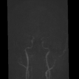
17 Dynamic MRA Conventional CEMRA Arterial phase Venous phase


18 4D TRAK CEMRA
19 MRI for Blood vessels None contrast Agent -Time of flight MRA ( 3D TOF MRA ) -Phase contrast MRA ( PC MRA ) Contrast Agent -Contrast-enhanced MRA ( CE MRA ) Contrast Agent + Acceleration - 4D-TRAK (Philips) - TWIST (Siemens) - TRICKS (GE)
20 Why should be accelerated? It avoids the vein interference - Contrast agent flows into vein rapidly from the artery It avoid that can not find arterial phase - Slow flow patient - Heart rate (Too slow) - Heart rate (Too fast) - Carotid artery Short (kids)
21 How do we avoid that vein is appeared It uses of accelerate scanning technology for reducing scan time
22 Parameters Compare (VGHTC) 3D MRA 4D-Trak matrix 380x280x x187x160 TR / TE (ms) 3.6/ /1 Flip angle (deg) Keyhole no yes dyn scans 2 30 Total scan duration Dyn. refscan time Dyn. keyhole scan duration - 1.6
23 4D-TRAK (Philips) 4D Time-Resolved Angiography using Keyhole Purpose Dynamic, high-resolution 4D CE-MRA Combination the speed of SENSE (parallel imaging with SENSitivity Encoding), CENTRA (Contrast-ENhanced Timing Robust Angiography), Keyhole halfscan
24 4D-TRAK (Philips) 4D Time-Resolved Angiography using Keyhole Purpose Dynamic, high-resolution 4D CE-MRA Combination the speed of SENSE (parallel imaging with SENSitivity Encoding), CENTRA (Contrast-ENhanced Timing Robust Angiography), Keyhole halfscan
25 SENSE SENSitivity Encoding (ipat,asset) Parallel Imaging technique: multi-element RF coils increase speed using in imaging Preparation: Determine sensitivity maps of individual RF coil elements Acquisition: Measure less data (k-profiles), results in faster scan Reconstruction: Decode images using coil element sensitivity maps
26 Sensitivity encoding Application with 2-element synergy coil Sensitivity Sensitivity Coil 1 Coil 2
 B")
27 Sensitivity encoding Scan with sense factor 2 (RFOV of 50%) B A
28 Sensitivity encoding C 1 = 0.85 A B Coil C 2 = 0.2 A B 1.0 B A Coil 2 B A

29 Sensitivity encoding Result -Accelerated Image Acquisition C 1 = 0.85 A B C 2 = 0.2 A B A and B B A
30 4D-TRAK (Philips) 4D Time-Resolved Angiography using Keyhole Purpose Dynamic, high-resolution 4D CE-MRA Combination the speed of SENSE (parallel imaging with SENSitivity Encoding), CENTRA (Contrast-ENhanced Timing Robust Angiography), Keyhole halfscan
31 Contrast-ENhanced Timing Robust Angiography Divide k-space in 2 segments: arterial window arterial + venous window Benefits: Optimal venous suppression longer scan times possible resulting in: Higher resolution High SNR k y k z

32 Contrast-ENhanced Timing Robust Angiography

33

34 Sequential view order (Fixed timing, Test bolus timing)
35 Elliptical centric view order (Real time imaging)
36 4D-TRAK (Philips) 4D Time-Resolved Angiography using Keyhole Purpose Dynamic, high-resolution 4D CE-MRA Combination the speed of SENSE (parallel imaging with SENSitivity Encoding), CENTRA (Contrast-ENhanced Timing Robust Angiography), Keyhole halfscan

 ADC 2D")
37 K space MR sampling K space MR image (Analogic signal) (Digital signal) (Digital image) ADC 2D FT
 k 1")
")
 k 4")
38 K space (a) k 1 (b) k 2 ky kx (c) k 3 38 (d) k 4
39 K space (a) k 1 (b) k 2 (c) k 3 (d) k 4 (e) k 5 (f) k 6 (g) k 8 (h) k (i) k 16 (j) k 24
40 K space (n) k 128 More K number that 1. Resolution will be better 2. Scanning time increases 40
 The central 64 64 points of k space")
41 K space The center of k space SNR and Contrast (a) The central points of k space The periphery of k space Resolution (b) The central points of k space 41

42 4D-TRAK (Keyhole) Dyn1 ~ Dyn(n-1) The center of k space Nth (Last scanning) The periphery of k space Keyhole : copying k-space information from reference scan to other dynamics.

43 Keyhole 4D-TRAK using keyhole acquisition and reconstruction. The last dynamic is fully sampled and the peripheral part of the k-space is used to reconstruct the contrast-enhanced timing-robust angiography (CENTRA) keyhole dynamics.
44 Keyhole Time-Resolved Angiography using Keyhole Arrival of contrast injection k y k z Fast dynamic scanning k z k z k z Reconstruction k y k y k y

45


46 Acquisition Order Order : ABACAD...
47 Acquisition Order Prior to the contrast injection, k-space is fully sampled. ABCD 4T ABCD ABCD ABACADABACAD.. ABACADABACAD.. 2T Posterior to the contrast injection ABACADABACAD.. 6T
48 MRM 36: (1996)
49 4D-TRAK Maximum acceleration Speedup of keyhole 6 High speedup thanks to simple segmentation in k-space Speedup of SENSE up to 8 Speedup of halfscan ~1.25 X
50 TWIST (Siemens) Time-resolved angiography With Interleaved Stochastic Trajectories Faster Scanning Options: Short TR Partial Fourier Reduction in spatial resolution In-plane resolution Slice resolution Parallel imaging (PAT²)
51 TWIST Sub-divide k-space into 2 regions: a central region A (with main information about image contrast) a peripheral region B (with main information about spatial resolution) Scan A with a higher frequency than B, in order to achieve a higher temporal resolution

52 TWIST

53 TWIST Region A is the center of k-space, determining the image contrast The size of Region A can be flexibly chosen, between 4% and 100% of total k-space

54 TWIST The key is to use a reduced sampling density of B (= SDB),


55 TWIST Sub-divide k-space into 2 regions and scan them in the alternating scheme A BT A BT ABT...

56 TWIST The diagram of TWIST k-space sampling strategy. In the dots show the ky kz plane of k-space, each dot represents onereadout line of k space data. Blue dots shows the center k space views that are acquired for each timepoint.

57 TWIST Higher (nominal) frame rate than conventional imaging: With the flexibility of Size of A Sampling Density of B the reconstructed temporal resolution can be set flexibly,

58 TWIST

59 3D TOF 4D CEMRA?! Image quality is good, but can not see... Image is poor, but can see

60


61

62

63 Vascular malformation


64 4D-TRAK MRA CTA Direct type CCF


65 DSA AV fistula MRA


66 AV fistula
 2~3 days at the")
67 4D-TRAK IV bolus injection One hour at the hospital DSA Arterial puncture (high risk) 2~3 days at the hospital
68 Conclusion 4D-TRAK (Philips) is combined the speed of keyhole, SENSE and halfscan that increases for high temporal resolution (Dynamic imaging). TWIST (Siemens) Time-resolved angiography With Interleaved Stochastic Trajectories, It is to use a reduced sampling density of B that increases for high temporal resolution For applications such as intracranial arteriovenous malformation (AVM),intracranial arteriovenous fistula (AVF) and intracranial venous thrombosis.

69
70 4D-TRAK Reconstruction Methods (VGHTC) Dyn1 Dyn2 Dyn3 Dyn4 Dyn5 Dyn slices Maximum intensity projection (MIP) We will Obtain the Dynamic MRA images
Outline: Contrast-enhanced MRA
 Outline: Contrast-enhanced MRA Background Technique Clinical Indications Future Directions Disclosures: GE Health Care: Research support Consultant: Bracco, Bayer The Basics During rapid IV infusion, Gadolinium
Module 5: Dynamic Imaging and Phase Sharing. (true-fisp, TRICKS, CAPR, DISTAL, DISCO, HYPR) Review. Improving Temporal Resolution.
 Review. Improving Temporal Resolution.") MRES 7005 - Fast Imaging Techniques Module 5: Dynamic Imaging and Phase Sharing (true-fisp, TRICKS, CAPR, DISTAL, DISCO, HYPR) Review Improving Temporal Resolution True-FISP (I) True-FISP (II) Keyhole
MRES 7005 - Fast Imaging Techniques Module 5: Dynamic Imaging and Phase Sharing (true-fisp, TRICKS, CAPR, DISTAL, DISCO, HYPR) Review Improving Temporal Resolution True-FISP (I) True-FISP (II) Keyhole
MR Advance Techniques. Vascular Imaging. Class III
 MR Advance Techniques Vascular Imaging Class III 1 Vascular Imaging There are several methods that can be used to evaluate the cardiovascular systems with the use of MRI. MRI will aloud to evaluate morphology
MRI. When to use What sequences. Outline 2012/09/19. Sequence: Definition. Basic Principles: Step 2. Basic Principles: Step 1. Govind Chavhan, MD
 MRI When to use What sequences Govind Chavhan, MD Assistant Professor and Staff Radiologist The Hospital For Sick Children, Toronto Planning Acquisition Post processing Interpretation Patient history and
Magnetic Resonance Angiography
 Magnetic Resonance Angiography Course: Advance MRI (BIOE 594) Instructors: Dr Xiaohong Joe Zhou Dr. Shadi Othman By, Nayan Pasad Phase Contrast Angiography By Moran 1982, Bryan et. Al. 1984 and Moran et.
MRI Physics II: Gradients, Imaging
 MRI Physics II: Gradients, Imaging Douglas C., Ph.D. Dept. of Biomedical Engineering University of Michigan, Ann Arbor Magnetic Fields in MRI B 0 The main magnetic field. Always on (0.5-7 T) Magnetizes
GE Healthcare CLINICAL GALLERY. Discovery * MR750w 3.0T. This brochure is intended for European healthcare professionals.
 GE Healthcare CLINICAL GALLERY Discovery * MR750w 3.0T This brochure is intended for European healthcare professionals. NEURO PROPELLER delivers high resolution, motion insensitive imaging in all planes.
Accelerated MRI Techniques: Basics of Parallel Imaging and Compressed Sensing
 Accelerated MRI Techniques: Basics of Parallel Imaging and Compressed Sensing Peng Hu, Ph.D. Associate Professor Department of Radiological Sciences PengHu@mednet.ucla.edu 310-267-6838 MRI... MRI has low
Applications Guide for Interleaved
 Applications Guide for Interleaved rephase/dephase MRAV Authors: Yongquan Ye, Ph.D. Dongmei Wu, MS. Tested MAGNETOM Systems : 7TZ, TRIO a Tim System, Verio MR B15A (N4_VB15A_LATEST_20070519) MR B17A (N4_VB17A_LATEST_20090307_P8)
Philips MRI Protocol Dump Created on Comment Software Stream
 Page 1 of 5 Philips MRI Protocol Dump Created on 2/17/2011 4:11:01 PM Comment Created by ExamCard_to_XML with inputs: "J:\ADNI GO - ADNI 2 Phantom5.ExamCard" on system (BU SCHOOL OF MEDICINE :: 192.168.71.10)
Compressed Sensing for Rapid MR Imaging
 Compressed Sensing for Rapid Imaging Michael Lustig1, Juan Santos1, David Donoho2 and John Pauly1 1 Electrical Engineering Department, Stanford University 2 Statistics Department, Stanford University rapid
Sampling, Ordering, Interleaving
 Sampling, Ordering, Interleaving Sampling patterns and PSFs View ordering Modulation due to transients Temporal modulations Slice interleaving Sequential, Odd/even, bit-reversed Arbitrary Other considerations:
Clinical Importance. Aortic Stenosis. Aortic Regurgitation. Ultrasound vs. MRI. Carotid Artery Stenosis
 Clinical Importance Rapid cardiovascular flow quantitation using sliceselective Fourier velocity encoding with spiral readouts Valve disease affects 10% of patients with heart disease in the U.S. Most
Lab Location: MRI, B2, Cardinal Carter Wing, St. Michael s Hospital, 30 Bond Street
 Lab Location: MRI, B2, Cardinal Carter Wing, St. Michael s Hospital, 30 Bond Street MRI is located in the sub basement of CC wing. From Queen or Victoria, follow the baby blue arrows and ride the CC south
Advanced MRI Techniques (and Applications)
") Advanced MRI Techniques (and Applications) Jeffry R. Alger, PhD Department of Neurology Ahmanson-Lovelace Brain Mapping Center Brain Research Institute Jonsson Comprehensive Cancer Center University of
Advanced MRI Techniques (and Applications) Jeffry R. Alger, PhD Department of Neurology Ahmanson-Lovelace Brain Mapping Center Brain Research Institute Jonsson Comprehensive Cancer Center University of
Fast Imaging Trajectories: Non-Cartesian Sampling (1)
") Fast Imaging Trajectories: Non-Cartesian Sampling (1) M229 Advanced Topics in MRI Holden H. Wu, Ph.D. 2018.05.03 Department of Radiological Sciences David Geffen School of Medicine at UCLA Class Business
Fast Imaging Trajectories: Non-Cartesian Sampling (1) M229 Advanced Topics in MRI Holden H. Wu, Ph.D. 2018.05.03 Department of Radiological Sciences David Geffen School of Medicine at UCLA Class Business
Diffusion MRI Acquisition. Karla Miller FMRIB Centre, University of Oxford
 Diffusion MRI Acquisition Karla Miller FMRIB Centre, University of Oxford karla@fmrib.ox.ac.uk Diffusion Imaging How is diffusion weighting achieved? How is the image acquired? What are the limitations,
HST.583 Functional Magnetic Resonance Imaging: Data Acquisition and Analysis Fall 2008
 MIT OpenCourseWare http://ocw.mit.edu HST.583 Functional Magnetic Resonance Imaging: Data Acquisition and Analysis Fall 2008 For information about citing these materials or our Terms of Use, visit: http://ocw.mit.edu/terms.
Sampling, Ordering, Interleaving
 Sampling, Ordering, Interleaving Sampling patterns and PSFs View ordering Modulation due to transients Temporal modulations Timing: cine, gating, triggering Slice interleaving Sequential, Odd/even, bit-reversed
TOF-MRA Using Multi-Oblique-Stack Acquisition (MOSA)
") JOURNAL OF MAGNETIC RESONANCE IMAGING 26:432 436 (2007) Technical Note TOF-MRA Using Multi-Oblique-Stack Acquisition (MOSA) Ed X. Wu, PhD, 1,2 * Edward S. Hui, BEng, 1,2 and Jerry S. Cheung, BEng 1,2 Purpose:
JOURNAL OF MAGNETIC RESONANCE IMAGING 26:432 436 (2007) Technical Note TOF-MRA Using Multi-Oblique-Stack Acquisition (MOSA) Ed X. Wu, PhD, 1,2 * Edward S. Hui, BEng, 1,2 and Jerry S. Cheung, BEng 1,2 Purpose:
CHAPTER 9: Magnetic Susceptibility Effects in High Field MRI
 Figure 1. In the brain, the gray matter has substantially more blood vessels and capillaries than white matter. The magnified image on the right displays the rich vasculature in gray matter forming porous,
SIEMENS MAGNETOM TrioTim syngo MR B17
 \\USER\KNARRGROUP\MultiBand\LavretskyMultiBand\trufi localizer 3-plane TA: 5.1 s PAT: Voxel size: 1.2 1.2 5. Rel. SNR: 1.00 SIEMENS: trufi Load to stamp Slice group 1 Slices 1 Dist. factor 20 % Phase enc.
Role of Parallel Imaging in High Field Functional MRI
 Role of Parallel Imaging in High Field Functional MRI Douglas C. Noll & Bradley P. Sutton Department of Biomedical Engineering, University of Michigan Supported by NIH Grant DA15410 & The Whitaker Foundation
TOPICS 2/5/2006 8:17 PM. 2D Acquisition 3D Acquisition
 TOPICS 2/5/2006 8:17 PM 2D Acquisition 3D Acquisition 2D Acquisition Involves two main steps : Slice Selection Slice selection is accomplished by spatially saturating (single or multi slice imaging) or
Scan Acceleration with Rapid Gradient-Echo
 Scan Acceleration with Rapid Gradient-Echo Hsiao-Wen Chung ( 鍾孝文 ), Ph.D., Professor Dept. Electrical Engineering, National Taiwan Univ. Dept. Radiology, Tri-Service General Hospital 1 of 214 The Need
HST.583 Functional Magnetic Resonance Imaging: Data Acquisition and Analysis Fall 2006
 MIT OpenCourseWare http://ocw.mit.edu HST.583 Functional Magnetic Resonance Imaging: Data Acquisition and Analysis Fall 2006 For information about citing these materials or our Terms of Use, visit: http://ocw.mit.edu/terms.
Head motion in diffusion MRI
 Head motion in diffusion MRI Anastasia Yendiki HMS/MGH/MIT Athinoula A. Martinos Center for Biomedical Imaging 11/06/13 Head motion in diffusion MRI 0/33 Diffusion contrast Basic principle of diffusion
SIEMENS MAGNETOM Verio syngo MR B15V
 \\USER\ZAHID_RESEARCH\MS\No Name\3D SWI TA: 6:39 PAT: 2 Voxel size: 1.0 0.5 2.0 mm Rel. SNR: 1.00 SIEMENS: gre Properties Prio Recon Before measurement After measurement Load to viewer Inline movie Auto
Initial Experience of Applying TWIST Dixon with Flexible View Sharing in Breast DCE-MRI
 Initial Experience of Applying TWIST Dixon with Flexible View Sharing in Breast DCE-MRI Yuan Le PhD 1, Hal D. Kipfer MD 1, Dominik M. Nickel PhD 2, Randall Kroeker PhD 2, Brian Dale PhD 2, Stephanie P.
SIEMENS MAGNETOM Avanto syngo MR B15
 \\USER\INVESTIGATORS\Ravi\ADNI-phantom\QC Phantom-Localizer TA: 0:10 PAT: Voxel size: 1.9 1.5 8.0 mm Rel. SNR: 1.00 SIEMENS: gre Properties Prio Recon Before measurement After measurement Load to viewer
Sparse sampling in MRI: From basic theory to clinical application. R. Marc Lebel, PhD Department of Electrical Engineering Department of Radiology
 Sparse sampling in MRI: From basic theory to clinical application R. Marc Lebel, PhD Department of Electrical Engineering Department of Radiology Objective Provide an intuitive overview of compressed sensing
COBRE Scan Information
 COBRE Scan Information Below is more information on the directory structure for the COBRE imaging data. Also below are the imaging parameters for each series. Directory structure: var/www/html/dropbox/1139_anonymized/human:
3D Radial Undersampling 7/19/2012. Artifact Removal SNR Restoration-- HYPR. Background: Time Resolved MR Angiography 4D DSA AND 4D FLUOROSCOPY:
 4D DSA AND 4D FLUOROSCOPY: Accelerated Applications using Undersampled Acquisition and Constrained Reconstruction Background: Time Resolved MR Angiography During the past 12 years we have been investigating
Motion Artifacts and Suppression in MRI At a Glance
 Motion Artifacts and Suppression in MRI At a Glance Xiaodong Zhong, PhD MR R&D Collaborations Siemens Healthcare MRI Motion Artifacts and Suppression At a Glance Outline Background Physics Common Motion
Functional MRI. Jerry Allison, Ph. D. Medical College of Georgia
 Functional MRI Jerry Allison, Ph. D. Medical College of Georgia BOLD Imaging Technique Blood Oxygen Level Dependent contrast can be used to map brain function Right Hand Motor Task Outline fmri BOLD Contrast
HST.583 Functional Magnetic Resonance Imaging: Data Acquisition and Analysis Fall 2008
 MIT OpenCourseWare http://ocw.mit.edu HST.583 Functional Magnetic Resonance Imaging: Data Acquisition and Analysis Fall 2008 For information about citing these materials or our Terms of Use, visit: http://ocw.mit.edu/terms.
SPECIFICATIONS FOR A NEW STATE OF ART 16 SLICE ALL PURPOSE C. T. SCANNER
 SPECIFICATIONS FOR A NEW STATE OF ART 16 SLICE ALL PURPOSE C. T. SCANNER A) Scanner Design X-Ray generator and tube: 1. Scanner: Whole body spiral CT scanner (16 slices) of latest technology. 2. X-Ray
SIEMENS MAGNETOM Avanto syngo MR B15
 \\USER\INVESTIGATORS\Ravi\ADNI-Subject\Localizer TA: 0:10 PAT: Voxel size: 1.9 1.5 8.0 mm Rel. SNR: 1.00 SIEMENS: gre Properties Prio Recon Before measurement After measurement Load to viewer Inline movie
(a Scrhon5 R2iwd b. P)jc%z 5. ivcr3. 1. I. ZOms Xn,s. 1E IDrAS boms. EE225E/BIOE265 Spring 2013 Principles of MRI. Assignment 8 Solutions
jc%z 5. ivcr3. 1. I. ZOms Xn,s. 1E IDrAS boms. EE225E/BIOE265 Spring 2013 Principles of MRI. Assignment 8 Solutions") EE225E/BIOE265 Spring 2013 Principles of MRI Miki Lustig Assignment 8 Solutions 1. Nishimura 7.1 P)jc%z 5 ivcr3. 1. I Due Wednesday April 10th, 2013 (a Scrhon5 R2iwd b 0 ZOms Xn,s r cx > qs 4-4 8ni6 4
EE225E/BIOE265 Spring 2013 Principles of MRI Miki Lustig Assignment 8 Solutions 1. Nishimura 7.1 P)jc%z 5 ivcr3. 1. I Due Wednesday April 10th, 2013 (a Scrhon5 R2iwd b 0 ZOms Xn,s r cx > qs 4-4 8ni6 4
CTA HEAD Perfusion AqONE without and with IV Contrast
 CTA HEAD Perfusion AqONE without and with IV Contrast Patient Position Adult Contrast Adult Injection Rate Supine IOML perpendicular to table top. IV: 100 ml with helical head CTA 50 ml without helical
2/5/2006 8:26 PM TOPICS
 TOPICS 2/5/2006 8:26 PM Key Hole Acquisition Block Regional Interpolation Scheme for K-space (BRISK) Time Resolved Imaging of Contrast Kinetics (TRICKS) Real Time Imaging Introduction 2/5/2006 8:26 PM
Parallel Imaging. Marcin.
 Parallel Imaging Marcin m.jankiewicz@gmail.com Parallel Imaging initial thoughts Over the last 15 years, great progress in the development of pmri methods has taken place, thereby producing a multitude
High-Resolution Time-Resolved Contrast-Enhanced MR Abdominal and Pulmonary Angiography Using a Spiral- TRICKS Sequence
 High-Resolution Time-Resolved Contrast-Enhanced MR Abdominal and Pulmonary Angiography Using a Spiral- TRICKS Sequence Jiang Du* and Mark Bydder Magnetic Resonance in Medicine 58:631 635 (2007) Both high
8/11/2009. Common Areas of Motion Problem. Motion Compensation Techniques and Applications. Type of Motion. What s your problem
 Common Areas of Motion Problem Motion Compensation Techniques and Applications Abdominal and cardiac imaging. Uncooperative patient, such as pediatric. Dynamic imaging and time series. Chen Lin, PhD Indiana
Module 4. K-Space Symmetry. Review. K-Space Review. K-Space Symmetry. Partial or Fractional Echo. Half or Partial Fourier HASTE
 MRES 7005 - Fast Imaging Techniques Module 4 K-Space Symmetry Review K-Space Review K-Space Symmetry Partial or Fractional Echo Half or Partial Fourier HASTE Conditions for successful reconstruction Interpolation
SIEMENS MAGNETOM Skyra syngo MR D13
 Page 1 of 8 SIEMENS MAGNETOM Skyra syngo MR D13 \\USER\CIND\StudyProtocols\PTSA\*dm_ep2d_mono70_b0_p2_iso2.0 TA:1:05 PAT:2 Voxel size:2.0 2.0 2.0 mm Rel. SNR:1.00 :epse Properties Routine Prio Recon Load
Slide 1. Technical Aspects of Quality Control in Magnetic Resonance Imaging. Slide 2. Annual Compliance Testing. of MRI Systems.
 Slide 1 Technical Aspects of Quality Control in Magnetic Resonance Imaging Slide 2 Compliance Testing of MRI Systems, Ph.D. Department of Radiology Henry Ford Hospital, Detroit, MI Slide 3 Compliance Testing
Image Acquisition Systems
 Image Acquisition Systems Goals and Terminology Conventional Radiography Axial Tomography Computer Axial Tomography (CAT) Magnetic Resonance Imaging (MRI) PET, SPECT Ultrasound Microscopy Imaging ITCS
SIEMENS MAGNETOM Verio syngo MR B17
 \\USER\Dr. Behrmann\routine\Ilan\ep2d_bold_PMU_resting TA: 8:06 PAT: Voxel size: 3.03.03.0 mm Rel. SNR: 1.00 USER: ep2d_bold_pmu Properties Special sat. Prio Recon System Before measurement Body After
White Pixel Artifact. Caused by a noise spike during acquisition Spike in K-space <--> sinusoid in image space
 White Pixel Artifact Caused by a noise spike during acquisition Spike in K-space sinusoid in image space Susceptibility Artifacts Off-resonance artifacts caused by adjacent regions with different
New Technology Allows Multiple Image Contrasts in a Single Scan
 These images were acquired with an investigational device. PD T2 T2 FLAIR T1 MAP T1 FLAIR PSIR T1 New Technology Allows Multiple Image Contrasts in a Single Scan MR exams can be time consuming. A typical
A Novel Iterative Thresholding Algorithm for Compressed Sensing Reconstruction of Quantitative MRI Parameters from Insufficient Data
 A Novel Iterative Thresholding Algorithm for Compressed Sensing Reconstruction of Quantitative MRI Parameters from Insufficient Data Alexey Samsonov, Julia Velikina Departments of Radiology and Medical
M R I Physics Course
 M R I Physics Course Multichannel Technology & Parallel Imaging Nathan Yanasak, Ph.D. Jerry Allison Ph.D. Tom Lavin, B.S. Department of Radiology Medical College of Georgia References: 1) The Physics of
ACQUIRING AND PROCESSING SUSCEPTIBILITY WEIGHTED IMAGING (SWI) DATA ON GE 3.0T
 DATA ON GE 3.0T") ACQUIRING AND PROCESSING SUSCEPTIBILITY WEIGHTED IMAGING (SWI) DATA ON GE 3.0T Revision date: 12/13/2010 Overview Susceptibility Weighted Imaging (SWI) is a relatively new data acquisition and processing
ACQUIRING AND PROCESSING SUSCEPTIBILITY WEIGHTED IMAGING (SWI) DATA ON GE 3.0T Revision date: 12/13/2010 Overview Susceptibility Weighted Imaging (SWI) is a relatively new data acquisition and processing
Supplementary Figure 1
 Supplementary Figure 1 BOLD and CBV functional maps showing EPI versus line-scanning FLASH fmri. A. Colored BOLD and CBV functional maps are shown in the highlighted window (green frame) of the raw EPI
Siemens AG, Healthcare Sector. syngo MR D13 0. Supplement - Parameters and image text 0.
 Siemens AG, Healthcare Sector 0 0 n.a. English Cs2 syngo Neuro Operator MR-05014 630 05/2010 01 02 Informatik, Manual D11 Cape syngo MR D13 0.0 Supplement - Parameters and image text 0. syngo MR D13 0.
MRI image formation 8/3/2016. Disclosure. Outlines. Chen Lin, PhD DABR 3. Indiana University School of Medicine and Indiana University Health
 MRI image formation Indiana University School of Medicine and Indiana University Health Disclosure No conflict of interest for this presentation 2 Outlines Data acquisition Spatial (Slice/Slab) selection
Basic fmri Design and Analysis. Preprocessing
 Basic fmri Design and Analysis Preprocessing fmri Preprocessing Slice timing correction Geometric distortion correction Head motion correction Temporal filtering Intensity normalization Spatial filtering
Redundancy Encoding for Fast Dynamic MR Imaging using Structured Sparsity
 Redundancy Encoding for Fast Dynamic MR Imaging using Structured Sparsity Vimal Singh and Ahmed H. Tewfik Electrical and Computer Engineering Dept., The University of Texas at Austin, USA Abstract. For
Vessel Explorer: a tool for quantitative measurements in CT and MR angiography
 Clinical applications Vessel Explorer: a tool for quantitative measurements in CT and MR angiography J. Oliván Bescós J. Sonnemans R. Habets J. Peters H. van den Bosch T. Leiner Healthcare Informatics/Patient
Multi-slice CT Image Reconstruction Jiang Hsieh, Ph.D.
 Multi-slice CT Image Reconstruction Jiang Hsieh, Ph.D. Applied Science Laboratory, GE Healthcare Technologies 1 Image Generation Reconstruction of images from projections. textbook reconstruction advanced
Imaging Notes, Part IV
 BME 483 MRI Notes 34 page 1 Imaging Notes, Part IV Slice Selective Excitation The most common approach for dealing with the 3 rd (z) dimension is to use slice selective excitation. This is done by applying
What is pmri? Overview. The Need for Speed: A Technical and Clinical Primer for Parallel MR Imaging 8/1/2011
 The Need for Speed: A Technical and Clinical Primer for Parallel MR Imaging Nathan Yanasak, Ph.D. Chair, AAPM TG118 Assistant Professor Department of Radiology Director, Core Imaging Facility for Small
Advanced Imaging Trajectories
 Advanced Imaging Trajectories Cartesian EPI Spiral Radial Projection 1 Radial and Projection Imaging Sample spokes Radial out : from k=0 to kmax Projection: from -kmax to kmax Trajectory design considerations
Breast MRI Accreditation Program Clinical Image Quality Guide
 Breast MRI Accreditation Program Clinical Image Quality Guide Introduction This document provides guidance on breast MRI clinical image quality and describes the criteria used by the ACR Breast MRI Accreditation
Use of MRI in Radiotherapy: Technical Consideration
 Use of MRI in Radiotherapy: Technical Consideration Yanle Hu, PhD Department of Radiation Oncology, Mayo Clinic Arizona 04/07/2018 2015 MFMER slide-1 Conflict of Interest: None 2015 MFMER slide-2 Objectives
SPM8 for Basic and Clinical Investigators. Preprocessing. fmri Preprocessing
 SPM8 for Basic and Clinical Investigators Preprocessing fmri Preprocessing Slice timing correction Geometric distortion correction Head motion correction Temporal filtering Intensity normalization Spatial
E. Mark Haacke, PhD. The MRI Institute for Biomedical Research Detroit, Michigan Wayne State University Detroit, Michigan 48201
 E. Mark Haacke, PhD The MRI Institute for Biomedical Research Detroit, Michigan 48202 Wayne State University Detroit, Michigan 48201 Acknowledgements The testing and establishment of these protocols has
Parallel Magnetic Resonance Imaging (pmri): How Does it Work, and What is it Good For?
: How Does it Work, and What is it Good For?") Parallel Magnetic Resonance Imaging (pmri): How Does it Work, and What is it Good For? Nathan Yanasak, Ph.D. Chair, AAPM TG118 Department of Radiology Georgia Regents University Overview Phased-array coils
Parallel Magnetic Resonance Imaging (pmri): How Does it Work, and What is it Good For? Nathan Yanasak, Ph.D. Chair, AAPM TG118 Department of Radiology Georgia Regents University Overview Phased-array coils
3D Vascular Segmentation using MRA Statistics and Velocity Field Information in PC-MRA
 3D Vascular Segmentation using MRA Statistics and Velocity Field Information in PC-MRA Albert C. S. Chung 1, J. Alison Noble 1, Paul Summers 2 and Michael Brady 1 1 Department of Engineering Science, Oxford
Fast Imaging UCLA. Class Business. Class Business. Daniel B. Ennis, Ph.D. Magnetic Resonance Research Labs. Tuesday (3/7) from 6-9pm HW #1 HW #2
 from 6-9pm HW #1 HW #2") Fast Imaging Daniel B. Ennis, Ph.D. Magnetic Resonance Research Labs Class Business Tuesday (3/7) from 6-9pm 6:00-7:30pm Groups Avanto Sara Said, Yara Azar, April Pan Skyra Timothy Marcum, Diana Lopez,
Fast Imaging Daniel B. Ennis, Ph.D. Magnetic Resonance Research Labs Class Business Tuesday (3/7) from 6-9pm 6:00-7:30pm Groups Avanto Sara Said, Yara Azar, April Pan Skyra Timothy Marcum, Diana Lopez,
ADNI, ADNI_QH, SURVEY. Geometry. connection
 ADNI, ADNI_QH, SURVEY Geometry Coil selection = Head connection = d Multi coil Homogeneity correction ne FOV (mm) = 250.00 RFOV (%) = 100.00 Foldover suppression Matrix scan = 256 reconstruction = 256
I.e. Sex differences in child appetitive traits and Eating in the Absence of Hunger:
 Supplementary Materials I. Evidence of sex differences on eating behavior in children I.e. Sex differences in child appetitive traits and Eating in the Absence of Hunger: Table 2. Parent Report for Child
Functional MRI in Clinical Research and Practice Preprocessing
 Functional MRI in Clinical Research and Practice Preprocessing fmri Preprocessing Slice timing correction Geometric distortion correction Head motion correction Temporal filtering Intensity normalization
Error! Bookmark not defined. Error! Bookmark not defined.
 MRI-Neuro Protocols Brain Protocols... 2 WITH AND WITHOUT... 3 WITHOUT CONTRT... 4 HHT... 5 MS WITH AND WITHOUT CONTRT... 6 MS WITHOUT CONTRT... 7 PEDIATRIC... 8 PEDIATRIC WITH CONTRT... 9 PITUITARY NO
Abbie M. Diak, PhD Loyola University Medical Center Dept. of Radiation Oncology
 Abbie M. Diak, PhD Loyola University Medical Center Dept. of Radiation Oncology Outline High Spectral and Spatial Resolution MR Imaging (HiSS) What it is How to do it Ways to use it HiSS for Radiation
EPI Data Are Acquired Serially. EPI Data Are Acquired Serially 10/23/2011. Functional Connectivity Preprocessing. fmri Preprocessing
 Functional Connectivity Preprocessing Geometric distortion Head motion Geometric distortion Head motion EPI Data Are Acquired Serially EPI Data Are Acquired Serially descending 1 EPI Data Are Acquired
Super-resolution Reconstruction of Fetal Brain MRI
 Super-resolution Reconstruction of Fetal Brain MRI Ali Gholipour and Simon K. Warfield Computational Radiology Laboratory Children s Hospital Boston, Harvard Medical School Worshop on Image Analysis for
CT Basics Principles of Spiral CT Dose. Always Thinking Ahead.
 1 CT Basics Principles of Spiral CT Dose 2 Who invented CT? 1963 - Alan Cormack developed a mathematical method of reconstructing images from x-ray projections Sir Godfrey Hounsfield worked for the Central
XI Signal-to-Noise (SNR)
") XI Signal-to-Noise (SNR) Lecture notes by Assaf Tal n(t) t. Noise. Characterizing Noise Noise is a random signal that gets added to all of our measurements. In D it looks like this: while in D
XI Signal-to-Noise (SNR) Lecture notes by Assaf Tal n(t) t. Noise. Characterizing Noise Noise is a random signal that gets added to all of our measurements. In D it looks like this: while in D
SPM8 for Basic and Clinical Investigators. Preprocessing
 SPM8 for Basic and Clinical Investigators Preprocessing fmri Preprocessing Slice timing correction Geometric distortion correction Head motion correction Temporal filtering Intensity normalization Spatial
Fast methods for magnetic resonance angiography (MRA)
") Fast methods for magnetic resonance angiography (MRA) Bahareh Vafadar Department of Electrical and Computer Engineering A thesis presented for the degree of Doctor of Philosophy University of Canterbury,
Fast methods for magnetic resonance angiography (MRA) Bahareh Vafadar Department of Electrical and Computer Engineering A thesis presented for the degree of Doctor of Philosophy University of Canterbury,
New Enhanced Multi-frame DICOM CT and MR Objects to Enhance Performance and Image Processing on PACS and Workstations
 New Enhanced Multi-frame DICOM CT and MR Objects to Enhance Performance and Image Processing on PACS and Workstations SCAR 2004 Hot Topics - 22 May 2004 David Clunie, RadPharm Charles Parisot,, GE Healthcare
Evaluations of k-space Trajectories for Fast MR Imaging for project of the course EE591, Fall 2004
 Evaluations of k-space Trajectories for Fast MR Imaging for project of the course EE591, Fall 24 1 Alec Chi-Wah Wong Department of Electrical Engineering University of Southern California 374 McClintock
Single Breath-hold Abdominal T 1 Mapping using 3-D Cartesian Sampling and Spatiotemporally Constrained Reconstruction
 Single Breath-hold Abdominal T 1 Mapping using 3-D Cartesian Sampling and Spatiotemporally Constrained Reconstruction Felix Lugauer 1,3, Jens Wetzl 1, Christoph Forman 2, Manuel Schneider 1, Berthold Kiefer
CP Generalize Concepts in Abstract Multi-dimensional Image Model Component Semantics. David Clunie.
 CP-1390 - Generalize Concepts in Abstract Multi-dimensional Image Model Semantics Page 1 STATUS Date of Last Update Person Assigned Submitter Name Submission Date Assigned 2014/06/09 David Clunie mailto:dclunie@dclunie.com
Compressed Sensing And Joint Acquisition Techniques In Mri
 Wayne State University Wayne State University Theses 1-1-2013 Compressed Sensing And Joint Acquisition Techniques In Mri Rouhollah Hamtaei Wayne State University, Follow this and additional works at: http://digitalcommons.wayne.edu/oa_theses
Lucy Phantom MR Grid Evaluation
 Lucy Phantom MR Grid Evaluation Anil Sethi, PhD Loyola University Medical Center, Maywood, IL 60153 November 2015 I. Introduction: The MR distortion grid, used as an insert with Lucy 3D QA phantom, is
MRI Hot Topics Motion Correction for MR Imaging. medical
 MRI Hot Topics Motion Correction for MR Imaging s medical Motion Correction for MR Imaging Kyle A. Salem, PhD Motion Artifacts in a Clinical Setting Patient motion is probably the most common cause of
ADNI GO - ADNI 2 Human7 (9) 38:30.1
 38:30.1") Philips RI Protocol Dump Create on 11/25/2013 11:40:24 A Comment Create by ExamCar_to_XL with inputs: "K:\ADNI GO - ADNI 2 Human7.ExamCar" on system (BU SCHOOL OF EDICINE :: 192.168.71.10) Software Stream
Philips RI Protocol Dump Create on 11/25/2013 11:40:24 A Comment Create by ExamCar_to_XL with inputs: "K:\ADNI GO - ADNI 2 Human7.ExamCar" on system (BU SCHOOL OF EDICINE :: 192.168.71.10) Software Stream
DICOM Correction Item
 DICOM Correction Item Correction Number CP-567 Log Summary: Type of Modification Clarification Name of Standard PS 3.3 2004 Rationale for Correction The order of signficance of dimension indices (i.e.,which
FOREWORD TO THE SPECIAL ISSUE ON MOTION DETECTION AND COMPENSATION
 Philips J. Res. 51 (1998) 197-201 FOREWORD TO THE SPECIAL ISSUE ON MOTION DETECTION AND COMPENSATION This special issue of Philips Journalof Research includes a number of papers presented at a Philips
Improved Spatial Localization in 3D MRSI with a Sequence Combining PSF-Choice, EPSI and a Resolution Enhancement Algorithm
 Improved Spatial Localization in 3D MRSI with a Sequence Combining PSF-Choice, EPSI and a Resolution Enhancement Algorithm L.P. Panych 1,3, B. Madore 1,3, W.S. Hoge 1,3, R.V. Mulkern 2,3 1 Brigham and
Constrained Reconstruction of Sparse Cardiac MR DTI Data
 Constrained Reconstruction of Sparse Cardiac MR DTI Data Ganesh Adluru 1,3, Edward Hsu, and Edward V.R. DiBella,3 1 Electrical and Computer Engineering department, 50 S. Central Campus Dr., MEB, University
MB-EPI PCASL. Release Notes for Version February 2015
 MB-EPI PCASL Release Notes for Version 1.0 20 February 2015 1 Background High-resolution arterial spin labeling (ASL) imaging is highly desirable in both neuroscience research and clinical applications
Analysis of Cerebral Blood Flow from Small Rodents
 Analysis of Cerebral Blood Flow from Small Rodents Phase Contrast Angiography of Vascular Geometry Monika Lehmpfuhl 1,2, Andre Gaudnek 3,4, Andreas Hess 3,5, Michael Sibila 3,4 1 Dep. of Electronics and
Enhao Gong, PhD Candidate, Electrical Engineering, Stanford University Dr. John Pauly, Professor in Electrical Engineering, Stanford University Dr.
 Enhao Gong, PhD Candidate, Electrical Engineering, Stanford University Dr. John Pauly, Professor in Electrical Engineering, Stanford University Dr. Greg Zaharchuk, Associate Professor in Radiology, Stanford
A Transmission Line Matrix Model for Shielding Effects in Stents
 A Transmission Line Matrix Model for hielding Effects in tents Razvan Ciocan (1), Nathan Ida (2) (1) Clemson University Physics and Astronomy Department Clemson University, C 29634-0978 ciocan@clemon.edu
Development of fast imaging techniques in MRI From the principle to the recent development
 980-8575 2-1 2012 10 13 Development of fast imaging techniques in MRI From the principle to the recent development Yoshio MACHIDA and Issei MORI Health Sciences, Tohoku University Graduate School of Medicine
UNIVERSITY OF SOUTHAMPTON
 UNIVERSITY OF SOUTHAMPTON PHYS2007W1 SEMESTER 2 EXAMINATION 2014-2015 MEDICAL PHYSICS Duration: 120 MINS (2 hours) This paper contains 10 questions. Answer all questions in Section A and only two questions