Computer-Tomography I: Principles, History, Technology
|
|
|
- Scarlett Wade
- 5 years ago
- Views:
Transcription
 Im Neuenheimer Feld 280 69120 Heidelberg, Germany u.oelfke@dkfz.de History of CT")

1 Computer-Tomography I: Principles, History, Technology Prof. Dr. U. Oelfke DKFZ Heidelberg Department of Medical Physics (E040) Im Neuenheimer Feld Heidelberg, Germany History of CT 1895 W. C. Röntgen: Discovery of x-rays 1917 J.H. Radon: Radon transform, reconstruction algorithm 1963 A.M. Cormack: CT reconstruction algorithm 1972 G.N. Hounsfield und J. Ambrose: first clinical CT clinical CT scanners (head scanners) 1975 First clinical whole body CT 1979 Nobelprize for Hounsfield and Cormack 1989 Spiral CT 1998 Multi-slice CT 2000 > clinical CT installations Seite 1 1
2 Contents Motivation for CT Basic Physics Hounsfield Units Technical aspects: CT scanner CT reconstruction Spiral- and multi-slice CT Image quality Clinical applications Motivation Limitations of 2d x-ray imaging: Loss of depth information, superposition of all structures (projection on one plane) Very limited contrast for different soft tissue contrast Seite 2 2


3 Problems with planar x-ray imaging 2D image: superposition of all structures in depth No differentiation of soft tissues CT vs. planar X-ray imaging X-ray image: 2D-projection Good contrast for bone-air interfaces CT = Reconstruction of anatomical images from x-ray images obtained from different directions Seite 3 3
 I 0 exp ( ( E, Z ) ds ) d 0 (, Z ) E")
4 Principle of CT: Basic physics Absorption of x-rays in matter Intensity Measured: absorption coefficient I ( d ) I 0 exp ( ( E, Z ) ds ) d 0 (, Z ) E Seite 4 4
5 CT-Numbers: Hounsfield Units HU ( c Wasser Wasser ) 1000 HU = Hounsfield Unit Water: 0 HU; Air: HU Range of measured HUs: HU bis HU 4096 = 2 12 Gray values Hounsfield Units Contrast resolution: ~ 3 HU Seite 5 5


6 Windowing of CT images Observer can differentiate between gray values windowing C = Center W = Width CT: Technical aspects Seite 6 6
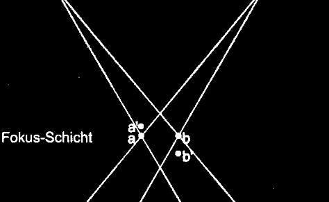
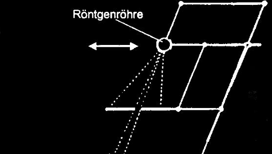
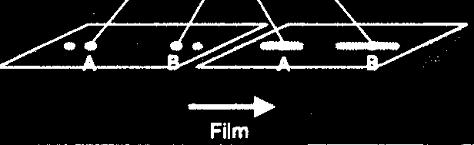

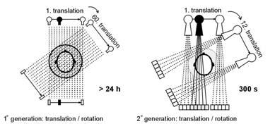
7 Planar -tomography Scanner generations Seite 7 7


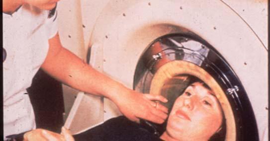

8 Scanner 1. und 2. Generation EMI-Scanner (1. Generation) Seite 8 8


9 2. Generation scanner Multi-Detector- Translation-rotation Seite 9 9

 continuos rotation of")
10 3. Generation Scanners Realisation of short acquisition time Problem: high voltage supply Solution: sliding ring technology (1987) continuos rotation of gantry Seite 10 10

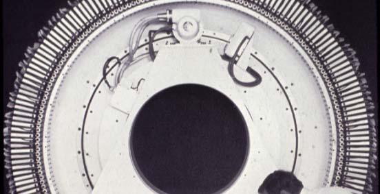

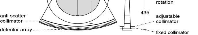
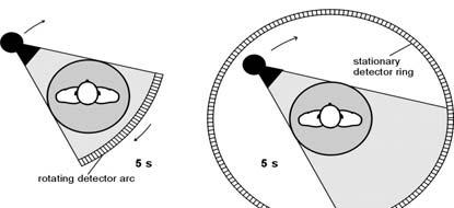
11 Scanner 3. und 4. generation Scanner setup - 3rd generation Seite 11 11


 Tube Detector")
12 First SIEMENS scanner in SIRETOM SIRETOM (in 1974) Acquisition time 7 min., image matrix 80x80 pixels, scan field 25 cm, spatial resolution 1,3 mm (4LP/cm) SIEMENS SOMATOM Plus 4 (1996) Tube Detector Seite 12 12
")

13 Progress in Image Quality SIRETOM (1974) SOMATOM Plus 4 UFC (1996) CT: Technical development - detectors Seite 13 13
 High quantum efficieny Longer image lag, slower response")
14 Detectors Xenon-ionisation chambers Good time response small dead time, small image lag Poor quantum efficiency Detectors Szintillation-detectors (Cs-Iodid, Cd-Wolframat) High quantum efficieny Longer image lag, slower response Seite 14 14
15 Detectors Today: Ceramic Szintillation- detectors High quantum efficiency Fast response, short image lag Combined advantages of previous detector types Time response of different detectors CT: Basic principle Seite 15 15

16 Image acquisition "1. Generation" CT-Scanner: Translation Rotation with pencilbeam x Seite 16 16

17 Seite 17 17
18 Raw data 1. Projection (Translationsscan) 160 measured values Time (Projections) Line-integrals Raw data 2. Projection (Translation scan after small rotation) 2 x 160 measured values Time (Projections) Line-integrals Seite 18 18


19 Raw data 180 Rotation Raw data 180 x 160 = Time (Projections) Line-integrals Image reconstruction Image Reconstruction Sinogram CT image Seite 19 19
Computer-Tomography II: Image reconstruction and applications
 Computer-Tomography II: Image reconstruction and applications Prof. Dr. U. Oelfke DKFZ Heidelberg Department of Medical Physics (E040) Im Neuenheimer Feld 280 69120 Heidelberg, Germany u.oelfke@dkfz.de
BME I5000: Biomedical Imaging
 1 Lucas Parra, CCNY BME I5000: Biomedical Imaging Lecture 4 Computed Tomography Lucas C. Parra, parra@ccny.cuny.edu some slides inspired by lecture notes of Andreas H. Hilscher at Columbia University.
Radiology. Marta Anguiano Millán. Departamento de Física Atómica, Molecular y Nuclear Facultad de Ciencias. Universidad de Granada
 Departamento de Física Atómica, Molecular y Nuclear Facultad de Ciencias. Universidad de Granada Overview Introduction Overview Introduction Tecniques of imaging in Overview Introduction Tecniques of imaging
Corso di laurea in Fisica A.A Fisica Medica 4 TC
 Corso di laurea in Fisica A.A. 2007-2008 Fisica Medica 4 TC Computed Tomography Principles 1. Projection measurement 2. Scanner systems 3. Scanning modes Basic Tomographic Principle The internal structure
Shadow casting. What is the problem? Cone Beam Computed Tomography THE OBJECTIVES OF DIAGNOSTIC IMAGING IDEAL DIAGNOSTIC IMAGING STUDY LIMITATIONS
 Cone Beam Computed Tomography THE OBJECTIVES OF DIAGNOSTIC IMAGING Reveal pathology Reveal the anatomic truth Steven R. Singer, DDS srs2@columbia.edu IDEAL DIAGNOSTIC IMAGING STUDY Provides desired diagnostic
CT Basics Principles of Spiral CT Dose. Always Thinking Ahead.
 1 CT Basics Principles of Spiral CT Dose 2 Who invented CT? 1963 - Alan Cormack developed a mathematical method of reconstructing images from x-ray projections Sir Godfrey Hounsfield worked for the Central
Moscow-Bavarian Joint Advanced Student School 2006 / Medical Imaging Principles of Computerized Tomographic Imaging and Cone-Beam Reconstruction
 Line Integrals Line integrals represent the integral of some parameter of the object along the line (e.g. attenuation of x-rays) Object: f(x,y) Line: x cosθ + y sinθ = t Line integral / Radon transform:
Image Acquisition Systems
 Image Acquisition Systems Goals and Terminology Conventional Radiography Axial Tomography Computer Axial Tomography (CAT) Magnetic Resonance Imaging (MRI) PET, SPECT Ultrasound Microscopy Imaging ITCS
MEDICAL IMAGING 2nd Part Computed Tomography
 MEDICAL IMAGING 2nd Part Computed Tomography Introduction 2 In the last 30 years X-ray Computed Tomography development produced a great change in the role of diagnostic imaging in medicine. In convetional
Computed tomography - outline
 Computed tomography - outline Computed Tomography Systems Jørgen Arendt Jensen and Mikael Jensen (DTU Nutech) October 6, 216 Center for Fast Ultrasound Imaging, Build 349 Department of Electrical Engineering
Introduction to Biomedical Imaging
 Alejandro Frangi, PhD Computational Imaging Lab Department of Information & Communication Technology Pompeu Fabra University www.cilab.upf.edu X-ray Projection Imaging Computed Tomography Digital X-ray
Ch. 4 Physical Principles of CT
 Ch. 4 Physical Principles of CT CLRS 408: Intro to CT Department of Radiation Sciences Review: Why CT? Solution for radiography/tomography limitations Superimposition of structures Distinguishing between
Radon Transform and Filtered Backprojection
 Radon Transform and Filtered Backprojection Jørgen Arendt Jensen October 13, 2016 Center for Fast Ultrasound Imaging, Build 349 Department of Electrical Engineering Center for Fast Ultrasound Imaging Department
Spiral CT. Protocol Optimization & Quality Assurance. Ge Wang, Ph.D. Department of Radiology University of Iowa Iowa City, Iowa 52242, USA
 Spiral CT Protocol Optimization & Quality Assurance Ge Wang, Ph.D. Department of Radiology University of Iowa Iowa City, Iowa 52242, USA Spiral CT Protocol Optimization & Quality Assurance Protocol optimization
MEDICAL IMAGING 2nd Part Computed Tomography
 MEDICAL IMAGING 2nd Part Computed Tomography Introduction 2 In the last 30 years X-ray Computed Tomography development produced a great change in the role of diagnostic imaging in medicine. In convetional
CT: Physics Principles & Equipment Design
 CT: Physics Principles & Equipment Design James Kofler, Ph.D Radiology Mayo Clinic Rochester, MN June 27, 2012 Disclosures Nothing to disclose Learning Objectives Understand fundamental concepts of - CT
Computed Tomography. Principles of Medical Imaging. Contents. Prof. Dr. Philippe Cattin. MIAC, University of Basel. Sep 26th/Oct 3rd, 2016
 Computed Tomography Principles of Medical Imaging Prof. Dr. Philippe Cattin MIAC, University of Basel Contents Abstract 1 Computed Tomography Basics Introduction Computed Tomography Hounsfield's CT Prototype
CLASS HOURS: 4 CREDIT HOURS: 4 LABORATORY HOURS: 0
 Revised 10/10 COURSE SYLLABUS TM 220 COMPUTED TOMOGRAPHY PHYSICS CLASS HOURS: 4 CREDIT HOURS: 4 LABORATORY HOURS: 0 CATALOG COURSE DESCRIPTION: This course is one of a three course set in whole body Computed
Tomographic Reconstruction
 Tomographic Reconstruction 3D Image Processing Torsten Möller Reading Gonzales + Woods, Chapter 5.11 2 Overview Physics History Reconstruction basic idea Radon transform Fourier-Slice theorem (Parallel-beam)
TEP Hounsfield units. Related topics Attenuation coefficient, Hounsfield units
 Hounsfield units TEP Related topics Attenuation coefficient, Hounsfield units Principle Depending on the type of CT scanner and the settings, the result of a CT scan of the same material can be different
Physical bases of X-ray diagnostics
 Physical bases of X-ray diagnostics Dr. István Voszka Possibilities of X-ray production (X-ray is produced, when charged particles of high velocity are stopped) X-ray tube: Relatively low accelerating
Micro-CT Methodology Hasan Alsaid, PhD
 Micro-CT Methodology Hasan Alsaid, PhD Preclinical & Translational Imaging LAS, PTS, GlaxoSmithKline 20 April 2015 Provide basic understanding of technical aspects of the micro-ct Statement: All procedures
Computed tomography (Item No.: P )
") Computed tomography (Item No.: P2550100) Curricular Relevance Area of Expertise: Biology Education Level: University Topic: Modern Imaging Methods Subtopic: X-ray Imaging Experiment: Computed tomography
Computed tomography (Item No.: P2550100) Curricular Relevance Area of Expertise: Biology Education Level: University Topic: Modern Imaging Methods Subtopic: X-ray Imaging Experiment: Computed tomography
First CT Scanner. How it Works. Contemporary CT. Before and After CT. Computer Tomography: How It Works. Medical Imaging and Pattern Recognition
 Computer Tomography: How t Works Medical maging and Pattern Recognition Lecture 7 Computed Tomography Oleh Tretiak Only one plane is illuminated. Source-subject motion provides added information. 2 How
Computed tomography of simple objects. Related topics. Principle. Equipment TEP Beam hardening, artefacts, and algorithms
 Related topics Beam hardening, artefacts, and algorithms Principle The CT principle is demonstrated with the aid of simple objects. In the case of very simple targets, only a few images need to be taken
Digital Image Processing
 Digital Image Processing SPECIAL TOPICS CT IMAGES Hamid R. Rabiee Fall 2015 What is an image? 2 Are images only about visual concepts? We ve already seen that there are other kinds of image. In this lecture
Cardiac Dual Energy CT: Technique
 RSNA 2013, VSCA51-01, Chicago, Dec. 5, 2013 Cardiac Radiology Series Cardiac Dual Energy CT: Technique Willi A. Kalender, Ph.D. Institute of Medical Physics University of Erlangen www.imp.uni-erlangen.de
Reduction of Metal Artifacts in Computed Tomographies for the Planning and Simulation of Radiation Therapy
 Reduction of Metal Artifacts in Computed Tomographies for the Planning and Simulation of Radiation Therapy T. Rohlfing a, D. Zerfowski b, J. Beier a, P. Wust a, N. Hosten a, R. Felix a a Department of
Fundamentals of CT imaging
 SECTION 1 Fundamentals of CT imaging I History In the early 1970s Sir Godfrey Hounsfield s research produced the first clinically useful CT scans. Original scanners took approximately 6 minutes to perform
MEDICAL EQUIPMENT: COMPUTED TOMOGRAPHY. Prof. Yasser Mostafa Kadah
 MEDICAL EQUIPMENT: COMPUTED TOMOGRAPHY Prof. Yasser Mostafa Kadah www.k-space.org Recommended Textbook X-Ray Computed Tomography in Biomedical Engineering, by Robert Cierniak, Springer, 211 Computed Tomography
DUE to beam polychromacity in CT and the energy dependence
 1 Empirical Water Precorrection for Cone-Beam Computed Tomography Katia Sourbelle, Marc Kachelrieß, Member, IEEE, and Willi A. Kalender Abstract We propose an algorithm to correct for the cupping artifact
8/7/2017. Disclosures. MECT Systems Overview and Quantitative Opportunities. Overview. Computed Tomography (CT) CT Numbers. Polyenergetic Acquisition
 CT Numbers. Polyenergetic Acquisition") Quantitative Multi-Energy Computed Tomography: Imaging and Therapy Advancements Disclosures MECT Systems Overview and Quantitative Opportunities The speaker receives research funding from GE Healthcare
Quantitative Multi-Energy Computed Tomography: Imaging and Therapy Advancements Disclosures MECT Systems Overview and Quantitative Opportunities The speaker receives research funding from GE Healthcare
Empirical cupping correction: A first-order raw data precorrection for cone-beam computed tomography
 Empirical cupping correction: A first-order raw data precorrection for cone-beam computed tomography Marc Kachelrieß, a Katia Sourbelle, and Willi A. Kalender Institute of Medical Physics, University of
Joint ICTP-TWAS Workshop on Portable X-ray Analytical Instruments for Cultural Heritage. 29 April - 3 May, 2013
 2455-5 Joint ICTP-TWAS Workshop on Portable X-ray Analytical Instruments for Cultural Heritage 29 April - 3 May, 2013 Lecture NoteBasic principles of X-ray Computed Tomography Diego Dreossi Elettra, Trieste
Motion Compensation from Short-Scan Data in Cardiac CT
 Motion Compensation from Short-Scan Data in Cardiac CT Juliane Hahn 1,2, Thomas Allmendinger 1, Herbert Bruder 1, and Marc Kachelrieß 2 1 Siemens Healthcare GmbH, Forchheim, Germany 2 German Cancer Research
Computational Medical Imaging Analysis
 Computational Medical Imaging Analysis Chapter 1: Introduction to Imaging Science Jun Zhang Laboratory for Computational Medical Imaging & Data Analysis Department of Computer Science University of Kentucky
Medical Image Processing: Image Reconstruction and 3D Renderings
 Medical Image Processing: Image Reconstruction and 3D Renderings 김보형 서울대학교컴퓨터공학부 Computer Graphics and Image Processing Lab. 2011. 3. 23 1 Computer Graphics & Image Processing Computer Graphics : Create,
Medical Imaging Projects
 NSF REU MedIX Summer 2006 Medical Imaging Projects Daniela Stan Raicu, PhD http://facweb.cs.depaul.edu/research draicu@cs.depaul.edu Outline Medical Informatics Imaging Modalities Computed Tomography Medical
Biomedical Imaging. Computed Tomography. Patrícia Figueiredo IST
 Biomedical Imaging Computed Tomography Patrícia Figueiredo IST 2013-2014 Overview Basic principles X ray attenuation projection Slice selection and line projections Projection reconstruction Instrumentation
Computed Tomography January 2002 KTH A.K.
 CT A.K. Computed Tomography January KTH 1 Introduction X-ray was discovered (accidentally) by a German physicist, Wilhelm Konrad Röntgen in 1895. A few years later, in 191, Röntgen was awarded the first
Optimization of CT Simulation Imaging. Ingrid Reiser Dept. of Radiology The University of Chicago
 Optimization of CT Simulation Imaging Ingrid Reiser Dept. of Radiology The University of Chicago Optimization of CT imaging Goal: Achieve image quality that allows to perform the task at hand (diagnostic
X-ray Tomography. A superficial introduction, but sufficient enough to get us started in surgical navigation.
 X-ray Tomography A superficial introduction, but sufficient enough to get us started in surgical navigation. X-ray absorption in homogeneous tissue I o I o / I d m = density I I=I o e -kdm k= constant
Diagnostic imaging techniques. Krasznai Zoltán. University of Debrecen Medical and Health Science Centre Department of Biophysics and Cell Biology
 Diagnostic imaging techniques Krasznai Zoltán University of Debrecen Medical and Health Science Centre Department of Biophysics and Cell Biology 1. Computer tomography (CT) 2. Gamma camera 3. Single Photon
CT vs. VolumeScope: image quality and dose comparison
 CT vs. VolumeScope: image quality and dose comparison V.N. Vasiliev *a, A.F. Gamaliy **b, M.Yu. Zaytsev b, K.V. Zaytseva ***b a Russian Sci. Center of Roentgenology & Radiology, 86, Profsoyuznaya, Moscow,
Annexure XII SPECIFICATIONS FOR A NEW STATE OF ART 16 SLICE ALL PURPOSE C. T. SCANNER
 Annexure XII SPECIFICATIONS FOR A NEW STATE OF ART 16 SLICE ALL PURPOSE C. T. SCANNER A) Scanner Design X-Ray generator and tube: 1. Scanner: Whole body spiral CT scanner (16 slices) of latest technology.
Enhanced material contrast by dual-energy microct imaging
 Enhanced material contrast by dual-energy microct imaging Method note Page 1 of 12 2 Method note: Dual-energy microct analysis 1. Introduction 1.1. The basis for dual energy imaging Micro-computed tomography
Fits you like no other
 Fits you like no other BrightView X and XCT specifications The new BrightView X system is a fully featured variableangle camera that is field-upgradeable to BrightView XCT without any increase in room
Computed Tomography. Principles, Design, Artifacts, and Recent Advances. Jiang Hsieh THIRD EDITION. SPIE PRESS Bellingham, Washington USA
 Computed Tomography Principles, Design, Artifacts, and Recent Advances THIRD EDITION Jiang Hsieh SPIE PRESS Bellingham, Washington USA Table of Contents Preface Nomenclature and Abbreviations xi xv 1 Introduction
CT Reconstruction with Good-Orientation and Layer Separation for Multilayer Objects
 17th World Conference on Nondestructive Testing, 25-28 Oct 2008, Shanghai, China CT Reconstruction with Good-Orientation and Layer Separation for Multilayer Objects Tong LIU 1, Brian Stephan WONG 2, Tai
Fits you like no other
 Fits you like no other Philips BrightView X and XCT specifications The new BrightView X system is a fully featured variableangle camera that is field-upgradeable to BrightView XCT without any increase
Digital Image Processing
 Digital Image Processing Image Restoration and Reconstruction (Image Reconstruction from Projections) Christophoros Nikou cnikou@cs.uoi.gr University of Ioannina - Department of Computer Science and Engineering
Some reference material
 Some reference material Physics reference book on medical imaging: A good one is The Essential Physics of Medical Imaging, 3 rd Ed. by Bushberg et al. ($170! new). However, there are several similar books
1970 Projection Radiography 2D projection of 3D anatomy
 Speakers: L. N. Rothenberg, Ph.D. Computed Tomography G. D. Clarke, Ph.D. Magnetic Resonance Imaging J. A. Zagzebski, Ph.D. Ultrasonic Imaging August 1, 2012 Lawrence N. Rothenberg, Ph.D. Keith S. Pentlow,
A closer look at CT scanning
 Vet Times The website for the veterinary profession https://www.vettimes.co.uk A closer look at CT scanning Author : Charissa Lee, Natalie Webster Categories : General, Vets Date : April 3, 2017 A basic
Spiral ASSR Std p = 1.0. Spiral EPBP Std. 256 slices (0/300) Kachelrieß et al., Med. Phys. 31(6): , 2004
 Kachelrieß et al., Med. Phys. 31(6): , 2004") Spiral ASSR Std p = 1.0 Spiral EPBP Std p = 1.0 Kachelrieß et al., Med. Phys. 31(6): 1623-1641, 2004 256 slices (0/300) Advantages of Cone-Beam Spiral CT Image quality nearly independent of pitch Increase
Spiral ASSR Std p = 1.0 Spiral EPBP Std p = 1.0 Kachelrieß et al., Med. Phys. 31(6): 1623-1641, 2004 256 slices (0/300) Advantages of Cone-Beam Spiral CT Image quality nearly independent of pitch Increase
Design and performance characteristics of a Cone Beam CT system for Leksell Gamma Knife Icon
 Design and performance characteristics of a Cone Beam CT system for Leksell Gamma Knife Icon WHITE PAPER Introduction Introducing an image guidance system based on Cone Beam CT (CBCT) and a mask immobilization
The Near Future in Cardiac CT Image Reconstruction
 SCCT 2010 The Near Future in Cardiac CT Image Reconstruction Marc Kachelrieß Institute of Medical Physics (IMP) Friedrich-Alexander Alexander-University Erlangen-Nürnberg rnberg www.imp.uni-erlangen.de
[PDR03] RECOMMENDED CT-SCAN PROTOCOLS
![[PDR03] RECOMMENDED CT-SCAN PROTOCOLS](/thumbs/72/66454100.jpg "[PDR03] RECOMMENDED CT-SCAN PROTOCOLS") SURGICAL & PROSTHETIC DESIGN [PDR03] RECOMMENDED CT-SCAN PROTOCOLS WORK-INSTRUCTIONS DOCUMENT (CUSTOMER) RECOMMENDED CT-SCAN PROTOCOLS [PDR03_V1]: LIVE 1 PRESCRIBING SURGEONS Patient-specific implants,
SURGICAL & PROSTHETIC DESIGN [PDR03] RECOMMENDED CT-SCAN PROTOCOLS WORK-INSTRUCTIONS DOCUMENT (CUSTOMER) RECOMMENDED CT-SCAN PROTOCOLS [PDR03_V1]: LIVE 1 PRESCRIBING SURGEONS Patient-specific implants,
Biophysical Techniques (BPHS 4090/PHYS 5800)
") Biophysical Techniques (BPHS 4090/PHYS 5800) Instructors: Prof. Christopher Bergevin (cberge@yorku.ca) Schedule: MWF 1:30-2:30 (CB 122) Website: http://www.yorku.ca/cberge/4090w2017.html York University
Biophysical Techniques (BPHS 4090/PHYS 5800) Instructors: Prof. Christopher Bergevin (cberge@yorku.ca) Schedule: MWF 1:30-2:30 (CB 122) Website: http://www.yorku.ca/cberge/4090w2017.html York University
Material for Chapter 6: Basic Principles of Tomography M I A Integral Equations in Visual Computing Material
 Material for Chapter : Integral Equations in Visual Computing Material Basic Principles of Tomography c 00 Bernhard Burgeth 0 Source: Images Figure : Radon Transform: ttenuation http://en.wikimedia.org/wiki/image:radon_transform.png
Image Reconstruction from Projection
 Image Reconstruction from Projection Reconstruct an image from a series of projections X-ray computed tomography (CT) Computed tomography is a medical imaging method employing tomography where digital
Medical Imaging BMEN Spring 2016
 Name Medical Imaging BMEN 420-501 Spring 2016 Homework #4 and Nuclear Medicine Notes All questions are from the introductory Powerpoint (based on Chapter 7) and text Medical Imaging Signals and Systems,
3/27/2012 WHY SPECT / CT? SPECT / CT Basic Principles. Advantages of SPECT. Advantages of CT. Dr John C. Dickson, Principal Physicist UCLH
 3/27/212 Advantages of SPECT SPECT / CT Basic Principles Dr John C. Dickson, Principal Physicist UCLH Institute of Nuclear Medicine, University College London Hospitals and University College London john.dickson@uclh.nhs.uk
CT NOISE POWER SPECTRUM FOR FILTERED BACKPROJECTION AND ITERATIVE RECONSTRUCTION
 CT NOISE POWER SPECTRUM FOR FILTERED BACKPROJECTION AND ITERATIVE RECONSTRUCTION Frank Dong, PhD, DABR Diagnostic Physicist, Imaging Institute Cleveland Clinic Foundation and Associate Professor of Radiology
MEDICAL IMAGE ANALYSIS
 SECOND EDITION MEDICAL IMAGE ANALYSIS ATAM P. DHAWAN g, A B IEEE Engineering in Medicine and Biology Society, Sponsor IEEE Press Series in Biomedical Engineering Metin Akay, Series Editor +IEEE IEEE PRESS
Continuous and Discrete Image Reconstruction
 25 th SSIP Summer School on Image Processing 17 July 2017, Novi Sad, Serbia Continuous and Discrete Image Reconstruction Péter Balázs Department of Image Processing and Computer Graphics University of
Contrast Enhancement with Dual Energy CT for the Assessment of Atherosclerosis
 Contrast Enhancement with Dual Energy CT for the Assessment of Atherosclerosis Stefan C. Saur 1, Hatem Alkadhi 2, Luca Regazzoni 1, Simon Eugster 1, Gábor Székely 1, Philippe Cattin 1,3 1 Computer Vision
Implementation and evaluation of a fully 3D OS-MLEM reconstruction algorithm accounting for the PSF of the PET imaging system
 Implementation and evaluation of a fully 3D OS-MLEM reconstruction algorithm accounting for the PSF of the PET imaging system 3 rd October 2008 11 th Topical Seminar on Innovative Particle and Radiation
GPR IMAGING USING COMPRESSED MEASUREMENTS
 GPR IMAGING USING COMPRESSED MEASUREMENTS A presentation by Prof. James H. McClellan School of Electrical and Computer Engineering Georgia Institute of Technology 26-Feb-09 Acknowledgements Dr. Ali Cafer
COMPARATIVE STUDIES OF DIFFERENT SYSTEM MODELS FOR ITERATIVE CT IMAGE RECONSTRUCTION
 COMPARATIVE STUDIES OF DIFFERENT SYSTEM MODELS FOR ITERATIVE CT IMAGE RECONSTRUCTION BY CHUANG MIAO A Thesis Submitted to the Graduate Faculty of WAKE FOREST UNIVERSITY GRADUATE SCHOOL OF ARTS AND SCIENCES
Deep Scatter Estimation (DSE): Feasibility of using a Deep Convolutional Neural Network for Real-Time X-Ray Scatter Prediction in Cone-Beam CT
: Feasibility of using a Deep Convolutional Neural Network for Real-Time X-Ray Scatter Prediction in Cone-Beam CT") Deep Scatter Estimation (DSE): Feasibility of using a Deep Convolutional Neural Network for Real-Time X-Ray Scatter Prediction in Cone-Beam CT Joscha Maier 1,2, Yannick Berker 1, Stefan Sawall 1,2 and
Deep Scatter Estimation (DSE): Feasibility of using a Deep Convolutional Neural Network for Real-Time X-Ray Scatter Prediction in Cone-Beam CT Joscha Maier 1,2, Yannick Berker 1, Stefan Sawall 1,2 and
Reconstruction Methods for Coplanar Translational Laminography Applications
 Reconstruction Methods for Coplanar Translational Laminography Applications U. EWERT, K.-U. THIESSENHUSEN, A. DERESCH, C. BELLON, S. HOHENDORF, S. KOLKOORI, N. WROBEL, B. REDMER, M. TSCHAIKNER, BAM, Berlin
The VesselGlyph: Focus & Context Visualization in CT-Angiography
 The VesselGlyph: Focus & Context Visualization in CT-Angiography Matúš Straka M. Šrámek, A. La Cruz E. Gröller, D. Fleischmann Contents Motivation:» Why again a new visualization method for vessel data?
Medical Image Analysis, Assignment 3
 Centre for Mathematical Sciences, Mathematics, LTH, December 2014 Medical Image Analysis, Assignment 3 1 Introduction The purpose of this assignment is to give hands-on experience with handling DICOM images
Trans-skull Imaging System by Ultrasonic Array Probe
 Proceedings of the 9 IEEE International Conference on Systems, Man, and Cybernetics San Antonio, TX, USA - October 9 Trans-skull Imaging System by Ultrasonic Array Probe Genta Hiramatsu, Yuichiro Ikeda,
Background. Outline. Radiographic Tomosynthesis: Image Quality and Artifacts Reduction 1 / GE /
 Radiographic Tomosynthesis: Image Quality and Artifacts Reduction Baojun Li, Ph.D Department of Radiology Boston University Medical Center 2012 AAPM Annual Meeting Background Linear Trajectory Tomosynthesis
University of Lübeck, Medical Laser Center Lübeck GmbH Optical Coherence Tomography
 University of Lübeck, Medical Laser Center Lübeck GmbH Optical Coherence Tomography. Theory Dr. Gereon Hüttmann / 009 What is OCT? ( for the MD ) Lichtquelle Probe Detektor Display OCT is Ultrasound with
C a t p h a n / T h e P h a n t o m L a b o r a t o r y
 C a t p h a n 5 0 0 / 6 0 0 T h e P h a n t o m L a b o r a t o r y C a t p h a n 5 0 0 / 6 0 0 Internationally recognized for measuring the maximum obtainable performance of axial, spiral and multi-slice
DUAL energy CT (DECT) is a modality where one and. Empirical Dual Energy Calibration (EDEC) for Cone-Beam Computed Tomography
 is a modality where one and. Empirical Dual Energy Calibration (EDEC) for Cone-Beam Computed Tomography") Empirical Dual Energy Calibration (EDEC) for Cone-Beam Computed Tomography Marc Kachelrieß, Member, IEEE, Timo Berkus, Philip Stenner, Willi A. Kalender Abstract Material selective imaging using dual energy
Empirical Dual Energy Calibration (EDEC) for Cone-Beam Computed Tomography Marc Kachelrieß, Member, IEEE, Timo Berkus, Philip Stenner, Willi A. Kalender Abstract Material selective imaging using dual energy
TomoTherapy Related Projects. An image guidance alternative on Tomo Low dose MVCT reconstruction Patient Quality Assurance using Sinogram
 TomoTherapy Related Projects An image guidance alternative on Tomo Low dose MVCT reconstruction Patient Quality Assurance using Sinogram Development of A Novel Image Guidance Alternative for Patient Localization
icatvision Quick Reference
 icatvision Quick Reference Navigating the i-cat Interface This guide shows how to: View reconstructed images Use main features and tools to optimize an image. REMINDER Images are displayed as if you are
Introduction to Emission Tomography
 Introduction to Emission Tomography Gamma Camera Planar Imaging Robert Miyaoka, PhD University of Washington Department of Radiology rmiyaoka@u.washington.edu Gamma Camera: - collimator - detector (crystal
Scatter Correction Methods in Dimensional CT
 Scatter Correction Methods in Dimensional CT Matthias Baer 1,2, Michael Hammer 3, Michael Knaup 1, Ingomar Schmidt 3, Ralf Christoph 3, Marc Kachelrieß 2 1 Institute of Medical Physics, Friedrich-Alexander-University
LAB DEMONSTRATION COMPUTED TOMOGRAPHY USING DESKCAT Lab Manual: 0
 LAB DEMONSTRATION COMPUTED TOMOGRAPHY USING DESKCAT Lab Manual: 0 Introduction This lab demonstration explores the physics and technology of Computed Tomography (CT) and guides the student and instructor
Low-Dose Dual-Energy CT for PET Attenuation Correction with Statistical Sinogram Restoration
 Low-Dose Dual-Energy CT for PET Attenuation Correction with Statistical Sinogram Restoration Joonki Noh, Jeffrey A. Fessler EECS Department, The University of Michigan Paul E. Kinahan Radiology Department,
Loma Linda University Medical Center Dept. of Radiation Medicine
 Loma Linda University Medical Center Dept. of Radiation Medicine and Northern Illinois University Dept. of Physics and Dept. of Computer Science Presented by George Coutrakon, PhD NIU Physics Dept. Collaborators
Principles of Computerized Tomographic Imaging
 Principles of Computerized Tomographic Imaging Parallel CT, Fanbeam CT, Helical CT and Multislice CT Marjolein van der Glas August 29, 2000 Abstract The total attenuation suffered by one beam of x-rays
Computed Tomography (CT) Scan Image Reconstruction on the SRC-7 David Pointer SRC Computers, Inc.
 Scan Image Reconstruction on the SRC-7 David Pointer SRC Computers, Inc.") Computed Tomography (CT) Scan Image Reconstruction on the SRC-7 David Pointer SRC Computers, Inc. CT Image Reconstruction Herman Head Sinogram Herman Head Reconstruction CT Image Reconstruction for all
Computed Tomography (CT) Scan Image Reconstruction on the SRC-7 David Pointer SRC Computers, Inc. CT Image Reconstruction Herman Head Sinogram Herman Head Reconstruction CT Image Reconstruction for all
Clarify behavioral factor of X-ray scatter in MDCT scanners based on evaluation data by a wide variety of MDCT scanners
 Clarify behavioral factor of X-ray scatter in MDCT scanners based on evaluation data by a wide variety of MDCT scanners Poster No.: C-1748 Congress: ECR 2012 Type: Educational Exhibit Authors: S. Miyashita
Brilliance CT Big Bore.
 1 2 2 There are two methods of RCCT acquisition in widespread clinical use: cine axial and helical. In RCCT with cine axial acquisition, repeat CT images are taken each couch position while recording respiration.
Medical Imaging and Virtual Medicine
 Medical Imaging and Virtual Medicine Lecture 1: Volume data, Sampling theorem, Artifacts, Filtering Dirk Bartz, Visual Computing for Medicine bartz@gris.uni-tuebingen.de University of Tübingen 1 University
Evaluation of Spectrum Mismatching using Spectrum Binning Approach for Statistical Polychromatic Reconstruction in CT
 Evaluation of Spectrum Mismatching using Spectrum Binning Approach for Statistical Polychromatic Reconstruction in CT Qiao Yang 1,4, Meng Wu 2, Andreas Maier 1,3,4, Joachim Hornegger 1,3,4, Rebecca Fahrig
SPECT QA and QC. Bruce McBride St. Vincent s Hospital Sydney.
 SPECT QA and QC Bruce McBride St. Vincent s Hospital Sydney. SPECT QA and QC What is needed? Why? How often? Who says? QA and QC in Nuclear Medicine QA - collective term for all the efforts made to produce
Modifications for P551 Fall 2014
 LAB DEMONSTRATION COMPUTED TOMOGRAPHY USING DESKCAT 1 Modifications for P551 Fall 2014 Introduction This lab demonstration explores the physics and technology of Computed Tomography (CT) and guides the
Computational Medical Imaging Analysis
 Computational Medical Imaging Analysis Chapter 2: Image Acquisition Systems Jun Zhang Laboratory for Computational Medical Imaging & Data Analysis Department of Computer Science University of Kentucky
FINDING THE TRUE EDGE IN CTA
 FINDING THE TRUE EDGE IN CTA by: John A. Rumberger, PhD, MD, FACC Your patient has chest pain. The Cardiac CT Angiography shows plaque in the LAD. You adjust the viewing window trying to evaluate the stenosis
DEVELOPMENT OF CONE BEAM TOMOGRAPHIC RECONSTRUCTION SOFTWARE MODULE
 Rajesh et al. : Proceedings of the National Seminar & Exhibition on Non-Destructive Evaluation DEVELOPMENT OF CONE BEAM TOMOGRAPHIC RECONSTRUCTION SOFTWARE MODULE Rajesh V Acharya, Umesh Kumar, Gursharan
PURE. ViSION Edition PET/CT. Patient Comfort Put First.
 PURE ViSION Edition PET/CT Patient Comfort Put First. 2 System features that put patient comfort and safety first. Oncology patients deserve the highest levels of safety and comfort during scans. Our Celesteion
X-ray tomography. X-ray tomography. Applications in Science. X-Rays. Notes. Notes. Notes. Notes
 X-ray tomography Important application of the Fast Fourier transform: X-ray tomography. Also referred to as CAT scan (Computerized Axial Tomography) X-ray tomography This has revolutionized medical diagnosis.
GE s Revolution CT MATLAB III: CT. Kathleen Chen March 20, 2018
 GE s Revolution CT MATLAB III: CT Kathleen Chen chens18@rpi.edu March 20, 2018 https://www.zmescience.com/medicine/inside-human-body-real-time-gifs-demo-power-ct-scan/ Reminders Make sure you have MATLAB