CT: Physics Principles & Equipment Design
|
|
|
- Shauna Bradley
- 6 years ago
- Views:
Transcription
1 CT: Physics Principles & Equipment Design James Kofler, Ph.D Radiology Mayo Clinic Rochester, MN June 27, 2012
2 Disclosures Nothing to disclose
3 Learning Objectives Understand fundamental concepts of - CT image acquisition - CT image formation Know basic CT scanner components and functions Implementation and implications of multi-slice CT But first


4 Jim s One Minute History of CT Johan Radon mathematically demonstrated reconstruction of an object from multiple projections Allan Cormack introduced the Fourier transform in the calculations. But, FALSE URBAN LEGEND says it all started with

5 Jim s One Minute History of CT

6 Jim s One Minute History of CT


")
7 Jim s One Minute History of CT Godfrey Hounsfield Nobel Award 1979 (with Cormack) Physiology or Medicine
:161-4.")
8 Jim s Mythical One Minute History of CT Maizlin ZV, Vos PM. Do we really need to thank the Beatles for the financing of the development of the computed tomography scanner? J Comput Assist Tomogr Mar-Apr;36(2): No evidence of Beatles CT connection EMI music did not fund EMI Medical Most of the funding came from British Department of Health and Social Security (DHSS)
9 CT Image Basics Image acquisition and reconstruction

10 System Introduction
11 System Introduction
: Total attenuation along 1 ray Projection (or view): Set of ray sums at a fixed angle N=N 0")
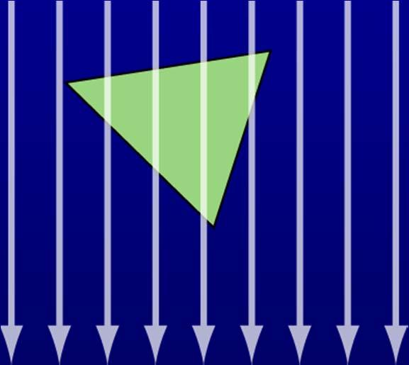
12 Goal Determine attenuation coefs within a section of the body Ray: One x-ray path Ray sum (or line integral): Total attenuation along 1 ray Projection (or view): Set of ray sums at a fixed angle N=N 0 e - t

13 Image Creation


14 Image Creation Call this 0 degrees Backprojection

15 45 Degrees


16 90 Degrees


17 135 Degrees


18 Image Creation 4 projections 16 projections 8 projections 32 projections

19 Why So Blurry? Inherent in back projection scheme Solution: Filter prior to back-projecting Called the Reconstruction Algorithm or Kernel
 100 views (filtered) 600 views")

20 Computer Generated 4 views (filtered) 20 views (filtered) 100 views (filtered) 600 views (filtered) 600 views (not filtered) Images courtesy of Lifeng Yu, Ph.D., Mayo Clinic, Rochester, Minnesota


21 Reconstruction Algorithm Affects frequency and noise in image Can reprocess (if have raw data) no need to re-scan Very Smooth Kernel Very Sharp Kernel


22 A Brief Sidetrack The elusive Sinogram


23 A Brief Sidetrack Sinogram Reconstructed Image Images courtesy of Shuai Leng, Ph.D., Mayo Clinic, Rochester, Minnesota


24 A Common Artifact Bad Detector
25 What We Are Actually Measuring Number of photons after traveling though thickness t N = N o e - t Starting number of photons Linear attenuation coefficient Know N (measured during scan) Know N o (from calibration) Know t (from matrix size and SFOV) Solve for μ


26 Linear Attenuation Coefficient Physical property of the material (z, ) Much more at

27 Linear Attenuation Coefficients Dependence on energy of photons


28 X-Ray Energy Spectra Adapted from Johnson, et. al., Eur Radiol (2007) 17: 1510Ð1517 and from R. Raupach,
 17: 1510Ð1517 and from R. Raupach, www.aapm.")
29 X-Ray Energy Spectra - Filtration HVL 8-10 mm Al (120 kv) Adapted from Johnson, et. al., Eur Radiol (2007) 17: 1510Ð1517 and from R. Raupach,


30 A Simple Example Lather, rinse, repeat solve for μ s Clinical CT (512 x 512 squares, 1000s of projections)
31 Reconstructions FFT techniques Iterative techniques More information AAPM Virtual Library (view on-line) Terry M. Peters. CT Reconstruction Presented at the 44th AAPM Annual Meeting. Montreal, Quebec, Canada. July 18, 2002.
32 Hounsfield Units CT # (HU) = ( material - water ) water X 1000 Water: 0 HU (by definition) Air: HU Soft Tissue: +30 to +60 HU Fat: -80 to -40 HU Bone: HU WW and WL control displayed range.

33 Hounsfield Units Assigned to pixel values -987 HU 142 HU 12 HU 1010 HU
34 (1 of 6)
35 Which is true regarding the reconstruction algorithm/kernel? 29% 1. Applied after back-projection. 2% 8% 62% 0% 2. Reduces ring artifact. 3. Used only during data acquisition. 4. Affects image noise. 5. Used for scanner calibration.
36 Which is true regarding the reconstruction algorithm/kernel? 1. Applied after back-projection. 2. Reduces ring artifact. 3. Used only during data acquisition. 4. Affects image noise. 5. Used for scanner calibration. Reference: Bushberg JT, Seibert JA, Leidholt EM, and Boone JM, The Essential Physics of Medical Imaging, 2 nd edition, Lippincott Wiliams & Wilkins, 2001, ISBN
37 (2 of 6)
38 Which is true regarding CT Numbers? 4% 1. CT # of water depends on the kv. 85% 3% 4% 3% 2. Independent of window/level setting. 3. Range from HU. 4. Equal to linear attenuation coefficient. 5. Scaling factors for back-projection.
39 Which is true regarding CT Numbers? 1. CT # of water depends on the kv. 2. Independent of window/level setting. 3. Range from HU. 4. Equal to linear attenuation coefficient. 5. Scaling factors for back-projection. Reference: Bushberg JT, Seibert JA, Leidholt EM, and Boone JM, The Essential Physics of Medical Imaging, 2 nd edition, Lippincott Wiliams & Wilkins, 2001, ISBN
40 AAPM 2012 Summer School on Medical Imaging using Ionizing Radiation CT Systems
41 CT System Gantry X-ray Tube High Voltage Generator Bow-tie & Flat Filter Pre-Patient Collimation Patient Post-Patient Collimation Operational Control Computer Operator Console Detectors Data Acquisition System (DAS) Image Generation
42 Tube and Generator Demanding on X-ray tube - >500 max ma - Scan times can be 30 sec or more - Heat capacities up to 30 MHU - Water or oil-cooled Generator kw - kv settings: (discrete steps)

43 Bowtie and Flat Filter X-ray source Patient Flat Filter Uniform (AL or Cu) Removes soft x-rays Bowtie Filter Compensates for different path lengths Reduces dose at periphery Different for different apps (body, head, peds, cardiac)

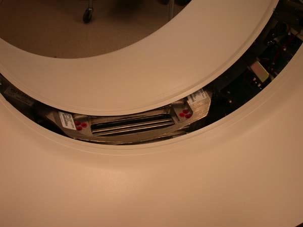
44 Bowtie Filter
45 Collimation Single-Slice Multi-Slice Total Coll. = Slice thickness No. detectors used x detector width


46 Detectors Electrical Signal Output Need very short afterglow Scintillating Material CdWO 4 Yttrium & gadolinium ceramics Others Reflective coating Photodiodes
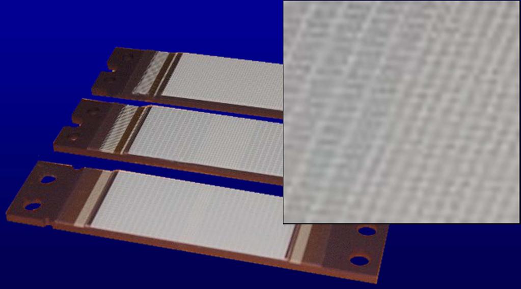

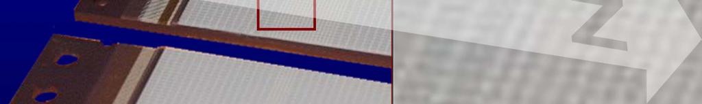
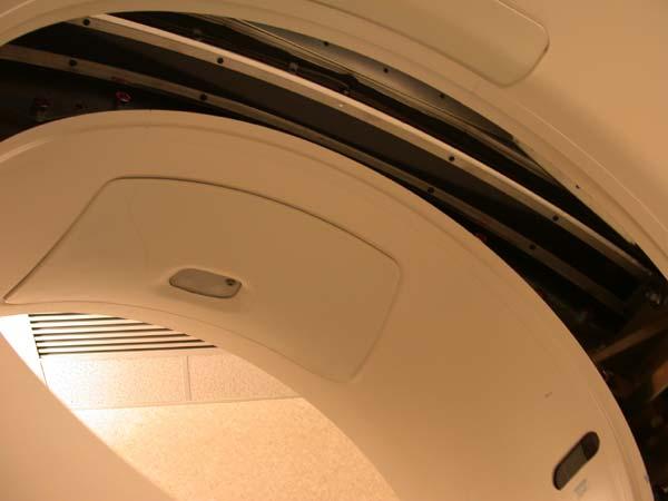
47 Detectors
48 CT System Gantry X-ray Tube High Voltage Generator Bow-tie & Flat Filter Pre-Patient Collimation Patient Post-Patient Collimation Operational Control Computer Operator Console Detectors Data Acquisition System (DAS) Image Generation
49 Data Acquisition System Amplifies detector signals Integrates detector signals over sampling period - Sample and hold, reset of each sample - Potential for cross-talk (imperfect reset) Analog to digital conversion Want output to be linear to input Output contains electronic and quantum noise - Electronic noise should be small fraction
50 (3 of 6)
51 Which is true regarding the bowtie filter? 67% 1. Reduces patient dose. 13% 14% 4% 3% 2. Uniformly removes low energy x-rays. 3. Applied prior to back-projection. 4. Between the patient and detectors. 5. Same filter used for all scans.
52 Which is true regarding the bowtie filter? 1. Reduces patient dose. 2. Uniformly removes low energy x-rays. 3. Applied prior to back-projection. 4. Between the patient and detectors. 5. Same filter used for all scans. Reference: Hsieh J, Computed Tomography: Principles, Design, Artifacts, and Recent Advances, SPIE The International Society of Optical Engineering, Bellingham, WA, 2003, ISBN
53 AAPM 2012 Summer School on Medical Imaging using Ionizing Radiation CT Implementations: Spiral Scanning

54 Some CT Milestones 1972: First CT scanner (EMI), heads only 1975: First body CT scanner 1989: Spiral CT introduced 1992: Dual Slice CT 1998: 4-Slice CT 2002: 8 and 16-Slice CT 2012: 64, 128, 320-Slice CT


55 Before Spiral Only axial (or sequential) One rotation, stop, increment table, repeat (still can do this) Needed to reverse rotation to unwind cables Data from each rotation in same plane Overlap Contiguous Gaps


56 Slip Rings Metal Contacts Transfer power to gantry Transfer data to and from gantry No need to unwind cables Allow for continuous table motion (spiral scanning) Wolbarst A B, Hendee W R Radiology 2006;238: by Radiological Society of North America

57 Slip rings Contacts are metal blocks (not bristles) Voltage Control signals/data Wolbarst A B, Hendee W R Radiology 2006;238: by Radiological Society of North America
58 Spiral Scanning Continuous acquisition Greatly reduced scan time Data not in same reconstruction plane New Term: Pitch Spiral Data Acquisition Path


59 Pitch Pitch < 1 Pitch = 1 Pitch > 1 The amount of overlap of spiral slices Pitch = Table index per rotation Beam width* *Single slice scanners = slice thickness *Multi-slice scanners = # images per rot n x image width

60 Beam Width Single-Slice Multi-Slice Total = Beam Width Slice thickness No. images per rot n x image width
61 Spiral Reconstruction Incomplete data in any plane Need to interpolate from nearest points Can use full 360 or 180 data Arbitrary reconstruction planes One data point in plane Reconstruction plane
62 (4 of 6)
63 What is the function of the slip ring? 1% 1. Reduces ring artifact. 2% 94% 3% 0% 2. Provides precise table movement. 3. Transfers power to the gantry. 4. Stabilizes unit during gantry rotation. 5. Determines scan field of view.
64 What is the function of the slip ring? 1. Reduces ring artifact. 2. Provides precise table movement. 3. Transfers power to the gantry. 4. Stabilizes unit during gantry rotation. 5. Determines scan field of view. Reference: Wolbarst AB, Hendee WR, Evolving and Experimental Technologies in Medical Imaging, Radiology 2006; 238:16-39.

65 AAPM 2012 Summer School on Medical Imaging using Ionizing Radiation CT Implementations: Multi-Slice CT
 GE QX/i")
66 Detector Differences Single Slice Multi-slice GE CT/i (single slice) GE QX/i (4-slice)


67 Multi-slice Detectors
 1.")
 1.")
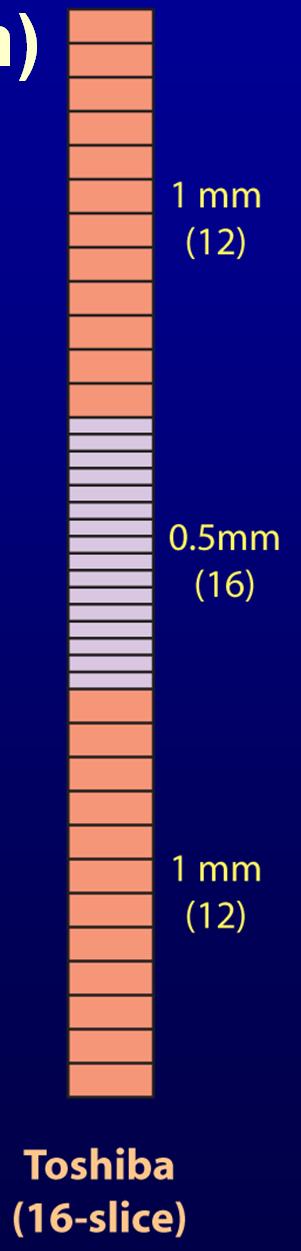
68 16 Data Channels (24 detector rows) 1.25 mm detectors (4) mm detectors (16) 1.25 mm detectors (4)
")


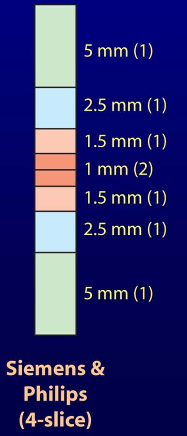
69 4 Data Channels (16 detector rows) # Slices Width Detectors 4 x 1.25 mm 4 x 2.50 mm 4 x 3.75 mm 4 x 5.00 mm Can recon down to width of one CHANNEL

70 16 Data Channels (24 detector rows) Number of slices x slice thickness 8 x x x x x x x x 5 2 x x x 5 1 x 1.25 * Doesn t consider recons, not all available in helical


71 Various Configurations (32 mm) (24 mm) (20 mm) (20 mm)
 360 0.5 mm (160 mm) 128 0.")
72 Various Configurations mm (38.4 mm) mm (40 mm) mm (160 mm) mm (80 mm)
73 Multi-Slice, Many Choices Not all configurations available under all circumstances. Depends on image thickness, table speed, pitch, axial or helical. Still many choices. Current trend appears to be to limit options.

74 Multi-Slice Advantages over Single Slice Single Slice Limited sampling for interpolation Scan width increases with pitch Image noise independent of pitch Multi-slice Much better sampling Scan width nearly independent of pitch Image noise depends on pitch
. Many possible recon thicknesses. Thinner images possible.")
75 Multi-Slice Advantages over Single Slice Single Slice Limited scan coverage (per rotation). One recon image thickness. Multi-slice More coverage per rotation (shorter scan times). Many possible recon thicknesses. Thinner images possible.
76 (5 of 6)
77 What is an advantage of single-slice scanners over multi-slice scanners? 3% 1. Shorter scan times. 2% 4% 5% 87% 2. Improved patient coverage. 3. Better sampling. 4. More image thickness options. 5. Easier reconstructions.
78 What is an advantage of single-slice scanners over multi-slice scanners? 1. Shorter scan times. 2. Improved patient coverage. 3. Better sampling. 4. More image thickness options 5. Easier reconstructions. Reference: Bushberg JT, Seibert JA, Leidholt EM, and Boone JM, The Essential Physics of Medical Imaging, 2 nd edition, Lippincott Wiliams & Wilkins, 2001, ISBN

79 (6 of 6)
80 What is true regarding multi-slice detectors? 4% 1. Data channels equals rows of elements. 2. Smallest data channel determines thinnest 90% image width. 1% 3% 2% 3. Most use high pressure xenon gas. 4. Pre-patient collimation determines image thickness. 5. Changing image thickness implies rescanning the patient.
81 What is an advantage of single-slice scanners over multi-slice scanners? 1. Data channels equals rows of elements. 2. Smallest data channel determines thinnest image width. 3. Most use high pressure xenon gas. 4. Pre-patient collimation determines image thickness. 5. Changing image thickness implies rescanning the patient. Reference: Hsieh J, Computed Tomography: Principles, Design, Artifacts, and Recent Advances, SPIE The International Society of Optical Engineering, Bellingham, WA, 2003, ISBN
82 Some Typical Specifications kv settings: (discrete steps) ma Range: ma Rotation times: sec Recon rate: 4 40 images/sec Bore opening: cm (80-90 cm wide bore ) Max. couch weight: kg ( lbs) Generator power rating: kw Much much more info at
83 Detailed Specifications & Measurements
84 Scanners in Motion


85 The Scanner Story (parts 1 and 2) Transfer from cine film of a documentary by EMI covering the development and early years of medical x-ray CT scanning

86 Thank You!
BME I5000: Biomedical Imaging
 1 Lucas Parra, CCNY BME I5000: Biomedical Imaging Lecture 4 Computed Tomography Lucas C. Parra, parra@ccny.cuny.edu some slides inspired by lecture notes of Andreas H. Hilscher at Columbia University.
CLASS HOURS: 4 CREDIT HOURS: 4 LABORATORY HOURS: 0
 Revised 10/10 COURSE SYLLABUS TM 220 COMPUTED TOMOGRAPHY PHYSICS CLASS HOURS: 4 CREDIT HOURS: 4 LABORATORY HOURS: 0 CATALOG COURSE DESCRIPTION: This course is one of a three course set in whole body Computed
MEDICAL IMAGING 2nd Part Computed Tomography
 MEDICAL IMAGING 2nd Part Computed Tomography Introduction 2 In the last 30 years X-ray Computed Tomography development produced a great change in the role of diagnostic imaging in medicine. In convetional
Corso di laurea in Fisica A.A Fisica Medica 4 TC
 Corso di laurea in Fisica A.A. 2007-2008 Fisica Medica 4 TC Computed Tomography Principles 1. Projection measurement 2. Scanner systems 3. Scanning modes Basic Tomographic Principle The internal structure
Computer-Tomography II: Image reconstruction and applications
 Computer-Tomography II: Image reconstruction and applications Prof. Dr. U. Oelfke DKFZ Heidelberg Department of Medical Physics (E040) Im Neuenheimer Feld 280 69120 Heidelberg, Germany u.oelfke@dkfz.de
Computed Tomography. Principles, Design, Artifacts, and Recent Advances. Jiang Hsieh THIRD EDITION. SPIE PRESS Bellingham, Washington USA
 Computed Tomography Principles, Design, Artifacts, and Recent Advances THIRD EDITION Jiang Hsieh SPIE PRESS Bellingham, Washington USA Table of Contents Preface Nomenclature and Abbreviations xi xv 1 Introduction
Multi-slice CT Image Reconstruction Jiang Hsieh, Ph.D.
 Multi-slice CT Image Reconstruction Jiang Hsieh, Ph.D. Applied Science Laboratory, GE Healthcare Technologies 1 Image Generation Reconstruction of images from projections. textbook reconstruction advanced
Computer-Tomography I: Principles, History, Technology
 Computer-Tomography I: Principles, History, Technology Prof. Dr. U. Oelfke DKFZ Heidelberg Department of Medical Physics (E040) Im Neuenheimer Feld 280 69120 Heidelberg, Germany u.oelfke@dkfz.de History
Optimization of CT Simulation Imaging. Ingrid Reiser Dept. of Radiology The University of Chicago
 Optimization of CT Simulation Imaging Ingrid Reiser Dept. of Radiology The University of Chicago Optimization of CT imaging Goal: Achieve image quality that allows to perform the task at hand (diagnostic
1970 Projection Radiography 2D projection of 3D anatomy
 Speakers: L. N. Rothenberg, Ph.D. Computed Tomography G. D. Clarke, Ph.D. Magnetic Resonance Imaging J. A. Zagzebski, Ph.D. Ultrasonic Imaging August 1, 2012 Lawrence N. Rothenberg, Ph.D. Keith S. Pentlow,
8/7/2017. Disclosures. MECT Systems Overview and Quantitative Opportunities. Overview. Computed Tomography (CT) CT Numbers. Polyenergetic Acquisition
 CT Numbers. Polyenergetic Acquisition") Quantitative Multi-Energy Computed Tomography: Imaging and Therapy Advancements Disclosures MECT Systems Overview and Quantitative Opportunities The speaker receives research funding from GE Healthcare
Quantitative Multi-Energy Computed Tomography: Imaging and Therapy Advancements Disclosures MECT Systems Overview and Quantitative Opportunities The speaker receives research funding from GE Healthcare
Radiology. Marta Anguiano Millán. Departamento de Física Atómica, Molecular y Nuclear Facultad de Ciencias. Universidad de Granada
 Departamento de Física Atómica, Molecular y Nuclear Facultad de Ciencias. Universidad de Granada Overview Introduction Overview Introduction Tecniques of imaging in Overview Introduction Tecniques of imaging
Biomedical Imaging. Computed Tomography. Patrícia Figueiredo IST
 Biomedical Imaging Computed Tomography Patrícia Figueiredo IST 2013-2014 Overview Basic principles X ray attenuation projection Slice selection and line projections Projection reconstruction Instrumentation
MEDICAL EQUIPMENT: COMPUTED TOMOGRAPHY. Prof. Yasser Mostafa Kadah
 MEDICAL EQUIPMENT: COMPUTED TOMOGRAPHY Prof. Yasser Mostafa Kadah www.k-space.org Recommended Textbook X-Ray Computed Tomography in Biomedical Engineering, by Robert Cierniak, Springer, 211 Computed Tomography
Introduction to Biomedical Imaging
 Alejandro Frangi, PhD Computational Imaging Lab Department of Information & Communication Technology Pompeu Fabra University www.cilab.upf.edu X-ray Projection Imaging Computed Tomography Digital X-ray
MEDICAL IMAGING 2nd Part Computed Tomography
 MEDICAL IMAGING 2nd Part Computed Tomography Introduction 2 In the last 30 years X-ray Computed Tomography development produced a great change in the role of diagnostic imaging in medicine. In convetional
3/27/2012 WHY SPECT / CT? SPECT / CT Basic Principles. Advantages of SPECT. Advantages of CT. Dr John C. Dickson, Principal Physicist UCLH
 3/27/212 Advantages of SPECT SPECT / CT Basic Principles Dr John C. Dickson, Principal Physicist UCLH Institute of Nuclear Medicine, University College London Hospitals and University College London john.dickson@uclh.nhs.uk
Shadow casting. What is the problem? Cone Beam Computed Tomography THE OBJECTIVES OF DIAGNOSTIC IMAGING IDEAL DIAGNOSTIC IMAGING STUDY LIMITATIONS
 Cone Beam Computed Tomography THE OBJECTIVES OF DIAGNOSTIC IMAGING Reveal pathology Reveal the anatomic truth Steven R. Singer, DDS srs2@columbia.edu IDEAL DIAGNOSTIC IMAGING STUDY Provides desired diagnostic
Some reference material
 Some reference material Physics reference book on medical imaging: A good one is The Essential Physics of Medical Imaging, 3 rd Ed. by Bushberg et al. ($170! new). However, there are several similar books
CT Basics Principles of Spiral CT Dose. Always Thinking Ahead.
 1 CT Basics Principles of Spiral CT Dose 2 Who invented CT? 1963 - Alan Cormack developed a mathematical method of reconstructing images from x-ray projections Sir Godfrey Hounsfield worked for the Central
Spiral CT. Protocol Optimization & Quality Assurance. Ge Wang, Ph.D. Department of Radiology University of Iowa Iowa City, Iowa 52242, USA
 Spiral CT Protocol Optimization & Quality Assurance Ge Wang, Ph.D. Department of Radiology University of Iowa Iowa City, Iowa 52242, USA Spiral CT Protocol Optimization & Quality Assurance Protocol optimization
Computed Tomography. Principles of Medical Imaging. Contents. Prof. Dr. Philippe Cattin. MIAC, University of Basel. Sep 26th/Oct 3rd, 2016
 Computed Tomography Principles of Medical Imaging Prof. Dr. Philippe Cattin MIAC, University of Basel Contents Abstract 1 Computed Tomography Basics Introduction Computed Tomography Hounsfield's CT Prototype
Image Acquisition Systems
 Image Acquisition Systems Goals and Terminology Conventional Radiography Axial Tomography Computer Axial Tomography (CAT) Magnetic Resonance Imaging (MRI) PET, SPECT Ultrasound Microscopy Imaging ITCS
Tomographic Reconstruction
 Tomographic Reconstruction 3D Image Processing Torsten Möller Reading Gonzales + Woods, Chapter 5.11 2 Overview Physics History Reconstruction basic idea Radon transform Fourier-Slice theorem (Parallel-beam)
Cardiac Dual Energy CT: Technique
 RSNA 2013, VSCA51-01, Chicago, Dec. 5, 2013 Cardiac Radiology Series Cardiac Dual Energy CT: Technique Willi A. Kalender, Ph.D. Institute of Medical Physics University of Erlangen www.imp.uni-erlangen.de
A closer look at CT scanning
 Vet Times The website for the veterinary profession https://www.vettimes.co.uk A closer look at CT scanning Author : Charissa Lee, Natalie Webster Categories : General, Vets Date : April 3, 2017 A basic
Computed tomography - outline
 Computed tomography - outline Computed Tomography Systems Jørgen Arendt Jensen and Mikael Jensen (DTU Nutech) October 6, 216 Center for Fast Ultrasound Imaging, Build 349 Department of Electrical Engineering
Joint ICTP-TWAS Workshop on Portable X-ray Analytical Instruments for Cultural Heritage. 29 April - 3 May, 2013
 2455-5 Joint ICTP-TWAS Workshop on Portable X-ray Analytical Instruments for Cultural Heritage 29 April - 3 May, 2013 Lecture NoteBasic principles of X-ray Computed Tomography Diego Dreossi Elettra, Trieste
TEP Hounsfield units. Related topics Attenuation coefficient, Hounsfield units
 Hounsfield units TEP Related topics Attenuation coefficient, Hounsfield units Principle Depending on the type of CT scanner and the settings, the result of a CT scan of the same material can be different
Acknowledgments and financial disclosure
 AAPM 2012 Annual Meeting Digital breast tomosynthesis: basic understanding of physics principles James T. Dobbins III, Ph.D., FAAPM Director, Medical Physics Graduate Program Ravin Advanced Imaging Laboratories
Digital Image Processing
 Digital Image Processing SPECIAL TOPICS CT IMAGES Hamid R. Rabiee Fall 2015 What is an image? 2 Are images only about visual concepts? We ve already seen that there are other kinds of image. In this lecture
Ch. 4 Physical Principles of CT
 Ch. 4 Physical Principles of CT CLRS 408: Intro to CT Department of Radiation Sciences Review: Why CT? Solution for radiography/tomography limitations Superimposition of structures Distinguishing between
CT NOISE POWER SPECTRUM FOR FILTERED BACKPROJECTION AND ITERATIVE RECONSTRUCTION
 CT NOISE POWER SPECTRUM FOR FILTERED BACKPROJECTION AND ITERATIVE RECONSTRUCTION Frank Dong, PhD, DABR Diagnostic Physicist, Imaging Institute Cleveland Clinic Foundation and Associate Professor of Radiology
Fundamentals of CT imaging
 SECTION 1 Fundamentals of CT imaging I History In the early 1970s Sir Godfrey Hounsfield s research produced the first clinically useful CT scans. Original scanners took approximately 6 minutes to perform
RADIOLOGY AND DIAGNOSTIC IMAGING
 Day 2 part 2 RADIOLOGY AND DIAGNOSTIC IMAGING Dr hab. Zbigniew Serafin, MD, PhD serafin@cm.umk.pl 2 3 4 5 CT technique CT technique 6 CT system Kanal K: RSNA/AAPM web module: CT Systems & CT Image Quality
Enhancement Image Quality of CT Using Single Slice Spiral Technique
 Enhancement Image Quality of CT Using Single Slice Spiral Technique Doaa. N. Al Sheack 1 and Dr.Mohammed H. Ali Al Hayani 2 1 2 Electronic and Communications Engineering Department College of Engineering,
First CT Scanner. How it Works. Contemporary CT. Before and After CT. Computer Tomography: How It Works. Medical Imaging and Pattern Recognition
 Computer Tomography: How t Works Medical maging and Pattern Recognition Lecture 7 Computed Tomography Oleh Tretiak Only one plane is illuminated. Source-subject motion provides added information. 2 How
Optimisation of Toshiba Aquilion ONE Volume Imaging
 Optimisation of Toshiba Aquilion ONE Volume Imaging Jane Edwards, RPRSG Royal Free London NHS Foundation Trust Dr Mufudzi Maviki, Plymouth Hospitals NHS Trust Background In 2011/12 Radiology at RFH was
Empirical cupping correction: A first-order raw data precorrection for cone-beam computed tomography
 Empirical cupping correction: A first-order raw data precorrection for cone-beam computed tomography Marc Kachelrieß, a Katia Sourbelle, and Willi A. Kalender Institute of Medical Physics, University of
Annexure XII SPECIFICATIONS FOR A NEW STATE OF ART 16 SLICE ALL PURPOSE C. T. SCANNER
 Annexure XII SPECIFICATIONS FOR A NEW STATE OF ART 16 SLICE ALL PURPOSE C. T. SCANNER A) Scanner Design X-Ray generator and tube: 1. Scanner: Whole body spiral CT scanner (16 slices) of latest technology.
Scatter Correction for Dual source Cone beam CT Using the Pre patient Grid. Yingxuan Chen. Graduate Program in Medical Physics Duke University
 Scatter Correction for Dual source Cone beam CT Using the Pre patient Grid by Yingxuan Chen Graduate Program in Medical Physics Duke University Date: Approved: Lei Ren, Supervisor Fang Fang Yin, Chair
Micro-CT Methodology Hasan Alsaid, PhD
 Micro-CT Methodology Hasan Alsaid, PhD Preclinical & Translational Imaging LAS, PTS, GlaxoSmithKline 20 April 2015 Provide basic understanding of technical aspects of the micro-ct Statement: All procedures
ML reconstruction for CT
 ML reconstruction for CT derivation of MLTR rigid motion correction resolution modeling polychromatic ML model dual energy ML model Bruno De Man, Katrien Van Slambrouck, Maarten Depypere, Frederik Maes,
8/1/2017. Current Technology: Energy Integrating Detectors. Principles, Pitfalls and Progress in Photon-Counting-Detector Technology.
 Photon Counting Detectors and Their Applications in Medical Imaging Principles, Pitfalls and Progress in Photon-Counting-Detector Technology Taly Gilat Schmidt, PhD Associate Professor Department of Biomedical
Moscow-Bavarian Joint Advanced Student School 2006 / Medical Imaging Principles of Computerized Tomographic Imaging and Cone-Beam Reconstruction
 Line Integrals Line integrals represent the integral of some parameter of the object along the line (e.g. attenuation of x-rays) Object: f(x,y) Line: x cosθ + y sinθ = t Line integral / Radon transform:
COMPARATIVE STUDIES OF DIFFERENT SYSTEM MODELS FOR ITERATIVE CT IMAGE RECONSTRUCTION
 COMPARATIVE STUDIES OF DIFFERENT SYSTEM MODELS FOR ITERATIVE CT IMAGE RECONSTRUCTION BY CHUANG MIAO A Thesis Submitted to the Graduate Faculty of WAKE FOREST UNIVERSITY GRADUATE SCHOOL OF ARTS AND SCIENCES
Physical bases of X-ray diagnostics
 Physical bases of X-ray diagnostics Dr. István Voszka Possibilities of X-ray production (X-ray is produced, when charged particles of high velocity are stopped) X-ray tube: Relatively low accelerating
Philips SPECT/CT Systems
 Philips SPECT/CT Systems Ling Shao, PhD Director, Imaging Physics & System Analysis Nuclear Medicine, Philips Healthcare June 14, 2008 *Presented SNM08 Categorical Seminar - Quantitative SPECT and PET
Artifacts in Spiral X-ray CT Scanners: Problems and Solutions
 Artifacts in Spiral X-ray CT Scanners: Problems and Solutions Mehran Yazdi, and Luc Beaulieu International Science Index, Electrical and Computer Engineering waset.org/publication/12966 Abstract Artifact
Equipment Specification
 MULTISLICE CT SCANNER ( 16 Slices ) Merk : Hitachi Japan Model : SUPRIA 5 MHU Price : Rp 6.512.360.215,27 No Equipment Specification 1 2 3 Scanner Gantry Object for scanning : Whole body including head
8/2/2016. Measures the degradation/distortion of the acquired image (relative to an ideal image) using a quantitative figure-of-merit
 using a quantitative figure-of-merit") Ke Li Assistant Professor Department of Medical Physics and Department of Radiology School of Medicine and Public Health, University of Wisconsin-Madison This work is partially supported by an NIH Grant
Ke Li Assistant Professor Department of Medical Physics and Department of Radiology School of Medicine and Public Health, University of Wisconsin-Madison This work is partially supported by an NIH Grant
ImPACT. Information Leaflet No. 1: CT Scanner Acceptance Testing
 ImPACT Information Leaflet No. 1: CT Scanner Acceptance Testing Version 1.02, 18/05/01 CONTENTS: 1. SCOPE OF LEAFLET 2. GENERAL PRINCIPLES OF ACCEPTANCE AND COMMISSIONING 2.1 PHANTOMS 2.2 EXPOSURE AND
Principles of Computerized Tomographic Imaging
 Principles of Computerized Tomographic Imaging Parallel CT, Fanbeam CT, Helical CT and Multislice CT Marjolein van der Glas August 29, 2000 Abstract The total attenuation suffered by one beam of x-rays
DUE to beam polychromacity in CT and the energy dependence
 1 Empirical Water Precorrection for Cone-Beam Computed Tomography Katia Sourbelle, Marc Kachelrieß, Member, IEEE, and Willi A. Kalender Abstract We propose an algorithm to correct for the cupping artifact
Metal Artifact Reduction CT Techniques. Tobias Dietrich University Hospital Balgrist University of Zurich Switzerland
 Metal Artifact Reduction CT Techniques R S S S Tobias Dietrich University Hospital Balgrist University of Zurich Switzerland N. 1 v o 4 1 0 2. Postoperative CT Metal Implants CT is accurate for assessment
Enhanced material contrast by dual-energy microct imaging
 Enhanced material contrast by dual-energy microct imaging Method note Page 1 of 12 2 Method note: Dual-energy microct analysis 1. Introduction 1.1. The basis for dual energy imaging Micro-computed tomography
Computed Tomography January 2002 KTH A.K.
 CT A.K. Computed Tomography January KTH 1 Introduction X-ray was discovered (accidentally) by a German physicist, Wilhelm Konrad Röntgen in 1895. A few years later, in 191, Röntgen was awarded the first
S. Guru Prasad, Ph.D., DABR
 PURPOSE S. Guru Prasad, Ph.D., DABR Director of Medical Physics IAEA Consultant NorthShore University Health System and University of Chicago, Pritzker School of Medicine Current TPS utilize more information
The Near Future in Cardiac CT Image Reconstruction
 SCCT 2010 The Near Future in Cardiac CT Image Reconstruction Marc Kachelrieß Institute of Medical Physics (IMP) Friedrich-Alexander Alexander-University Erlangen-Nürnberg rnberg www.imp.uni-erlangen.de
O-ARM IMAGING SYSTEM TECHNICAL SPECIFICATION GUIDE
 IMAGING SYSTEM TECHNICAL SPECIFICATION GUIDE SYSTEM FEATURES CATEGORY PHYSICAL DIMENSIONS IMAGING MODALITY PERFORMANCE SPECIFICATION Length Width Height Weight Gantry Opening Bore Diameter Single Plane
Radon Transform and Filtered Backprojection
 Radon Transform and Filtered Backprojection Jørgen Arendt Jensen October 13, 2016 Center for Fast Ultrasound Imaging, Build 349 Department of Electrical Engineering Center for Fast Ultrasound Imaging Department
Photon counting spectral CT versus conventional CT: comparative evaluation for breast imaging application
 Physics in Medicine & Biology Photon counting spectral CT versus conventional CT: comparative evaluation for breast imaging application To cite this article: Polad M Shikhaliev and Shannon G Fritz 2011
Continuous and Discrete Image Reconstruction
 25 th SSIP Summer School on Image Processing 17 July 2017, Novi Sad, Serbia Continuous and Discrete Image Reconstruction Péter Balázs Department of Image Processing and Computer Graphics University of
CBCT Equivalent Source Generation Using HVL and Beam Profile Measurements. Johnny Little PSM - Medical Physics Graduate Student University of Arizona
 CBCT Equivalent Source Generation Using HVL and Beam Profile Measurements. Johnny Little PSM - Medical Physics Graduate Student University of Arizona Introduction CBCT has become a routine procedure for
C a t p h a n / T h e P h a n t o m L a b o r a t o r y
 C a t p h a n 5 0 0 / 6 0 0 T h e P h a n t o m L a b o r a t o r y C a t p h a n 5 0 0 / 6 0 0 Internationally recognized for measuring the maximum obtainable performance of axial, spiral and multi-slice
Evaluation of Spectrum Mismatching using Spectrum Binning Approach for Statistical Polychromatic Reconstruction in CT
 Evaluation of Spectrum Mismatching using Spectrum Binning Approach for Statistical Polychromatic Reconstruction in CT Qiao Yang 1,4, Meng Wu 2, Andreas Maier 1,3,4, Joachim Hornegger 1,3,4, Rebecca Fahrig
PURE. ViSION Edition PET/CT. Patient Comfort Put First.
 PURE ViSION Edition PET/CT Patient Comfort Put First. 2 System features that put patient comfort and safety first. Oncology patients deserve the highest levels of safety and comfort during scans. Our Celesteion
Introduction to Emission Tomography
 Introduction to Emission Tomography Gamma Camera Planar Imaging Robert Miyaoka, PhD University of Washington Department of Radiology rmiyaoka@u.washington.edu Gamma Camera: - collimator - detector (crystal
Technological Advances and Challenges: Experience with Time-Of-Flight PET Combined with 3T MRI. Floris Jansen, GE Healthcare July, 2015
 Technological Advances and Challenges: Experience with Time-Of-Flight PET Combined with 3T MRI Floris Jansen, GE Healthcare July, 2015 PET/MR 101 : challenges Thermal Workflow & Apps RF interactions?!!
Fits you like no other
 Fits you like no other BrightView X and XCT specifications The new BrightView X system is a fully featured variableangle camera that is field-upgradeable to BrightView XCT without any increase in room
DEVELOPMENT OF CONE BEAM TOMOGRAPHIC RECONSTRUCTION SOFTWARE MODULE
 Rajesh et al. : Proceedings of the National Seminar & Exhibition on Non-Destructive Evaluation DEVELOPMENT OF CONE BEAM TOMOGRAPHIC RECONSTRUCTION SOFTWARE MODULE Rajesh V Acharya, Umesh Kumar, Gursharan
International Journal of Innovative Research in Advanced Engineering (IJIRAE) ISSN:
 ISSN:") COMPARISON OF RADIATION DOSE AND IMAGE QUALITY BETWEEN THE SINGLE DETECTOR CT AND MULTI DETECTOR CT ON HEAD EXAMINATION: STUDY PHANTOM Samburi Master of Physics, University of Diponegoro, Indonesia Agency
COMPARISON OF RADIATION DOSE AND IMAGE QUALITY BETWEEN THE SINGLE DETECTOR CT AND MULTI DETECTOR CT ON HEAD EXAMINATION: STUDY PHANTOM Samburi Master of Physics, University of Diponegoro, Indonesia Agency
Thickness Measurement of Metal Plate Using CT Projection Images and Nominal Shape
 Thickness Measurement of Metal Plate Using CT Projection Images and Nominal Shape Tasuku Ito 1, Yutaka Ohtake 1, Yukie Nagai 2, Hiromasa Suzuki 1 More info about this article: http://www.ndt.net/?id=23662
1. Deployment of a framework for drawing a correspondence between simple figure of merits (FOM) and quantitative imaging performance in CT.
 and quantitative imaging performance in CT.") Progress report: Development of assessment and predictive metrics for quantitative imaging in chest CT Subaward No: HHSN6801000050C (4a) PI: Ehsan Samei Reporting Period: month 1-18 Deliverables: 1. Deployment
Progress report: Development of assessment and predictive metrics for quantitative imaging in chest CT Subaward No: HHSN6801000050C (4a) PI: Ehsan Samei Reporting Period: month 1-18 Deliverables: 1. Deployment
X-ray Computed Tomography: Principle and Recent Advancements
 X-ray Computed Tomography: Principle and Recent Advancements Jiang Hsieh, Ph.D. GE Medical Systems, Milwaukee, WI Jiang Hsieh SPIE MI 2003 Course Note 1 Principle and Recent Advancements in X-ray Computed
Simulation of Mammograms & Tomosynthesis imaging with Cone Beam Breast CT images
 Simulation of Mammograms & Tomosynthesis imaging with Cone Beam Breast CT images Tao Han, Chris C. Shaw, Lingyun Chen, Chao-jen Lai, Xinming Liu, Tianpeng Wang Digital Imaging Research Laboratory (DIRL),
Financial disclosure. Onboard imaging modality for IGRT
 Tetrahedron Beam Computed Tomography Based On Multi-Pixel X- Ray Source and Its Application in Image Guided Radiotherapy Tiezhi Zhang, Ph.D. Advanced X-ray imaging Lab Financial disclosure Patent royalty
Reduction of Metal Artifacts in Computed Tomographies for the Planning and Simulation of Radiation Therapy
 Reduction of Metal Artifacts in Computed Tomographies for the Planning and Simulation of Radiation Therapy T. Rohlfing a, D. Zerfowski b, J. Beier a, P. Wust a, N. Hosten a, R. Felix a a Department of
Total Variation and Tomographic Imaging from Projections
 Total Variation and Tomographic Imaging from Projections Per Christian Hansen & Jakob Heide Jørgensen Technical University of Denmark Joint work with Dr. Emil Sidky & Prof. Xiaochuan Pan Univ. of Chicago
GPU implementation for rapid iterative image reconstruction algorithm
 GPU implementation for rapid iterative image reconstruction algorithm and its applications in nuclear medicine Jakub Pietrzak Krzysztof Kacperski Department of Medical Physics, Maria Skłodowska-Curie Memorial
AIDR 3D Iterative Reconstruction:
 Iterative Reconstruction: Integrated, Automated and Adaptive Dose Reduction Erin Angel, PhD Manager, Clinical Sciences, CT Canon Medical Systems USA Iterative Reconstruction 1 Since the introduction of
Motion Compensation from Short-Scan Data in Cardiac CT
 Motion Compensation from Short-Scan Data in Cardiac CT Juliane Hahn 1,2, Thomas Allmendinger 1, Herbert Bruder 1, and Marc Kachelrieß 2 1 Siemens Healthcare GmbH, Forchheim, Germany 2 German Cancer Research
MEDICAL IMAGE ANALYSIS
 SECOND EDITION MEDICAL IMAGE ANALYSIS ATAM P. DHAWAN g, A B IEEE Engineering in Medicine and Biology Society, Sponsor IEEE Press Series in Biomedical Engineering Metin Akay, Series Editor +IEEE IEEE PRESS
Evaluation of AutoQA Lite TM Image Quality Measurement Software
 Evaluation of AutoQA Lite TM Image Quality Measurement Software Andrew J Reilly Imaging Physicist Oncology Physics Edinburgh Cancer Centre Western General Hospital EDINBURGH EH4 2XU Phone: 0131 537 1161
Introduction to Positron Emission Tomography
 Planar and SPECT Cameras Summary Introduction to Positron Emission Tomography, Ph.D. Nuclear Medicine Basic Science Lectures srbowen@uw.edu System components: Collimator Detector Electronics Collimator
Diagnostic imaging techniques. Krasznai Zoltán. University of Debrecen Medical and Health Science Centre Department of Biophysics and Cell Biology
 Diagnostic imaging techniques Krasznai Zoltán University of Debrecen Medical and Health Science Centre Department of Biophysics and Cell Biology 1. Computer tomography (CT) 2. Gamma camera 3. Single Photon
Metal Streak Artifacts in X-ray Computed Tomography: A Simulation Study
 Metal Streak Artifacts in X-ray Computed Tomography: A Simulation Study B. De Man, J. Nuyts, P. Dupont, G. Marchal and P. Suetens Medical Image Computing, ESAT-PSI, K.U.Leuven, B-3000 Leuven, Belgium Department
Fits you like no other
 Fits you like no other Philips BrightView X and XCT specifications The new BrightView X system is a fully featured variableangle camera that is field-upgradeable to BrightView XCT without any increase
The protocols used to scan the musculoskeletal system are tailored to each patient and
 Chapter 22. Musculoskeletal Protocols The protocols used to scan the musculoskeletal system are tailored to each patient and region being examined. The clinical indication for the examination will also
An approximate cone beam reconstruction algorithm for gantry-tilted CT
 An approximate cone beam reconstruction algorithm for gantry-tilted CT Ming Yan a, Cishen Zhang ab, Hongzhu Liang a a School of Electrical & Electronic Engineering, Nanyang Technological University, Singapore;
Medical Image Processing: Image Reconstruction and 3D Renderings
 Medical Image Processing: Image Reconstruction and 3D Renderings 김보형 서울대학교컴퓨터공학부 Computer Graphics and Image Processing Lab. 2011. 3. 23 1 Computer Graphics & Image Processing Computer Graphics : Create,
Improvement of Efficiency and Flexibility in Multi-slice Helical CT
 J. Shanghai Jiaotong Univ. (Sci.), 2008, 13(4): 408 412 DOI: 10.1007/s12204-008-0408-x Improvement of Efficiency and Flexibility in Multi-slice Helical CT SUN Wen-wu 1 ( ), CHEN Si-ping 2 ( ), ZHUANG Tian-ge
Effect of Scattering on the Image. Reducing Compton Scatter with a Grid
 Effect of Scattering on the Image Increasing Compton scattering degrades image. Webb 21 Reducing Compton Scatter with a Grid Grids Parallel (focused at infinity) Linear Focused (see figure) Moving grids
Computed tomography (Item No.: P )
") Computed tomography (Item No.: P2550100) Curricular Relevance Area of Expertise: Biology Education Level: University Topic: Modern Imaging Methods Subtopic: X-ray Imaging Experiment: Computed tomography
Computed tomography (Item No.: P2550100) Curricular Relevance Area of Expertise: Biology Education Level: University Topic: Modern Imaging Methods Subtopic: X-ray Imaging Experiment: Computed tomography
Correlation between Model and Human Observer Performance on a Lesion Shape Discrimination Task in CT
 Correlation between Model and Human Observer Performance on a Lesion Shape Discrimination Task in CT Yi Zhang, Shuai Leng, Lifeng Yu and Cynthia McCollough Department of Radiology Mayo Clinic, Rochester
Digital Image Processing
 Digital Image Processing Image Restoration and Reconstruction (Image Reconstruction from Projections) Christophoros Nikou cnikou@cs.uoi.gr University of Ioannina - Department of Computer Science and Engineering
Introduction to Medical Imaging. Lecture 6: X-Ray Computed Tomography. CT number (in HU) = Overview. Klaus Mueller
 = Overview. Klaus Mueller") Overview Introduction to Medical Imaging Lecture 6: X-Ray Computed Tomography Scanning: rotate source-detector pair around the patient Klaus Mueller data Computer Science Department Stony Brook University
Overview Introduction to Medical Imaging Lecture 6: X-Ray Computed Tomography Scanning: rotate source-detector pair around the patient Klaus Mueller data Computer Science Department Stony Brook University
Fujifilm DR Solution. FDR AcSelerate. The new pinnacle in diagnostic imaging from Fujifilm ISS. CsI. Dynamic Visualization. Technology.
 Fujifilm DR Solution FDR AcSelerate The new pinnacle in diagnostic imaging from Fujifilm CsI Scintillator ISS Technology Dynamic Visualization Welcome to the X-ray room of the future! A streamlined solution
Deviceless respiratory motion correction in PET imaging exploring the potential of novel data driven strategies
 g Deviceless respiratory motion correction in PET imaging exploring the potential of novel data driven strategies Presented by Adam Kesner, Ph.D., DABR Assistant Professor, Division of Radiological Sciences,
Medical Image Reconstruction Term II 2012 Topic 6: Tomography
 Medical Image Reconstruction Term II 2012 Topic 6: Tomography Professor Yasser Mostafa Kadah Tomography The Greek word tomos means a section, a slice, or a cut. Tomography is the process of imaging a cross