Micro-CT in geosciences: Getting the inside story. Wesley De Boever
|
|
|
- Kathlyn Shepherd
- 5 years ago
- Views:
Transcription


1 Micro-CT in geosciences: Getting the inside story Wesley De Boever November 24, 2016


2 Bruker microct Micro-CT scanner manufacturer Life science and material science Hardware and software Headquarters in Kontich, Belgium Part of Bruker group Worldwide supplier of analytical equipment 6000 employees November 24,
3 Introduction What is (micro)-ct? Computed tomography is a non-destructive 3D imaging and analysis technique Study the internal structure of all types of samples Organic material to metals Living organisms to fossils Sample diameters from 1 mm to 20 cm Micro-CT X-rays from an X-ray tube with a spot size of a few micrometres November 24, 2016
4 Introduction What is micro-ct? X-rays can penetrate matter Differential absorption reveals internal structure 2D projections = radiographies Sample X-ray source X-ray source November 24, 2016


5 Introduction What is micro-ct? X-rays can penetrate matter Differential absorption reveals internal structure 2D projections = radiographies X-ray source X-ray source November 24, 2016


6 Introduction What is micro-ct? X-rays can penetrate matter Differential absorption reveals internal structure Computed tomography 100s to 1000s of radiographies over many angles November 24, 2016
7 Introduction What is micro-ct? Laboratory micro-ct Source and detector are fixed. Sample rotates X-ray source X-ray source R d 1 1 M M s M SDD SOD R: Voxel size d: resolution detector s: spot size X-ray source M: magnification November 24, 2016
8 Introduction What is micro-ct? Laboratory micro-ct Resolutions down to ± 400 nm Most systems rely on geometric magnification. X-ray source X-ray source R d 1 1 M M s M SDD SOD R: Voxel size d: resolution detector s: spot size X-ray source M: magnification November 24, 2016
9 Introduction What is micro-ct? Laboratory micro-ct Resolution depends on sample size X-ray source X-ray source R d 1 1 M M s M SDD SOD R: Voxel size d: resolution detector s: spot size X-ray source M: magnification November 24, 2016

10 Introduction What is micro-ct? Laboratory micro-ct Resolution depends on sample size But region of interest scanning is possible X-ray source X-ray source R d 1 1 M M s M SDD SOD R: Voxel size d: resolution detector s: spot size X-ray source M: magnification November 24, 2016


11 Introduction What is micro-ct? Laboratory micro-ct Take 1000s of projections Backprojection into virtual space November 24, 2016

12 Introduction What is micro-ct? Fe oxides Reconstructed slices grey values represent differences in attenuation density and atomic number. Quartz (SiO 2 ) Feldspars (Ca,Na)Al 2 Si 2 O 8 Pores 2 mm 500 µm Vosges Sandstone 2 mm sample size 1 µm voxel size November 24, 2016

13 Introduction What is micro-ct? Reconstructed slices grey values represent differences in attenuation density and atomic number. November 24, mm Estaillades limestone 4 mm sample size 5 µm voxel size

14 Introduction What is micro-ct? Reconstructed slices Stack of slices forms a 3D volume 1 mm November 24, 2016
15 Applications Micro-CT in geosciences Mature technology applications already 20 years ago. Throughout all fields of geosciences Oil and gas Sedimentology Structural geology Construction materials Geochemistry Paleontology November 24,
16 Applications Micro-CT in geosciences Review papers on the technology Ketcham & Carlson (2001). Acquisition, optimiziation and interpretation of {X}-ray computed tomography imagery: applications to the geosciences. Computers & Geosciences, 27, Mees, Swennen, Van Geet, Jacobs (2003). Applications of X-ray computed tomography in the Geosciences. Geological society, London, Special Publications Volume 215. Wildenschild & Sheppard (2013). X-ray imaging and analysis techniques for quantifying porescale structure and processes in subsurface porous medium systems. Advances in Water Resources, 51, Cnudde & Boone (2013). High-resolution X-ray computed tomography in geosciences: A review of the current technology and applications. Earth-Science Reviews, 123, Noiriel (2015). Resolving time-dependent evolution of Pore-Scale Structure, Permeability and Reactivity using X-ray Microtomography. Reviews in Mineralogy and Geochemistry, 80, Similar papers can be found for each sub-domain of geosciences. November 24,

17 Micro-CT Systems November 24,
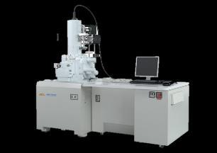
18 Micro-CT Systems Desktop micro-ct systems Small footprint Maintenance free High ease of use Laboratory micro-ct systems Large cabinet High flexibility Best resolution Differences between systems: X-ray energy Resolution (pixel size and X-ray spot size) Maximum sample size November 24,

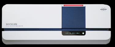
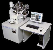
19 Micro-CT Systems Desktop micro-ct systems Skyscan1272 High resolution Skyscan1173 High energy Skyscan1275 Automated, high throughput Laboratory micro-ct systems Large cabinet High flexibility Best resolution Voxel size down to 0.4 µm X-ray energy 100 kev November 24,

20 Micro-CT Systems Desktop micro-ct systems Skyscan1272 High resolution Skyscan1173 High energy Skyscan1275 Automated, high throughput Laboratory micro-ct systems Large cabinet High flexibility Best resolution X-ray energy 130 kev Voxel size down to ~5 µm November 24,
21 Micro-CT Systems Desktop micro-ct systems Skyscan1272 High resolution Skyscan1173 High energy Skyscan1275 Automated, high throughput Laboratory micro-ct systems Large cabinet High flexibility Best resolution X-ray energy 100 kev Voxel size down to 4.5 µm November 24,


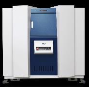
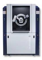
22 200 cm Micro-CT Systems Desktop micro-ct systems 1272 High resolution 1173 High energy 1275 Automated, high throughput Laboratory micro-ct systems SkyScan2211 X-ray energy up to 190 kev Voxel size down to 0.1 µm X-ray spot size 500 nm 2 X-ray detectors November 24,
23 Results Quantitative 3D analysis Acquisition and reconstruction Image processing filtering Segmentation Dividing images into different phases Determination of parameters of interest in 3D Porosity and pore network properties Amount of mineral phase X Grain size shape orientation sphericity Results November 24,


24 Results Quantitative 3D analysis November 24,
25 Micro-CT Main advantages for geological applications November 24,
26 Micro-CT Main advantages for geological applications 3D structural information Porosity analysis Pore networks connectivity flow paths 3D mineral distribution 3D analysis of shape and morphometries Grains, sedimentary patterns, fossils, etc. November 24,


27 Application examples 3D structural information - porosity Rocks can be segmented into binary pore vs. solid models. Reconstructed slice Binarized image 1275 November 24,

28 Application examples 3D structural information - porosity Rocks can be segmented into binary pore vs. solid models. At each point, 3D pore network properties are known November 24,
29 Application examples 3D structural information - porosity Rocks can be segmented into binary pore vs. solid models. At each point, 3D pore network properties are known. Local thickness of the pore network 2211 November 24,


30 Application examples 3D structural information - porosity 2 mm microplug - 1 µm resolution SkyScan mm microplug - 1 µm resolution SkyScan November 24,

31 Application examples 3D structural information - porosity Rocks can be segmented into binary pore vs. solid models. At each point, 3D pore network properties are known. Local thickness of the pore network Open versus closed porosity 2211 November 24,


32 Application examples 3D structural information - porosity Based on 3D imaging, direct modelling of transport properties can be done. Permeability Multiphase flow properties Capillary pressure curves Resistivity index Etc. For conventional reservoirs simulated values are very close to laboratory measured values. More and more methods available for unconventional reservoirs Simulation and image created with November 24,
33 Application examples Grain size analysis 1275 November 24,

34 Application examples Grain size analysis 1275 November 24,
35 Application examples 3D mineral distribution If sufficient contrast rocks can be segmented into different mineral fractions. November 24,


36 Application examples 3D mineral distribution ore geology Gold deposit 9 µm pixel size 29 minutes scan time 1275 November 24,


37 Application examples 3D mineral distribution ore geology Cu-ore 5 cm sample size 25 µm resolution 1275 Original sample Volume render November 24,


38 Application examples 3D mineral distribution ore geology Cu-ore 5 cm sample size 25 µm resolution 57 % 34 % 7.5 % 1.5 % 1275 November 24,


39 Application examples No sample preparation fast results 10 mm microplug 6 µm resolution SkyScan1275 Scanned in 30 minutes 1275 November 24,

40 Application examples No sample preparation fast results 5 mm microplug 5 µm resolution SkyScan minutes per scan 5 vertical scans stitched together 1275 November 24,


41 Application examples Sedimentology 50 mm sediment core 35 µm resolution SkyScan November 24,

42 Application examples Sedimentology 80 mm clay core 50 µm resolution SkyScan November 24,

43 Application examples Imaging of fossils Arthropod larva Leanchoilia illecebrosa (fossil length: 2 mm) SkyScan1272 Voxel size 1.5 µm 1272 Courtesy of Dr. Yu Liu Yunnan Key Laboratory for Paleobiology Yunnan University - China November 24,


44 Application examples Imaging of fossils insect in amber Mosquito in amber Voxel size 7 um Scan time 20 minutes 1275 November 24,
45 Micro-CT Main advantages for geological applications 3D structural information Porosity analysis Pore networks connectivity flow paths 3D mineral distribution 3D analysis of shape and morphometries Grains, sedimentary patterns, fossils, etc. Non-destructive imaging Multi-scale analysis Monitoring of dynamic processes / changing samples Imaging (and digital preservation) of precious samples November 24,

46 Application examples Multi-scale analysis Multi-scale analysis Large samples at low resolution Small samples at high resolution Region of interest scanning No physical subsampling 10 mm sample 5.5 µm resolution Zoom in scan (4 mm FOV) 2 µm resolution 2211 November 24,


47 Application examples Multi-scale analysis Very high resolutions on reasonable sample sizes. 4 mm 1 µm resolution 2 mm 500 nm resolution 2211 November 24,
48 Application examples Monitoring of dynamic processes November 24,
49 Application examples Monitoring of dynamic processes Compression test of a Noyant limestone using SkyScan MTS November 24,


50 Application examples Monitoring of dynamic processes Compression test of a Noyant limestone using SkyScan MTS3 SkyScan MTS2 - Compression / tensile up to 440 N SkyScan MTS3 - Compression up to 4400 N Custom design / third party 1275 November 24,

51 Application examples Monitoring of dynamic processes Crystallization in porous media Time-lapse CT Salt crust formation on Bentheimer sandstone Scanned after each absorption drying cycle SkyScan µm voxel size - 30 minutes per scan 1275 November 24,

52 Application examples Monitoring of dynamic processes Moisture transport in porous media Real-time CT Liquid displacement in Bentheimer sandstone. 1 scan = 80 seconds Pixel size = 10 µm SkyScan November 24,
53 Micro-CT Main advantages for geological applications 3D structural information Porosity analysis Pore networks connectivity flow paths 3D mineral distribution 3D analysis of shape and morphometries Grains, sedimentary patterns, fossils, etc. Non-destructive imaging Multi-scale analysis Monitoring of dynamic processes / changing samples Imaging (and digital preservation) of precious samples Complementary to traditional methods E.g. optical or electron microscopy, XRF, XRD November 24,
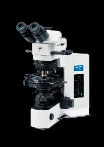
54 Application examples Data-fusion with traditional research methods X-ray fluorescence imaging SEM / EDS FIB Nanotomography X-ray diffraction Optical microscopy November 24,

55 Application examples Data-fusion with traditional research methods Data fusion combination of micro-ct with EDS / XRD / XRF X-ray MicroCT in Conjunction with Other Techniques for Core Analysis I.V. Yakimchuk 1, A.S. Denisenko 1, B.D. Sharchilev 1, and I.A. Varfolomeev 1, 2 1 Schlumberger, , 13 Pudovkina str., Moscow, Russia, 2 Moscow Institute of Physics and Technology, , 9 Institutskiy per., Dolgoprudny, Russia 1272 November 24,

56 Application examples Data-fusion with traditional research methods A drill core was scanned using the 1275 and an XRF mapping was performed using the Bruker M4 Tornado From left to right: - XRF data - µ-ct data - Overlay of XRF on µ-ct November 24,


57 Application examples Data-fusion with traditional research methods A drill core was scanned using the 1275 and an XRF mapping was performed using the Bruker M4 Tornado Image registration allows 3D alignment of both image modalities and inspection of the sample. November 24,
58 Conclusions Micro-CT provides 3D information on Porosity Mineral distribution Structure All kinds of samples Sample sizes from mm to full core Mature and accepted method Complementary method to traditional analysis techniques November 24,

59 Q & A session Thank you for your attention More information: Wesley De Boever Applications.bmct@bruker.com Phone: +32 (0) November 24,


60 Copyright Bruker Corporation. All rights reserved. Innovation with Integrity
X-ray MicroCT in Conjunction with Other Techniques for Core Analysis
 X-ray MicroCT in Conjunction with Other Techniques for Core Analysis I.V. Yakimchuk 1, A.S. Denisenko 1, B.D. Sharchilev 1, and I.A. Varfolomeev 1,2 1 Schlumberger, 119285, 13 Pudovkina str., Moscow, Russia,
Exa DigitalROCK: A Virtual Lab for Fast, Reliable Permeability Data
 Exa DigitalROCK: A Virtual Lab for Fast, Reliable Permeability Data Exa Corporation 2017 CONTACT Exa Corporation 55 Network Drive Burlington, MA USA 01803 1.781.564.0200 www.exa.com Copyright 2017 Exa
HIGH RESOLUTION COMPUTED TOMOGRAPHY FOR METROLOGY
 HIGH RESOLUTION COMPUTED TOMOGRAPHY FOR METROLOGY David K. Lehmann 1, Kathleen Brockdorf 1 and Dirk Neuber 2 1 phoenix x-ray Systems + Services Inc. St. Petersburg, FL, USA 2 phoenix x-ray Systems + Services
microct Systems for Life Science Innovation with Integrity High Resolution Microtomography for in vivo and ex vivo Applications Microtomography
 microct Systems for Life Science High Resolution Microtomography for in vivo and ex vivo Applications Innovation with Integrity Microtomography SkyScan 1278 High Throughput Large Field of View in vivo
Enhanced material contrast by dual-energy microct imaging
 Enhanced material contrast by dual-energy microct imaging Method note Page 1 of 12 2 Method note: Dual-energy microct analysis 1. Introduction 1.1. The basis for dual energy imaging Micro-computed tomography
Product Information Version 1.1. ZEISS Xradia 510 Versa Submicron X-ray Imaging: Maintain High Resolution Even at Large Working Distances
 Product Information Version 1.1 ZEISS Xradia 510 Versa Submicron X-ray Imaging: Maintain High Resolution Even at Large Working Distances Breakthrough Flexibility for 3D Submicron Imaging Achieve new levels
The Electrochemical Innovation Lab X-ray Suite: from macro- to nano-ct
 The Electrochemical Innovation Lab X-ray Suite: from macro- to nano-ct Dr. Francesco Iacoviello, Toby Neville & the Electrochemical Innovation Lab f.iacoviello@ucl.ac.uk MANIFEST - 2017 November 10 th,
μct for the Pharmaceutical Industries Innovation with Integrity From R&D to Production, Inspection and Failure Analysis Microtomography
 μct for the Pharmaceutical Industries From R&D to Production, Inspection and Failure Analysis Innovation with Integrity Microtomography Find out What's Inside Porosity and Pore Size Distribution Micro-cracks
X-rays see all X-ray computed microtomography (µct) a novel technique available at CERN for material and metrological inspection
 a novel technique available at CERN for material and metrological inspection") X-rays see all X-ray computed microtomography (µct) a novel technique available at CERN for material and metrological inspection Mariusz Jedrychowski EN/MME-MM Introduction of new technique at CERN 12/12/2017
X-rays see all X-ray computed microtomography (µct) a novel technique available at CERN for material and metrological inspection Mariusz Jedrychowski EN/MME-MM Introduction of new technique at CERN 12/12/2017
Latest developments in 3D analysis of geomaterials by Morpho+
 Latest developments in 3D analysis of geomaterials by Morpho+ Cnudde, Veerle *,**, Vlassenbroeck, Jelle **, De Witte, Yoni **, Brabant, Loes **, Boone, Matthieu **, Dewanckele, Jan *,**, Van Hoorebeke,
Using X-Ray Micro Tomography to assess the quality of Packaging Foil
 Using X-Ray Micro Tomography to assess the quality of Packaging Foil Martin Koster 1, Gerard van Dalen 2, Robert Hoeve 3 1 Unilever Research 2 Unilever Research 3 Unilever Research, Olivier van Noortlaaan
Applying Hounsfield unit density calibration in SkyScan CT-analyser
 1 Bruker-microCT Method note Applying Hounsfield unit density calibration in SkyScan CT-analyser Hounsfield units (HU) are a standard unit of x-ray CT density, in which air and water are ascribed values
How does the ROI affect the thresholding?
 How does the ROI affect the thresholding? Micro-computed tomography can be applied for the visualization of the inner structure of a material or biological tissue in a non-destructive manner. Besides visualization,
LABORATORY SYSTEM FOR X-RAY NANOTOMOGRAPHY
 79 LABORATORY SYSTEM FOR X-RAY NANOTOMOGRAPHY Alexander Sasov, SkyScan, Vluchtenburgstraat 3, Aartselaar B2630, Belgium, www.skyscan.be. ABSTRACT Using advanced X-ray technologies and X-ray scattering
Quality control phantoms and protocol for a tomography system
 Quality control phantoms and protocol for a tomography system Lucía Franco 1 1 CT AIMEN, C/Relva 27A O Porriño Pontevedra, Spain, lfranco@aimen.es Abstract Tomography systems for non-destructive testing
MicroCT for bone and biomaterials characterization. Lea Vaiana PARALAB SL
 MicroCT for bone and biomaterials characterization Lea Vaiana PARALAB SL Distribución de equipamiento Científico para laboratorio y proceso Oficinas en Barcelona, Madrid y Bilbao. Asesoramiento pre-venta
Micro-CT in situ study of carbonate rock microstructural evolution for geologic CO2 storage
 Downloaded from orbit.dtu.dk on: Apr 10, 2018 Micro-CT in situ study of carbonate rock microstructural evolution for geologic CO2 storage Zheng, Yi; Yang, Yan; Rogowska, M.; Gundlach, Carsten Published
Image Acquisition Systems
 Image Acquisition Systems Goals and Terminology Conventional Radiography Axial Tomography Computer Axial Tomography (CAT) Magnetic Resonance Imaging (MRI) PET, SPECT Ultrasound Microscopy Imaging ITCS
Improvement of the bimodal parameterization of particle size distribution using laser diffraction
 Improvement of the bimodal parameterization of particle size distribution using laser diffraction Marco Bittelli Department of Agro-Environmental Science and Technology, University of Bologna, Italy. Soil
QUICK CORE ASSESSMENT FROM CT IMAGING: FROM PETROPHYSICAL PROPERTIES TO LOG EVALUATION
 SCA2016-031 1/12 QUICK CORE ASSESSMENT FROM CT IMAGING: FROM PETROPHYSICAL PROPERTIES TO LOG EVALUATION Olivier Lopez, Carl F. Berg, Lars Rennan, Gunnar Digranes, Thibaut Forest, Anders Kristoffersen &
Werkstoffmodellierung und eigenschaftsberechnung auf Basis von CT-Aufnahmen
 Werkstoffmodellierung und eigenschaftsberechnung auf Basis von CT-Aufnahmen Fachtagung Computertomografie, 27.09.2010 Erik Glatt, Jürgen Becker, Stefan Rief und Andreas Wiegmann Fraunhofer Institut Techno
Digital Core study of Wanaea and Perseus Core Fragments:
 Digital Core study of Wanaea and Perseus Core Fragments: Summary for Woodside Energy Mark A. Knackstedt,2, A. Ghous 2, C. H. Arns, H. Averdunk, F. Bauget, A. Sakellariou, T.J. Senden, A.P. Sheppard,R.
Effect of an initial solution in iterative reconstruction of dynamically changing objects
 Effect of an initial solution in iterative reconstruction of dynamically changing objects M. HEYNDRICKX 1, T. DE SCHRYVER 1, M. DIERICK 1, M. N. BOONE* 1, T. BULTREYS 2, V. CNUDDE 2, L. VAN HOOREBEKE 1
SUPPLEMENTARY INFORMATION
 SUPPLEMENTARY INFORMATION doi:10.1038/nature10934 Supplementary Methods Mathematical implementation of the EST method. The EST method begins with padding each projection with zeros (that is, embedding
Arion: a realistic projection simulator for optimizing laboratory and industrial micro-ct
 Arion: a realistic projection simulator for optimizing laboratory and industrial micro-ct J. DHAENE* 1, E. PAUWELS 1, T. DE SCHRYVER 1, A. DE MUYNCK 1, M. DIERICK 1, L. VAN HOOREBEKE 1 1 UGCT Dept. Physics
A new concept for high-speed atline and inline CT for up to 100% mass production process control allowing both 3D metrology and failure analysis
 A new concept for high-speed atline and inline CT for up to 100% mass production process control allowing both 3D metrology and failure analysis Oliver Brunke 1, Ferdinand Hansen 2, Ingo Stuke 3, Friedrich
BME I5000: Biomedical Imaging
 1 Lucas Parra, CCNY BME I5000: Biomedical Imaging Lecture 4 Computed Tomography Lucas C. Parra, parra@ccny.cuny.edu some slides inspired by lecture notes of Andreas H. Hilscher at Columbia University.
Optiv CT160 Accurate measurement for every detail. Computed Tomography System
 Optiv CT160 Accurate measurement for every detail Computed Tomography System Computed Tomography CT Technology. New prospects for metrology. Computed tomography (CT), a technology with unique advantages,
Ch. 4 Physical Principles of CT
 Ch. 4 Physical Principles of CT CLRS 408: Intro to CT Department of Radiation Sciences Review: Why CT? Solution for radiography/tomography limitations Superimposition of structures Distinguishing between
High-resolution X-ray CT Inspection of Honeycomb Composites Using Planar Computed Tomography Technology
 2nd International Symposium on NDT in Aerospace 2010 - We.4.B.4 High-resolution X-ray CT Inspection of Honeycomb Composites Using Planar Computed Tomography Technology Tong LIU, Andrew A. MALCOLM, and
3D X-ray Microscopy Characterization. Metal Additively Manufactured Parts. Application Note
 3D X-ray Microscopy Characterization Metal Additively Manufactured Parts Application Note 3D X-ray Microscopy Characterization Metal Additively Manufactured Parts Author: Luke Hunter ZEISS X-ray Microscopy
Digital Image Processing
 Digital Image Processing SPECIAL TOPICS CT IMAGES Hamid R. Rabiee Fall 2015 What is an image? 2 Are images only about visual concepts? We ve already seen that there are other kinds of image. In this lecture
Product Information Version 3.0. ZEISS Xradia 520 Versa Submicron X-ray Imaging: Extending the Limits of Your Exploration
 Product Information Version 3.0 ZEISS Xradia 520 Versa Submicron X-ray Imaging: Extending the Limits of Your Exploration Submicron X-ray Imaging: Extending the Limits of Your Exploration Unlock new degrees
PREDICTION OF ADSORPTION AND BREAK THROUGH CURVES BY DIRECT NUMERICAL SIMULATION ABSTRACT
 PREDICTION OF ADSORPTION AND BREAK THROUGH CURVES BY DIRECT NUMERICAL SIMULATION E. Glatt and A. Wiegmann Fraunhofer-Institut Techno- und Wirtschaftsmathematik, Fraunhofer-Platz 1, 67663 Kaiserslautern,
A Lattice-Boltzmann Based Method Applied to Digital Rock Characterization of Perforation Tunnel Damage
 SCA2016-058 1/6 A Lattice-Boltzmann Based Method Applied to Digital Rock Characterization of Perforation Tunnel Damage Bernd Crouse, David M Freed, Nils Koliha, Gana Balasubramanian EXA CORP Rajani Satti,
Image Processing, Quantification and Model Reconstructions in SEM/FIB
 Image Processing, Quantification and Model Reconstructions in SEM/FIB Case studies Daniel Lichau Visualization Science Group Bordeaux, France EFUG 2010 - Gatea, Italy 16 October, 2010 About VSG Formerly
Non-destructive, High-resolution Fault Imaging for Package Failure Analysis. with 3D X-ray Microscopy. Application Note
 Non-destructive, High-resolution Fault Imaging for Package Failure Analysis with 3D X-ray Microscopy Application Note Non-destructive, High-resolution Fault Imaging for Package Failure Analysis with 3D
Onyx. XwinSys. In-line Non-Destructive Inspection and Metrology for the Semiconductor and Micro-Electronic Industries
 Onyx In-line Non-Destructive Inspection and Metrology for the Semiconductor and Micro-Electronic Industries Hybrid Configuration: X-Ray Analysis Automated 3D Scanning 2D Microscope XwinSys IN-LINE NON-DESTRUCTIVE
Supplementary Materials for
 www.advances.sciencemag.org/cgi/content/full/1/3/e1400199/dc1 Supplementary Materials for Life and death of a single catalytic cracking particle Florian Meirer, Sam Kalirai, Darius Morris, Santosh Soparawalla,
Phase-Contrast Imaging and Tomography at 60 kev using a Conventional X-ray Tube
 Phase-Contrast Imaging and Tomography at 60 kev using a Conventional X-ray Tube T. Donath* a, F. Pfeiffer a,b, O. Bunk a, W. Groot a, M. Bednarzik a, C. Grünzweig a, E. Hempel c, S. Popescu c, M. Hoheisel
Copyright JCPDS-International Centre for Diffraction Data 2006 ISSN Advances in X-ray Analysis, Volume 49
 X-RAY MICROTOMOGRAPHIC IMAGING AND ANALYSIS FOR BASIC RESEARCH J. H. Dunsmuir, S. Bennett, L. Fareria, A. Mingino, M. Sansone ExxonMobil Research and Engineering Company Corporate Strategic Research Annandale,
A new Concept for High-Speed atline and inlinect for up to 100% Mass Production Process Control
 18th World Conference on Nondestructive Testing, 16-20 April 2012, Durban, South Africa A new Concept for High-Speed atline and inlinect for up to 100% Mass Production Process Control Oliver BRUNKE 1,
CORRELATION BETWEEN CORE LINEAR X-RAY AND WIRELINE BULK DENSITY IN TWO CLASTIC RESERVOIRS: AN EXAMPLE OF CORE-LOG INTEGRATION
 SCA2017-072 1/9 CORRELATION BETWEEN CORE LINEAR X-RAY AND WIRELINE BULK DENSITY IN TWO CLASTIC RESERVOIRS: AN EXAMPLE OF CORE-LOG INTEGRATION Yu Jin and David K. Potter Department of Physics, and Department
Diffraction Enhanced X-ray Imaging (DEI) Benjamin Reinhart Physics 570 Final Presentation December 6 th, 2013
 Benjamin Reinhart Physics 570 Final Presentation December 6 th, 2013") Diffraction Enhanced X-ray Imaging (DEI) Benjamin Reinhart Physics 570 Final Presentation December 6 th, 2013 Bullet Points of Presentation Radiography vs. Diffraction Enhanced Imaging (DEI) Basic Principles
Diffraction Enhanced X-ray Imaging (DEI) Benjamin Reinhart Physics 570 Final Presentation December 6 th, 2013 Bullet Points of Presentation Radiography vs. Diffraction Enhanced Imaging (DEI) Basic Principles
A new Concept for High-Speed atline and inlinect for up to 100% Mass Production Process Control
 6 th International Congress of Metrology, 06003 (203) DOI: 0.05/ metrology/20306003 C Owned by the authors, published by EDP Sciences, 203 A new Concept for High-Speed atline and inlinect for up to 00%
3D Energy Dispersive Spectroscopy Elemental Tomography in the Scanning Transmission Electron Microscope
 3D Energy Dispersive Spectroscopy Elemental Tomography in the Scanning Transmission Electron Microscope Brian Van Devener Topics 1.Introduction to EDS in the STEM 2.Extending EDS into three dimensions
Porosity and Pore-Size Distribution of Geomaterials from X-Ray CT Scans
 Porosity and Pore-Size Distribution of Geomaterials from X-Ray CT Scans Hyu-Soung Shin & Kwang-Yeom Kim Geotechnical Eng. Research Division, Korea Institute of Construction Technology (KICT), South Korea
Supporting Information for Critical Factors of the 3D Microstructural Formation. in Hybrid Conductive Adhesive Materials by X-ray Nano-tomography
 Electronic Supplementary Material (ESI) for Nanoscale. This journal is The Royal Society of Chemistry 2014 Supporting Information for Critical Factors of the 3D Microstructural Formation in Hybrid Conductive
Micro-CT Methodology Hasan Alsaid, PhD
 Micro-CT Methodology Hasan Alsaid, PhD Preclinical & Translational Imaging LAS, PTS, GlaxoSmithKline 20 April 2015 Provide basic understanding of technical aspects of the micro-ct Statement: All procedures
Dr. Javier Santillan, San Carlos, CA
 X-ray diffraction as a tool for automated residual st ress analysis & a non-synchrotron based nanofocus x-ray computed tomography technique for materials characterization and metrology Dr. Javier Santillan,
3D Computed Tomography (CT) Its Application to Aerospace Industry
 Its Application to Aerospace Industry") 3D Computed Tomography (CT) Its Application to Aerospace Industry C. Muralidhar, M. P. Subramanian, V. Ravi Shankar and G. Chandrasekhar Directorate of Non Destructive Evaluation, Defence Research & Development
3D Computed Tomography (CT) Its Application to Aerospace Industry C. Muralidhar, M. P. Subramanian, V. Ravi Shankar and G. Chandrasekhar Directorate of Non Destructive Evaluation, Defence Research & Development
ML reconstruction for CT
 ML reconstruction for CT derivation of MLTR rigid motion correction resolution modeling polychromatic ML model dual energy ML model Bruno De Man, Katrien Van Slambrouck, Maarten Depypere, Frederik Maes,
Deformation of granular texture media studied by X-ray CT & 3D DIC at the continuous and microstructure scales
 Deformation of granular texture media studied by X-ray CT & 3D DIC at the continuous and microstructure scales N. Lenoir, S.A. Hall, J. Desrues, G. Viggiani and P. Bésuelle (Laboratoire 3S-R) Michel Bornert
X-ray tomographic study of wetting bentonite
 UNIVERSITY OF JYVÄSKYLÄ X-ray tomographic study of wetting bentonite Tero Harjupatana Department of Physics, University of Jyväskylä BOA seminar 19.8.2014 Goal To develop and apply experimental techniques
Optimization of scanner parameters for dual energy micro-ct
 Optimization of scanner parameters for dual energy micro-ct E. PAUWELS* 1, J. DHAENE 1, A. DE MUYNCK 1 E., M. DIERICK 1, L. VAN HOOREBEKE 1 1 UGCT Dept. Physics and Astronomy, Ghent University, Proeftuinstraat
CLASS HOURS: 4 CREDIT HOURS: 4 LABORATORY HOURS: 0
 Revised 10/10 COURSE SYLLABUS TM 220 COMPUTED TOMOGRAPHY PHYSICS CLASS HOURS: 4 CREDIT HOURS: 4 LABORATORY HOURS: 0 CATALOG COURSE DESCRIPTION: This course is one of a three course set in whole body Computed
EarthStudy 360. Full-Azimuth Angle Domain Imaging and Analysis
 EarthStudy 360 Full-Azimuth Angle Domain Imaging and Analysis 1 EarthStudy 360 A New World of Information for Geoscientists Expanding the Frontiers of Subsurface Exploration Paradigm EarthStudy 360 is
Accuracy of the Two Common Semi- Analytical Equations in Predicting Asphalt Permeability
 Accuracy of the Two Common Semi- Analytical Equations in Predicting Asphalt Permeability M. Emin Kutay * Ahmet H. Aydilek** * Research Engineer, Turner-Fairbank Highway Research Center (TFHRC) 6300 Georgetown
Advanced automated High Speed Computed Tomography for production process control. DGZfP 2011, Bremen
 DGZfP-Jahrestagung 2011 - Mo.3.B.3 Advanced automated High Speed Computed Tomography for production process control DGZfP 2011, Bremen Dr. Ingo Stuke* and Dr. Oliver Brunke** GE Sensing & Inspection Technologies
SEM topography. Live quantitative surface topography in SEM
 SEM topography Live quantitative surface topography in SEM 2 Measure surface topography with SEM. n Use conventional segmented BSE signals. n Get immediate feedback with automated topographic reconstruction.
TEP Hounsfield units. Related topics Attenuation coefficient, Hounsfield units
 Hounsfield units TEP Related topics Attenuation coefficient, Hounsfield units Principle Depending on the type of CT scanner and the settings, the result of a CT scan of the same material can be different
Digital Volume Correlation for Materials Characterization
 19 th World Conference on Non-Destructive Testing 2016 Digital Volume Correlation for Materials Characterization Enrico QUINTANA, Phillip REU, Edward JIMENEZ, Kyle THOMPSON, Sharlotte KRAMER Sandia National
Investigation of Two-Phase Flow Mechanisms in Porous Media Using Micro-Particle Image Velocimetry
 Investigation of Two-Phase Flow Mechanisms in Porous Media Using Micro-Particle Image Velocimetry oil solid water Sophie Roman*, Cyprien Soulaine, Moataz Abu AlSaud, Hamdi Tchelepi, Anthony Kovscek *sroman@stanford.edu
AN AUTOMATED MACHINE-LEARNING PROCEDURE FOR ROBUST CLASSIFICATION OF SEM IMAGES OF CROSS-LAMINATED SANDSTONES FOR DIGITAL ROCK ANALYSIS
 SCA2014-034 1/6 AN AUTOMATED MACHINE-LEARNING PROCEDURE FOR ROBUST CLASSIFICATION OF SEM IMAGES OF CROSS-LAMINATED SANDSTONES FOR DIGITAL ROCK ANALYSIS Chen Jin and Jingsheng Ma Institute of Petroleum
Displacement in sand under triaxial compression by tracking soil particles on X-ray CT data
 Soils and Foundations 212;52(2):312 32 The Japanese Geotechnical Society Soils and Foundations www.sciencedirect.com journal homepage: www.elsevier.com/locate/sandf Displacement in sand under triaxial
Tomography. Introduction to Tomography TEM Tilt-Series Tomography in Life Science STEM Tomography in Materials Science
 Tomography Introduction to Tomography TEM Tilt-Series Tomography in Life Science STEM Tomography in Materials Science Introduction to Tomography Tomography is imaging by sections or sectioning. A device
3/27/2012 WHY SPECT / CT? SPECT / CT Basic Principles. Advantages of SPECT. Advantages of CT. Dr John C. Dickson, Principal Physicist UCLH
 3/27/212 Advantages of SPECT SPECT / CT Basic Principles Dr John C. Dickson, Principal Physicist UCLH Institute of Nuclear Medicine, University College London Hospitals and University College London john.dickson@uclh.nhs.uk
Corso di laurea in Fisica A.A Fisica Medica 4 TC
 Corso di laurea in Fisica A.A. 2007-2008 Fisica Medica 4 TC Computed Tomography Principles 1. Projection measurement 2. Scanner systems 3. Scanning modes Basic Tomographic Principle The internal structure
Comparison of phase contrast X-ray computed tomography methods for non-destructive testing of materials
 Comparison of phase contrast X-ray computed tomography methods for non-destructive testing of materials Johann KASTNER 1, Bernhard PLANK 1, Christian KOTTLER 2, Vincent REVOL 2 1 University of Applied
Fast X-ray Micro-Tomography of Multiphase Flow in Berea Sandstone: A Sensitivity Study on Image Processing
 Transp Porous Med (2014) 105:451 469 DOI 10.1007/s11242-014-0378-4 Fast X-ray Micro-Tomography of Multiphase Flow in Berea Sandstone: A Sensitivity Study on Image Processing L. Leu S. Berg F. Enzmann R.
Industrial Computed Tomography Innovations
 GE Inspection Technologies Industrial Computed Tomography Innovations Premium performance for premium quality and speed. gemeasurement.com/ct Premium CT technologies. Faster than ever before. Every industrial
*Corresponding author:
 Comparison of X-ray computed tomography and immersion ultrasonic nondestructive testing techniques in the case of qualitative and quantitative assessment of brazing quality level More info about this article:
XRF MAPPING: NEW TOOLS FOR DISTRIBUTION ANALYSIS
 19 19 XRF MAPNG: NEW TOOLS FOR DISTRIBUTION ANALYSIS B. Scruggd, M. Haschke2, L. Herczeg, J. Nicolosi Edax Inc; Mahwah, NJ 2Riintgenanalytik MeJtechnik GmbH; Taunusstein, Germany INTRODUCTION With the
New Generation Explorer Handheld XRF. EXPLORER 7000 Mineral Ore Analyzer
 New Generation Explorer Handheld XRF EXPLORER 7000 Mineral Ore Analyzer EXPLORER 7000 Mineral Ore XRF Based on ten-year research and development experience in Handheld X-Ray Fluorescense, Skyray Instruments
Advanced Non-Destructive Testing by High Resolution Computed Tomography for 3D analysis of Automotive Components
 Advanced Non-Destructive Testing by High Resolution Computed Tomography for 3D analysis of Automotive Components J. Luebbehuesen GE Sensing & Inspection Technologies GmbH, phoenix x-ray, Niels-Bohr-Str.
DEVELOPMENT OF CONE BEAM TOMOGRAPHIC RECONSTRUCTION SOFTWARE MODULE
 Rajesh et al. : Proceedings of the National Seminar & Exhibition on Non-Destructive Evaluation DEVELOPMENT OF CONE BEAM TOMOGRAPHIC RECONSTRUCTION SOFTWARE MODULE Rajesh V Acharya, Umesh Kumar, Gursharan
Robot-based real-time ultrasonic tomography for industrial NDT applications
 1 Robot-based real-time ultrasonic tomography for industrial NDT applications Dr. Andrey Bulavinov I-Deal Technologies GmbH 2 Content I. Introduction of I-Deal Technologies GmbH I-Deal Spin-Off of Fraunhofer-IZFP
Foolproof AvO. Abstract
 Foolproof AvO Dr. Ron Masters, Geoscience Advisor, Headwave, Inc Copyright 2013, The European Association of Geoscientists and Engineers This paper was prepared for presentation during the 75 th EAGE Conference
SYSTEM LINEARITY LAB MANUAL: 2 Modifications for P551 Fall 2013 Medical Physics Laboratory
 SYSTEM LINEARITY LAB MANUAL: 2 Modifications for P551 Fall 2013 Medical Physics Laboratory Introduction In this lab exercise, you will investigate the linearity of the DeskCAT scanner by making measurements
Computed Tomography & 3D Metrology Application of the VDI/VDE Directive 2630 and Optimization of the CT system
 Computed Tomography & 3D Metrology Application of the VDI/VDE Directive 2630 and Optimization of the CT system ECNDT 2014 Prague October 6-10, 2014 Dr. Eberhard Neuser Dr. Alexander Suppes Imagination
INDUSTRIAL SYSTEM DEVELOPMENT FOR VOLUMETRIC INTEGRITY
 INDUSTRIAL SYSTEM DEVELOPMENT FOR VOLUMETRIC INTEGRITY VERIFICATION AND ANALYSIS M. L. Hsiao and J. W. Eberhard CR&D General Electric Company Schenectady, NY 12301 J. B. Ross Aircraft Engine - QTC General
Advanced materials research using the Real-Time 3D Analytical FIB-SEM 'NX9000'
 SCIENTIFIC INSTRUMENT NEWS 2017 Vol. 9 SEPTEMBER Technical magazine of Electron Microscope and Analytical Instruments. Technical Explanation Advanced materials research using the Real-Time 3D Analytical
Spherical Crystal X-ray Imaging for MTW, OMEGA, and OMEGA EP
 Spherical Crystal X-ray Imaging for MTW, OMEGA, and OMEGA EP C.STOECKL, G. FISKEL, R. K. JUNGQUIST, P. M. NILSON, AND W. THEOBALD University of Rochester, Laboratory for Laser Energetics Spherical Crystal
Dragonfly Pro. Visual Pathway to Quantitative Answers ORS. Exclusive to ZEISS OBJECT RESEARCH SYSTEMS
 Dragonfly Pro Exclusive to ZEISS Visual Pathway to Quantitative Answers ORS OBJECT RESEARCH SYSTEMS Visualization and analysis software without bounds Dragonfly Pro by Object Research Systems (ORS) is
Characterization of Powder Injection Molded Components Using X-Ray CT. David P. Harding, Zhigang Zak Fang, C.L. Lin, Jan D. Miller
 Characterization of Powder Injection Molded Components Using X-Ray CT David P. Harding, Zhigang Zak Fang, C.L. Lin, Jan D. Miller Department of Metallurgical Engineering University of Utah 135 South 1450
ZEISS Launches New High-resolution 3D X-ray Imaging Solutions for Advanced Semiconductor Packaging Failure Analysis
 Press Release ZEISS Launches New High-resolution 3D X-ray Imaging Solutions for Advanced Semiconductor Packaging Failure Analysis New submicron and nanoscale XRM systems and new microct system provide
Recognition and Measurement of Small Defects in ICT Testing
 19 th World Conference on Non-Destructive Testing 2016 Recognition and Measurement of Small Defects in ICT Testing Guo ZHIMIN, Ni PEIJUN, Zhang WEIGUO, Qi ZICHENG Inner Mongolia Metallic Materials Research
Stress and Texture by XRD Bob He, Bruker AXS
 Stress and Texture by XRD Bob He, Bruker AXS Intensity Conventional X-ray Diffractometer Divergence slit Antiscatter slit Monochromator Bragg-Brentano Geometry. Scanning over range to collect XRD pattern.
Washability Monitor for Coal Utilizing Optical and X-Ray Analysis Techniques
 Washability Monitor for Coal Utilizing Optical and X-Ray Analysis Techniques Jan F. Bachmann, Claus C. Bachmann, Michael P. Cipold, Helge B. Wurst J&C Bachmann GmbH, Bad Wildbad, Germany Mel J. Laurila
A Geostatistical and Flow Simulation Study on a Real Training Image
 A Geostatistical and Flow Simulation Study on a Real Training Image Weishan Ren (wren@ualberta.ca) Department of Civil & Environmental Engineering, University of Alberta Abstract A 12 cm by 18 cm slab
The Determination of Inner Surfaces in Composites by X-Ray Refraction
 The Determination of Inner Surfaces in Composites by X-Ray Refraction A. H. Hampe, K.-W. Harbich, M. P. Hentschel and H.-V. Rudolph Bundesanstalt für Materialforschung und -prüfung (BAM), 12200 Berlin,
Supporting Information: FIB-SEM Tomography Probes the Meso-Scale Pore Space of an Individual Catalytic Cracking Particle
 Supporting Information: FIB-SEM Tomography Probes the Meso-Scale Pore Space of an Individual Catalytic Cracking Particle D.A.Matthijs de Winter, Florian Meirer, Bert M. Weckhuysen*, Inorganic Chemistry
From Eye to Insight HOW TO ANALYZE PREPARED AND UNPREPARED GEOLOGICAL SAMPLES WITH ONE DIGITAL MICROSCOPE. A Leica DVM6 M Case Study.
 From Eye to Insight Earth Science Applications HOW TO ANALYZE PREPARED AND UNPREPARED GEOLOGICAL SAMPLES WITH ONE DIGITAL MICROSCOPE A Leica DVM6 M Case Study Author Michael Doppler Sales Manager, EEMEA,
Fast Z-stacking 3D Microscopy Extended Depth of Field Autofocus Z Depth Measurement 3D Surface Analysis
 MICROSCOPE 3D ADD-ON FAST PRECISE AFFORDABLE 3D ADD-ON FOR MICROSCOPY Fast Z-stacking 3D Microscopy Extended Depth of Field Autofocus Z Depth Measurement 3D Surface Analysis Compatible With Transmitted
Méthodes d imagerie pour les écoulements et le CND
 Méthodes d imagerie pour les écoulements et le CND Journée scientifique FED3G CEA LIST/Lab Imagerie Tomographie et Traitement Samuel Legoupil 15 juin 2012 2D/3D imaging tomography Example Petrochemical
Design and performance characteristics of a Cone Beam CT system for Leksell Gamma Knife Icon
 Design and performance characteristics of a Cone Beam CT system for Leksell Gamma Knife Icon WHITE PAPER Introduction Introducing an image guidance system based on Cone Beam CT (CBCT) and a mask immobilization
Grey value images as a basis for finite element models and their mechanical properties
 Grey value images as a basis for finite element models and their mechanical properties K. Szlazak 1, J. Jaroszewicz 1, J. Idaszek 1, A. Dejaco 2, P. Hasslinger 2, V. Vass 2, C. Hellmich 2, W. Swieszkowski
HIGH-SPEED THEE-DIMENSIONAL TOMOGRAPHIC IMAGING OF FRAGMENTS AND PRECISE STATISTICS FROM AN AUTOMATED ANALYSIS
 23 RD INTERNATIONAL SYMPOSIUM ON BALLISTICS TARRAGONA, SPAIN 16-20 APRIL 2007 HIGH-SPEED THEE-DIMENSIONAL TOMOGRAPHIC IMAGING OF FRAGMENTS AND PRECISE STATISTICS FROM AN AUTOMATED ANALYSIS P. Helberg 1,
Mining Engineering Technician: Speciality Mine Exploitation
 Mining Engineering Technician: Speciality Mine Exploitation FIRST YEAR PHYSICAL BASES OF ENGINEERING 9220001 Core 1st 1st semester 6 7.2 Mechanics. Electricity. Thermodynamics. Fluid mechanics. Classes
Introduction to Biomedical Imaging
 Alejandro Frangi, PhD Computational Imaging Lab Department of Information & Communication Technology Pompeu Fabra University www.cilab.upf.edu X-ray Projection Imaging Computed Tomography Digital X-ray