ML reconstruction for CT
|
|
|
- Beatrice McGee
- 5 years ago
- Views:
Transcription
1 ML reconstruction for CT derivation of MLTR rigid motion correction resolution modeling polychromatic ML model dual energy ML model Bruno De Man, Katrien Van Slambrouck, Maarten Depypere, Frederik Maes, Jung-ha Kim, Roger Fulton, Johan Nuyts MIRC, KU Leuven & Univ of Sydney 1 Tomography 2 1
2 CT data recon 3 Maximum Likelihood one wishes to find recon that maximizes p(recon data) data recon computing p(recon data) computing p(data recon) difficult inverse problem easy forward problem Bayes: p(recon data) = ~ p(data recon) p(recon) p(data) 4 2
 j = 1..J i = 1.")
 find recon: Iterative inversion")
3 Maximum Likelihood recon p(recon data) ~ p(data recon) data projection Poisson µ j p(data recon) j = 1..J i = 1..I ln(p(data recon)) = ~ L(data recon) = 5 Maximum Likelihood L(data recon) find recon: Iterative inversion needed 6 3
 T 2 (µ, Δµ) T 1 (µ, Δµ) µ 8")
4 MLTR Likelihood L(µ+Δµ) T 1 (µ, Δµ) µ 7 MLTR Likelihood L(µ+Δµ) T 2 (µ, Δµ) T 1 (µ, Δµ) µ 8 4
5 MLTR Likelihood L(µ+Δµ) T 2 (µ, Δµ) T 1 (µ, Δµ) µ 9 MLTR Likelihood L(µ+Δµ) T 2 (µ, Δµ) T 1 (µ, Δµ) 10 5


6 MLTR 11 MLTR MEASUREMENT COMPARE UPDATE RECON REPROJECTION 12 6


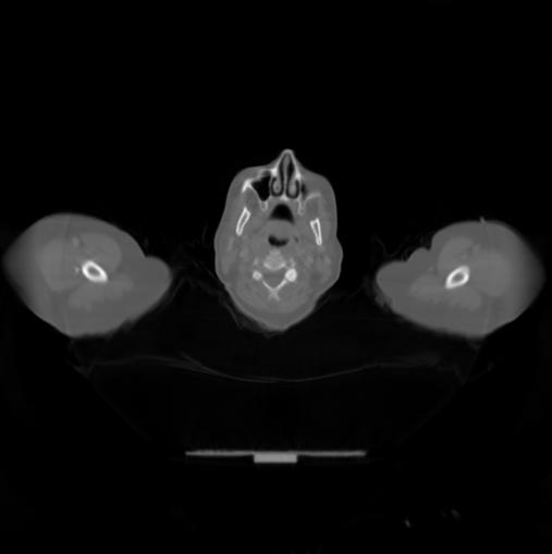
7 MLTR FBP MLTR 13 MLTR metal artifact reduction projection truncation FBP FBP MLTR MLTR 7


")

8 ML reconstruction for CT derivation of MLTR rigid motion correction resolution modeling polychromatic ML model dual energy ML model 15 MLTR for rigid motion correction 1) validation Siemens Sensation 16 Siemens MLTR J-H Kim, Z Kuncic, R Fulton, J Nuyts 16 8


9 MLTR for rigid motion correction 2) simulation motion rotations: in transaxial plane in sagittal plane in coronal plane 5 s measured rat motion software phantom trans cor CT protocol sag proj translations: along column along row along plane high pitch narrow collimation low tube current high rotation speed low dose 17 MLTR for rigid motion correction relativity : assign inverse motion to CT MLTR modified to support stationary object rigid view-dependent displacement of CT detector-source assembly 18 9


")


10 MLTR for rigid motion correction pitch = 2 trans cor sag proj MLTR w/o correction MLTR with correction pitch = 0.5 MLTR w/o correction MLTR with correction 19 3) phantom measurement MLTR for rigid motion correction 20 10

11 MLTR for rigid motion correction 21 MLTR 22 11
12 MLTR 23 ML reconstruction for CT derivation of MLTR rigid motion correction resolution modeling polychromatic ML model dual energy ML model 24 12


13 MLTR 25 MLTR microct Ex vivo - global FBP - global FBP - adap1ve MAPTR global Recon Segment 26 13


 Mouse bone and titanium screw")
14 ML reconstruction for CT derivation of MLTR rigid motion correction resolution modeling polychromatic ML model dual energy ML model 27 metal artifacts Double knee prosthesis Double hip prosthesis Dental fillings Cause of metal artifacts: Beam hardening Scatter (Non) linear partial volume effects Noise (Motion) Mouse bone and titanium screw (microct) 28 14

15 metal artifact reduction (MAR) Projection completion Initial filtered backprojection (FBP) reconstruction Segment the metals and project Remove metal projections for sinogram Interpolate (e.g. linear, polynomial, ) Reconstruct (FBP) and paste metal parts 29 Models for iterative reconstruction Poisson Likelihood: Update: Projection model: SKYSCAN SPECTRUM Black = without filter Blue = 0.5 mm Al and mm Cu monochromatic: 1 material polychromatic: 30 15
16 Models for iterative reconstruction Full Polychromatic Model IMPACT SKYSCAN SPECTRUM Black = without filter Blue = 0.5 mm Al and mm Cu 31 Models for iterative reconstruction Full Polychromatic Model IMPACT Base substances Material dependence Energy dependence 32 16


17 Models for iterative reconstruction Φ and θ (1/cm) Base substances µ mono (1/cm) 33 Local models IMPACT is complex and slow, MLTR and MLTR_C are simpler and faster Find the metals PATCH 3 Define patches IMPACT in metals MLTR_C elsewhere PATCH 2 PATCH

 + (a-1)*view(k) detector 500 µs 35 results")
18 simulations Geometry based on Siemens Sensation 16 Included: polychromatic spectrum detector, source and view subsampling afterglow crosstalk source view(k) = a*view(k-1) + (a-1)*view(k) detector 500 µs 35 results PMMA Al Fe 36 18


19 clinical CT (Siemens Sensation 16) Circular phantom PMMA Al Fe Siemens Sensation 16 (part of Biograph 16 PET/CT) 120 kv, 300 ma 2 x 1.00 mm Circular scan, 0.5 s per rotation (no flying focal spot) 2D reconstruction of 1 slice 37 clinical CT (Siemens Sensation 16) Body shaped phantom 38 19


20 clinical CT (Siemens Sensation 16) Body shaped phantom 39 SKYSCAN SPECTRUM Black = without filter Blue = 0.5 mm Al and mm Cu FDK iterative reconstruction for microct IMPACT Ti-cage, culture of soft tissue and cartilage 40 20


21 ML reconstruction for CT derivation of MLTR rigid motion correction resolution modeling polychromatic ML model dual energy ML model 41 Dual energy CT Dual energy CT: exploits dependence of linear attenuation coefficient on photon energy to discriminate between materials. Dual energy CT has been widely used to discriminate bone from contrast agent


22 Dual energy microct applications MicroCT: imaging bone and contrast agents in small animals, such as mice. Bone development and repair requires a normal vascular system to supply oxygen and nutrients. Rat skull Detail of trabecular bone structure Mouse bone fracture MicroCT Imaging X-ray energy range: kev 43 Post-reconstruction: microct post-reconstruction dual energy for microct problems: Perfused mouse tibia E1: 56 minutes beam hardening due to dense materials contrast agent metal implants Noise. Signal-to-noise ratio is limited by In vivo microct: dose concerns Ex vivo microct: cumbersome long scan times Voxel by voxel comparison is sensitive to erroneous intensity values Noise robustness can be increased by incorporating a noise model resorting to statistical approaches 22


23 Polychromatic attenuation model Dual energy algorithms exploit the dependency of the linear attenuation coefficient µ on the photon energy E The attenuation can be modeled as a linear combination of b basis functions A well known combination of basis functions is the Compton scatter and the photoelectric effect. Water Bone 45 IMPACT extension to dual energy microct Iodine Iodine Our model consists of a third basis function that models the attenuation of a single contrast material (barium, iodine, lead): 46 23


 0.1455 g/ml + 0.0021 (0.")


24 Results Noiseless simulation Noiseless simulation of water, bone and 0.15 and 0.20 g/ml mixtures of barium sulfate Post reconstruction Beam hardening affects tissue decomposition Polychromatic model accounts for beam hardening Iterative Decomposition g/ml (0.20) g/ml (0.15) 47 IMPACT extension to dual energy microct Measurement of polypropene tube, water, bone equivalent material CaHA and a barium sulfate mixture Noisy Post reconstruction Coefficient of variation in BaSO4 region: 0.36 Coeffecient of variation in BaSO4 region: 0.15 IMPACT Decomposition 48 24


25 IMPACT extension to dual energy microct Measurement of a mouse bone perfused with barium sulfate Post reconstruction Barium fractions Iterative decomposition Barium coefficients Iterative decomposition Coloured overlay 49 thanks 50 25
Enhanced material contrast by dual-energy microct imaging
 Enhanced material contrast by dual-energy microct imaging Method note Page 1 of 12 2 Method note: Dual-energy microct analysis 1. Introduction 1.1. The basis for dual energy imaging Micro-computed tomography
Evaluation of Spectrum Mismatching using Spectrum Binning Approach for Statistical Polychromatic Reconstruction in CT
 Evaluation of Spectrum Mismatching using Spectrum Binning Approach for Statistical Polychromatic Reconstruction in CT Qiao Yang 1,4, Meng Wu 2, Andreas Maier 1,3,4, Joachim Hornegger 1,3,4, Rebecca Fahrig
Computer-Tomography II: Image reconstruction and applications
 Computer-Tomography II: Image reconstruction and applications Prof. Dr. U. Oelfke DKFZ Heidelberg Department of Medical Physics (E040) Im Neuenheimer Feld 280 69120 Heidelberg, Germany u.oelfke@dkfz.de
Ch. 4 Physical Principles of CT
 Ch. 4 Physical Principles of CT CLRS 408: Intro to CT Department of Radiation Sciences Review: Why CT? Solution for radiography/tomography limitations Superimposition of structures Distinguishing between
Fast iterative beam hardening correction based on frequency splitting in computed tomography
 Fast iterative beam hardening correction based on frequency splitting in computed tomography Qiao Yang a,b, Matthias Elter b, Ingo Schasiepen b, Nicole Maass b and Joachim Hornegger a,c a Pattern Recognition
Corso di laurea in Fisica A.A Fisica Medica 4 TC
 Corso di laurea in Fisica A.A. 2007-2008 Fisica Medica 4 TC Computed Tomography Principles 1. Projection measurement 2. Scanner systems 3. Scanning modes Basic Tomographic Principle The internal structure
Metal Artifact Reduction CT Techniques. Tobias Dietrich University Hospital Balgrist University of Zurich Switzerland
 Metal Artifact Reduction CT Techniques R S S S Tobias Dietrich University Hospital Balgrist University of Zurich Switzerland N. 1 v o 4 1 0 2. Postoperative CT Metal Implants CT is accurate for assessment
Metal Streak Artifacts in X-ray Computed Tomography: A Simulation Study
 Metal Streak Artifacts in X-ray Computed Tomography: A Simulation Study B. De Man, J. Nuyts, P. Dupont, G. Marchal and P. Suetens Medical Image Computing, ESAT-PSI, K.U.Leuven, B-3000 Leuven, Belgium Department
Some reference material
 Some reference material Physics reference book on medical imaging: A good one is The Essential Physics of Medical Imaging, 3 rd Ed. by Bushberg et al. ($170! new). However, there are several similar books
Optimization of CT Simulation Imaging. Ingrid Reiser Dept. of Radiology The University of Chicago
 Optimization of CT Simulation Imaging Ingrid Reiser Dept. of Radiology The University of Chicago Optimization of CT imaging Goal: Achieve image quality that allows to perform the task at hand (diagnostic
Biomedical Imaging. Computed Tomography. Patrícia Figueiredo IST
 Biomedical Imaging Computed Tomography Patrícia Figueiredo IST 2013-2014 Overview Basic principles X ray attenuation projection Slice selection and line projections Projection reconstruction Instrumentation
DUE to beam polychromacity in CT and the energy dependence
 1 Empirical Water Precorrection for Cone-Beam Computed Tomography Katia Sourbelle, Marc Kachelrieß, Member, IEEE, and Willi A. Kalender Abstract We propose an algorithm to correct for the cupping artifact
3/27/2012 WHY SPECT / CT? SPECT / CT Basic Principles. Advantages of SPECT. Advantages of CT. Dr John C. Dickson, Principal Physicist UCLH
 3/27/212 Advantages of SPECT SPECT / CT Basic Principles Dr John C. Dickson, Principal Physicist UCLH Institute of Nuclear Medicine, University College London Hospitals and University College London john.dickson@uclh.nhs.uk
Computed Tomography. Principles, Design, Artifacts, and Recent Advances. Jiang Hsieh THIRD EDITION. SPIE PRESS Bellingham, Washington USA
 Computed Tomography Principles, Design, Artifacts, and Recent Advances THIRD EDITION Jiang Hsieh SPIE PRESS Bellingham, Washington USA Table of Contents Preface Nomenclature and Abbreviations xi xv 1 Introduction
RADIOLOGY AND DIAGNOSTIC IMAGING
 Day 2 part 2 RADIOLOGY AND DIAGNOSTIC IMAGING Dr hab. Zbigniew Serafin, MD, PhD serafin@cm.umk.pl 2 3 4 5 CT technique CT technique 6 CT system Kanal K: RSNA/AAPM web module: CT Systems & CT Image Quality
Digital Image Processing
 Digital Image Processing SPECIAL TOPICS CT IMAGES Hamid R. Rabiee Fall 2015 What is an image? 2 Are images only about visual concepts? We ve already seen that there are other kinds of image. In this lecture
Low-Dose Dual-Energy CT for PET Attenuation Correction with Statistical Sinogram Restoration
 Low-Dose Dual-Energy CT for PET Attenuation Correction with Statistical Sinogram Restoration Joonki Noh, Jeffrey A. Fessler EECS Department, The University of Michigan Paul E. Kinahan Radiology Department,
Superiorized polyenergetic reconstruction algorithm for reduction of metal artifacts in CT images
 1 Superiorized polyenergetic reconstruction algorithm for reduction of metal artifacts in CT images T. Humphries 1 and A. Gibali 2 Abstract Artifacts caused by metal objects such as dental fillings, hip
FAST KVP-SWITCHING DUAL ENERGY CT FOR PET ATTENUATION CORRECTION
 2009 IEEE Nuclear Science Symposium Conference Record M03-7 FAST KVP-SWITCHING DUAL ENERGY CT FOR PET ATTENUATION CORRECTION Wonseok Huh, Jeffrey A. Fessler, Adam M. Alessio, and Paul E. Kinahan Department
Moscow-Bavarian Joint Advanced Student School 2006 / Medical Imaging Principles of Computerized Tomographic Imaging and Cone-Beam Reconstruction
 Line Integrals Line integrals represent the integral of some parameter of the object along the line (e.g. attenuation of x-rays) Object: f(x,y) Line: x cosθ + y sinθ = t Line integral / Radon transform:
Multi-slice CT Image Reconstruction Jiang Hsieh, Ph.D.
 Multi-slice CT Image Reconstruction Jiang Hsieh, Ph.D. Applied Science Laboratory, GE Healthcare Technologies 1 Image Generation Reconstruction of images from projections. textbook reconstruction advanced
CT Reconstruction Using Spectral and Morphological Prior Knowledge: Application to Imaging the Prosthetic Knee
 CT Reconstruction Using Spectral and Morphological Prior Knowledge: Application to Imaging the Prosthetic Knee Wojciech Zbijewski, J. Webster Stayman, Abdullah Muhit, John Yorkston, John A. Carrino and
Spiral ASSR Std p = 1.0. Spiral EPBP Std. 256 slices (0/300) Kachelrieß et al., Med. Phys. 31(6): , 2004
 Kachelrieß et al., Med. Phys. 31(6): , 2004") Spiral ASSR Std p = 1.0 Spiral EPBP Std p = 1.0 Kachelrieß et al., Med. Phys. 31(6): 1623-1641, 2004 256 slices (0/300) Advantages of Cone-Beam Spiral CT Image quality nearly independent of pitch Increase
Spiral ASSR Std p = 1.0 Spiral EPBP Std p = 1.0 Kachelrieß et al., Med. Phys. 31(6): 1623-1641, 2004 256 slices (0/300) Advantages of Cone-Beam Spiral CT Image quality nearly independent of pitch Increase
Introduction to Biomedical Imaging
 Alejandro Frangi, PhD Computational Imaging Lab Department of Information & Communication Technology Pompeu Fabra University www.cilab.upf.edu X-ray Projection Imaging Computed Tomography Digital X-ray
Segmentation-Free Quasi-Newton Method for Polyenergetic CT Reconstruction
 Segmentation-Free Quasi-Newton Method for Polyenergetic CT Reconstruction T. Humphries and A. Faridani Abstract X-ray polychromaticity is a well-known source of artifacts in clinical CT imaging. As a polyenergetic
Image Acquisition Systems
 Image Acquisition Systems Goals and Terminology Conventional Radiography Axial Tomography Computer Axial Tomography (CAT) Magnetic Resonance Imaging (MRI) PET, SPECT Ultrasound Microscopy Imaging ITCS
Optimization of scanner parameters for dual energy micro-ct
 Optimization of scanner parameters for dual energy micro-ct E. PAUWELS* 1, J. DHAENE 1, A. DE MUYNCK 1 E., M. DIERICK 1, L. VAN HOOREBEKE 1 1 UGCT Dept. Physics and Astronomy, Ghent University, Proeftuinstraat
Reduction of metal streak artifacts in x-ray computed tomography using a transmission maximum a posteriori algorithm
 Reduction of metal streak artifacts in x-ray computed tomography using a transmission maximum a posteriori algorithm B. De Man 1, Student Member, IEEE, J. Nuyts 2, Member, IEEE,. Dupont 2, G. Marchal 3,
Radon Transform and Filtered Backprojection
 Radon Transform and Filtered Backprojection Jørgen Arendt Jensen October 13, 2016 Center for Fast Ultrasound Imaging, Build 349 Department of Electrical Engineering Center for Fast Ultrasound Imaging Department
8/7/2017. Disclosures. MECT Systems Overview and Quantitative Opportunities. Overview. Computed Tomography (CT) CT Numbers. Polyenergetic Acquisition
 CT Numbers. Polyenergetic Acquisition") Quantitative Multi-Energy Computed Tomography: Imaging and Therapy Advancements Disclosures MECT Systems Overview and Quantitative Opportunities The speaker receives research funding from GE Healthcare
Quantitative Multi-Energy Computed Tomography: Imaging and Therapy Advancements Disclosures MECT Systems Overview and Quantitative Opportunities The speaker receives research funding from GE Healthcare
Simplified statistical image reconstruction algorithm for polyenergetic X-ray CT. y i Poisson I i (E)e R } where, b i
e R } where, b i") implified statistical image reconstruction algorithm for polyenergetic X-ray C omesh rivastava, tudent member, IEEE, and Jeffrey A. Fessler, enior member, IEEE Abstract In X-ray computed tomography (C),
implified statistical image reconstruction algorithm for polyenergetic X-ray C omesh rivastava, tudent member, IEEE, and Jeffrey A. Fessler, enior member, IEEE Abstract In X-ray computed tomography (C),
CT NOISE POWER SPECTRUM FOR FILTERED BACKPROJECTION AND ITERATIVE RECONSTRUCTION
 CT NOISE POWER SPECTRUM FOR FILTERED BACKPROJECTION AND ITERATIVE RECONSTRUCTION Frank Dong, PhD, DABR Diagnostic Physicist, Imaging Institute Cleveland Clinic Foundation and Associate Professor of Radiology
Spiral CT. Protocol Optimization & Quality Assurance. Ge Wang, Ph.D. Department of Radiology University of Iowa Iowa City, Iowa 52242, USA
 Spiral CT Protocol Optimization & Quality Assurance Ge Wang, Ph.D. Department of Radiology University of Iowa Iowa City, Iowa 52242, USA Spiral CT Protocol Optimization & Quality Assurance Protocol optimization
Empirical cupping correction: A first-order raw data precorrection for cone-beam computed tomography
 Empirical cupping correction: A first-order raw data precorrection for cone-beam computed tomography Marc Kachelrieß, a Katia Sourbelle, and Willi A. Kalender Institute of Medical Physics, University of
Spectral CT reconstruction with an explicit photon-counting detector model: a one-step approach
 Spectral CT reconstruction with an explicit photon-counting detector model: a one-step approach Pierre-Antoine Rodesch, V. Rebuffel, C. Fournier, Florence Forbes, L. Verger To cite this version: Pierre-Antoine
A closer look at CT scanning
 Vet Times The website for the veterinary profession https://www.vettimes.co.uk A closer look at CT scanning Author : Charissa Lee, Natalie Webster Categories : General, Vets Date : April 3, 2017 A basic
BME I5000: Biomedical Imaging
 1 Lucas Parra, CCNY BME I5000: Biomedical Imaging Lecture 4 Computed Tomography Lucas C. Parra, parra@ccny.cuny.edu some slides inspired by lecture notes of Andreas H. Hilscher at Columbia University.
Carestream s 2 nd Generation Metal Artifact Reduction Software (CMAR 2)
") Carestream s 2 nd Generation Metal Artifact Reduction Software (CMAR 2) Author: Levon Vogelsang Introduction Cone beam computed tomography (CBCT), or cone beam CT technology, offers considerable promise
Carestream s 2 nd Generation Metal Artifact Reduction Software (CMAR 2) Author: Levon Vogelsang Introduction Cone beam computed tomography (CBCT), or cone beam CT technology, offers considerable promise
The Near Future in Cardiac CT Image Reconstruction
 SCCT 2010 The Near Future in Cardiac CT Image Reconstruction Marc Kachelrieß Institute of Medical Physics (IMP) Friedrich-Alexander Alexander-University Erlangen-Nürnberg rnberg www.imp.uni-erlangen.de
Workshop on Quantitative SPECT and PET Brain Studies January, 2013 PUCRS, Porto Alegre, Brasil Corrections in SPECT and PET
 Workshop on Quantitative SPECT and PET Brain Studies 14-16 January, 2013 PUCRS, Porto Alegre, Brasil Corrections in SPECT and PET Físico João Alfredo Borges, Me. Corrections in SPECT and PET SPECT and
CT vs. VolumeScope: image quality and dose comparison
 CT vs. VolumeScope: image quality and dose comparison V.N. Vasiliev *a, A.F. Gamaliy **b, M.Yu. Zaytsev b, K.V. Zaytseva ***b a Russian Sci. Center of Roentgenology & Radiology, 86, Profsoyuznaya, Moscow,
Principles of Computerized Tomographic Imaging
 Principles of Computerized Tomographic Imaging Parallel CT, Fanbeam CT, Helical CT and Multislice CT Marjolein van der Glas August 29, 2000 Abstract The total attenuation suffered by one beam of x-rays
Acknowledgments and financial disclosure
 AAPM 2012 Annual Meeting Digital breast tomosynthesis: basic understanding of physics principles James T. Dobbins III, Ph.D., FAAPM Director, Medical Physics Graduate Program Ravin Advanced Imaging Laboratories
Index. aliasing artifacts and noise in CT images, 200 measurement of projection data, nondiffracting
 Index Algebraic equations solution by Kaczmarz method, 278 Algebraic reconstruction techniques, 283-84 sequential, 289, 293 simultaneous, 285-92 Algebraic techniques reconstruction algorithms, 275-96 Algorithms
Joint ICTP-TWAS Workshop on Portable X-ray Analytical Instruments for Cultural Heritage. 29 April - 3 May, 2013
 2455-5 Joint ICTP-TWAS Workshop on Portable X-ray Analytical Instruments for Cultural Heritage 29 April - 3 May, 2013 Lecture NoteBasic principles of X-ray Computed Tomography Diego Dreossi Elettra, Trieste
Applying Hounsfield unit density calibration in SkyScan CT-analyser
 1 Bruker-microCT Method note Applying Hounsfield unit density calibration in SkyScan CT-analyser Hounsfield units (HU) are a standard unit of x-ray CT density, in which air and water are ascribed values
Quality control phantoms and protocol for a tomography system
 Quality control phantoms and protocol for a tomography system Lucía Franco 1 1 CT AIMEN, C/Relva 27A O Porriño Pontevedra, Spain, lfranco@aimen.es Abstract Tomography systems for non-destructive testing
Artifacts in Spiral X-ray CT Scanners: Problems and Solutions
 Artifacts in Spiral X-ray CT Scanners: Problems and Solutions Mehran Yazdi, and Luc Beaulieu International Science Index, Electrical and Computer Engineering waset.org/publication/12966 Abstract Artifact
DUAL energy CT (DECT) is a modality where one and. Empirical Dual Energy Calibration (EDEC) for Cone-Beam Computed Tomography
 is a modality where one and. Empirical Dual Energy Calibration (EDEC) for Cone-Beam Computed Tomography") Empirical Dual Energy Calibration (EDEC) for Cone-Beam Computed Tomography Marc Kachelrieß, Member, IEEE, Timo Berkus, Philip Stenner, Willi A. Kalender Abstract Material selective imaging using dual energy
Empirical Dual Energy Calibration (EDEC) for Cone-Beam Computed Tomography Marc Kachelrieß, Member, IEEE, Timo Berkus, Philip Stenner, Willi A. Kalender Abstract Material selective imaging using dual energy
TEP Hounsfield units. Related topics Attenuation coefficient, Hounsfield units
 Hounsfield units TEP Related topics Attenuation coefficient, Hounsfield units Principle Depending on the type of CT scanner and the settings, the result of a CT scan of the same material can be different
Cardiac Dual Energy CT: Technique
 RSNA 2013, VSCA51-01, Chicago, Dec. 5, 2013 Cardiac Radiology Series Cardiac Dual Energy CT: Technique Willi A. Kalender, Ph.D. Institute of Medical Physics University of Erlangen www.imp.uni-erlangen.de
Reconstruction of CT Images from Sparse-View Polyenergetic Data Using Total Variation Minimization
 1 Reconstruction of CT Images from Sparse-View Polyenergetic Data Using Total Variation Minimization T. Humphries and A. Faridani Abstract Recent work in CT image reconstruction has seen increasing interest
Introduction to Positron Emission Tomography
 Planar and SPECT Cameras Summary Introduction to Positron Emission Tomography, Ph.D. Nuclear Medicine Basic Science Lectures srbowen@uw.edu System components: Collimator Detector Electronics Collimator
Arion: a realistic projection simulator for optimizing laboratory and industrial micro-ct
 Arion: a realistic projection simulator for optimizing laboratory and industrial micro-ct J. DHAENE* 1, E. PAUWELS 1, T. DE SCHRYVER 1, A. DE MUYNCK 1, M. DIERICK 1, L. VAN HOOREBEKE 1 1 UGCT Dept. Physics
A Comparative Study of Two Methods for the Correction of Beam Hardening Artifacts in X-ray Computed Tomography
 A Comparative Study of Two Methods for the Correction of Beam Hardening Artifacts in X-ray Computed Tomography by Maryam Khalid Alarfaj submitted to Oregon State University in partial fulfillment of the
Radiology. Marta Anguiano Millán. Departamento de Física Atómica, Molecular y Nuclear Facultad de Ciencias. Universidad de Granada
 Departamento de Física Atómica, Molecular y Nuclear Facultad de Ciencias. Universidad de Granada Overview Introduction Overview Introduction Tecniques of imaging in Overview Introduction Tecniques of imaging
CT Basics Principles of Spiral CT Dose. Always Thinking Ahead.
 1 CT Basics Principles of Spiral CT Dose 2 Who invented CT? 1963 - Alan Cormack developed a mathematical method of reconstructing images from x-ray projections Sir Godfrey Hounsfield worked for the Central
Contrast Enhancement with Dual Energy CT for the Assessment of Atherosclerosis
 Contrast Enhancement with Dual Energy CT for the Assessment of Atherosclerosis Stefan C. Saur 1, Hatem Alkadhi 2, Luca Regazzoni 1, Simon Eugster 1, Gábor Székely 1, Philippe Cattin 1,3 1 Computer Vision
Computed Tomography January 2002 KTH A.K.
 CT A.K. Computed Tomography January KTH 1 Introduction X-ray was discovered (accidentally) by a German physicist, Wilhelm Konrad Röntgen in 1895. A few years later, in 191, Röntgen was awarded the first
Iterative and analytical reconstruction algorithms for varying-focal-length cone-beam
 Home Search Collections Journals About Contact us My IOPscience Iterative and analytical reconstruction algorithms for varying-focal-length cone-beam projections This content has been downloaded from IOPscience.
Tomographic Reconstruction
 Tomographic Reconstruction 3D Image Processing Torsten Möller Reading Gonzales + Woods, Chapter 5.11 2 Overview Physics History Reconstruction basic idea Radon transform Fourier-Slice theorem (Parallel-beam)
Review of PET Physics. Timothy Turkington, Ph.D. Radiology and Medical Physics Duke University Durham, North Carolina, USA
 Review of PET Physics Timothy Turkington, Ph.D. Radiology and Medical Physics Duke University Durham, North Carolina, USA Chart of Nuclides Z (protons) N (number of neutrons) Nuclear Data Evaluation Lab.
Background. Outline. Radiographic Tomosynthesis: Image Quality and Artifacts Reduction 1 / GE /
 Radiographic Tomosynthesis: Image Quality and Artifacts Reduction Baojun Li, Ph.D Department of Radiology Boston University Medical Center 2012 AAPM Annual Meeting Background Linear Trajectory Tomosynthesis
Design and performance characteristics of a Cone Beam CT system for Leksell Gamma Knife Icon
 Design and performance characteristics of a Cone Beam CT system for Leksell Gamma Knife Icon WHITE PAPER Introduction Introducing an image guidance system based on Cone Beam CT (CBCT) and a mask immobilization
CT: Physics Principles & Equipment Design
 CT: Physics Principles & Equipment Design James Kofler, Ph.D Radiology Mayo Clinic Rochester, MN June 27, 2012 Disclosures Nothing to disclose Learning Objectives Understand fundamental concepts of - CT
MEDICAL EQUIPMENT: COMPUTED TOMOGRAPHY. Prof. Yasser Mostafa Kadah
 MEDICAL EQUIPMENT: COMPUTED TOMOGRAPHY Prof. Yasser Mostafa Kadah www.k-space.org Recommended Textbook X-Ray Computed Tomography in Biomedical Engineering, by Robert Cierniak, Springer, 211 Computed Tomography
Advanced Multi Material Decomposition of Dual Energy in Computed Tomography Image
 Advanced Multi Material Decomposition of Dual Energy in Computed Tomography Image A.Prema 1, M.Priyadharshini 2, S.Renuga 3, K.Radha 4 UG Students, Department of CSE, Muthayammal Engineering College, Rasipuram,
Financial disclosure. Onboard imaging modality for IGRT
 Tetrahedron Beam Computed Tomography Based On Multi-Pixel X- Ray Source and Its Application in Image Guided Radiotherapy Tiezhi Zhang, Ph.D. Advanced X-ray imaging Lab Financial disclosure Patent royalty
Efficient Monte Carlo based scatter artefact reduction in cone-beam micro-ct
 Chapter 6 Efficient Monte Carlo based scatter artefact reduction in cone-beam micro-ct Abstract Cupping and streak artefacts caused by the detection of scattered photons may severely degrade the quantitative
A Curvelet based Sinogram Correction Method for Metal Artifact Reduction
 A based Sinogram Correction Method for Metal Artifact Reduction Kiwan Jeon 1 and Hyoung Suk Park 1 More info about this article: http://www.ndt.net/?id=3715 1 National Institute for Mathematical Sciences,
Image Quality Assessment and Quality Assurance of Advanced Imaging Systems for IGRT. AAPM Penn-Ohio Chapter Sep 25, 2015 Soyoung Lee, PhD
 Image Quality Assessment and Quality Assurance of Advanced Imaging Systems for IGRT AAPM Penn-Ohio Chapter Sep 25, 2015 Soyoung Lee, PhD 1 Outline q Introduction q Imaging performances in 4D-CBCT Image
Iterative correction of beam hardening artifacts in CT.
 Iterative correction of beam hardening artifacts in CT. G. Van Gompel 1,2, K. Van Slambrouck 3, M. Defrise 4, K. J. Batenburg 1,5, J. de Mey 2, J. Sijbers 1, and J. Nuyts 3 1 IBBT-Vision Lab, Universiteit
Introduc)on to PET Image Reconstruc)on. Tomographic Imaging. Projec)on Imaging. Types of imaging systems
on to PET Image Reconstruc)on. Tomographic Imaging. Projec)on Imaging. Types of imaging systems") Introduc)on to PET Image Reconstruc)on Adam Alessio http://faculty.washington.edu/aalessio/ Nuclear Medicine Lectures Imaging Research Laboratory Division of Nuclear Medicine University of Washington Fall
Introduc)on to PET Image Reconstruc)on Adam Alessio http://faculty.washington.edu/aalessio/ Nuclear Medicine Lectures Imaging Research Laboratory Division of Nuclear Medicine University of Washington Fall
Digital Scatter Removal in Mammography to enable Patient Dose Reduction
 Digital Scatter Removal in Mammography to enable Patient Dose Reduction Mary Cocker Radiation Physics and Protection Oxford University Hospitals NHS Trust Chris Tromans, Mike Brady University of Oxford
A Comparison of the Uniformity Requirements for SPECT Image Reconstruction Using FBP and OSEM Techniques
 IMAGING A Comparison of the Uniformity Requirements for SPECT Image Reconstruction Using FBP and OSEM Techniques Lai K. Leong, Randall L. Kruger, and Michael K. O Connor Section of Nuclear Medicine, Department
Acknowledgments. High Performance Cone-Beam CT of Acute Traumatic Brain Injury
 A. Sisniega et al. (presented at RSNA 214) High Performance Cone-Beam CT of Acute Traumatic Brain Injury A. Sisniega 1 W. Zbijewski 1, H. Dang 1, J. Xu 1 J. W. Stayman 1, J. Yorkston 2, N. Aygun 3 V. Koliatsos
GPU implementation for rapid iterative image reconstruction algorithm
 GPU implementation for rapid iterative image reconstruction algorithm and its applications in nuclear medicine Jakub Pietrzak Krzysztof Kacperski Department of Medical Physics, Maria Skłodowska-Curie Memorial
Application of 450 kv Computed Tomography to Engine Blocks with Steel Liners
 Application of 450 kv Computed Tomography to Engine Blocks with Steel Liners Charles R. Smith, Kevin Holt BIR, Inc. Uwe Bischoff, Bernd Georgi, Ferdinand Hansen, Frank Jeltsch Volkswagen Commercial Vehicles
Physical bases of X-ray diagnostics
 Physical bases of X-ray diagnostics Dr. István Voszka Possibilities of X-ray production (X-ray is produced, when charged particles of high velocity are stopped) X-ray tube: Relatively low accelerating
Quantitative Attenuation Correction for PET/CT Using Iterative Reconstruction of Low-Dose Dual-Energy CT
 Quantitative Attenuation Correction for PET/CT Using Iterative Reconstruction of Low-Dose Dual-Energy CT Paul E. Kinahan, Senior Member, IEEE, Jeffrey A. Fessler, Senior Member, IEEE, Adam M. Alessio,
An Iterative Approach to the Beam Hardening Correction in Cone Beam CT (Proceedings)
") Marquette University e-publications@marquette Biomedical Engineering Faculty Research and Publications Engineering, College of 1-1-1999 An Iterative Approach to the Beam Hardening Correction in Cone Beam
Marquette University e-publications@marquette Biomedical Engineering Faculty Research and Publications Engineering, College of 1-1-1999 An Iterative Approach to the Beam Hardening Correction in Cone Beam
arxiv: v1 [physics.med-ph] 18 Apr 2016
![arxiv: v1 [physics.med-ph] 18 Apr 2016](/thumbs/89/97491883.jpg "arxiv: v1 [physics.med-ph] 18 Apr 2016") Segmentation-free x-ray energy spectrum estimation for computed tomography arxiv:164.4986v1 [physics.med-ph] 18 Apr 216 Wei Zhao a, Qiude Zhang a, Tianye Niu b a Department of Biomedical Engineering, Huazhong
Segmentation-free x-ray energy spectrum estimation for computed tomography arxiv:164.4986v1 [physics.med-ph] 18 Apr 216 Wei Zhao a, Qiude Zhang a, Tianye Niu b a Department of Biomedical Engineering, Huazhong
Shadow casting. What is the problem? Cone Beam Computed Tomography THE OBJECTIVES OF DIAGNOSTIC IMAGING IDEAL DIAGNOSTIC IMAGING STUDY LIMITATIONS
 Cone Beam Computed Tomography THE OBJECTIVES OF DIAGNOSTIC IMAGING Reveal pathology Reveal the anatomic truth Steven R. Singer, DDS srs2@columbia.edu IDEAL DIAGNOSTIC IMAGING STUDY Provides desired diagnostic
Photon counting spectral CT versus conventional CT: comparative evaluation for breast imaging application
 Physics in Medicine & Biology Photon counting spectral CT versus conventional CT: comparative evaluation for breast imaging application To cite this article: Polad M Shikhaliev and Shannon G Fritz 2011
Frequency split metal artifact reduction (FSMAR) in computed tomography
 in computed tomography") The Johns Hopkins University Advanced Computer Integrated Surgery Group 4 Metal Artifact Removal in C-arm Cone-Beam CT Paper Seminar Critical Review of Frequency split metal artifact reduction (FSMAR)
The Johns Hopkins University Advanced Computer Integrated Surgery Group 4 Metal Artifact Removal in C-arm Cone-Beam CT Paper Seminar Critical Review of Frequency split metal artifact reduction (FSMAR)
Segmentation-free statistical image reconstruction for polyenergetic x-ray computed tomography with experimental validation
 Segmentation-free statistical image reconstruction for polyenergetic x-ray computed tomography with experimental validation Idris A. Elbakri and Jeffrey A. Fessler Electrical Engineering and Computer Science
Fluorescence Tomography Source Reconstruction and Analysis
 TECHNICAL NOTE Pre-clinical in vivo imaging Fluorescence Tomography Source Reconstruction and Analysis Note: This Technical Note is part of a series for Fluorescence Imaging Tomography (FLIT). The user
Introduction to Emission Tomography
 Introduction to Emission Tomography Gamma Camera Planar Imaging Robert Miyaoka, PhD University of Washington Department of Radiology rmiyaoka@u.washington.edu Gamma Camera: - collimator - detector (crystal
Simulation of Mammograms & Tomosynthesis imaging with Cone Beam Breast CT images
 Simulation of Mammograms & Tomosynthesis imaging with Cone Beam Breast CT images Tao Han, Chris C. Shaw, Lingyun Chen, Chao-jen Lai, Xinming Liu, Tianpeng Wang Digital Imaging Research Laboratory (DIRL),
Scatter Correction Methods in Dimensional CT
 Scatter Correction Methods in Dimensional CT Matthias Baer 1,2, Michael Hammer 3, Michael Knaup 1, Ingomar Schmidt 3, Ralf Christoph 3, Marc Kachelrieß 2 1 Institute of Medical Physics, Friedrich-Alexander-University
DICOM. Supplement 188 Multi-Energy CT Imaging. DICOM Working Group 21 Computed Tomography
 DICOM Supplement 188 Multi-Energy CT Imaging DICOM Working Group 21 Computed Tomography Rationale Short introduction of Multi Energy (ME) s Overview: Imaging techniques, including scanning, reconstruction,
Unmatched Projector/Backprojector Pairs in an Iterative Reconstruction Algorithm
 548 IEEE TRANSACTIONS ON MEDICAL IMAGING, VOL. 19, NO. 5, MAY 2000 Unmatched Projector/Backprojector Pairs in an Iterative Reconstruction Algorithm Gengsheng L. Zeng*, Member, IEEE, and Grant T. Gullberg,
Splitting-Based Statistical X-Ray CT Image Reconstruction with Blind Gain Correction
 Splitting-Based Statistical X-Ray CT Image Reconstruction with Blind Gain Correction Hung Nien and Jeffrey A. Fessler Department of Electrical Engineering and Computer Science University of Michigan, Ann
Quantitative image-based spectral reconstruction for computed tomography
 Quantitative image-based spectral reconstruction for computed tomography B. Heismann a Friedrich-Alexander-University Erlangen-Nuremberg, 9152 Erlangen, Germany and Siemens Healthcare, Computed Tomography,
CT Systems and their standards
 CT Systems and their standards Stephen Brown Engineering Measurement 11 th April 2012 Industrial X-ray computed tomography: The future of co-ordinate metrology? Burleigh Court, Loughborough University
Streak artifacts arising from metal implants such as dental fillings, surgical clips, coils, wires, and orthopedic hardware may obscure important diag
 Note: This copy is for your personal, non-commercial use only. To order presentation-ready copies for distribution to your colleagues or clients, contact us at www.rsna.org/rsnarights. ORIGINAL RESEARCH
Fundamentals of CT imaging
 SECTION 1 Fundamentals of CT imaging I History In the early 1970s Sir Godfrey Hounsfield s research produced the first clinically useful CT scans. Original scanners took approximately 6 minutes to perform
A new calibration-free beam hardening reduction method for industrial CT
 A new calibration-free beam hardening reduction method for industrial CT Tobias Würfl 1, Nicole Maaß 2, Frank Dennerlein 2, Andreas K. Maier 1 1 Pattern Recognition Lab, Friedrich-Alexander-Universität
Phys. 428, Lecture 5:
 Phys. 428, Lecture 5: Plans for the mid-term and final exam will be communicated later this week. Your assignment will be emailed this evening. It will include asking two questions about today s lecture.
Artifact Mitigation in High Energy CT via Monte Carlo Simulation
 PIERS ONLINE, VOL. 7, NO. 8, 11 791 Artifact Mitigation in High Energy CT via Monte Carlo Simulation Xuemin Jin and Robert Y. Levine Spectral Sciences, Inc., USA Abstract The high energy (< 15 MeV) incident