2D spatially selective excitation pulse design and the artifact evaluation
|
|
|
- Solomon Wheeler
- 5 years ago
- Views:
Transcription
1 EE 591 Project 2D spatially selective excitation pulse design and the artifact evaluation 12/08/2004 Zungho Zun
2 Two-dimensional spatially selective excitation is used to excite a volume such as pencil beam or cylinder-like shape (1). It can also be used to obtain reduced FOV in slice selective excitation, which corresponds to a rectangular stick in the object domain (2). This study is to explore how to design RF and gradient pulses in order to excite a desired volume given gradient limitations, and to see how off-resonance and gradient delay affect the excitation profile. In the present work, echo-planar RF pulse with forward-backward design scheme is adopted (3). MATERIALS AND METHODS PULSE DESIGN As Pauly et al. developed (4), the relationship between the spatial weighting deposited by RF pulse and the 2D excitation volume is Fourier transform formula. M xy (x,y) = i γ M o integral kx ky P(k x, k y ) e ixkx e iyky dk x dk y [1] where P(k x, k y ) = W(k x, k y ) S(k x, k y ). S(k x, k y ) is a sampling structure that is determined by the k-space trajectory, and W(k x, k y ) is a spatial weighting function defined by W(k x, k y ) = B1(t)/ γg(t). Also, k x (t) = -γ t T G x (s)ds [2] k y (t) = -γ t T G y (s)ds [3] More exactly, P(k x, k y )=1/(2πγ) FT{ M xy (x,y) } [4] The desired excitation shape in the object domain is set to rect(ax)cos 2 (by) as shown in Fig.1 so that the spatial weighting function in the excitation k-space is 1/a sinc(1/a k/(2π)) π 2 (δ (k y +2b) + 2δ( k y ) + δ( k y -2b), i.e.,!(ax)cos 2 (by) 1/a sinc(1/a k/(2π)) π 2 (δ (k y +2b) + 2δ( k y ) + δ( k y -2b) [5] which leads to binomial weighting (5) with sub-pulses, as shown in Fig.1. The subpulses used here are only the main-lobe of sinc pulse which covers the k-space from 2π

3 to 2π in k x -axis, and 2b from to 2b in k y -axis as visualized in Fig.2. The truncation in the coverage of k x -axis will cause blurring in x-axis in the object domain. Fig.3 shows the forward-backward design trajectory also known as non-flyback. Since Fourier relationship holds true mostly for a small tip-angle, the RF pulse amplitude should not be chosen too large. In this experiment, the parameter a and b are both set to 0.05, and RF pulse duration is 5.5ms. The designed RF and gradient pulses are shown in Fig.4, and the resultant excitation in Fig.5. In real MR scanner, the slew rate and maximum value in gradient pulse should be considered. Therefore, the gradient pulse should be trapezoid shape as opposed to rectangular due to the slew rate limitation, and the slope of transition should be designed making the most of the maximum slew rate in order to reduce the time duration. Also, the use of maximum limit value in the gradient strength will save the time duration, enabling the k-space scanning velocity fast. Pulse duration time is a critical issue because offresonance and flow artifact show more impact in longer time duration. And longer TE degrades imaging quality as well. Fig.1. Desired excitation shape in the object domain.

4 Fig.2. RF pulse deposited in the excitation k-space. Weighting function is truncated along k x axis. Fig.3. Echo-planar forward-backward trajectory in the k-space.

5 B1(t) Gx(t) Gy(t) 5 x Fig.4. Designed pulse sequences. Sampling period is set to 4µs and total pulse duration is 5.5ms. Slew rate and maximum limitation of gradient is not considered in the simulation. y x
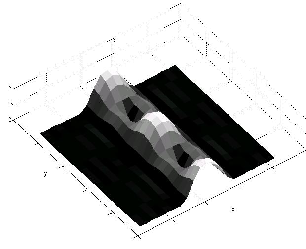
6 Fig.5. Numerically calculated excitation profile in the object domain. Since cos 2 (y) is a period of π, two peaks can be seen within a FOV. OFF-RESONANCE AND GRADIENT DELAY ARTIFACT While the forward-backward scheme in echo-planar pulse helps to save the time duration, it also causes serious artifact in the presence of off-resonance and gradient delay compared to flyback design. A generic non-flyback echo-planar trajectory can be viewed as a sum of two flyback trajectory scans as shown in Fig.6 in which case, the off-resonance is a negative value. With halved amplitudes, the final object is shifted along x-axis as well as y-axis because there is a linear phase not only in x-direction but also in y-direction. The even line scan and odd line scan cause the opposite shift along x-axis due to the reversed scan direction while the shift along y-direction is the same. And also, odd line scan in the k- space causes alternating sign of replicas in object domain because of the shifted sampling. The final result is a sum of two excitation shapes. Matlab simulation shows the expected results in Fig.7. Off-resonance of 50Hz, -100Hz, and 440Hz are used here. For 50Hz and 100Hz, artifact in x-axis is not noticeable because the scan time along x-axis is short compared to y-axis scanning. Artifact in x-axis is more visible with off-resonance of 440Hz. The object is separated in x-direction and also, incomplete destructive interference shows up halfway between Nyquist replicas. Phase inconsistency caused by eddy current, gradient imperfection or anisotropic gradient delay in echo-planar excitation also generates so-called N/2 ghosts as in echo-planar imaging. In a similar way to the above analysis, echo-planar trajectory can be thought of as a sum of even and odd line trajectories. Since gradient delay causes alternating shift in even and odd lines in the k-space, the corresponding object domain excitation has the opposite phase shift to each other as shown in Fig.8. As time delay gets greater, N/2 ghosts get bigger. Matlab simulation shows the consistent result as visualized in Fig.9.
7 Fig.6. Off-resonance artifact analysis in non-flyback echo-planar design. White circle denotes the opposite sign of the back circle.

")
8 y x (a) For f = -50Hz y x (b) For f = -100Hz

9 y x (c) For f = -440Hz Fig.7. Excitation shape change in the presence of off-resonance of 50Hz, -100Hz, and 440Hz.
10 Fig.8. Gradient delay artifact analysis in non-flyback echo-planar design. For a small time delay a, cos(a) is almost 1 and sin(a) is close to 0. As the delay gets greater, N/2 ghosting grows up.


 When the")
11 y x (a) When the gradient is delayed by 8% of the subpulse duration. y x (b) When the gradient is delayed by 12% of the subpulse duration.


12 y x (c) When the gradient is delayed by 16% of the subpulse duration. Fig.9. The artifact caused by gradient delay. With longer time delay, N/2 ghosting grows up. DISCUSSION The major drawback of 2D selective excitation is the longer RF pulse duration, which causes many artifacts in the presence of flow, motion, or off-resonance. To reduce the time duration, slew rate and maximum gradient limit should be utilized at the most, and echoplanar trajectory can be adopted. Especially, forward-backward scheme saves pulse duration twice as the flyback design because it deposits RF energy in the k-space during both positive and negative gradient lobes. At the same time, however, it is very susceptible to system imperfections such as off-resonance and gradient delay. In this study, how these artifacts affect the 2D excitation pulse in non-flyback echo-planar design has been examined. They show almost the same effect as in echo-planar imaging. Off-resonance imposes linear phase in the excitation k-space, which corresponds to shift in the object domain. Conversely, gradient delay generates shift in the k-space domain resulting in linear phase in the object domain. Due to the alternating shift (or linear phase) in the non-flyback k-space lines, the desired interference destruction in N/2 position of FOV becomes
13 incomplete, and therefore produces unwanted excitation. These artifacts can be resolved by the similar solutions from echo-planar imaging, and could be improved in further study. ACKNOWLEDGMENT binomial weighting has been adopted in this work at Kyunghyun Sung s suggestion. REFERENCES 1. Schroder C, Bornert P, Aldefeld B. Spatial excitation using variable-density spiral trajectories. J Magn Reson 2003;18: Rieseberg S, Frahm J, Finsterbusch J. Two-dimensional spatially-selective RF excitation pulses in echo-planar imaging. Magn Reson Med 2002;47: Oelhafen M, Pruessmann KP, Kozerke S, Boesiger P. Calibration of echo-planar 2Dselective RF excitation pulses. Magn Reson Med 2004;52: Pauly J, Nishimura D, Macovski A. A k-space analysis of small-tip angle excitation. J Magn Reson 1989;81: Hore PJ. Solvent suppression in Fourier transform nuclear magnetic resonance. J Magn Reson 1983;55: APPENDIX : MATLAB CODE close all; clear all; a=0.05; b=0.05; D=0; df=0;
14 Morig=[0;0;1]; gamma=26752; m=10^(-3); T=5.5*m; dt= 4*10^(-6); N=T/dt; %%%%% gx(t) generation for k=1:n t=dt*k; if (0<t& t<=m) (4*m<t& t<=5*m) gx(k)=4*pi*a/m/gamma; elseif (2*m<t& t<= 3*m) (5*m<t& t<=5.5*m) gx(k)=-4*pi*a/m/gamma; else gx(k)=0; %%%%% gy(t) generation for k=1:n t=dt*k; if (m<t& t<=2*m) (3*m<t & t<=4*m) gy(k)=2*b/m/gamma; elseif 5*m<t & t<=5.5*m gy(k)=-4*b/m/gamma; else gy(k)=0;
15 %%%%% b1(t) generation for k=1:n t=dt*k; if 0<t & t<=m b1(k)= sinc((t-0.5*m)/(m/2)) * sqrt( gx(k)^2+gy(k)^2 ); elseif 2*m<t & t<=3*m b1(k)= 2*sinc((t-2.5*m)/(m/2))* sqrt( gx(k)^2+gy(k)^2 ); elseif 4*m<t & t<=5*m b1(k)= sinc((t-4.5*m)/(m/2))* sqrt( gx(k)^2+gy(k)^2 ); else b1(k)= 0; %%%% gradient delay generation if D~=0 for k=1:d gxd(k)=0; gyd(k)=0; for k=d+1:n gxd(k)=gx(k-d); gyd(k)=gy(k-d);
16 for k=1:n gx(k)=gxd(k); gy(k)=gyd(k); %%%% excitation process x=-2/a: 4/a/20 :2/a; y=-pi/b: 2*pi/b/20 :pi/b; for i=1:length(x) for j=1:length(y) M=simpulse2(b1,gx,gy,dt,Morig,x(i),y(j),df); Mxy(j,i)=sqrt( M(1)^2 + M(2)^2 ); %%%% pulses and Mxy plotting figure(1); subplot(3,1,1); plot(b1); subplot(3,1,2); plot(gx); subplot(3,1,3); plot(gy); figure(2); dispimage(mxy); xlabel('x'); ylabel('y');
Spatially selective RF excitation using k-space analysis
 Spatially selective RF excitation using k-space analysis Dimitrios Pantazis a, a Signal and Image Processing Institute, University of Southern California, Los Angeles, CA 90089-2564 Abstract This project
Evaluations of k-space Trajectories for Fast MR Imaging for project of the course EE591, Fall 2004
 Evaluations of k-space Trajectories for Fast MR Imaging for project of the course EE591, Fall 24 1 Alec Chi-Wah Wong Department of Electrical Engineering University of Southern California 374 McClintock
MRI Physics II: Gradients, Imaging
 MRI Physics II: Gradients, Imaging Douglas C., Ph.D. Dept. of Biomedical Engineering University of Michigan, Ann Arbor Magnetic Fields in MRI B 0 The main magnetic field. Always on (0.5-7 T) Magnetizes
K-Space Trajectories and Spiral Scan
 K-Space and Spiral Scan Presented by: Novena Rangwala nrangw2@uic.edu 1 Outline K-space Gridding Reconstruction Features of Spiral Sampling Pulse Sequences Mathematical Basis of Spiral Scanning Variations
Field Maps. 1 Field Map Acquisition. John Pauly. October 5, 2005
 Field Maps John Pauly October 5, 25 The acquisition and reconstruction of frequency, or field, maps is important for both the acquisition of MRI data, and for its reconstruction. Many of the imaging methods
(a Scrhon5 R2iwd b. P)jc%z 5. ivcr3. 1. I. ZOms Xn,s. 1E IDrAS boms. EE225E/BIOE265 Spring 2013 Principles of MRI. Assignment 8 Solutions
jc%z 5. ivcr3. 1. I. ZOms Xn,s. 1E IDrAS boms. EE225E/BIOE265 Spring 2013 Principles of MRI. Assignment 8 Solutions") EE225E/BIOE265 Spring 2013 Principles of MRI Miki Lustig Assignment 8 Solutions 1. Nishimura 7.1 P)jc%z 5 ivcr3. 1. I Due Wednesday April 10th, 2013 (a Scrhon5 R2iwd b 0 ZOms Xn,s r cx > qs 4-4 8ni6 4
EE225E/BIOE265 Spring 2013 Principles of MRI Miki Lustig Assignment 8 Solutions 1. Nishimura 7.1 P)jc%z 5 ivcr3. 1. I Due Wednesday April 10th, 2013 (a Scrhon5 R2iwd b 0 ZOms Xn,s r cx > qs 4-4 8ni6 4
Partial k-space Recconstruction
 Partial k-space Recconstruction John Pauly September 29, 2005 1 Motivation for Partial k-space Reconstruction a) Magnitude b) Phase In theory, most MRI images depict the spin density as a function of position,
Partial k-space Reconstruction
 Chapter 2 Partial k-space Reconstruction 2.1 Motivation for Partial k- Space Reconstruction a) Magnitude b) Phase In theory, most MRI images depict the spin density as a function of position, and hence
ADVANCED RECONSTRUCTION TECHNIQUES IN MRI - 2
 ADVANCED RECONSTRUCTION TECHNIQUES IN MRI - 2 Presented by Rahil Kothari PARTIAL FOURIER RECONSTRUCTION WHAT IS PARTIAL FOURIER RECONSTRUCTION? In Partial Fourier Reconstruction data is not collected symmetrically
Sampling, Ordering, Interleaving
 Sampling, Ordering, Interleaving Sampling patterns and PSFs View ordering Modulation due to transients Temporal modulations Slice interleaving Sequential, Odd/even, bit-reversed Arbitrary Other considerations:
k y 2k y,max k x 2k x,max
 EE225E/BIOE265 Spring 2013 Principles of MRI Miki Lustig Assignment 5 Solutions Due March 6th 2012 1. Finish reading Nishimura Ch. 5. 2. For the 16 turn spiral trajectory, plotted below, what is the a)
Advanced Imaging Trajectories
 Advanced Imaging Trajectories Cartesian EPI Spiral Radial Projection 1 Radial and Projection Imaging Sample spokes Radial out : from k=0 to kmax Projection: from -kmax to kmax Trajectory design considerations
Clinical Importance. Aortic Stenosis. Aortic Regurgitation. Ultrasound vs. MRI. Carotid Artery Stenosis
 Clinical Importance Rapid cardiovascular flow quantitation using sliceselective Fourier velocity encoding with spiral readouts Valve disease affects 10% of patients with heart disease in the U.S. Most
Exam 8N080 - Introduction MRI
 Exam 8N080 - Introduction MRI Friday January 23 rd 2015, 13.30-16.30h For this exam you may use an ordinary calculator (not a graphical one). In total there are 6 assignments and a total of 65 points can
Midterm Review
 Midterm Review - 2017 EE369B Concepts Noise Simulations with Bloch Matrices, EPG Gradient Echo Imaging 1 About the Midterm Monday Oct 30, 2017. CCSR 4107 Up to end of C2 1. Write your name legibly on this
Imaging Notes, Part IV
 BME 483 MRI Notes 34 page 1 Imaging Notes, Part IV Slice Selective Excitation The most common approach for dealing with the 3 rd (z) dimension is to use slice selective excitation. This is done by applying
Role of Parallel Imaging in High Field Functional MRI
 Role of Parallel Imaging in High Field Functional MRI Douglas C. Noll & Bradley P. Sutton Department of Biomedical Engineering, University of Michigan Supported by NIH Grant DA15410 & The Whitaker Foundation
EE225E/BIOE265 Spring 2011 Principles of MRI. Assignment 5. Solutions
 EE225E/BIOE265 Spring 211 Principles of MRI Miki Lustig Handout Assignment 5 Solutions 1. Matlab Exercise: 2DFT Pulse sequence design. In this assignment we will write functions to design a 2DFT pulse
Improved Spatial Localization in 3D MRSI with a Sequence Combining PSF-Choice, EPSI and a Resolution Enhancement Algorithm
 Improved Spatial Localization in 3D MRSI with a Sequence Combining PSF-Choice, EPSI and a Resolution Enhancement Algorithm L.P. Panych 1,3, B. Madore 1,3, W.S. Hoge 1,3, R.V. Mulkern 2,3 1 Brigham and
Development of fast imaging techniques in MRI From the principle to the recent development
 980-8575 2-1 2012 10 13 Development of fast imaging techniques in MRI From the principle to the recent development Yoshio MACHIDA and Issei MORI Health Sciences, Tohoku University Graduate School of Medicine
Fast Imaging Trajectories: Non-Cartesian Sampling (1)
") Fast Imaging Trajectories: Non-Cartesian Sampling (1) M229 Advanced Topics in MRI Holden H. Wu, Ph.D. 2018.05.03 Department of Radiological Sciences David Geffen School of Medicine at UCLA Class Business
Fast Imaging Trajectories: Non-Cartesian Sampling (1) M229 Advanced Topics in MRI Holden H. Wu, Ph.D. 2018.05.03 Department of Radiological Sciences David Geffen School of Medicine at UCLA Class Business
Compressed Sensing for Rapid MR Imaging
 Compressed Sensing for Rapid Imaging Michael Lustig1, Juan Santos1, David Donoho2 and John Pauly1 1 Electrical Engineering Department, Stanford University 2 Statistics Department, Stanford University rapid
Sources of Distortion in Functional MRI Data
 Human Brain Mapping 8:80 85(1999) Sources of Distortion in Functional MRI Data Peter Jezzard* and Stuart Clare FMRIB Centre, Department of Clinical Neurology, University of Oxford, Oxford, UK Abstract:
HST.583 Functional Magnetic Resonance Imaging: Data Acquisition and Analysis Fall 2008
 MIT OpenCourseWare http://ocw.mit.edu HST.583 Functional Magnetic Resonance Imaging: Data Acquisition and Analysis Fall 2008 For information about citing these materials or our Terms of Use, visit: http://ocw.mit.edu/terms.
Removal of EPI Nyquist Ghost Artifacts With Two- Dimensional Phase Correction
 Removal of EPI Nyquist Ghost Artifacts With Two- Dimensional Phase Correction Nan-kuei Chen 1,5 and Alice M. Wyrwicz 4 * Magnetic Resonance in Medicine 51:147 153 (004) Odd even echo inconsistencies result
Slide 1. Technical Aspects of Quality Control in Magnetic Resonance Imaging. Slide 2. Annual Compliance Testing. of MRI Systems.
 Slide 1 Technical Aspects of Quality Control in Magnetic Resonance Imaging Slide 2 Compliance Testing of MRI Systems, Ph.D. Department of Radiology Henry Ford Hospital, Detroit, MI Slide 3 Compliance Testing
k y 2k y,max k x 2k x,max
 EE225E/BIOE265 Spring 2012 Principles of MRI Miki Lustig Assignment 5 Due Feb 26, 2012 1. Finish reading Nishimura Ch. 5. 2. For the 16 turn spiral trajectory, plotted below, what is the a) Spatial resolution,
Open file format for MR sequences
 Open file format for MR sequences Version 1.1 Kelvin Layton Maxim Zaitsev University Medical Centre Freiburg kelvin.layton@uniklinik-freiburg.de maxim.zaitsev@uniklinik-freiburg.de This file specification
TOPICS 2/5/2006 8:17 PM. 2D Acquisition 3D Acquisition
 TOPICS 2/5/2006 8:17 PM 2D Acquisition 3D Acquisition 2D Acquisition Involves two main steps : Slice Selection Slice selection is accomplished by spatially saturating (single or multi slice imaging) or
3-D Gradient Shimming
 3-D Gradient Shimming 3-D Gradient Shimming on imaging and micro-imaging systems Technical Overview Introduction Advantage statement The GE3DSHIM method is a 3-D gradient shimming procedure developed for
Diffusion MRI Acquisition. Karla Miller FMRIB Centre, University of Oxford
 Diffusion MRI Acquisition Karla Miller FMRIB Centre, University of Oxford karla@fmrib.ox.ac.uk Diffusion Imaging How is diffusion weighting achieved? How is the image acquired? What are the limitations,
surface Image reconstruction: 2D Fourier Transform
 2/1/217 Chapter 2-3 K-space Intro to k-space sampling (chap 3) Frequenc encoding and Discrete sampling (chap 2) Point Spread Function K-space properties K-space sampling principles (chap 3) Basic Contrast
Automatic Correction of Echo-Planar Imaging (EPI) Ghosting Artifacts in Real-Time Interactive Cardiac MRI Using Sensitivity Encoding
 Ghosting Artifacts in Real-Time Interactive Cardiac MRI Using Sensitivity Encoding") JOURNAL OF MAGNETIC RESONANCE IMAGING 27:239 245 (2008) Technical Note Automatic Correction of Echo-Planar Imaging (EPI) Ghosting Artifacts in Real-Time Interactive Cardiac MRI Using Sensitivity Encoding
JOURNAL OF MAGNETIC RESONANCE IMAGING 27:239 245 (2008) Technical Note Automatic Correction of Echo-Planar Imaging (EPI) Ghosting Artifacts in Real-Time Interactive Cardiac MRI Using Sensitivity Encoding
Module 4. K-Space Symmetry. Review. K-Space Review. K-Space Symmetry. Partial or Fractional Echo. Half or Partial Fourier HASTE
 MRES 7005 - Fast Imaging Techniques Module 4 K-Space Symmetry Review K-Space Review K-Space Symmetry Partial or Fractional Echo Half or Partial Fourier HASTE Conditions for successful reconstruction Interpolation
Motion Artifacts and Suppression in MRI At a Glance
 Motion Artifacts and Suppression in MRI At a Glance Xiaodong Zhong, PhD MR R&D Collaborations Siemens Healthcare MRI Motion Artifacts and Suppression At a Glance Outline Background Physics Common Motion
MR-Encephalography (MREG)
") MR-Encephalography (MREG) C2P package Version 2.0 For SIEMENS Magnetom Verio/TRIO/Skyra/Prisma Installation and User s Guide NUMARIS/4 VB17, VD11, VD13 Jakob Assländer Pierre LeVan, Ph.D. 10.09.2014 Magnetic
MR-Encephalography (MREG) C2P package Version 2.0 For SIEMENS Magnetom Verio/TRIO/Skyra/Prisma Installation and User s Guide NUMARIS/4 VB17, VD11, VD13 Jakob Assländer Pierre LeVan, Ph.D. 10.09.2014 Magnetic
MRI Imaging Options. Frank R. Korosec, Ph.D. Departments of Radiology and Medical Physics University of Wisconsin Madison
 MRI Imaging Options Frank R. Korosec, Ph.D. Departments of Radiolog and Medical Phsics Universit of Wisconsin Madison f.korosec@hosp.wisc.edu As MR imaging becomes more developed, more imaging options
Magnetic Resonance Imaging (MRI)
") C. A. Bouman: Digital Image Processing - January 12, 215 1 Magnetic Resonance Imaging (MRI) Can be very high resolution No radiation exposure Very flexible and programable Tends to be expensive, noisy,
C. A. Bouman: Digital Image Processing - January 12, 215 1 Magnetic Resonance Imaging (MRI) Can be very high resolution No radiation exposure Very flexible and programable Tends to be expensive, noisy,
Correction for EPI Distortions Using Multi-Echo Gradient-Echo Imaging
 Correction for EPI Distortions Using Multi-Echo Gradient-Echo Imaging Nan-kuei Chen and Alice M. Wyrwicz* Magnetic Resonance in Medicine 41:1206 1213 (1999) A novel and effective technique is described
White Pixel Artifact. Caused by a noise spike during acquisition Spike in K-space <--> sinusoid in image space
 White Pixel Artifact Caused by a noise spike during acquisition Spike in K-space sinusoid in image space Susceptibility Artifacts Off-resonance artifacts caused by adjacent regions with different
M R I Physics Course
 M R I Physics Course Multichannel Technology & Parallel Imaging Nathan Yanasak, Ph.D. Jerry Allison Ph.D. Tom Lavin, B.S. Department of Radiology Medical College of Georgia References: 1) The Physics of
Steen Moeller Center for Magnetic Resonance research University of Minnesota
 Steen Moeller Center for Magnetic Resonance research University of Minnesota moeller@cmrr.umn.edu Lot of material is from a talk by Douglas C. Noll Department of Biomedical Engineering Functional MRI Laboratory
VD-AUTO-SMASH Imaging
 Magnetic Resonance in Medicine 45:1066 1074 (2001) VD-AUTO-SMASH Imaging Robin M. Heidemann, Mark A. Griswold, Axel Haase, and Peter M. Jakob* Recently a self-calibrating SMASH technique, AUTO-SMASH, was
EE290T: Advanced Reconstruction Methods for Magnetic Resonance Imaging. Martin Uecker
 EE290T: Advanced Reconstruction Methods for Magnetic Resonance Imaging Martin Uecker Tentative Syllabus 01: Jan 27 Introduction 02: Feb 03 Parallel Imaging as Inverse Problem 03: Feb 10 Iterative Reconstruction
Dynamic Image and Fieldmap Joint Estimation Methods for MRI Using Single-Shot Trajectories
 Dynamic Image and Fieldmap Joint Estimation Methods for MRI Using Single-Shot Trajectories by Antonis Matakos A dissertation submitted in partial fulfillment of the requirements for the degree of Doctor
Dynamic Autocalibrated Parallel Imaging Using Temporal GRAPPA (TGRAPPA)
") Magnetic Resonance in Medicine 53:981 985 (2005) Dynamic Autocalibrated Parallel Imaging Using Temporal GRAPPA (TGRAPPA) Felix A. Breuer, 1 * Peter Kellman, 2 Mark A. Griswold, 1 and Peter M. Jakob 1 Current
Magnetic Resonance in Medicine 53:981 985 (2005) Dynamic Autocalibrated Parallel Imaging Using Temporal GRAPPA (TGRAPPA) Felix A. Breuer, 1 * Peter Kellman, 2 Mark A. Griswold, 1 and Peter M. Jakob 1 Current
Referenceless Interleaved Echo-Planar Imaging
 Referenceless Interleaved Echo-Planar Imaging Magnetic Resonance in Medicine 41:87 94 (1999) Scott B. Reeder, 1 Ergin Atalar, * Anthony Z. Faranesh, 1 and Elliot R. McVeigh 1, Interleaved echo-planar imaging
Spiral Imaging: A Critical Appraisal
 JOURNAL OF MAGNETIC RESONANCE IMAGING 21:657 668 (2005) Review Article Spiral Imaging: A Critical Appraisal Kai Tobias Block, MSc and Jens Frahm, PhD* In view of recent applications in cardiovascular and
An Intuitive Explanation of Fourier Theory
 An Intuitive Explanation of Fourier Theory Steven Lehar slehar@cns.bu.edu Fourier theory is pretty complicated mathematically. But there are some beautifully simple holistic concepts behind Fourier theory
High Spatial Resolution EPI Using an Odd Number of Interleaves
 Magnetic Resonance in Medicine 41:1199 1205 (1999) High Spatial Resolution EPI Using an Odd Number of Interleaves Michael H. Buonocore* and David C. Zhu Ghost artifacts in echoplanar imaging (EPI) arise
Classification of Subject Motion for Improved Reconstruction of Dynamic Magnetic Resonance Imaging
 1 CS 9 Final Project Classification of Subject Motion for Improved Reconstruction of Dynamic Magnetic Resonance Imaging Feiyu Chen Department of Electrical Engineering ABSTRACT Subject motion is a significant
Computational Aspects of MRI
 David Atkinson Philip Batchelor David Larkman Programme 09:30 11:00 Fourier, sampling, gridding, interpolation. Matrices and Linear Algebra 11:30 13:00 MRI Lunch (not provided) 14:00 15:30 SVD, eigenvalues.
XI Signal-to-Noise (SNR)
") XI Signal-to-Noise (SNR) Lecture notes by Assaf Tal n(t) t. Noise. Characterizing Noise Noise is a random signal that gets added to all of our measurements. In D it looks like this: while in D
XI Signal-to-Noise (SNR) Lecture notes by Assaf Tal n(t) t. Noise. Characterizing Noise Noise is a random signal that gets added to all of our measurements. In D it looks like this: while in D
Digital Image Processing. Lecture # 3 Image Enhancement
 Digital Image Processing Lecture # 3 Image Enhancement 1 Image Enhancement Image Enhancement 3 Image Enhancement 4 Image Enhancement Process an image so that the result is more suitable than the original
Sampling, Ordering, Interleaving
 Sampling, Ordering, Interleaving Sampling patterns and PSFs View ordering Modulation due to transients Temporal modulations Timing: cine, gating, triggering Slice interleaving Sequential, Odd/even, bit-reversed
Following on from the two previous chapters, which considered the model of the
 Chapter 5 Simulator validation Following on from the two previous chapters, which considered the model of the simulation process and how this model was implemented in software, this chapter is concerned
CHAPTER 9: Magnetic Susceptibility Effects in High Field MRI
 Figure 1. In the brain, the gray matter has substantially more blood vessels and capillaries than white matter. The magnified image on the right displays the rich vasculature in gray matter forming porous,
Applications Guide for Interleaved
 Applications Guide for Interleaved rephase/dephase MRAV Authors: Yongquan Ye, Ph.D. Dongmei Wu, MS. Tested MAGNETOM Systems : 7TZ, TRIO a Tim System, Verio MR B15A (N4_VB15A_LATEST_20070519) MR B17A (N4_VB17A_LATEST_20090307_P8)
IMAGE reconstruction in conventional magnetic resonance
 IEEE TRANSACTIONS ON MEDICAL IMAGING, VOL. 24, NO. 3, MARCH 2005 325 Conjugate Phase MRI Reconstruction With Spatially Variant Sample Density Correction Douglas C. Noll*, Member, IEEE, Jeffrey A. Fessler,
Fast Imaging UCLA. Class Business. Class Business. Daniel B. Ennis, Ph.D. Magnetic Resonance Research Labs. Tuesday (3/7) from 6-9pm HW #1 HW #2
 from 6-9pm HW #1 HW #2") Fast Imaging Daniel B. Ennis, Ph.D. Magnetic Resonance Research Labs Class Business Tuesday (3/7) from 6-9pm 6:00-7:30pm Groups Avanto Sara Said, Yara Azar, April Pan Skyra Timothy Marcum, Diana Lopez,
Fast Imaging Daniel B. Ennis, Ph.D. Magnetic Resonance Research Labs Class Business Tuesday (3/7) from 6-9pm 6:00-7:30pm Groups Avanto Sara Said, Yara Azar, April Pan Skyra Timothy Marcum, Diana Lopez,
FOCUS. Image Based Automatic Shimming Using B 0 Gradients. Installation and Users Guide Version 0.9
 FOCUS Image Based Automatic Shimming Using B 0 Gradients Installation and Users Guide Version 0.9 FOCUS - Field Optimization by Computed Update of Shims Joost A. B. Lohman, Bruker Spectrospin ltd, U.K.
Module 5: Dynamic Imaging and Phase Sharing. (true-fisp, TRICKS, CAPR, DISTAL, DISCO, HYPR) Review. Improving Temporal Resolution.
 Review. Improving Temporal Resolution.") MRES 7005 - Fast Imaging Techniques Module 5: Dynamic Imaging and Phase Sharing (true-fisp, TRICKS, CAPR, DISTAL, DISCO, HYPR) Review Improving Temporal Resolution True-FISP (I) True-FISP (II) Keyhole
MRES 7005 - Fast Imaging Techniques Module 5: Dynamic Imaging and Phase Sharing (true-fisp, TRICKS, CAPR, DISTAL, DISCO, HYPR) Review Improving Temporal Resolution True-FISP (I) True-FISP (II) Keyhole
Sparse sampling in MRI: From basic theory to clinical application. R. Marc Lebel, PhD Department of Electrical Engineering Department of Radiology
 Sparse sampling in MRI: From basic theory to clinical application R. Marc Lebel, PhD Department of Electrical Engineering Department of Radiology Objective Provide an intuitive overview of compressed sensing
Use of Multicoil Arrays for Separation of Signal from Multiple Slices Simultaneously Excited
 JOURNAL OF MAGNETIC RESONANCE IMAGING 13:313 317 (2001) Technical Note Use of Multicoil Arrays for Separation of Signal from Multiple Slices Simultaneously Excited David J. Larkman, PhD, 1 * Joseph V.
The SIMRI project A versatile and interactive MRI simulator *
 COST B21 Meeting, Lodz, 6-9 Oct. 2005 The SIMRI project A versatile and interactive MRI simulator * H. Benoit-Cattin 1, G. Collewet 2, B. Belaroussi 1, H. Saint-Jalmes 3, C. Odet 1 1 CREATIS, UMR CNRS
Edge and local feature detection - 2. Importance of edge detection in computer vision
 Edge and local feature detection Gradient based edge detection Edge detection by function fitting Second derivative edge detectors Edge linking and the construction of the chain graph Edge and local feature
HST.583 Functional Magnetic Resonance Imaging: Data Acquisition and Analysis Fall 2006
 MIT OpenCourseWare http://ocw.mit.edu HST.583 Functional Magnetic Resonance Imaging: Data Acquisition and Analysis Fall 2006 For information about citing these materials or our Terms of Use, visit: http://ocw.mit.edu/terms.
Dynamic Contrast enhanced MRA
 Dynamic Contrast enhanced MRA Speaker: Yung-Chieh Chang Date : 106.07.22 Department of Radiology, Taichung Veterans General Hospital, Taichung, Taiwan 1 Outline Basic and advanced principles of Diffusion
FOV. ] are the gradient waveforms. The reconstruction of this signal proceeds by an inverse Fourier Transform as:. [2] ( ) ( )
![FOV. ] are the gradient waveforms. The reconstruction of this signal proceeds by an inverse Fourier Transform as:. [2] ( ) ( )](/thumbs/86/94912904.jpg "FOV. ] are the gradient waveforms. The reconstruction of this signal proceeds by an inverse Fourier Transform as:. [2] ( ) ( )") Gridding Procedures for Non-Cartesian K-space Trajectories Douglas C. Noll and Bradley P. Sutton Dept. of Biomedical Engineering, University of Michigan, Ann Arbor, MI, USA 1. Introduction The data collected
Gridding Procedures for Non-Cartesian K-space Trajectories Douglas C. Noll and Bradley P. Sutton Dept. of Biomedical Engineering, University of Michigan, Ann Arbor, MI, USA 1. Introduction The data collected
Introduction to Digital Image Processing
 Introduction to Digital Image Processing Ranga Rodrigo June 9, 29 Outline Contents Introduction 2 Point Operations 2 Histogram Processing 5 Introduction We can process images either in spatial domain or
Non-Cartesian Parallel Magnetic Resonance Imaging
 Non-Cartesian Parallel Magnetic Resonance Imaging Dissertation zur Erlangung des naturwissenschaftlichen Doktorgrades der Bayerischen Julius-Maximilians-Universität Würzburg vorgelegt von Robin Heidemann
Computational Medical Imaging Analysis
 Computational Medical Imaging Analysis Chapter 2: Image Acquisition Systems Jun Zhang Laboratory for Computational Medical Imaging & Data Analysis Department of Computer Science University of Kentucky
ARTICLE IN PRESS YNIMG-03684; No. of pages: 14; 4C:
 YNIMG-03684; No. of pages: 14; 4C: DTD 5 Technical note Application of k-space energy spectrum analysis to susceptibility field mapping and distortion correction in gradient-echo EPI Nan-kuei Chen, a,
Automatic Gradient Preemphasis Adjustment: A 15-Minute Journey to Improved Diffusion-Weighted Echo-Planar Imaging
 Automatic Gradient Preemphasis Adjustment: A 15-Minute Journey to Improved Diffusion-Weighted Echo-Planar Imaging Vincent J. Schmithorst* and Bernard J. Dardzinski Magnetic Resonance in Medicine 47:208
CoE4TN4 Image Processing. Chapter 5 Image Restoration and Reconstruction
 CoE4TN4 Image Processing Chapter 5 Image Restoration and Reconstruction Image Restoration Similar to image enhancement, the ultimate goal of restoration techniques is to improve an image Restoration: a
HST.583 Functional Magnetic Resonance Imaging: Data Acquisition and Analysis Fall 2008
 MIT OpenCourseWare http://ocw.mit.edu HST.583 Functional Magnetic Resonance Imaging: Data Acquisition and Analysis Fall 2008 For information about citing these materials or our Terms of Use, visit: http://ocw.mit.edu/terms.
Zigzag Sampling for Improved Parallel Imaging
 Magnetic Resonance in Medicine 60:474 478 (2008) Zigzag Sampling for Improved Parallel Imaging Felix A. Breuer, 1 * Hisamoto Moriguchi, 2 Nicole Seiberlich, 3 Martin Blaimer, 1 Peter M. Jakob, 1,3 Jeffrey
3. Image formation, Fourier analysis and CTF theory. Paula da Fonseca
 3. Image formation, Fourier analysis and CTF theory Paula da Fonseca EM course 2017 - Agenda - Overview of: Introduction to Fourier analysis o o o o Sine waves Fourier transform (simple examples of 1D
Motion artifact reduction technique for dual-contrast FSE imaging
 Magnetic Resonance Imaging 20 (2002) 455 462 Motion artifact reduction technique for dual-contrast FSE imaging Eugene G. Kholmovski a, *, Alexei A. Samsonov a, Dennis L. Parker a,b a Department of Physics,
Unaliasing by Fourier-Encoding the Overlaps Using the Temporal Dimension (UNFOLD), Applied to Cardiac Imaging and fmri
, Applied to Cardiac Imaging and fmri") 1999 ISMRM YOUNG INVESTIGATORS MOORE AWARD PAPERS Magnetic Resonance in Medicine 42:813 828 (1999) Unaliasing by Fourier-Encoding the Overlaps Using the Temporal Dimension (UNFOLD), Applied to Cardiac
1999 ISMRM YOUNG INVESTIGATORS MOORE AWARD PAPERS Magnetic Resonance in Medicine 42:813 828 (1999) Unaliasing by Fourier-Encoding the Overlaps Using the Temporal Dimension (UNFOLD), Applied to Cardiac
CANAL FOLLOWING USING AR DRONE IN SIMULATION
 CANAL FOLLOWING USING AR DRONE IN SIMULATION ENVIRONMENT Ali Ahmad, Ahmad Aneeque Khalid Department of Electrical Engineering SBA School of Science & Engineering, LUMS, Pakistan {14060006, 14060019}@lums.edu.pk
Supplementary methods
 Supplementary methods This section provides additional technical details on the sample, the applied imaging and analysis steps and methods. Structural imaging Trained radiographers placed all participants
Lecture 4. Digital Image Enhancement. 1. Principle of image enhancement 2. Spatial domain transformation. Histogram processing
 Lecture 4 Digital Image Enhancement 1. Principle of image enhancement 2. Spatial domain transformation Basic intensity it tranfomation ti Histogram processing Principle Objective of Enhancement Image enhancement
MRI image formation 8/3/2016. Disclosure. Outlines. Chen Lin, PhD DABR 3. Indiana University School of Medicine and Indiana University Health
 MRI image formation Indiana University School of Medicine and Indiana University Health Disclosure No conflict of interest for this presentation 2 Outlines Data acquisition Spatial (Slice/Slab) selection
Accelerated MRI Techniques: Basics of Parallel Imaging and Compressed Sensing
 Accelerated MRI Techniques: Basics of Parallel Imaging and Compressed Sensing Peng Hu, Ph.D. Associate Professor Department of Radiological Sciences PengHu@mednet.ucla.edu 310-267-6838 MRI... MRI has low
COBRE Scan Information
 COBRE Scan Information Below is more information on the directory structure for the COBRE imaging data. Also below are the imaging parameters for each series. Directory structure: var/www/html/dropbox/1139_anonymized/human:
General and Efficient Super-Resolution Method for Multi-slice MRI
 General and Efficient Super-Resolution Method for Multi-slice MRI D.H.J. Poot 1,2,V.VanMeir 2, and J. Sijbers 2 1 BIGR, Erasmus Medical Center, Rotterdam 2 Visionlab, University of Antwerp, Antwerp Abstract.
Spread Spectrum Using Chirp Modulated RF Pulses for Incoherent Sampling Compressive Sensing MRI
 Spread Spectrum Using Chirp Modulated RF Pulses for Incoherent Sampling Compressive Sensing MRI Sulaiman A. AL Hasani Department of ECSE, Monash University, Melbourne, Australia Email: sulaiman.alhasani@monash.edu
Experimental Competition. Sample Solution
 The 37th International Physics Olympiad Singapore Experimental Competition Wednesday, 1 July, 006 Sample Solution a. A sketch of the experimental setup Part 1 Receiver Rotating table Goniometer Fixed arm
SIMULATION D'IRM ANGIOGRAPHIQUE PAR EXTENSION DU LOGICIEL JEMRIS
 SIMULATION D'IRM ANGIOGRAPHIQUE PAR EXTENSION DU LOGICIEL JEMRIS Alexandre FORTIN Supervised by Emmanuel DURAND and Stéphanie SALMON Laboratoire de Mathématiques de Reims Model : clipart-fr.com VIVABRAIN
Magnetic Resonance Angiography
 Magnetic Resonance Angiography Course: Advance MRI (BIOE 594) Instructors: Dr Xiaohong Joe Zhou Dr. Shadi Othman By, Nayan Pasad Phase Contrast Angiography By Moran 1982, Bryan et. Al. 1984 and Moran et.
Single-Shot Spiral Imaging Enabled by an Expanded Encoding Model: Demonstration in Diffusion MRI
 RAPID COMMUNICATION Magnetic Resonance in Medicine 77:83 91 (2017) Single-Shot Spiral Imaging Enabled by an Expanded Encoding Model: Demonstration in Diffusion MRI Bertram J. Wilm, 1 * Christoph Barmet,
SUPPLEMENTARY FILE S1: 3D AIRWAY TUBE RECONSTRUCTION AND CELL-BASED MECHANICAL MODEL. RELATED TO FIGURE 1, FIGURE 7, AND STAR METHODS.
 SUPPLEMENTARY FILE S1: 3D AIRWAY TUBE RECONSTRUCTION AND CELL-BASED MECHANICAL MODEL. RELATED TO FIGURE 1, FIGURE 7, AND STAR METHODS. 1. 3D AIRWAY TUBE RECONSTRUCTION. RELATED TO FIGURE 1 AND STAR METHODS
11.2 RECTANGULAR COORDINATES IN THREE DIMENSIONS
 11.2 Rectangular Coordinates in Three Dimensions Contemporary Calculus 1 11.2 RECTANGULAR COORDINATES IN THREE DIMENSIONS In this section we move into 3 dimensional space. First we examine the 3 dimensional
Single-sided Magnet Diffusion Measurements
 Single-sided Magnet Diffusion Measurements Kuldeep S. Panesar April 2013 (kuldeep.panesar@gmail.com) Contents 1. Initial configuration... 2 2. Preparation for diffusion measurement... 5 3. Starting a diffusion
Fast Isotropic Volumetric Coronary MR Angiography Using Free-Breathing 3D Radial Balanced FFE Acquisition
 Fast Isotropic Volumetric Coronary MR Angiography Using Free-Breathing 3D Radial Balanced FFE Acquisition C. Stehning, 1 * P. Börnert, 2 K. Nehrke, 2 H. Eggers, 2 and O. Dössel 1 Magnetic Resonance in
Lab Location: MRI, B2, Cardinal Carter Wing, St. Michael s Hospital, 30 Bond Street
 Lab Location: MRI, B2, Cardinal Carter Wing, St. Michael s Hospital, 30 Bond Street MRI is located in the sub basement of CC wing. From Queen or Victoria, follow the baby blue arrows and ride the CC south
MRI Reconstruction using Real-Time Motion Tracking: A Simulation Study
 MRI Reconstruction using Real-Time Motion Tracking: A Simulation Study Jeff Orchard David R. Cheriton School of Computer Science University of Waterloo Waterloo, ON Canada, N2L 3G1 e-mail: jorchard@uwaterloo.ca
Motion Robust Magnetic Susceptibility and Field Inhomogeneity Estimation Using Regularized Image Restoration Techniques for fmri
 Motion Robust Magnetic Susceptibility and Field Inhomogeneity Estimation Using Regularized Image Restoration Techniques for fmri Desmond Tec Beng Yeo 1,, Jeffrey A. Fessler 1,, and Bolye Kim 1 1 Department
2D Fan Beam Reconstruction 3D Cone Beam Reconstruction
 2D Fan Beam Reconstruction 3D Cone Beam Reconstruction Mario Koerner March 17, 2006 1 2D Fan Beam Reconstruction Two-dimensional objects can be reconstructed from projections that were acquired using parallel