Lorad FFDM QC Procedures for Medical Physicist. Tao Wu, Ph.D. Hologic, Inc.
|
|
|
- Sibyl Newman
- 5 years ago
- Views:
Transcription
1 Lorad FFDM QC Procedures for Medical Physicist Tao Wu, Ph.D. Hologic, Inc.

2 Lorad Selenia FFDM
3 Tests Following Lorad QC Manual Collimation Assessment Artifact Evaluation System Resolution Phantom Image Quality Evaluation Automatic Exposure Control Function Performance Automatic Exposure Control Reproducibility Signal-To-Noise and Contrast-To-Noise Measurements Softcopy Workstation QC
4 Acquisition Workstation Software


5 OS Login Medical Physicist username: phys password: phys

6 Detector Warm-Up After logging in OS, a dialog showing detector warm-up status appears If the system has been running - Dismiss If the system has been shut down - Wait for min (or dismiss it and start with tests in which detector is not involved, e.g. HVL, tube output)

7 Application Login Username:Andrea Password: Andrea
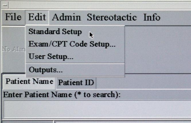
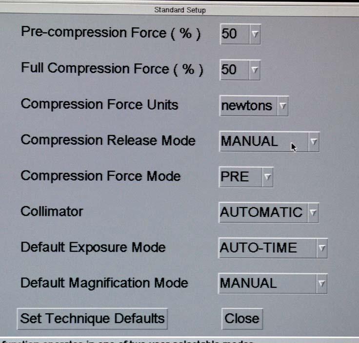
8 Turn Off Auto-decompression

9 Send Images to Output Devices


10 Send Images to Output Devices


11 Send Images to Output Devices
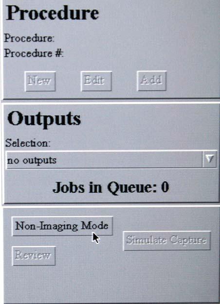
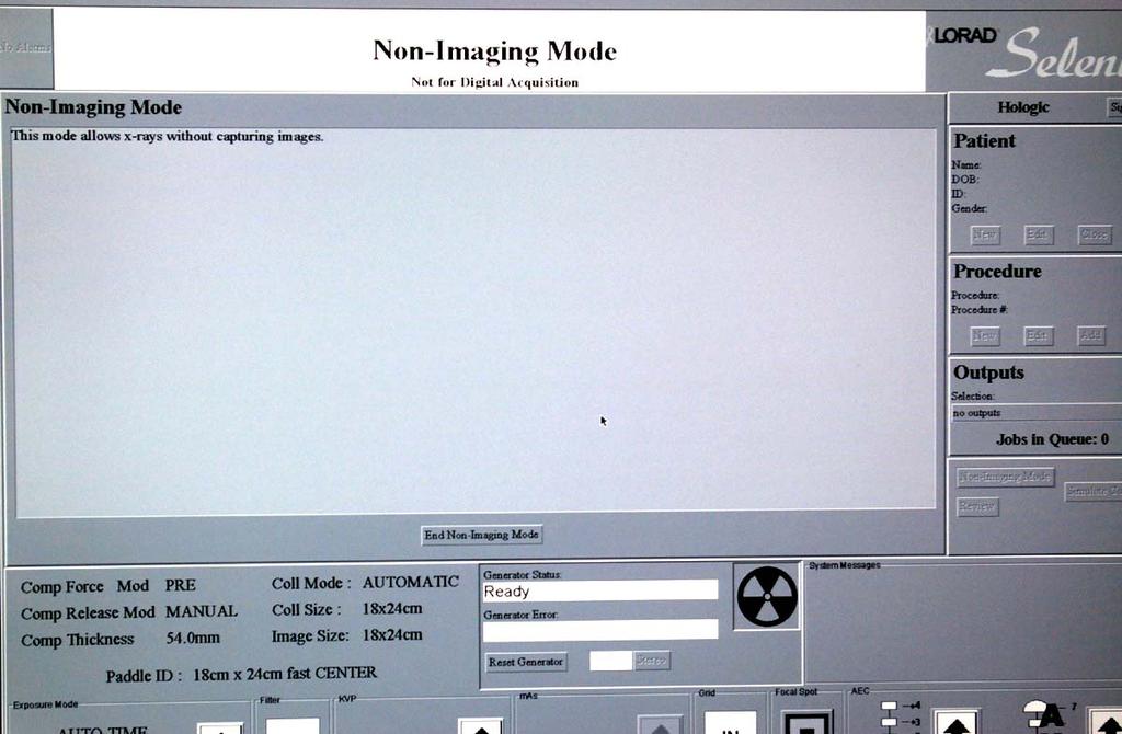
12 Non-Imaging Mode

13 Create a New Patient Account Select No Output

14 Create a New Patient Account

15 Create a New Patient Account
16 Selenia QC Tests
17 Tests Following Lorad QC Manual Collimation Assessment Artifact Evaluation System Resolution Phantom Image Quality Evaluation Automatic Exposure Control Function Performance Signal-To-Noise and Contrast-To-Noise Measurements Softcopy Workstation QC
18 Order of Testing Artifact Evaluation Phantom Image Quality Evaluation SNR and CNR Measurements AEC Function Performance Other Tests
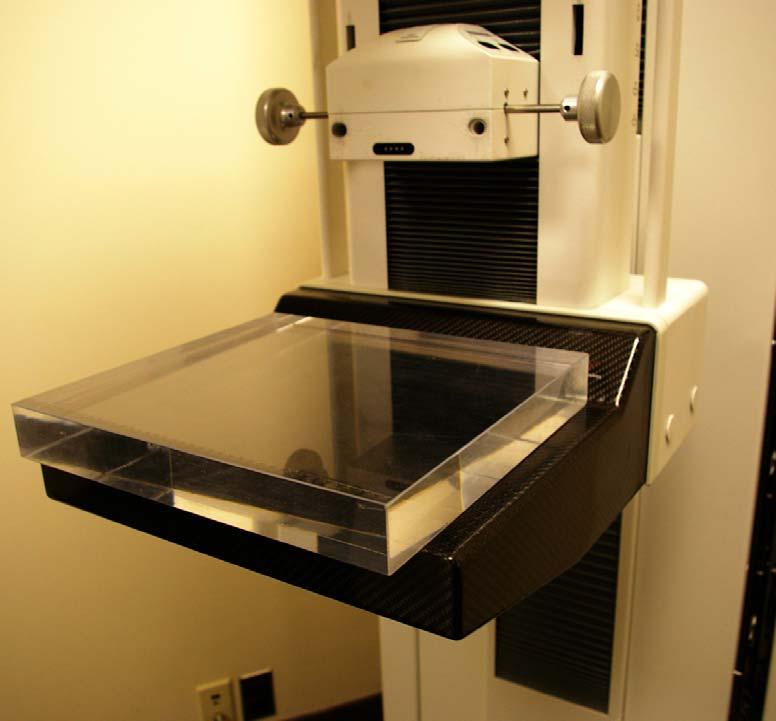
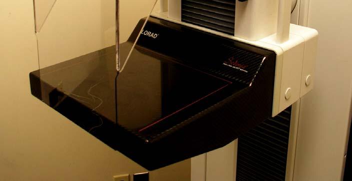
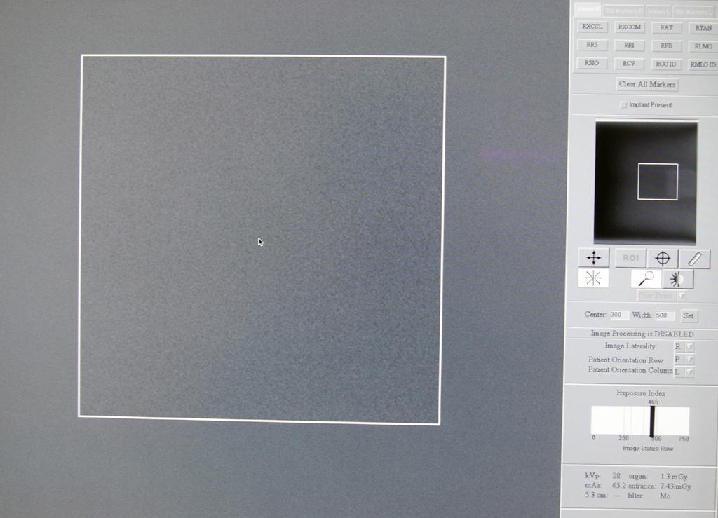
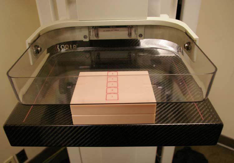
19 Artifact Evaluation Evaluation of detector, x-ray tube, filter etc 4 cm thick acrylic block No compression paddle Four flat field images Mo/Mo, contact mode, LFS with grid Mo/Rh, contact mode, LFS with grid Mo/Mo, mag mode, SFS without grid Mo/Rh, mag mode, SFS without grid Review the images on acquisition workstation monitor Window width = 500, Level = 350

20 Artifact Evaluation
21 Artifact Evaluation
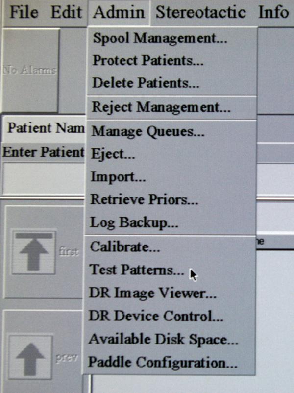
22 Evaluation of Printer Artifact Evaluation Select a computer-generated flat-field image Print the image and evaluate it Repeat the test for each printer If a printer is connected to multiple Selenia systems, it only needs to be tested once from one Selenia.

23 Artifact Evaluation
24 Phantom Image Quality Evaluation ACR phantom with acrylic disc AEC position 2 Print film Measure film densities - Background density > 1.20, with control limit ± Density difference over the acrylic disk > 0.40, with control limit ± 0.05 Phantom scoring (hard copy and softcopy station) 5 fibers, 4 speck groups, 4 masses

25 Phantom Image Quality Evaluation
26 SNR and CNR Measurements
27 SNR and CNR Measurements SNR 40 Establish the reference CNR value during the acceptance test CNR should stay within ±15% of the reference
28 Automatic Exposure Control (AEC) AEC modes Auto-Filter: Auto-kV: Auto-Time: filter, kvp, mas all automatically determined filter manually selected kvp and mas automatically determined filter and kvp manual selected mas automatically determined AEC Method - Filter and kvp are determined by breast thickness (in Auto- Filter and Auto-kV modes) - A pre-exposure is used to determine mas AEC positions Auto-AEC (The densest tissue gets enough exposure) Seven manual positions (marked on compression paddle)
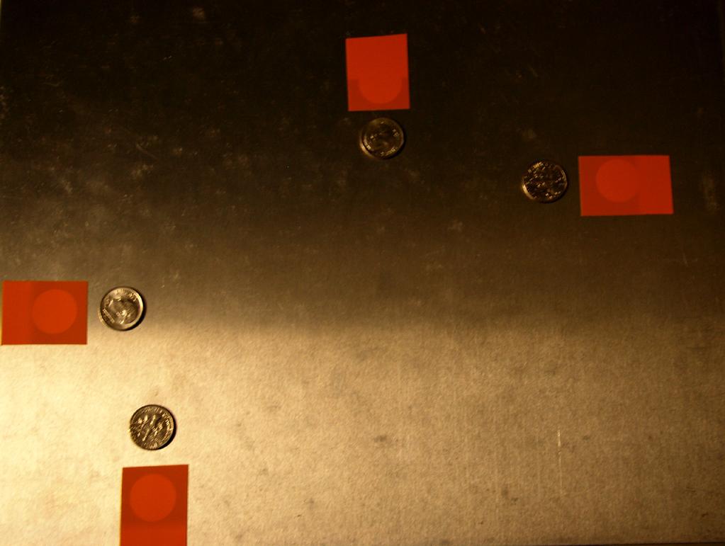
29 AEC Function Performance 1. Performance at different thickness and imaging modes 2. Performance at different compensation levels
30 AEC Function Performance 1. Performance at different thickness and imaging modes Acrylic or BR-12 blocks AEC position 2 Contact mode (LFS, grid): 2, 4, 6, 8 cm phantom Mag mode (SFS, no grid): 4 cm phantom Variation of target pixel values 10% of the mean target pixel value

31 AEC Function Performance
32 AEC Function Performance 2. Performance at different compensation levels 4 cm acrylic or BR-12 Compensation levels: Step -5 to Step +5 Use average target count at Step 0 as reference Compare target count at other steps to the reference Step Step Step Step Step Step Step Step Step Step
33 Collimation Assessment 1. X-Ray Field to Light Field 2. X-Ray Field to Image Receptor 3. Compression Paddle to Image Receptor

34 Collimation Assessment One test for cm imaging mode Three tests for cm imaging modes Left imaging position Center imaging position Right imaging position L M R

35 Collimation Assessment Use coin method as described in ACR manual
36 Collimation Assessment 1. X-Ray Field to Light Field Measure deviations between the edges of the light field (exterior edge of the coin) and the x- ray field at al four sides Sum of the magnitude of deviations on opposite sides 2% of the SID SID = 66 cm 2% of the SID = 1.32 cm
37 Collimation Assessment 2. X-Ray Field to Image Receptor d 1 : measured on film distance from the inner edge of the coin to the edge of the x-ray field d 2 : measured on digital image distance from the inner edge of the coin to the edge of the detector (d ) - d 2 The x-ray field shall extend beyond the chest wall side of the image receptor The x-ray field must NOT extend >2% of the SID at any of the four sides of the image receptor
38 Collimation Assessment 3. Compression Paddle to Image Receptor Measure distance from the outer edge of the coin to the edge of the image The anterior edge of the compression paddle must be aligned just beyond the chest wall edge of the image receptor, but 1% of the SID

39 Collimation Assessment
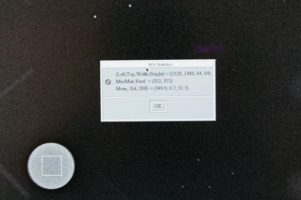
40 Collimation Assessment

41 Collimation Assessment

42 Evaluation of System Resolution Line pair phantom 4 cm acrylic block Line pair phantom placed at 45º to the detector row


43 Evaluation of System Resolution Limiting spatial resolution 7 c/mm (lp/mm)
44 Detector Ghosting (Optional) 1. Create ghosting effect 2. Quantify ghosting effect

45 Detector Ghosting (Optional) 1. Create ghosting effect Ghosting area

46 Detector Ghosting (Optional) 2. Quantify ghosting effect Ghosting area

47 Detector Ghosting (Optional) 2. Quantify ghosting effect Ghosting factor 0.3 ROI 1 in background without ghosting Ghosting Factor = ROI 2 in Al filter without ghosting mean region2 -mean region3 mean region1 -mean region2 ROI 3 in Al filter with ghosting
48 Thank you! Tao Wu Tel:
Unique Features of the GE Senoclaire Tomosynthesis System. Tyler Fisher, M.S., DABR Therapy Physics, Inc.
 Unique Features of the GE Senoclaire Tomosynthesis System Tyler Fisher, M.S., DABR Therapy Physics, Inc. Conflict of Interest Disclosure I have no conflicts to disclose. Learning Objectives Overview of
ACMP 25th Annual Meeting
 Surveying and QC of Stereotactic Breast Biopsy Units for ACR Accreditation ACMP 25th Annual Meeting Seattle, WA May 3, 2008 Melissa C. Martin, M.S., FACR Therapy Physics, Inc. 879 West 190th St., Ste 419,
Certificate Extension II 8/1/2016. Certification Extension Process for Digital Breast Tomosynthesis and Medical Physicists Role
 Certification Extension Process for Digital Breast Tomosynthesis and Medical Physicists Role Kish Chakrabarti, Ph.D., FAAPM Division of Mammography Quality Standards Center for Devices and Radiological
RECENTLY APPROVED GE FFDM DBT TESTING PROCEDURES.
 RECENTLY APPROVED GE FFDM DBT TESTING PROCEDURES. S. G U R U P R A S A D, P H. D., D A B R, E M E R I T U S, M AT H E W H A L L M. S. N O RT H S H O R E U N I V E R S I T Y H E A LT H S Y S T E M A N D
8/2/2016. Hands-on GE SenoClaire DBT: technical characteristics & quality control. Disclosures and Acknowledgements
 Hands-on GE SenoClaire DBT: technical characteristics & quality control Razvan Iordache, Ph.D. GE Healthcare Imagination at work Disclosures and Acknowledgements Razvan Iordache Employee of GE Healthcare
Z-MOTION. Universal Digital Radiographic System Z-MOTION. Control-X Medical CONTROL-X MEDICAL
 Control-X Medical Z-MOTION Compact design, low ceiling height requirement Motorized and manual movement capability Wide motion / SID range Best-in-class image quality Flexible connectivity to PACS systems
Assessment of 3D performance metrics. X-ray based Volumetric imaging systems: Fourier-based imaging metrics. The MTF in CT
 Assessment of 3D performance metrics D and 3D Metrics of Performance Towards Quality Index: Volumetric imaging systems X-ray based Volumetric imaging systems: CBCT/CT Tomosynthesis Samuel Richard and Ehsan
DISC QA Mamchex Tool Use Instructions with CARESTREAM DIRECTVIEW CR Mammography Systems
 DISC QA Mamchex Tool Use Instructions with CARESTREAM DIRECTVIEW CR Mammography Systems Purpose This document describes and provides instructions for use of the QA Mamchex Tool in calibrating a mammography
Scatter reduction for grid-less mammography using the convolutionbased image post-processing technique
 Scatter reduction for grid-less mammography using the convolutionbased image post-processing technique Elena Marimón *a,b, Hammadi Nait-Charif a, Asmar Khan b, Philip A. Marsden c, Oliver Diaz d a CDE
Introduction to Biomedical Imaging
 Alejandro Frangi, PhD Computational Imaging Lab Department of Information & Communication Technology Pompeu Fabra University www.cilab.upf.edu X-ray Projection Imaging Computed Tomography Digital X-ray
S. Guru Prasad, Ph.D., DABR
 PURPOSE S. Guru Prasad, Ph.D., DABR Director of Medical Physics IAEA Consultant NorthShore University Health System and University of Chicago, Pritzker School of Medicine Current TPS utilize more information
4/19/2016. Deborah Thames R.T. (R)(M)(QM) Theory & Technology and advancement in 3D imaging DBT
(M)(QM) Theory & Technology and advancement in 3D imaging DBT") Deborah Thames R.T. (R)(M)(QM) Theory & Technology and advancement in 3D imaging DBT 1 Three manufacturers approved for Tomo Hologic and GE, and Siemens Why 2D Digital Mammography 2D FFDM it appears to
Deborah Thames R.T. (R)(M)(QM) Theory & Technology and advancement in 3D imaging DBT 1 Three manufacturers approved for Tomo Hologic and GE, and Siemens Why 2D Digital Mammography 2D FFDM it appears to
SUMMARY OF DENTAL MEASUREMENT PROCEDURES (Abridged Protocol)
") SUMMARY OF DENTAL MEASUREMENT PROCEDURES (Abridged Protocol) INTRAORAL IMAGING PROCEDURE Entrance Skin Exposure / Air Kerma To measure the typical intraoral Entrance Skin Exposure (ESE) and Entrance Skin
SUMMARY OF DENTAL MEASUREMENT PROCEDURES (Abridged Protocol) INTRAORAL IMAGING PROCEDURE Entrance Skin Exposure / Air Kerma To measure the typical intraoral Entrance Skin Exposure (ESE) and Entrance Skin
Micro-CT Methodology Hasan Alsaid, PhD
 Micro-CT Methodology Hasan Alsaid, PhD Preclinical & Translational Imaging LAS, PTS, GlaxoSmithKline 20 April 2015 Provide basic understanding of technical aspects of the micro-ct Statement: All procedures
Nicholas Marshall Department of Radiology, University Hospitals Leuven, Herestraat 49, 3000 Leuven, Belgium
 Practical Methods for Assessing Image Quality in Diagnostic Radiology Nicholas Marshall Department of Radiology, University Hospitals Leuven, Herestraat 49, 3000 Leuven, Belgium general x-ray conebeam
Product data. Mammographic System
 Technologically advanced mammographic system, offering the best quality/price ratio. Lightweight and easy-to-use, facilitating the operator s job. Excellent diagnostic image quality, similar to the top-of
Image Quality Assessment and Quality Assurance of Advanced Imaging Systems for IGRT. AAPM Penn-Ohio Chapter Sep 25, 2015 Soyoung Lee, PhD
 Image Quality Assessment and Quality Assurance of Advanced Imaging Systems for IGRT AAPM Penn-Ohio Chapter Sep 25, 2015 Soyoung Lee, PhD 1 Outline q Introduction q Imaging performances in 4D-CBCT Image
Acknowledgments and financial disclosure
 AAPM 2012 Annual Meeting Digital breast tomosynthesis: basic understanding of physics principles James T. Dobbins III, Ph.D., FAAPM Director, Medical Physics Graduate Program Ravin Advanced Imaging Laboratories
Fujifilm DR Solution. FDR AcSelerate. The new pinnacle in diagnostic imaging from Fujifilm ISS. CsI. Dynamic Visualization. Technology.
 Fujifilm DR Solution FDR AcSelerate The new pinnacle in diagnostic imaging from Fujifilm CsI Scintillator ISS Technology Dynamic Visualization Welcome to the X-ray room of the future! A streamlined solution
RaySafe Solo. Specifications
 RaySafe Solo Specifications RAYSAFE SOLO GENERAL EMC TESTED According to EN 61000-6-1:2007 and EN 61000-6-3:2007 EXPOSURE NEEDED One RESET Automatic TEMP. 15 35 C (59 95 F) DETECTOR CABLE LENGTH 2 and
Monte Carlo simulation of scatter field for calculation of contrast of discs in synthetic CDMAM images
 Monte Carlo simulation of scatter field for calculation of contrast of discs in synthetic CDMAM images Oliver Díaz 1, Mary Yip 1, Jorge Cabello 1, David R. Dance 2,3, Kenneth C. Young 2,3, and Kevin Wells
LAMBDA DATA SHEET. ANALOGIC mammography unit. ANALOGIC mammography unit. Rev. n.05 of 15/01/13
 NOTE The manufacturer reserves the right to make further improvements while keeping main features unchanged MAIN CHARACTERISTICS Line voltage 220/230/240 VAC 10% @ 50/60 Hz Power 6.6 kva (0.5 kva stand-by)
Spiral CT. Protocol Optimization & Quality Assurance. Ge Wang, Ph.D. Department of Radiology University of Iowa Iowa City, Iowa 52242, USA
 Spiral CT Protocol Optimization & Quality Assurance Ge Wang, Ph.D. Department of Radiology University of Iowa Iowa City, Iowa 52242, USA Spiral CT Protocol Optimization & Quality Assurance Protocol optimization
Design Considerations in Optimizing a Breast Tomosynthesis System
 Design Considerations in Optimizing a Breast Tomosynthesis System Andrew Smith, Ph.D., Vice President - Imaging Science, Hologic Introduction Breast tomosynthesis, also referred to as three-dimensional
CT Protocol Review: Practical Tips for the Imaging Physicist Physicist
 CT Protocol Review: Practical Tips for the Imaging Physicist Physicist Dianna Cody, Ph.D., DABR, FAAPM U.T.M.D. Anderson Cancer Center August 8, 2013 AAPM Annual Meeting Goals Understand purpose and importance
Optimisation of Toshiba Aquilion ONE Volume Imaging
 Optimisation of Toshiba Aquilion ONE Volume Imaging Jane Edwards, RPRSG Royal Free London NHS Foundation Trust Dr Mufudzi Maviki, Plymouth Hospitals NHS Trust Background In 2011/12 Radiology at RFH was
10/9/2018. Deborah Thames BSRS RT (R)(M)(QM) Theory & Technology and advancement in 3D imaging DBT
(M)(QM) Theory & Technology and advancement in 3D imaging DBT") Deborah Thames BSRS RT (R)(M)(QM) Theory & Technology and advancement in 3D imaging DBT 1 Mammography Five FFDM approved for Tomo Hologic, Ge Senoclair and GE Pristina, Siemens, and Fujifilm 2 Why 2D Digital
Deborah Thames BSRS RT (R)(M)(QM) Theory & Technology and advancement in 3D imaging DBT 1 Mammography Five FFDM approved for Tomo Hologic, Ge Senoclair and GE Pristina, Siemens, and Fujifilm 2 Why 2D Digital
Lucy Phantom MR Grid Evaluation
 Lucy Phantom MR Grid Evaluation Anil Sethi, PhD Loyola University Medical Center, Maywood, IL 60153 November 2015 I. Introduction: The MR distortion grid, used as an insert with Lucy 3D QA phantom, is
Philips MicroDose L30
 English DICOM Conformance Statement Philips MicroDose L30 Software 8.3 P1 DICOM Conformance Statement - Philips MicroDose L30, Software 8.3 P1 Issued by: Philips Medical Systems Nederland BV, a Philips
DX-D 100 with WITH ITS EXCELLENT IMAGE QUALITY AND
 M O B I L E D R S O L U T I O N DX-D 100 with wireless detector Patients who most need imaging exams may lack the mobility necessary to move to the X-ray room or to position themselves properly for optimum
DX-D 300 FLEXIBLE DIRECT RADIOGRAPHY SYSTEM DX-D 300
 DX-D 300 FLEXIBLE DIRECT RADIOGRAPHY SYSTEM MUSICA processing provides excellent contrast detail and exam-independent, consistent image quality Cesium Iodide DR detector technology offers potential for
Digital Scatter Removal in Mammography to enable Patient Dose Reduction
 Digital Scatter Removal in Mammography to enable Patient Dose Reduction Mary Cocker Radiation Physics and Protection Oxford University Hospitals NHS Trust Chris Tromans, Mike Brady University of Oxford
DX-D 100 with wireless detector
 M O B I L E D R S O L U T I O N DX-D 100 with wireless detector Patients who most need imaging exams may lack the mobility necessary to move to the X-ray room or to position themselves properly for optimum
COMPREHENSIVE QUALITY CONTROL OF NMR TOMOGRAPHY USING 3D PRINTED PHANTOM
 COMPREHENSIVE QUALITY CONTROL OF NMR TOMOGRAPHY USING 3D PRINTED PHANTOM Mažena MACIUSOVIČ *, Marius BURKANAS *, Jonas VENIUS *, ** * Medical Physics Department, National Cancer Institute, Vilnius, Lithuania
SwissVision TR4000. DICOM Conformance Statement Storage Services. Program Version 9.3 or later Document Revision 1.6.
 SwissVision TR4000 DICOM Conformance Statement Storage Services Program Version 9.3 or later Document Revision 1.6 Date: 22-Mar-06 SwissVision TR4000 Storage Services 22-03-06 SHD_15_121_413_16_E Page
FLEXIBLE DIRECT RADIOGRAPHY SYSTEM DX-D 300
 FLEXIBLE DIRECT RADIOGRAPHY SYSTEM DX-D 300 The DX-D 300 DR system unites excellent image quality with complete convenience. It offers top-of-the-line technology, a single detector and a fully-motorized
연구용유방초음파질관리 원광대학병원김혜원
 연구용유방초음파질관리 원광대학병원김혜원 Why US QC? Quality control (QC) testing of ultrasound scanners is important to verify the proper and consistent operation of these devices. main goal ; quality improvement Guidelines
The smaller, simpler, safer alternative to radioisotope irradiators. Precision s MultiRad160 is ideal for irradiating larger cell cultures and/or
 The smaller, simpler, safer alternative to radioisotope irradiators. Precision s MultiRad160 is ideal for irradiating larger cell cultures and/or superficial small animal irradiation. STOP Off On The MultiRad160
DX-D 300 FLEXIBLE DIRECT RADIOGRAPHY SYSTEM DX-D 300 DR SYSTEM
 DX-D 300 FLEXIBLE DIRECT RADIOGRAPHY SYSTEM The DX-D 300 DR system unites excellent image quality with the ultimate convenience. It offers top-of-the-line technology: a fullymotorized positioner enabling
Digital Imaging and Communications in Medicine (DICOM) Supplement 116: 3D X-Ray Storage SOP Class
 Supplement 116: 3D X-Ray Storage SOP Class") 2 4 6 Digital Imaging and Communications in Medicine (DICOM) 8 Supplement 116: 3D X-Ray Storage SOP Class 10 12 14 16 18 20 22 Prepared by: 24 DICOM Standards Committee, Working Groups 02, Projection Imaging
2 4 6 Digital Imaging and Communications in Medicine (DICOM) 8 Supplement 116: 3D X-Ray Storage SOP Class 10 12 14 16 18 20 22 Prepared by: 24 DICOM Standards Committee, Working Groups 02, Projection Imaging
DX-D 300. Flexible Direct Radiography System
 DX-D 300 Flexible Direct Radiography System MUSICA² processing provides outstanding contrast detail and consistently excellent image quality Universal, flexible and affordable modality combines a single
Photon counting spectral CT versus conventional CT: comparative evaluation for breast imaging application
 Physics in Medicine & Biology Photon counting spectral CT versus conventional CT: comparative evaluation for breast imaging application To cite this article: Polad M Shikhaliev and Shannon G Fritz 2011
Hybrid. powered. 20BT lite / 20BT / 40BT. 20 lite / 60. Model mex+20bt lite mex+20bt mex+40bt. Model mex+20 lite mex+20 mex+40 mex+60 mex+100
 20BT lite / 20BT / 40BT 20 lite 20 Hybrid powered 40 / 60 Model mex+20bt lite mex+20bt mex+40bt Output Tube Vol. / Current 90 kv / 20 ma 100 kv / 20 ma 100 kv / 35 ma Voltage range 50-90 kv in 1 kv steps
Effective detective quantum efficiency (edqe) and effective noise equivalent quanta (eneq) for system optimization purposes in digital mammography
 and effective noise equivalent quanta (eneq) for system optimization purposes in digital mammography") Effective detective quantum efficiency (edqe) and effective noise equivalent quanta (eneq) for system optimization purposes in digital mammography Elena Salvagnini a,b, Hilde Bosmans a, Lara Struelens
Effective detective quantum efficiency (edqe) and effective noise equivalent quanta (eneq) for system optimization purposes in digital mammography Elena Salvagnini a,b, Hilde Bosmans a, Lara Struelens
CT NOISE POWER SPECTRUM FOR FILTERED BACKPROJECTION AND ITERATIVE RECONSTRUCTION
 CT NOISE POWER SPECTRUM FOR FILTERED BACKPROJECTION AND ITERATIVE RECONSTRUCTION Frank Dong, PhD, DABR Diagnostic Physicist, Imaging Institute Cleveland Clinic Foundation and Associate Professor of Radiology
DX-D 300. Flexible Direct Radiography System
 DX-D 300 Flexible Direct Radiography System MUSICA 2 processing provides superior contrast detail and exam-independent, consistent image quality Cesium Iodide DR detector technology offers potential for
Quality control phantoms and protocol for a tomography system
 Quality control phantoms and protocol for a tomography system Lucía Franco 1 1 CT AIMEN, C/Relva 27A O Porriño Pontevedra, Spain, lfranco@aimen.es Abstract Tomography systems for non-destructive testing
Photon beam dose distributions in 2D
 Photon beam dose distributions in 2D Sastry Vedam PhD DABR Introduction to Medical Physics III: Therapy Spring 2014 Acknowledgments! Narayan Sahoo PhD! Richard G Lane (Late) PhD 1 Overview! Evaluation
ImPACT. Information Leaflet No. 1: CT Scanner Acceptance Testing
 ImPACT Information Leaflet No. 1: CT Scanner Acceptance Testing Version 1.02, 18/05/01 CONTENTS: 1. SCOPE OF LEAFLET 2. GENERAL PRINCIPLES OF ACCEPTANCE AND COMMISSIONING 2.1 PHANTOMS 2.2 EXPOSURE AND
iray 1717M2.5 Configuration Introduction
 1 iray 1717M2.5 Configuration Introduction 1. Verifying Opal or Ultra configuration a. Opal Verify 2.4.0.7 or higher is installed. Verify Venu_2011 Driver pack is installed. Verify Use new venu Driver
3/27/2012 WHY SPECT / CT? SPECT / CT Basic Principles. Advantages of SPECT. Advantages of CT. Dr John C. Dickson, Principal Physicist UCLH
 3/27/212 Advantages of SPECT SPECT / CT Basic Principles Dr John C. Dickson, Principal Physicist UCLH Institute of Nuclear Medicine, University College London Hospitals and University College London john.dickson@uclh.nhs.uk
Experience Boundless Performance
 Experience Boundless Performance About Samsung Samsung Electronics Co., Ltd. inspires the world and shapes the future with transformative ideas and technologies, redefining the worlds of TVs, smartphones,
DX-D 100 WITH WIRELESS DETECTOR
 DX-D 100 WITH WIRELESS DETECTOR MOBILE DR SOLUTION WITH ITS EXCELLENT IMAGE QUALITY AND FLEXIBLE HANDLING, THE MOBILE DX-D 100 WITH WIRELESS DETECTOR OFFERS FAST IMAGING THAT CAN BE VALIDATED IMMEDIATELY.
Scatter Correction for Dual source Cone beam CT Using the Pre patient Grid. Yingxuan Chen. Graduate Program in Medical Physics Duke University
 Scatter Correction for Dual source Cone beam CT Using the Pre patient Grid by Yingxuan Chen Graduate Program in Medical Physics Duke University Date: Approved: Lei Ren, Supervisor Fang Fang Yin, Chair
Financial disclosure. Onboard imaging modality for IGRT
 Tetrahedron Beam Computed Tomography Based On Multi-Pixel X- Ray Source and Its Application in Image Guided Radiotherapy Tiezhi Zhang, Ph.D. Advanced X-ray imaging Lab Financial disclosure Patent royalty
Slide 1. Technical Aspects of Quality Control in Magnetic Resonance Imaging. Slide 2. Annual Compliance Testing. of MRI Systems.
 Slide 1 Technical Aspects of Quality Control in Magnetic Resonance Imaging Slide 2 Compliance Testing of MRI Systems, Ph.D. Department of Radiology Henry Ford Hospital, Detroit, MI Slide 3 Compliance Testing
Empirical cupping correction: A first-order raw data precorrection for cone-beam computed tomography
 Empirical cupping correction: A first-order raw data precorrection for cone-beam computed tomography Marc Kachelrieß, a Katia Sourbelle, and Willi A. Kalender Institute of Medical Physics, University of
Power Spectrum Analysis of an Anthropomorphic Breast Phantom Compared to Patient Data in 2D Digital Mammography and Breast Tomosynthesis
 Power Spectrum Analysis of an Anthropomorphic Breast Phantom Compared to Patient Data in 2D Digital Mammography and Breast Tomosynthesis Lesley Cockmartin 1,*, Predrag R. Bakic 2, Hilde Bosmans 1, Andrew
Philips SPECT/CT Systems
 Philips SPECT/CT Systems Ling Shao, PhD Director, Imaging Physics & System Analysis Nuclear Medicine, Philips Healthcare June 14, 2008 *Presented SNM08 Categorical Seminar - Quantitative SPECT and PET
Mammographic QC Kit Gammex 182B Routine Mammography QC Kit Gammex Processor QC Kit Gammex 185C... 13
 INDEX PHANTOMS Mammographic Accreditation Phantom 156... 1 Digital Mammographic Phantom 156S... 2 Rachel Anthropomorphic Breast Phantom 169... 2 Ultrasound Biopsy Phantom 429... 3 Stereotactic Breast Biopsy
Automated Image Analysis Software for Quality Assurance of a Radiotherapy CT Simulator
 Automated Image Analysis Software for Quality Assurance of a Radiotherapy CT Simulator Andrew J Reilly Imaging Physicist Oncology Physics Edinburgh Cancer Centre Western General Hospital EDINBURGH EH4
DX-D WITH DX-D 40/45 DETECTOR FAMILY MOBILE DR SOLUTION
 DX-D 100 + WITH DX-D 40/45 DETECTOR FAMILY MOBILE DR SOLUTION WITH ITS EXCELLENT IMAGE QUALITY AND FLEXIBLE HANDLING, THE MOBILE DX-D 100 + WITH WIRELESS DETECTOR OFFERS FAST IMAGING THAT CAN BE VALIDATED
Fluoroscan InSight V5.0.6 DICOM Conformance Statement
 Fluoroscan InSight V5.0.6 DICOM Conformance Statement MAN-02996 Rev 002 Page 1 of 30 1. INTRODUCTION... 4 1.1. Purpose of the Document... 4 1.2. References... 4 1.3. Definitions... 4 2. IMPLEMENTATION
CLASS HOURS: 4 CREDIT HOURS: 4 LABORATORY HOURS: 0
 Revised 10/10 COURSE SYLLABUS TM 220 COMPUTED TOMOGRAPHY PHYSICS CLASS HOURS: 4 CREDIT HOURS: 4 LABORATORY HOURS: 0 CATALOG COURSE DESCRIPTION: This course is one of a three course set in whole body Computed
Medical X-ray Imaging
 Medical X-ray Imaging HYBRID PORTABLE X-RAY 20BT lite / 20BT / 40BT The world s only hybrid powered portable X-ray Light weight, compact size and durable cover Light weight portable X-ray with high power
Medical X-ray Imaging
 Medical X-ray Imaging HYBRID PORTABLE X-RAY 20BT lite / 20BT / 40BT The world s only hybrid powered portable X-ray Light weight, compact size and durable cover Light weight portable X-ray with high power
Corso di laurea in Fisica A.A Fisica Medica 4 TC
 Corso di laurea in Fisica A.A. 2007-2008 Fisica Medica 4 TC Computed Tomography Principles 1. Projection measurement 2. Scanner systems 3. Scanning modes Basic Tomographic Principle The internal structure
Design and performance characteristics of a Cone Beam CT system for Leksell Gamma Knife Icon
 Design and performance characteristics of a Cone Beam CT system for Leksell Gamma Knife Icon WHITE PAPER Introduction Introducing an image guidance system based on Cone Beam CT (CBCT) and a mask immobilization
Radiology. Marta Anguiano Millán. Departamento de Física Atómica, Molecular y Nuclear Facultad de Ciencias. Universidad de Granada
 Departamento de Física Atómica, Molecular y Nuclear Facultad de Ciencias. Universidad de Granada Overview Introduction Overview Introduction Tecniques of imaging in Overview Introduction Tecniques of imaging
O-ARM IMAGING SYSTEM TECHNICAL SPECIFICATION GUIDE
 IMAGING SYSTEM TECHNICAL SPECIFICATION GUIDE SYSTEM FEATURES CATEGORY PHYSICAL DIMENSIONS IMAGING MODALITY PERFORMANCE SPECIFICATION Length Width Height Weight Gantry Opening Bore Diameter Single Plane
Future Topics. Projection Imaging Dose Reporting, XA 3D Volume Objects. for DICOM WG-02. Presented by Heinz Blendinger, Siemens Medical Solutions
 Future Topics for Projection Imaging Dose Reporting, XA 3D Volume Objects DICOM WG-02 Presented by Heinz Blendinger, Siemens Medical Solutions 1 Presentation outline Dose Reporting Why Dose Reporting?
CT vs. VolumeScope: image quality and dose comparison
 CT vs. VolumeScope: image quality and dose comparison V.N. Vasiliev *a, A.F. Gamaliy **b, M.Yu. Zaytsev b, K.V. Zaytseva ***b a Russian Sci. Center of Roentgenology & Radiology, 86, Profsoyuznaya, Moscow,
MEDICAL EQUIPMENT: COMPUTED TOMOGRAPHY. Prof. Yasser Mostafa Kadah
 MEDICAL EQUIPMENT: COMPUTED TOMOGRAPHY Prof. Yasser Mostafa Kadah www.k-space.org Recommended Textbook X-Ray Computed Tomography in Biomedical Engineering, by Robert Cierniak, Springer, 211 Computed Tomography
DIAGNOSTIC IMAGING QA & PATIENT ALIGNMENT
 2018 Product Solutions DIAGNOSTIC IMAGING QA & PATIENT ALIGNMENT CT Ultrasound Mammography DR/CR/Fluoro Radiation Oncology Patient Alignment PRODUCT SOLUTIONS // Introduction PROVIDING PROVEN SOLUTIONS
product data Raybow dr mobile digital radiographic system with wireless detector product data
 Raybow dr mobile digital radiographic system with wireless detector Integrating all the advanced technologies, the ergonomic and high quality system Raybow dr lets you perform digital radiographies anywhere:
A comparative study of limited-angle cone-beam reconstruction methods for breast tomosynthesis
 A comparative study of limited-angle cone-beam reconstruction methods for breast tomosynthesis Yiheng Zhang, a Heang-Ping Chan, Berkman Sahiner, Jun Wei, Mitchell M. Goodsitt, Lubomir M. Hadjiiski, Jun
Scattered radiation in DBT geometries with flexible breast compression paddles: A Monte Carlo simulation study
 Scattered radiation in DBT geometries with flexible breast compression paddles: A Monte Carlo simulation study Oliver Diaz, Eloy García, Arnau Oliver, Joan Martí, Robert Martí a a VICOROB research institute,
Detailed analysis of scatter contribution from different simulated geometries of X-ray detectors
 Detailed analysis of scatter contribution from different simulated geometries of X-ray detectors Elena Marimon 1,2*, Hammadi Nait-Charif 1, Asmar Khan 2, Philip A. Marsden 3, Oliver Diaz 4 1 Centre for
Design and development of a phantom for tomosynthesis with potential for automated analysis via the cloud
 Received: 21 August 2017 Revised: 25 October 2017 Accepted: 7 January 2018 DOI: 10.1002/acm2.12297 MEDICAL IMAGING Design and development of a phantom for tomosynthesis with potential for automated analysis
Ch. 4 Physical Principles of CT
 Ch. 4 Physical Principles of CT CLRS 408: Intro to CT Department of Radiation Sciences Review: Why CT? Solution for radiography/tomography limitations Superimposition of structures Distinguishing between
Measurement of Skin Dose
 Measurement of Skin Dose Sources of Uncertainty Kenneth A. Fetterly, Ph.D. William Pavlicek, Ph.D. Dan Bednarek, PhD 2014 AAPM Annual Meeting, Austin Texas 2013 MFMER slide-1 Purpose 1. Present a framework
Equipment Specification
 MULTISLICE CT SCANNER ( 16 Slices ) Merk : Hitachi Japan Model : SUPRIA 5 MHU Price : Rp 6.512.360.215,27 No Equipment Specification 1 2 3 Scanner Gantry Object for scanning : Whole body including head
Advanced Instrument Development, Inc Curtiss St. Downers Grove, IL U.S.A. Phone: (630) Fax: (630)
 Fax: (630)") Three Output Ionization Chamber Calibration Procedure for Pre-Amplifier board 61171D The following adjustments apply to the calibration of a 61171D pre-amplifier board for a stationary three-output ion
Three Output Ionization Chamber Calibration Procedure for Pre-Amplifier board 61171D The following adjustments apply to the calibration of a 61171D pre-amplifier board for a stationary three-output ion
RAD. Experiences Using the Wireless FPD-Equipped MobileDaRt Evolution and Its Usefulness. 1. Introduction
 RAD Experiences Using the Wireless FPD-Equipped MobileDaRt Evolution and Its Usefulness Department of Radiological Technology, Kitasato University Kitasato Institute Medical Center Hospital Satoshi Yanagita
REGIUS CONSOLE CS-2. Revision History. Date Version Description 02/08/2004 Ver This Conformance Statement applies to DICOM3.0 (2000 version).
.") Revision History Date Version Description 02/08/2004 Ver.1.00 This Conformance Statement applies to DICOM3.0 (2000 version). REGIUS CONSOLE CS-2 DICOM 3.0 Conformance Statement Ver.1.00 02/08/2004 1 Contents
Revision History Date Version Description 02/08/2004 Ver.1.00 This Conformance Statement applies to DICOM3.0 (2000 version). REGIUS CONSOLE CS-2 DICOM 3.0 Conformance Statement Ver.1.00 02/08/2004 1 Contents
Shadow casting. What is the problem? Cone Beam Computed Tomography THE OBJECTIVES OF DIAGNOSTIC IMAGING IDEAL DIAGNOSTIC IMAGING STUDY LIMITATIONS
 Cone Beam Computed Tomography THE OBJECTIVES OF DIAGNOSTIC IMAGING Reveal pathology Reveal the anatomic truth Steven R. Singer, DDS srs2@columbia.edu IDEAL DIAGNOSTIC IMAGING STUDY Provides desired diagnostic
Optimization of CT Simulation Imaging. Ingrid Reiser Dept. of Radiology The University of Chicago
 Optimization of CT Simulation Imaging Ingrid Reiser Dept. of Radiology The University of Chicago Optimization of CT imaging Goal: Achieve image quality that allows to perform the task at hand (diagnostic
Background. Outline. Radiographic Tomosynthesis: Image Quality and Artifacts Reduction 1 / GE /
 Radiographic Tomosynthesis: Image Quality and Artifacts Reduction Baojun Li, Ph.D Department of Radiology Boston University Medical Center 2012 AAPM Annual Meeting Background Linear Trajectory Tomosynthesis
MAMMOMAT 1000 / 3000 Nova. Accessories. Accessories (optional) Compression plate with low edge 18 cm x 24 cm #
 Compression plate with low edge 18 cm x 24 cm #") MAMMOMAT 1000 / 3000 Nova Accessories Accessories (optional) Compression plate with low edge 18 cm x 24 cm # 44 94 378 Standard compression plate with 40 mm high front edge. Compression plate with high
MAMMOMAT 1000 / 3000 Nova Accessories Accessories (optional) Compression plate with low edge 18 cm x 24 cm # 44 94 378 Standard compression plate with 40 mm high front edge. Compression plate with high
VXvue User Manual (For Human Use)
") VXvue User Manual (For Human Use) Page 2 of 90 Revision History Version Date Description 1.0 2012-03-20 Initial Release Page 3 of 90 Contents Safety and Regulatory... 8 Safety Notice... 8 1. Introduction...
VXvue User Manual (For Human Use) Page 2 of 90 Revision History Version Date Description 1.0 2012-03-20 Initial Release Page 3 of 90 Contents Safety and Regulatory... 8 Safety Notice... 8 1. Introduction...
8/2/2016. Measures the degradation/distortion of the acquired image (relative to an ideal image) using a quantitative figure-of-merit
 using a quantitative figure-of-merit") Ke Li Assistant Professor Department of Medical Physics and Department of Radiology School of Medicine and Public Health, University of Wisconsin-Madison This work is partially supported by an NIH Grant
Ke Li Assistant Professor Department of Medical Physics and Department of Radiology School of Medicine and Public Health, University of Wisconsin-Madison This work is partially supported by an NIH Grant
Fits you like no other
 Fits you like no other BrightView X and XCT specifications The new BrightView X system is a fully featured variableangle camera that is field-upgradeable to BrightView XCT without any increase in room
Chapter 1 General Information
 Chapter 1 General Information System Description Chapter 1 General Information 1.0 System Description The Affirm attaches to the Selenia Dimensions. A biopsy device attaches to the Affirm. X- and Y-axes
Simulation of Mammograms & Tomosynthesis imaging with Cone Beam Breast CT images
 Simulation of Mammograms & Tomosynthesis imaging with Cone Beam Breast CT images Tao Han, Chris C. Shaw, Lingyun Chen, Chao-jen Lai, Xinming Liu, Tianpeng Wang Digital Imaging Research Laboratory (DIRL),
Background 8/2/2011. Development of Breast Models for Use in Simulation of Breast Tomosynthesis and CT Breast Imaging. Stephen J.
 Development of Breast Models for Use in Simulation of Breast Tomosynthesis and CT Breast Imaging Stephen J. Glick* J. Michael O Connor**, Clay Didier**, Mini Das*, * University of Massachusetts Medical
Detector Noise evaluation by means of Continue Wavelet Transform. Comparison with Fourier Transform methods
 Detector Noise evaluation by means of Continue Wavelet Transform. Comparison with Fourier Transform methods Poster No.: C-0215 Congress: ECR 2014 Type: Scientific Exhibit Authors: N. Kalyvas 1, S. Angelakis
AIDR 3D Iterative Reconstruction:
 Iterative Reconstruction: Integrated, Automated and Adaptive Dose Reduction Erin Angel, PhD Manager, Clinical Sciences, CT Canon Medical Systems USA Iterative Reconstruction 1 Since the introduction of
Simulation Based POD Estimation for Radiographic Testing of Turbine Blades
 5 th European-American Workshop on Reliability of NDE Lecture 12 Simulation Based POD Estimation for Radiographic Testing of Turbine Blades Hans-Uwe BARON *, Benjamin HENKEL *, Carsten BELLON **, Andreas
Digital Breast Tomosynthesis: The Fundamentals
 Digital Breast Tomosynthesis: The Fundamentals 8:00 am Registration and Coffee 8:30 am Introduction What is DBT? Why do we need DBT? How does DBT work? Clinical issues Clinical advantages 10:10 am Informal
Ultra KDR NewCo Arm Installation Guide
 KDR NewCo Arm Installation Guide 2 Contents 1 KDR Panel Installation into a NewCo Arm... 3 1.1 Panel Installation... 3 2 Panel Configuration and Registration... 8 2.1 Configuring the KDR panel... 9 2.2