3D Echocardiography Development Timeline
|
|
|
- Beryl Foster
- 5 years ago
- Views:
Transcription


1 3D Acquisition Strategies and Display Lissa Sugeng, MD, MPH Associate Professor of Medicine Director of the Yale Echo Lab and Echo Core Lab (YCRG) And Peter Flueckiger, MD Advanced Imaging Cardiology Fellow Yale School of Medicine-Dept. Cardiovascular Medicine New Haven, CT A-Mode D Echocardiography Development Timeline Gated 3D Fully-Sampled Matrix 2003 Sparse Array Matrix


2 Real-Time Three-Dimensional Echocardiography Fully-Sampled Matrix 2003 Fully-Sampled Matrix TEE 2007 The Ideal Acquisition 1 Beat High Volume Rate High Spatial Resolution 2


3 Components of Acquisition Mode of Acquisition Beats (Single vs Multi-beat) Gain Settings Modes of Acquisition Narrow-Angled Zoom Full-Volume 3


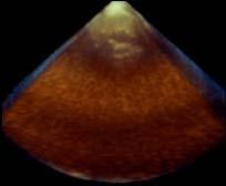
4 Narrow Angled Acquisition Narrow Angled Acquisition Narrow-Angled Partial Volume One beat acquisition Steerable sector Lateral Elevation 4
 Partial Volume 1-6")

5 Zoom Zoomed Acquisition (TTE) Partial Volume 1-6 beat acquisition Adjustable Sector Zoomed Acquisition (TEE) 5


6 Zoomed Acquisition Advantages: Focus on structural pathology Higher spatial and temporal resolution Disadvantage: Loss of spatial orientation Wide-Angled Acquisition 6

 Wide Sector Beats Single Beat Multibeat (2,4,6, HVR)")
7 Wide-Angled Acquisition Method: Matrix Array Probe Stitch Volume Wide-Angled Scan 1 beat 6 beat Components of Acquisition Mode of Acquisition Narrow Sector Focused Sector (Zoom) Wide Sector Beats Single Beat Multibeat (2,4,6, HVR) Gain Settings 7
8 1 Beat Acquisition 2 Beat Acquisition 8
9 4 Beats Acquisition Beats 6 Beats 9

")

10 High Volume Rate (HVR) Multibeat High Volume Rate (HVR) 10
11 Single vs Multi- Beat Single Beat Overcomes rhythm and respiratory gating artifacts Multi Beat Higher temporal resolution Poor temporal resolution Requires optimal patient, provider conditions Susceptible to gating issues Components of Acquisition Mode of Acquisition Narrow Sector Focused Sector (Zoom) Wide Sector Beats Single Beat Multibeat (2,4,6, HVR) Gain Settings 11
 Chamber (or large stlurctures) What is important? Spatial Resolution Volume Rate Or both Are there rhythm or respiratory issues?")
12 Optimize 2D and 3D (Gain and TGC) Reduce Overall Gain Adjust TGC Factors To Consider During Acquisition What is your region of interest? Valve (or small structures) Chamber (or large stlurctures) What is important? Spatial Resolution Volume Rate Or both Are there rhythm or respiratory issues? No ECG available/ Atrial fibrillation/ irregular rhythms Intubated patient Breath holds 12


13 Valvular Ds. Small structure Region of Interest ROI Small Volume High Volume Rate High Spatial Resolution Region of Interest ROI Valvular Ds. Chamber Small Volume High Volume Rate High Spatial Resolution Large Volume Highest Volume Rate Good Spatial Resolution 13
14 Optimizing 3DE Operator The Patient Sonographer Machine Acquisition Requirements Good Quality Volume Data Operator Well trained in TEE and 3D TEE Centered Region of Interest Find the right window Use Ante-, Retro-, Left and Right Flexion Steady Imaging 14
15 Acquisition Requirements Good Quality Volume Data The Patient Patient s Image Quality and Anatomy Presence of Air and Poor contact Hiatal Hernia Esophageal abnormalities Rhythm and Rate Irregular rhythms Heart rate extremes Sedation General Anesthesia Moderate or No Sedation Acquisition Requirements Good Quality Volume Data Operator (Sonographer) Well trained in 2D and 3D TTE Optimal 2D Imaging suboptimal 2D results in suboptimal 3D data sets Appropriate echo window for structure of interest Works within echo lab contraints (time, patient load, etc.) 15

16 Acquisition Requirements Good Quality Volume Data The Sonographer Knowledgeable about the TEE protocol in addition to 2D and 3D TEE: Collaboration between Echocardiographer and sonographer Good communication Understand 3D workflow Optimizes 2D and 3DE Gain and TGC Goal Optimum Acquisition The Machine Gain Depth Space-Time Adjustment Sector Size Zoom Display Pathology 16


17 Sector Change Wide Narrow Depth Change Higher Depth Lower Depth 17


18 Optimizing Spatial and Temporal Resolution MV (high depth) MV Zoom Space Time Balance T1 S1 S2 S3 18


19 Balancing Spatial Temporal Depth Sector size Zoom Space - Time 3D Color Acquisition Ideal Acquisition for 3D Color Flow 1 Beat High Temporal and Spatial Resolution Large 3D B-mode with Steerable Color Flow 19


20 Multibeat 3D Color Imaging Increase Volume Rate Single Beat Color Imaging Wide Angled Acquisition Large Color Sector Volume rate 13 vps Zoom Acquisition Narrow Color Sector VR = 23 vps 20


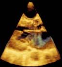
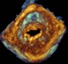
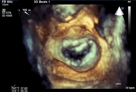
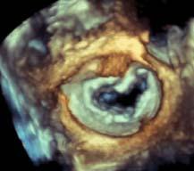
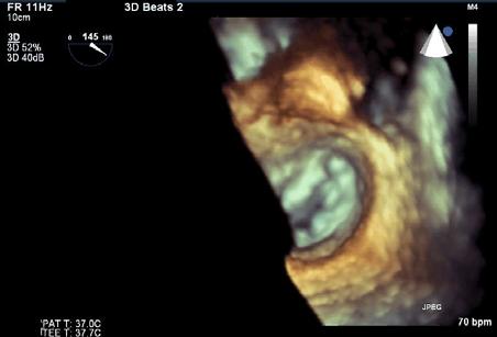
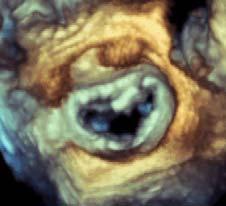
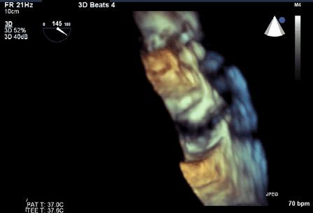
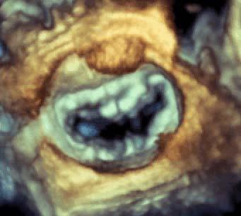
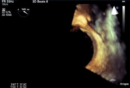
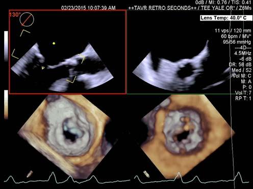
21 Goal of 3D Color Acquisition Visualize the origin of regurgitation Useful in Paravalvular regurgitation Bileaflet degenerative mitral valve disease Understand the severity of regurgitation or the size of the paravalvular leak Paravalvular Leak 21


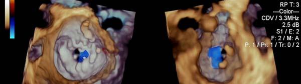
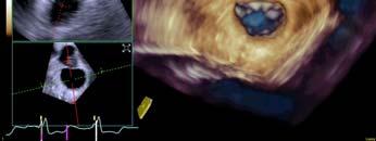
22 Steerable Color Sector (Anterior Paravalvular Leak) Steerable Color Sector (Medial Paravalvular Leak) 22
 Needs a high Volume Rate Narrow color")


23 3D Color Acquisition (Quantitation of Volume Flow) Needs a high Volume Rate Narrow color sector Narrow 3D B-mode sector 3D color can include: Both Mitral and Aortic flow Dedicated Valvular flow 3D Display (Types of Display) Volume Rendering Surface Rendering Wire-Frame 2D Tomographic Slices 23

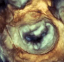
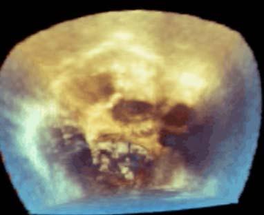
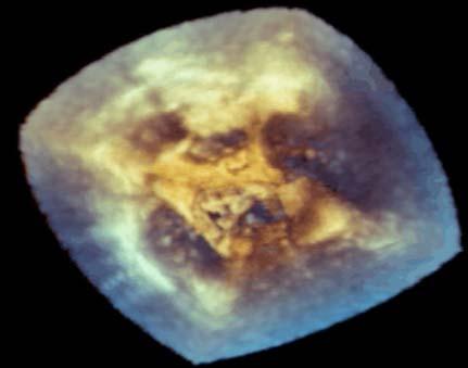
24 Volume Rendered Images Displays Anatomy and Pathology Dynamic images Thresholding/ Gain adjustments needed Ideal Volume Rendered Display Display that demonstrates the pathology Automated Orientation depending on the specific structure Quick Cut planes 24


25 Wire-Framed Rendered Tracing ventricular or atrial chambers Tracing valvular structures No anatomic information Results in length, circumference, area or volumetric measurements Surface Rendered Based on a wire-framed reconstruction Surface is applied over the wire-frame No anatomic detail Provide additional information Timing of events Motion Displacement 25

")
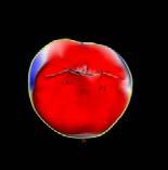

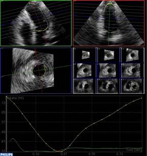
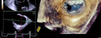
26 Surface Rendered (Mitral Valve) Multiplanar Reconstructions (MPR) Single MV slice Multiple LV slices 26


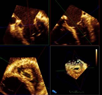
27 Segmentation or Cut Planes Sector Narrowing the Sector Zoom imaging Cropping Box Cut Planes Auto -crop Box-crop Anatomic cut plane D Art Plane icrop Segmentation 27


28 Cut-Planes 3D Volume Auto Crop Box Crop Cut-Planes 3D Volume icrop D art / 2 click Crop Plane Crop 28
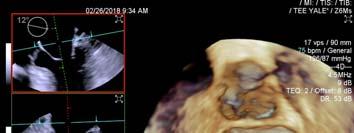
29 Dual View (Simultaneous Display of 2 Perspectives ) Stitch Artifact 29


30 Bovie Artifact Electrical Interference Solution: Stop Cautery Probe 30
This is your imaging dashboard. There is a bevy of valuable information to read from here. Let s break it up.
 The Volume Strikes Back Press the (4D) button. This is your imaging dashboard. There is a bevy of valuable information to read from here. Let s break it up. In the Left GUI (Graphic User Interface), you
Clinical Importance. Aortic Stenosis. Aortic Regurgitation. Ultrasound vs. MRI. Carotid Artery Stenosis
 Clinical Importance Rapid cardiovascular flow quantitation using sliceselective Fourier velocity encoding with spiral readouts Valve disease affects 10% of patients with heart disease in the U.S. Most
Ultrasound. Q-Station software. Streamlined workflow solutions. Philips Q-Station ultrasound workspace software
 Ultrasound Q-Station software Streamlined workflow solutions Philips Q-Station ultrasound workspace software Managing your off-cart workf low Everyone is being asked to do more with fewer resources it
Chapter 32 3-D and 4-D imaging in Obstetrics and Gynecology
 Objectives Define common terms related to 3D/4D ultrasound Chapter 32 3-D and 4-D imaging in Obstetrics and Gynecology Bridgette Lunsford Describe how 3D and 4D imaging differs from 2D ultrasound Identify
Volumetric Analysis of the Heart from Tagged-MRI. Introduction & Background
 Volumetric Analysis of the Heart from Tagged-MRI Dimitris Metaxas Center for Computational Biomedicine, Imaging and Modeling (CBIM) Rutgers University, New Brunswick, NJ Collaboration with Dr. Leon Axel,
GE Healthcare. Vivid E9 XDclear. with
 GE Healthcare Vivid E9 XDclear with capabilities for every day Whether in TTE or TEE, capture the entire heart in a single beat. Image a valve, or the entire ventricle with excellent image quality. And
GE Healthcare. Vivid 7 Dimension 08 Cardiovascular ultrasound system
 GE Healthcare Vivid 7 Dimension 08 Cardiovascular ultrasound system ltra Definition. Technology. Performance. Start with a system that s proven its worth in LV quantification and 4D imaging. Then add even
7D Cardiac Flow MRI: Techniques & Automation of Reconstruction
 Cleveland State University EngagedScholarship@CSU ETD Archive 2012 7D Cardiac Flow MRI: Techniques & Automation of Reconstruction Michael G. Ambrosia Cleveland State University Follow this and additional
High spatial and temporal resolution retrospective cine cardiovascular magnetic resonance from shortened free breathing real-time acquisitions
 Xue et al. Journal of Cardiovascular Magnetic Resonance 2013, 15:102 RESEARCH Open Access High spatial and temporal resolution retrospective cine cardiovascular magnetic resonance from shortened free breathing
Chapter 9 Conclusions
 Chapter 9 Conclusions This dissertation has described a new method for using local medial properties of shape to identify and measure anatomical structures. A bottom up approach based on image properties
Robust Live Tracking of Mitral Valve Annulus for Minimally-Invasive Intervention Guidance
 Robust Live Tracking of Mitral Valve Annulus for Minimally-Invasive Intervention Guidance Ingmar Voigt 1, Mihai Scutaru 2, Tommaso Mansi 3, Bogdan Georgescu 3, Noha El-Zehiry 3, Helene Houle 4, and Dorin
GE Healthcare. Vivid E9 XDclear. with. healthymagination. GE imagination at work
 GE Healthcare with Vivid E9 XDclear healthymagination GE imagination at work Extraordinary capabilities Whether in TTE or TEE, capture the entire heart in a single beat. Image a valve, or the entire ventricle
Technical Principles of Transthoracic Three-Dimensional Echocardiography
 Technical Principles of Transthoracic Three-Dimensional Echocardiography 2 Stein Inge Rabben 2.1 Introduction The last years we have experienced a rapid development in three-dimensional echocardiography
4D Auto MVQ (Mitral Valve Quantification)
") 4D Auto MVQ (Mitral Valve Quantification) Federico Veronesi, Glenn Reidar Lie, Stein Inge Rabben GE Healthcare Introduction As the number of mitral valve repairs is on the rise, so is the need for mitral
4D Auto MVQ (Mitral Valve Quantification) Federico Veronesi, Glenn Reidar Lie, Stein Inge Rabben GE Healthcare Introduction As the number of mitral valve repairs is on the rise, so is the need for mitral
IHE Cardiology Technical Framework Supplement. Evidence Documents Profile. Cardiology Options: Stress Testing CT/MR Angiography
 Integrating the Healthcare Enterprise 5 IHE Cardiology Technical Framework Supplement Evidence Documents Profile 10 Cardiology Options: Stress Testing CT/MR Angiography 15 Trial Implementation Date: October
WE DEVELOPED A NOVEL MULTI-BEAT image fusion technique using
 Interpolation of irregularly distributed sparse 4D ultrasound data using normalized convolution 4 WE DEVELOPED A NOVEL MULTI-BEAT image fusion technique using a special spatiotemporal interpolation for
Embedded Systems. Cristian Rotariu
 Embedded Systems Cristian Rotariu Dept. of of Biomedical Sciences Grigore T Popa University of Medicine and Pharmacy of Iasi, Romania cristian.rotariu@bioinginerie.ro May 2016 Introduction An embedded
Accurate Regression-based 4D Mitral Valve Segmentation from 2D MRI Slices
 Accurate Regression-based 4D Mitral Valve Segmentation from 2D MRI Slices Dime Vitanovski 2,3, Alexey Tsymbal 2, Razvan Ioan Ionasec 1, Andreas Greiser 4, Edgar Mueller 4, Xiaoguang Lu 1, Gareth Funka-Lea
Motion Compensation from Short-Scan Data in Cardiac CT
 Motion Compensation from Short-Scan Data in Cardiac CT Juliane Hahn 1,2, Thomas Allmendinger 1, Herbert Bruder 1, and Marc Kachelrieß 2 1 Siemens Healthcare GmbH, Forchheim, Germany 2 German Cancer Research
Lecture 6: Medical imaging and image-guided interventions
 ME 328: Medical Robotics Winter 2019 Lecture 6: Medical imaging and image-guided interventions Allison Okamura Stanford University Updates Assignment 3 Due this Thursday, Jan. 31 Note that this assignment
Corso di laurea in Fisica A.A Fisica Medica 4 TC
 Corso di laurea in Fisica A.A. 2007-2008 Fisica Medica 4 TC Computed Tomography Principles 1. Projection measurement 2. Scanner systems 3. Scanning modes Basic Tomographic Principle The internal structure
EchoPAC Software Only v201
 EchoPAC Software Only v201 Product Description The EchoPAC Application is a comprehensive clinical software package for viewing, analyzing, and reporting of multi-dimensional echo, vascular and abdominal
연구용유방초음파질관리 원광대학병원김혜원
 연구용유방초음파질관리 원광대학병원김혜원 Why US QC? Quality control (QC) testing of ultrasound scanners is important to verify the proper and consistent operation of these devices. main goal ; quality improvement Guidelines
VEVO 2100 Sole Source Specifications
 VEVO 2100 Sole Source Specifications Table of Contents 1.SYSTEM... 2 1-1 Licensing for the User... 2 1-2 Safety... 2 1-3 Portability... 2 2.TRANSDUCERS... 2 2-1 MicroScan transducers... 2 3. IMAGING STATION...
Using TeraRecon intuition Viewer with STAT Quick Reference Guide
 Using TeraRecon intuition Viewer with STAT Quick Reference Guide 1. Launching intuition 2. 3D Assessment of Coronary Arteries for Stenosis 3. Time Volume Analysis (TVA) for Determination of Left Ventricular
Tomographic Reconstruction
 Tomographic Reconstruction 3D Image Processing Torsten Möller Reading Gonzales + Woods, Chapter 5.11 2 Overview Physics History Reconstruction basic idea Radon transform Fourier-Slice theorem (Parallel-beam)
SONOS 7500/5500 Using 3-Dimensional and BiPlane Imaging And Other System Changes for Software Revision D.1
 SONOS 7500/5500 Using 3-Dimensional and BiPlane Imaging And Other System Changes for Software Revision D.1 User s Guide Using 3-Dimensional and BiPlane Imaging And Other System Changes for Software Revision
Mindray. Hand-carried Diagnostic Ultrasound System. Expanding the Envelope of Performance and Flexibility
 M7 Mindray M7 Hand-carried Diagnostic Ultrasound System Expanding the Envelope of Performance and Flexibility M7 Diagnostic Ultrasound System Equipped for Quality Radiology Cardiology Outpatient Emergency
Computational Medical Imaging Analysis Chapter 4: Image Visualization
 Computational Medical Imaging Analysis Chapter 4: Image Visualization Jun Zhang Laboratory for Computational Medical Imaging & Data Analysis Department of Computer Science University of Kentucky Lexington,
Multi-slice CT Image Reconstruction Jiang Hsieh, Ph.D.
 Multi-slice CT Image Reconstruction Jiang Hsieh, Ph.D. Applied Science Laboratory, GE Healthcare Technologies 1 Image Generation Reconstruction of images from projections. textbook reconstruction advanced
Robust Spatio-Temporal Registration of 4D Cardiac Ultrasound Sequences
 Robust Spatio-Temporal Registration of 4D Cardiac Ultrasound Sequences Jørn Bersvendsen a,b,c,matthewtoews d, Adriyana Danudibroto a,e, William M. Wells III f, Stig Urheim g,raúlsanjoséestépar f and Eigil
Medical Image Processing: Image Reconstruction and 3D Renderings
 Medical Image Processing: Image Reconstruction and 3D Renderings 김보형 서울대학교컴퓨터공학부 Computer Graphics and Image Processing Lab. 2011. 3. 23 1 Computer Graphics & Image Processing Computer Graphics : Create,
Digital Image Processing
 Digital Image Processing SPECIAL TOPICS CT IMAGES Hamid R. Rabiee Fall 2015 What is an image? 2 Are images only about visual concepts? We ve already seen that there are other kinds of image. In this lecture
Image Guidance and Beam Level Imaging in Digital Linacs
 Image Guidance and Beam Level Imaging in Digital Linacs Ruijiang Li, Ph.D. Department of Radiation Oncology Stanford University School of Medicine 2014 AAPM Therapy Educational Course Disclosure Research
4DM Packages. 4DM Packages & License Types. Information to help you order the appropriate licenses for your site.
 4DM Packages 4DM Packages & License Types. Information to help you order the appropriate licenses for your site. Nuclear Cardiac Quantification, Review, and Reporting Select Your 4DM Package and corresponding
A powerful, software-based beamformer image reconstruction platform
 csound A powerful, software-based beamformer image reconstruction platform Figure 1: Vivid S70 and E90/95; first GE systems built upon the csound platform Background GE s cardiovascular ultrasound imaging
Module 5: Dynamic Imaging and Phase Sharing. (true-fisp, TRICKS, CAPR, DISTAL, DISCO, HYPR) Review. Improving Temporal Resolution.
 Review. Improving Temporal Resolution.") MRES 7005 - Fast Imaging Techniques Module 5: Dynamic Imaging and Phase Sharing (true-fisp, TRICKS, CAPR, DISTAL, DISCO, HYPR) Review Improving Temporal Resolution True-FISP (I) True-FISP (II) Keyhole
MRES 7005 - Fast Imaging Techniques Module 5: Dynamic Imaging and Phase Sharing (true-fisp, TRICKS, CAPR, DISTAL, DISCO, HYPR) Review Improving Temporal Resolution True-FISP (I) True-FISP (II) Keyhole
Modeling Mitral Valve Leaflets from Three-Dimensional Ultrasound
 Modeling Mitral Valve Leaflets from Three-Dimensional Ultrasound Robert J. Schneider 1, William C. Burke 1, Gerald R. Marx 3, Pedro J. del Nido 2, and Robert D. Howe 1 1 Harvard School of Engineering and
4D Cardiac Reconstruction Using High Resolution CT Images
 4D Cardiac Reconstruction Using High Resolution CT Images Mingchen Gao 1, Junzhou Huang 1, Shaoting Zhang 1, Zhen Qian 2, Szilard Voros 2, Dimitris Metaxas 1, and Leon Axel 3 1 CBIM Center, Rutgers University,
Digital Imaging and Communications in Medicine (DICOM) Supplement 167: X-Ray 3D Angiographic IOD Informative Annex
 Supplement 167: X-Ray 3D Angiographic IOD Informative Annex") 5 Digital Imaging and Communications in Medicine (DICOM) Supplement 167: X-Ray 3D Angiographic IOD Informative Annex 10 15 Prepared by: DICOM Standards Committee, Working Group 2, Projection Radiography
5 Digital Imaging and Communications in Medicine (DICOM) Supplement 167: X-Ray 3D Angiographic IOD Informative Annex 10 15 Prepared by: DICOM Standards Committee, Working Group 2, Projection Radiography
Philips SPECT/CT Systems
 Philips SPECT/CT Systems Ling Shao, PhD Director, Imaging Physics & System Analysis Nuclear Medicine, Philips Healthcare June 14, 2008 *Presented SNM08 Categorical Seminar - Quantitative SPECT and PET
VEVO 1100 Sole Source Specifications
 VEVO 1100 Sole Source Specifications Table of Contents 1.SYSTEM... 2 1-1 Licensing for the User... 2 1-2 Safety... 2 1-3 Portability... 2 2.TRANSDUCERS... 2 2-1 MicroScan transducers... 2 3. IMAGING STATION...
Deep dive into SR: Key Object Selection and Radiation Dose Report
 THE DICOM 2013 INTERNATIONAL CONFERENCE & SEMINAR March 14-16 Bangalore, India Harry Solomon Deep dive into SR: Key Object Selection and Radiation Dose Report Interoperability Architect, GE Healthcare
Quick Guide. EPIQ 7 Ultrasound System. English
 Quick Guide EPIQ 7 Ultrasound System English Contents For easy reference, each section is color-coded. The color of each section title correlates with the color band on each page in that section. Controls
icatvision Quick Reference
 icatvision Quick Reference Navigating the i-cat Interface This guide shows how to: View reconstructed images Use main features and tools to optimize an image. REMINDER Images are displayed as if you are
Tissue Doppler gated (TDOG) dynamic three-dimensional ultrasound imaging of the fetal heart
 dynamic three-dimensional ultrasound imaging of the fetal heart") Ultrasound Obstet Gynecol 2004; 24: 192 198 Published online in Wiley InterScience (www.interscience.wiley.com). DOI: 10.1002/uog.1094 Tissue Doppler gated (TDOG) dynamic three-dimensional ultrasound imaging
Ultrasound Obstet Gynecol 2004; 24: 192 198 Published online in Wiley InterScience (www.interscience.wiley.com). DOI: 10.1002/uog.1094 Tissue Doppler gated (TDOG) dynamic three-dimensional ultrasound imaging
Total Variation Regularization Method for 3D Rotational Coronary Angiography
 Total Variation Regularization Method for 3D Rotational Coronary Angiography Haibo Wu 1,2, Christopher Rohkohl 1,3, Joachim Hornegger 1,2 1 Pattern Recognition Lab (LME), Department of Computer Science,
The Near Future in Cardiac CT Image Reconstruction
 SCCT 2010 The Near Future in Cardiac CT Image Reconstruction Marc Kachelrieß Institute of Medical Physics (IMP) Friedrich-Alexander Alexander-University Erlangen-Nürnberg rnberg www.imp.uni-erlangen.de
Respiratory Motion Estimation using a 3D Diaphragm Model
 Respiratory Motion Estimation using a 3D Diaphragm Model Marco Bögel 1,2, Christian Riess 1,2, Andreas Maier 1, Joachim Hornegger 1, Rebecca Fahrig 2 1 Pattern Recognition Lab, FAU Erlangen-Nürnberg 2
MR Advance Techniques. Vascular Imaging. Class III
 MR Advance Techniques Vascular Imaging Class III 1 Vascular Imaging There are several methods that can be used to evaluate the cardiovascular systems with the use of MRI. MRI will aloud to evaluate morphology
csound 2.0 Vivid E95 gehealthcare.com Background
 csound 2.0 Vivid E95 Background Through the Healthymagination initiative, GE Healthcare continuously invests in innovations that help lower the cost, increase access, and improve the quality of healthcare.
Deviceless respiratory motion correction in PET imaging exploring the potential of novel data driven strategies
 g Deviceless respiratory motion correction in PET imaging exploring the potential of novel data driven strategies Presented by Adam Kesner, Ph.D., DABR Assistant Professor, Division of Radiological Sciences,
Image Guidance of Intracardiac Ultrasound with Fusion of Pre-operative Images
 Image Guidance of Intracardiac Ultrasound with Fusion of Pre-operative Images Yiyong Sun 1, Samuel Kadoury 1,YongLi 1, Matthias John 2,JeffResnick 3, Gerry Plambeck 3,RuiLiao 1, Frank Sauer 1, and Chenyang
A Framework for Real-time Left Ventricular tracking in 3D+T Echocardiography, Using Nonlinear Deformable Contours and Kalman Filter Based Tracking
 1 A Framework for Real-time Left Ventricular tracking in 3D+T Echocardiography, Using Nonlinear Deformable Contours and Kalman Filter Based Tracking Fredrik Orderud Norwegian University of Science and
Cardiac Disease Recognition in Echocardiograms Using Spatio-temporal Statistical Models
 Cardiac Disease Recognition in Echocardiograms Using Spatio-temporal Statistical Models David Beymer and Tanveer Syeda-Mahmood Abstract In this paper we present a method of automatic disease recognition
Ch. 4 Physical Principles of CT
 Ch. 4 Physical Principles of CT CLRS 408: Intro to CT Department of Radiation Sciences Review: Why CT? Solution for radiography/tomography limitations Superimposition of structures Distinguishing between
Learning Anatomical Image Representations for Cardiac Imaging
 Imperial College of Science, Technology and Medicine Department of Computing Learning Anatomical Image Representations for Cardiac Imaging Ozan Oktay A dissertation submitted in part fulfilment of the
VieW 3D. 3D Post-Processing WorKstation THE THIRD DIMENSION. Version 3.1
 VieW 3D 3D Post-Processing WorKstation THE THIRD DIMENSION Version 3.1 iq-view 3D THE FULLY-FEATURED 3D MEDICAL IMAGING SOLUTION FOR RADIOLOGISTS iq-view 3D contains all components of iq-view with the
A THREE-DIMENSIONAL PHASED ARRAY ULTRASONIC TESTING TECHNIQUE
 A THREE-DIMENSIONAL PHASED ARRAY ULTRASONIC TESTING TECHNIQUE So KITAZAWA, Naoyuki KONO, Atsushi BABA and Yuji ADACHI HITACHI, Ltd., Japan Mitsuru ODAKURA HITACHI-GE Nuclear Energy, Ltd., Japan Introduction
Artefakt-resistente Bewegungsschätzung für die bewegungskompensierte CT
 Artefakt-resistente Bewegungsschätzung für die bewegungskompensierte CT Marcus Brehm 1,2, Thorsten Heußer 1, Pascal Paysan 3, Markus Oehlhafen 3, and Marc Kachelrieß 1,2 1 German Cancer Research Center
Total Variation Regularization Method for 3-D Rotational Coronary Angiography
 Total Variation Regularization Method for 3-D Rotational Coronary Angiography Haibo Wu 1,2, Christopher Rohkohl 1,3, Joachim Hornegger 1,2 1 Pattern Recognition Lab (LME), Department of Computer Science,
Certificate in Clinician Performed Ultrasound (CCPU)
") Certificate in Clinician Performed Ultrasound (CCPU) Syllabus Physics Tutorial Physics Tutorial Purpose: Training: Assessments: This unit is designed to cover the theoretical and practical curriculum for
Certificate in Clinician Performed Ultrasound (CCPU) Syllabus Physics Tutorial Physics Tutorial Purpose: Training: Assessments: This unit is designed to cover the theoretical and practical curriculum for
997-10, Daechi-dong, Gangnam-gu, Seoul, , Korea Tel: Fax:
 997-10, Daechi-dong, Gangnam-gu, Seoul, 135-280, Korea Tel:080-337-2003 Fax: 02-2194-1209 * Indicates feature in development. Some equipment and software mentioned or shown in this brochure may be optional
INTRODUCTION TO MEDICAL IMAGING- 3D LOCALIZATION LAB MANUAL 1. Modifications for P551 Fall 2013 Medical Physics Laboratory
 INTRODUCTION TO MEDICAL IMAGING- 3D LOCALIZATION LAB MANUAL 1 Modifications for P551 Fall 2013 Medical Physics Laboratory Introduction Following the introductory lab 0, this lab exercise the student through
03:47:29PM S3194 # MHz 180mm YNHH. MSc Thesis. 3D Transesophageal Echocardiography using a fast-rotating transducer. Kyriakos Nathanail
 03:47:29PM S3194 # 43 2.5MHz 180mm YNHH MSc Thesis 3D Transesophageal Echocardiography using a fast-rotating transducer Kyriakos Nathanail 3D Transesophageal Echocardiography using a fast-rotating transducer
The Supreme 3D/4D Ultrasound. The Supreme 3D/4D Ultrasound. CT-V20-ICM
 The Supreme 3D/4D Ultrasound The Supreme 3D/4D Ultrasound www.medison.com info@medison.com CT-V20-ICM-06.20.2008 The Supreme 3D/4D Ultrasound Since launching the first commercially available 3D ultrasound
Chapter 9 Field Shaping: Scanning Beam
 Chapter 9 Field Shaping: Scanning Beam X. Ronald Zhu, Ph.D. Department of Radiation Physics M. D. Anderson Cancer Center Houston, TX June 14-18, 2015 AAPM - Summer School 2015, Colorado Spring Acknowledgement
LOGIQ. V2 Ultrasound. Part of LOGIQ Vision Series. Imagination at work LOGIQ is a trademark of General Electric Company.
 TM LOGIQ V2 Ultrasound Part of LOGIQ Vision Series Imagination at work The brilliance of color. The simplicity of GE. Now you can add the advanced capabilities of color Doppler to patient care with the
The team. Disclosures. Ultrasound Guidance During Radiation Delivery: Confronting the Treatment Interference Challenge.
 Ultrasound Guidance During Radiation Delivery: Confronting the Treatment Interference Challenge Dimitre Hristov Radiation Oncology Stanford University The team Renhui Gong 1 Magdalena Bazalova-Carter 1
A Virtual Reality Training System for Pediatric Sonography
 1 A Virtual Reality Training System for Pediatric Sonography W. Arkhurst a, A. Pommert a, E. Richter b, H. Frederking a, S.-I. Kim ab, R. Schubert a, and K. H. Höhne a a Institute of Mathematics and Computer
FINDING THE TRUE EDGE IN CTA
 FINDING THE TRUE EDGE IN CTA by: John A. Rumberger, PhD, MD, FACC Your patient has chest pain. The Cardiac CT Angiography shows plaque in the LAD. You adjust the viewing window trying to evaluate the stenosis
3/27/2012 WHY SPECT / CT? SPECT / CT Basic Principles. Advantages of SPECT. Advantages of CT. Dr John C. Dickson, Principal Physicist UCLH
 3/27/212 Advantages of SPECT SPECT / CT Basic Principles Dr John C. Dickson, Principal Physicist UCLH Institute of Nuclear Medicine, University College London Hospitals and University College London john.dickson@uclh.nhs.uk
Combination of Markerless Surrogates for Motion Estimation in Radiation Therapy
 Combination of Markerless Surrogates for Motion Estimation in Radiation Therapy CARS 2016 T. Geimer, M. Unberath, O. Taubmann, C. Bert, A. Maier June 24, 2016 Pattern Recognition Lab (CS 5) FAU Erlangen-Nu
US 1.
 US 1 Sample image: Normal pancreas seen on sonogram. Looking up from abdomen toward the head of the patient. The liver is in front of the pancreas. A vein draining the spleen is behind the pancreas http://www.radiologyinfo.org/photocat/photos.cfm?image=abdo-us-pancr.jpg&&subcategory=abdomen&&stop=9
Compressed Sensing for Rapid MR Imaging
 Compressed Sensing for Rapid Imaging Michael Lustig1, Juan Santos1, David Donoho2 and John Pauly1 1 Electrical Engineering Department, Stanford University 2 Statistics Department, Stanford University rapid
Mohammad Baharvandy & Sina Fazelpour
 Mohammad Baharvandy & Sina Fazelpour Ultrasound Basics Data acquisition in 3D Reconstruction of 2D images 3D Ultrasound Modeling Medical applications 4D Ultrasound 2 Ultrasound consist of sound waves of
19 CP Replace uses of "data set" that do not refer to the PS3.5 defined meaning
 19 CP-1817 - Replace uses of "data set" that do not refer to the PS3.5 defined meaning Page 1 1 Status Assigned 2 Date of Last Update 2018/06/04 3 Person Assigned David Clunie 4 mailto:dclunie@dclunie.com
Computer-Aided Diagnosis in Abdominal and Cardiac Radiology Using Neural Networks
 Computer-Aided Diagnosis in Abdominal and Cardiac Radiology Using Neural Networks Du-Yih Tsai, Masaru Sekiya and Yongbum Lee Department of Radiological Technology, School of Health Sciences, Faculty of
Motion artifact detection in four-dimensional computed tomography images
 Motion artifact detection in four-dimensional computed tomography images G Bouilhol 1,, M Ayadi, R Pinho, S Rit 1, and D Sarrut 1, 1 University of Lyon, CREATIS; CNRS UMR 5; Inserm U144; INSA-Lyon; University
Computer-Tomography I: Principles, History, Technology
 Computer-Tomography I: Principles, History, Technology Prof. Dr. U. Oelfke DKFZ Heidelberg Department of Medical Physics (E040) Im Neuenheimer Feld 280 69120 Heidelberg, Germany u.oelfke@dkfz.de History
Visualisation : Lecture 1. So what is visualisation? Visualisation
 So what is visualisation? UG4 / M.Sc. Course 2006 toby.breckon@ed.ac.uk Computer Vision Lab. Institute for Perception, Action & Behaviour Introducing 1 Application of interactive 3D computer graphics to
HPC Infrastructure for and Simulations of Impact of Drug-Induced Arrhythmias in Living Hearts
 HPC User Forum Tucson, AZ, April 16 18, 2018 HPC Infrastructure for and Simulations of Impact of Drug-Induced Arrhythmias in Living Hearts Wolfgang Gentzsch The UberCloud Big Thanks To the HPC User Forum
Tomography at all Scales. Uccle, 7 April 2014
 Tomography at all Scales Uccle, 7 April 2014 Outline The Vision Lab ASTRA: All Scale Tomographic Reconstruction Antwerp Tomography Discrete Tomography In situ CT Superresolution Dynamic imaging The ASTRA
Image Quality Assessment and Quality Assurance of Advanced Imaging Systems for IGRT. AAPM Penn-Ohio Chapter Sep 25, 2015 Soyoung Lee, PhD
 Image Quality Assessment and Quality Assurance of Advanced Imaging Systems for IGRT AAPM Penn-Ohio Chapter Sep 25, 2015 Soyoung Lee, PhD 1 Outline q Introduction q Imaging performances in 4D-CBCT Image
Role of Parallel Imaging in High Field Functional MRI
 Role of Parallel Imaging in High Field Functional MRI Douglas C. Noll & Bradley P. Sutton Department of Biomedical Engineering, University of Michigan Supported by NIH Grant DA15410 & The Whitaker Foundation
10 Working With Setup Tools
 10 Working With Setup Tools The Setup window allows you to set defaults and control various functions of the Terason software. The Setup window tabs display sets of controls for defaults and other settings.
Lab Location: MRI, B2, Cardinal Carter Wing, St. Michael s Hospital, 30 Bond Street
 Lab Location: MRI, B2, Cardinal Carter Wing, St. Michael s Hospital, 30 Bond Street MRI is located in the sub basement of CC wing. From Queen or Victoria, follow the baby blue arrows and ride the CC south
Adaptive Multiscale Ultrasound Compounding Using Phase Information
 Adaptive Multiscale Ultrasound Compounding Using Phase Information Vicente Grau and J. Alison Noble Wolfson Medical Vision Laboratory, Department of Engineering Science, University of Oxford, Parks Road,
Hitachi Medical Systems America, Inc. Hitachi Medical Corporation
 Advanced 128 Advanced 128 128 Slice Capability Enlarged 75cm Aperture Thin Gantry Profile 88cm Intelli IP Advanced, Iterative Reconstruction IntelliCenter Auto-Lateral Shifting Table, up to 16cm Range
GE Healthcare CLINICAL GALLERY. Discovery * MR750w 3.0T. This brochure is intended for European healthcare professionals.
 GE Healthcare CLINICAL GALLERY Discovery * MR750w 3.0T This brochure is intended for European healthcare professionals. NEURO PROPELLER delivers high resolution, motion insensitive imaging in all planes.
Robust Segmentation and Volumetric Registration in
 Robust Segmentation and Volumetric Registration in a Multi-view 3D Freehand Ultrasound Reconstruction System Honggang Yu, Marios S. Pattichis image and video Processing and Communications Lab Department
CLASS HOURS: 4 CREDIT HOURS: 4 LABORATORY HOURS: 0
 Revised 10/10 COURSE SYLLABUS TM 220 COMPUTED TOMOGRAPHY PHYSICS CLASS HOURS: 4 CREDIT HOURS: 4 LABORATORY HOURS: 0 CATALOG COURSE DESCRIPTION: This course is one of a three course set in whole body Computed
Magnetic Resonance Elastography (MRE) of Liver Disease
 of Liver Disease") Magnetic Resonance Elastography (MRE) of Liver Disease Authored by: Jennifer Dolan Fox, PhD VirtualScopics Inc. jennifer_fox@virtualscopics.com 1-585-249-6231 1. Overview of MRE Imaging MRE is a magnetic
Magnetic Resonance Elastography (MRE) of Liver Disease Authored by: Jennifer Dolan Fox, PhD VirtualScopics Inc. jennifer_fox@virtualscopics.com 1-585-249-6231 1. Overview of MRE Imaging MRE is a magnetic
Basic principles of MR image analysis. Basic principles of MR image analysis. Basic principles of MR image analysis
 Basic principles of MR image analysis Basic principles of MR image analysis Julien Milles Leiden University Medical Center Terminology of fmri Brain extraction Registration Linear registration Non-linear
ECG MACHINES. Kizlon Medical. Website:
 ECG MACHINES Email: info@kizlonmedical.com Website: www.kizlonmedical.com ECG Machine KEC-A100 This ECG machine is a 3 channel model with synchronous acquisition and display of 12 leads. Features: A color
10 Working With Setup Tools
 10 Working With Setup Tools Setup Tools Overview The Setup window allows you to set defaults and control various functions of the Terason software. The Setup window tabs display sets of controls for defaults
Brilliance CT Big Bore.
 1 2 2 There are two methods of RCCT acquisition in widespread clinical use: cine axial and helical. In RCCT with cine axial acquisition, repeat CT images are taken each couch position while recording respiration.
Acknowledgments and financial disclosure
 AAPM 2012 Annual Meeting Digital breast tomosynthesis: basic understanding of physics principles James T. Dobbins III, Ph.D., FAAPM Director, Medical Physics Graduate Program Ravin Advanced Imaging Laboratories
SISCOM (Subtraction Ictal SPECT CO-registered to MRI)
") SISCOM (Subtraction Ictal SPECT CO-registered to MRI) Introduction A method for advanced imaging of epilepsy patients has been developed with Analyze at the Mayo Foundation which uses a combination of
SISCOM (Subtraction Ictal SPECT CO-registered to MRI) Introduction A method for advanced imaging of epilepsy patients has been developed with Analyze at the Mayo Foundation which uses a combination of
The Usage of Optical Flow Algorithm to the Problem of Recovery Contour of the Left Ventricle of the Human Heart on the Ultrasound Image Data
 The Usage of Optical Flow Algorithm to the Problem of Recovery Contour of the Left Ventricle of the Human Heart on the Ultrasound Image Data Andrey A. Mukhtarov 1, Sergey V. Porshnev 1, Vasiliy V. Zyuzin