ISO ISO ISO OHSAS ISO
|
|
|
- Julia Franklin
- 5 years ago
- Views:
Transcription


1 ISO 9001 ISO ISO OHSAS ISO 27001


2


 optional heavy duty carrying case (08-110)")
3 Pro-NM Performance standard version version with the PET Lid Phantom for NM and PET systems performance evaluation (collimator, artifacts, calibration, reconstruction parameters). Among other things, it can be used to evaluate: center-of-rotation error, non-uniformity artifacts, changes of radius-of-rotation on spatial resolution, reconstruction filters on spatial resolution, attenuation and scatter compensation. main cylinder: inside cylinder diameter: 206 mm inside cylinder height: 186 mm cylinder wall thickness: 7 mm cold rods insert: rod diameters: 4.8, 6.4, 7.9, 9.5, 11.1 and 12.7 mm height of rods: 88 mm cold spheres: solid sphere diameters: 9.5, 12.7, 15.9, 19.1, 25.4 and 31.8 mm height of the center of the spheres from the base plate: 127 mm optional PET Lid with cylindrical samples (08-102): refillable thin-walled cylinders, diameters: 8, 12, 16 and 25 mm water filled cylinder diameter: 25 mm air filled cylinder diameter: 25 mm PTFE solid cylinder diameter: 25 mm cylinder height: 38 mm optional hollow spheres set (08-104) optional heavy duty carrying case (08-110) NEMA Standards Publication (NU ) Performance Measurements of Scintillation Cameras AAPM Report No. 9 - Computer Aided Scintillation Camera Acceptance Testing AAPM Report No Rotating Scintillation Camera SPECT Acceptance Testing and Quality Control ACR SNM (Res ) technical standard for diagnostic procedures using radiopharmaceuticals the Manual provides detailed guidelines for carrying out each test, results assessment and registration
.")


")
4 Pro-NM Performance HD standard version version with the PET Lid Phantom for High resolution NM and PET systems performance evaluation (collimator, artifacts, calibration, reconstruction parameters). Among other things, it can be used to evaluate: center-of-rotation error, non-uniformity artifacts, changes of radius-of-rotation on spatial resolution, reconstruction filters on spatial resolution, attenuation and scatter compensation. (can be modified to customer specifications): main cylinder: inside cylinder diameter: 206 mm inside cylinder height: 186 mm cylinder wall thickness: 7 mm cold rods insert: rod diameters: 3.2, 4.8, 6.4, 7.9, 9.5 and 11.1 mm height of rods: 88 mm cold spheres: solid sphere diameters: 9.5, 12.7, 15.9, 19.1, 25.4 and 31.8 mm height of the center of the spheres from the base plate: 127 mm optional PET Lid with cylindrical samples: refillable thin-walled cylinders, diameters: 8, 12, 16 and 25 mm water filled cylinder diameter: 25 mm air filled cylinder diameter: 25 mm PTFE solid cylinder diameter: 25 mm cylinder height: 38 mm optional heavy duty carrying case NEMA Standards Publication (NU ) Performance Measurements of Scintillation Cameras AAPM Report No. 9 - Computer Aided Scintillation Camera Acceptance Testing AAPM Report No Rotating Scintillation Camera SPECT Acceptance Testing and Quality Control ACR SNM (Res ) technical standard for diagnostic procedures using radiopharmaceuticals the Manual provides detailed guidelines for carrying out each test, results assessment and registration
.")

5 Pro-NM Performance SMALL standard version version with the PET Lid Phantom for small FOV NM and PET systems performance evaluation (collimator, artifacts, calibration, reconstruction parameters). Among other things, it can be used to evaluate: center-of-rotation error, non-uniformity artifacts, changes of radius-of-rotation on spatial resolution, reconstruction filters on spatial resolution, attenuation and scatter compensation. (can be modified to customer specifications): main cylinder: inside cylinder diameter: 140 mm inside cylinder height: 150 mm cylinder wall thickness: 6 mm cold rods insert: rod diameters: 3.2, 4.8, 6.4, 7.9, 9.5 and 11.1mm height of rods: 40 mm cold spheres: solid sphere diameters: 3.2, 4.8, 9.5, 12.7, 15.9 and 19.1mm height of the center of the spheres from the base plate: 78.5 mm optional PET Lid with cylindrical samples: refillable thin-walled cylinders, diameters: 4, 6, 8, 12, 16mm PTFE solid cylinder diameter: 16 mm cylinder height: 25.4 mm optional heavy duty carrying case NEMA Standards Publication (NU ) Performance Measurements of Scintillation Cameras AAPM Report No. 9 - Computer Aided Scintillation Camera Acceptance Testing AAPM Report No Rotating Scintillation Camera SPECT Acceptance Testing and Quality Control ACR SNM (Res ) technical standard for diagnostic procedures using radiopharmaceuticals the Manual provides detailed guidelines for carrying out each test, results assessment and registration


 Performance Measurements of Scintillation Cameras AAPM Report No.")

6 Pro-NM Performance MINI Phantom for performance evaluation of small animal systems or ultra-high resolution ECT systems. It can be used to evaluate: center-of-rotation error, non-uniformity artifacts and spatial resolution. Available in two sizes for FOV at least 45mm or 75mm. (can be modified to customer specifications): at least 75mm FOV version: main cylinder: - outside cylinder diameter: 80 mm - inside cylinder diameter: 74 mm - inside cylinder height: 65 mm cold rods insert: - rod diameters: 1.2, 1.6, 2.4, 3.2, 4.0 and 4.8 mm - height of rods: 34 mm at least 45mm FOV version: main cylinder: - outside cylinder diameter: 50 mm - inside cylinder diameter: 45 mm - inside cylinder height: 65 mm cold rods insert: - rod diameters: 1.2, 1.6, 2.4, 3.2, 4.0 and 4.8 mm - height of rods: 34 mm - optional carrying case NEMA Standards Publication (NU ) Performance Measurements of Scintillation Cameras AAPM Report No. 9 - Computer Aided Scintillation Camera Acceptance Testing AAPM Report No Rotating Scintillation Camera SPECT Acceptance Testing and Quality Control ACR SNM (Res ) technical standard for diagnostic procedures using radiopharmaceuticals the Manual provides detailed guidelines for carrying out each test, results assessment and registration
, slice width, uniformity, spatial resolution, point spread function, slice position verification,")
: external diameter 200 mm")
7 Pro-NM Performance ECT Phantom for NM systems performance evaluation: routine quality assurance tests, as well as extensive acceptance tests. It can be used to evaluate: pixel size, spatial linearity, RMS noise, signal to noise ratio (SNR), slice width, uniformity, spatial resolution, point spread function, slice position verification, slice incrementation, accuracy, center of rotation, verification, volume sensitivity and low contrast sensitivity. main cylinder: internal cylinder diameter: 206 mm internal cylinder height: 186 mm cylinder wall thickness: 7 mm main insert (slice width, pixel size and high resolution): external diameter 200 mm free square internal 80 x 80 mm consists of 10 x 5 mm discs, 2 spacers and 3 mounting screws contains a pair of channels 20 x 10 mm thick forming two hot ramps whose slope angle tangent is equal to 0.5 contains four hot holes 5mm in diameter that are located in corners of the 120 x 120 mm square contains four groups of hot and cold resolution patterns that are 2, 4, 6 and 8 mm thick and correspond to 2.5, 1.25, 0.83 and LP/cm point source insert (PSF - point spread function): the fill plug can be positioned at the center or at radial plug location source screw contains a well (ø3 x 5 mm) that can be filled with appropriate solution source screw can be mounted outside of or inside the phantom (for in air or scatter measurements) low contrast inserts: can be threaded into the phantom in a radial pattern 75 mm of the center axis three cold low contrast rods comprised of three parts: 10, 15 and 20 mm in diameter and 40 mm long three optional hot low contrast rods comprised of three parts: 10, 15 and 20 mm in diameter and 40 mm long that can be filled from the outside heavy duty carrying case NEMA Standards Publication (NU ) Performance Measurements of Scintillation Cameras NEMA Standards Publication (NU ) Gamma Cameras AAPM Report No. 9 - Computer Aided Scintillation Camera Acceptance Testing AAPM Report No Rotating Scintillation Camera SPECT Acceptance Testing and Quality Control ACR SNM (Res ) technical standard for diagnostic procedures using radiopharmaceuticals the Manual provides detailed guidelines for carrying out each test, results assessment and registration


8 Pro-NM Linear Source Module This module can be used as a standalone in air or in water if mounted in the Pro-NM Performance cylinder. It can be used to evaluate changes of radius-of rotation on spatial resolution, spatial resolution measurement in air and in water, quantitative evaluation of reconstruction filters and scatter compensation methods. insert diameter: 186 mm diameter of line sources: 1 mm spacing of line sources: 75 mm useful height of line sources: 70 mm stopcocks with luer connection allow easy and safe filling and draining of line sources NEMA Standards Publication (NU ) Performance Measurements of Scintillation Cameras AAPM Report No. 9 - Computer Aided Scintillation Camera Acceptance Testing AAPM Report No Rotating Scintillation Camera SPECT Acceptance Testing and Quality Control ACR SNM (Res ) technical standard for diagnostic procedures using radiopharmaceuticals the Manual provides detailed guidelines for carrying out


9 Pro-NM NEMA NU A phantom for evaluating performance of positron emission tomographs (PET). Recommended for use in the evaluation of reconstructed image quality in whole body PET imaging. For simulation of whole-body imaging, especially that using PET and camera-based coincidence imaging techniques. Can also be used for determination of the coincidence count rate characteristics in brain and cardiac imaging, evaluation of the relationship between true coincidence count rate and radioactivity, determination of the address errors caused by address pile up, evaluation of the count loss correction scheme. It has been designed in accordance with the recommendations by the International Electrotechnical Commission (IEC) and modified by the National Electrical Manufacturers Association (NEMA). interior length of the phantom: 180 mm volume of empty cylinder: 9.7 laters 6 fillable spheres: inner diameter: 10 mm, 13 mm, 17 mm, 22 mm, 28 mm, and 37 mm distance from sphere plane to inside wall: 70 mm cylindrical insert dimension: outside diameter: 51 mm length: 180 mm optional heavy duty carrying case (08-110) International Standard: Radionuclide imaging devices Characteristics and test conditions Part 1: Positron emission tomographs, International Electrotechnical Commission (IEC), , Geneva, Switzerland, Performance Measurements of Positron Emission Tomographs, NEMA Standards Publication No. NU2-2012, National Electrical Manufacturers Association (NEMA), NEMA2007/IEC2008 NEMA 2012/IEC 2008 manual provides detailed guidelines for carrying out each test, results assessment and registration
 of compact radioactive sources. It has been designed in accordance with the NEMA Nu 2-2012.")
, 10 cm, and 20 cm from the center of the plane middle of the capillaries is marked to provide guidance while the")
10 Pro-NM NEMA NU 2 Resolution A phantom for evaluation of spatial resolution of positron emission tomographs (PET). It is used to characterize the widths of the reconstructed image point spread functions (PSF) of compact radioactive sources. It has been designed in accordance with the NEMA Nu five capillaries with an inside diameter of 1 mm and an outside diameter of less than 2 mm in the transverse direction the capillary is positioned at 1 cm (to represent the center of the FOV, but positioned to avoid any possible inconsistent results at the very center of the FOV), 10 cm, and 20 cm from the center of the plane middle of the capillaries is marked to provide guidance while the radionuclide is introduced markers and convenient base with a spirit level for easy and accurate positioning optional carrying case International Standard: Radionuclide imaging devices Characteristics and test conditions Part 1: Positron emission tomographs, International Electrotechnical Commission (IEC), , Geneva, Switzerland, Performance Measurements of Positron Emission Tomographs, NEMA Standards Publication No. NU2-2012, National Electrical Manufacturers Association (NEMA), NEMA2007/IEC2008 NEMA 2012/IEC 2008 manual provides detailed guidelines for carrying out each test, results assessment and registration

 Performance Measurements of Scintillation Cameras AAPM Report No.")
11 Pro-NM Resolution holders for different cameras are available Bar phantom for determination of resolution of Scintillation Cameras. The four-quadrant phantom offers precise determination of camera intrinsic resolution, collimator spatial resolution, field size and linearity. Apart from this standard size phantom, we offer different sizes and configurations manufactured to the highest quality standards. dimensions: 564 x 432 x 15 mm lead bar widths: 2.0, 2.5, 3.0 and 3.5 mm field across bar configuration: 533 x 405 mm NEMA Standards Publication (NU ) Performance Measurements of Scintillation Cameras AAPM Report No. 9 - Computer Aided Scintillation Camera Acceptance Testing AAPM Report No Rotating Scintillation Camera SPECT Acceptance Testing and Quality Control ACR SNM (Res ) technical standard for diagnostic procedures using radiopharmaceuticals the Manual provides detailed guidelines for carrying out


12 Pro-NM ResL x 21 version x 18 version Pro-NM ResL for determination of resolution of Scintillation Cameras. The phantom offers precise determination of camera intrinsic resolution, collimator spatial resolution, field size and linearity. In addition to the standard size phantoms, we offer different sizes and configurations manufactured to the highest quality standards. The most cost-effective means of performing routine quality control checks of gamma camera resolution or linearity on the market today One image per detector head is all that s needed to equally and effectively test all quadrants of the gamma camera Perform routine quality control tests of spatial resolution and linearity in approximately one quarter of the time needed at present, which will make it possible to save time and money! Quickly and easily perform extrinsic testing and intrinsic visual evaluation Outperforms any 90 bar phantom, single-frequency Parallel-Line Equal-Space (PLES), Hine-Duley or orthogonal hole test pattern Meets mandatory requirements of state quality control Optimized for dual and triple-head gamma cameras Ideal for large detectors. Its large size covers UFOV Increase patient throughput dimensions: mm (18 ) x mm (24 ) x 15 mm (0.59 ) dimensions: mm (21 ) x mm (21 ) x 15 mm (0.59 ) lead bar widths: 6.35 mm (1/4 ), mm (3/16 ), mm (5/32 ), 2.54 mm (1/10 ) NEMA Standards Publication (NU ) Performance Measurements of Scintillation Cameras AAPM Report No. 9 - Computer Aided Scintillation Camera Acceptance Testing AAPM Report No Rotating Scintillation Camera SPECT Acceptance Testing and Quality Control ACR SNM (Res ) technical standard for diagnostic procedures using radiopharmaceuticals the Manual provides detailed guidelines for carrying out
 - spatial resolution - at each point and its")
 Performance")
13 Pro-NM Distortion A simple phantom for quality assurance of geometric distortion and spatial resolution of gamma cameras. An array of holes, which when filled with activity, allow measuring point-to-point distances and Point Spread Function (PSF) - spatial resolution - at each point and its homogeneity across the entire Field of View. dimensions 500 x 400 x 5 mm series of holes - 1 mm in diameter and 3 mm deep, at 80 mm pitch - to be filled with activity NEMA Standards Publication (NU ) Performance Measurements of Scintillation Cameras NEMA Standards Publication (NU ) Performance Measurements of Scintillation Cameras manual provides detailed guidelines for carrying out

 Performance Measurements of Scintillation Cameras NEMA Standards Publication (NU 1-2012) Performance Measurements of Scintillation Cameras the")
14 Pro-NM Slits The slits phantom for Intrinsic Spatial Resolution evaluation (Quantitative technique) according to NEMA Standards Publication NU In addition to this standard size phantom, we offer different sizes and configurations manufactured to the highest quality standards. dimensions: 620 x 520 x 15 mm lead size 600 x 500 x 3 mm parallel 1mm thick slits with 30 mm spacing cover made of PMMA uniform PMMA part of the phantom NEMA Standards Publication (NU ) Performance Measurements of Scintillation Cameras NEMA Standards Publication (NU ) Performance Measurements of Scintillation Cameras the Manual provides detailed guidelines for carrying out

15 Pro-NM PETsensi The PET sensitivity phantom is used to measure the sensitivity or ability of positron emission tomographs to detect positrons. The phantom used for this purpose is a set of five metal tubes with a similar wall thickness. A plastic tube homogeneously filled with 18F liquid is inserted for the measurement. Successive measurements are made by accumulating the sleeve wall thickness with the uniform line source surrounded by known absorbers. From these measurements, the sensitivity without absorbers can be extrapolated to arrive at an attenuation free measurement. The measurement setup, data collection and analysis are described in section 5 of the NEMA standard NU Five internally stacked concentric aluminium tubes all 700 mm in length. 1st Tube Inside Diameter: 3.9 mm Outside Diameter: 6.4 mm 2nd Tube Inside Diameter: 7.0 mm Outside Diameter: 9.5 mm 3rd Tube Inside Diameter: 10.2 mm Outside Diameter: 12.7 mm 4th Tube Inside Diameter: 13.4 mm Outside Diameter: 15.9 mm 5th Tube Inside Diameter: 16.6 mm Outside Diameter: 19.1 mm 6th Innermost Tube (a fillable polyethylene tube) Inside Diameter: 2 mm Outside Diameter: 3.2 mm NEMA Standard (NU ) manual provides detailed guidelines for carrying out


16 Pro-NM PETscatter The PET scatter phantom is an acceptance testing tool used to determine the imaging systems relative sensitivity to scatter radiation. It can be used to measure the effects of dead-time and the effects of random events generated at different levels of activity of the line source. The test phantom is a right circular cylinder composed of polyethylene. For ease of handling, it consists of 3 segments that are assembled together during testing. A hole is drilled parallel to the central axis of the cylinder, at a radial distance of 45mm. cylinder outside diameter: 203 mm length: 700mm hole diameter: 6.4 mm hole offset from the central axis: 45 mm line source outside diameter: 4.8 mm length: 800 mm inside diameter: 3.2 mm carrying case with a trolley NEMA Standard (NU ) NEMA 2012 Standard manual provides detailed guidelines for carrying out


17 Pro-NM MultiAlign A phantom designed for a simple and cost effective verification of image alignment and distortion in hybrid scanning systems like PET/CT or NM/CT. It consists of a cylinder that can be filled with a variety of fluids. Several non-parallel rods of varying diameter and at certain angles in relation to the phantom s axes run the entire length of the cylinder. Images produced with different modalities allow quick and simple identification of misalignments on fused studies. Additionally, the phantom contains a 2 cm calibration module that is used by Pro-Control.online software to accurately identify the phantom s position in space and accurately calculate any mismatches. PMMA cylinder dimensions: 250 x 260 x 410 mm 2 cm thick calibration section with four through holes 4 PMMA rods: 30, 25, 20, 15 mm filling plugs and vent ports carrying handle AAPM Report No Rotating Scintillation Camera SPECT Acceptance Testing and Quality Control the Manual provides detailed guidelines for carrying out

18 Pro-NM DualSource This scatter phantom simulates in-vivo forward and backscatter characteristics of 99 mtc gamma rays for the extrinsic measurement of a scintillation camera s deadtime. The phantom produces a spectrum typical of that observed from 99 mtc in the myocardium. Reference: Ralph Adams, Gerald J. Hine, and C. Duane Zimmerman, Deadtime Measurements in Scintillation Cameras Under Scatter Conditions Simulating Quantitative Nuclear Cardiography, The Journal of Nuclear Medicine, 19 (1978), made of PMMA dimensions: 150 x 200 x 200 mm the two holes are used to hold the radioactive sources hole dimensions: ø17 mm x 120 mm deep spaced 50 mm apart (center-to-center) distance from the face of the phantom: 50 mm NEMA Standards Publication (NU ) Performance Measurements of Scintillation Cameras NEMA Standards Publication (NU ) Gamma Cameras AAPM Report No. 9 - Computer Aided Scintillation Camera Acceptance Testing AAPM Report No Rotating Scintillation Camera SPECT Acceptance Testing and Quality Control ACR SNM (Res ) technical standard for diagnostic procedures using radiopharmaceuticals the Manual provides detailed guidelines for carrying out


19 Pro-NM FloodRECT Pro-NM FloodRECT Pro-NM FloodRECT XL Flood phantoms provide a simple and efficient means of obtaining optimum camera performance with respect to uniformity of response over the entire crystal area. These phantoms are designed to be filled in horizontal position thus preventing slight bulging caused by water pressure during vertical filling. Therefore, better uniformity in distribution of activity can be achieved. Pro-NM FloodRECT: outside dimensions: 460 x 580 x 23 mm cavity dimensions: 410 x 530 x 13 mm Pro-NM FloodRECT XL: outside dimensions: 710 x 570 x 33 mm cavity dimensions: 660 x 520 x 13 mm NEMA Standards Publication (NU ) Performance Measurements of Scintillation Cameras AAPM Report No. 9 - Computer Aided Scintillation Camera Acceptance Testing AAPM Report No Rotating Scintillation Camera SPECT Acceptance Testing and Quality Control ACR SNM (Res ) technical standard for diagnostic procedures using radiopharmaceuticals the Manual provides detailed guidelines for carrying out


20 Pro-NM FloodROUND Pro-NM FloodROUND Pro-NM FloodROUND XL Flood phantoms provide a simple and efficient means of obtaining optimum camera performance with respect to uniformity of response over the entire crystal area. These phantoms are designed to be filled in horizontal position thus preventing slight bulging caused by water pressure during vertical filling. Therefore, better uniformity in distribution of activity can be achieved. Pro-NM FloodROUND: outside dimensions: 460 x 460 x 23 mm cavity dimensions: ø430 x 13 mm Pro-NM FloodROUND XL: outside dimensions: 580 x 580 x 23 mm cavity dimensions: ø560 x 13 mm NEMA Standards Publication (NU ) Performance Measurements of Scintillation Cameras AAPM Report No. 9 - Computer Aided Scintillation Camera Acceptance Testing AAPM Report No Rotating Scintillation Camera SPECT Acceptance Testing and Quality Control ACR SNM (Res ) technical standard for diagnostic procedures using radiopharmaceuticals the Manual provides detailed guidelines for carrying out
21 RADIOTHERAPY

 heavy duty carrying case Product")
22 RADIOTHERAPY Pro-Dose Small Water The stationary water phantom for high energy photon dosimetry with all types of ionization chambers. dimensions: 200 x 200 x 110 mm cover made of PMMA solid construction ensures durability and no leaks entrance wall made of milky / opaque 3 mm thick PMMA fixed measuring depth at 50 mm positioning markers on the top and side surfaces phantom can be filled with water through a seal plug - a funnel is provided to facilitate the process two elastic extension vessels compensate changes of the water volume caused by ambient temperature changes well fitted holders are available for different chamber types optional interchangeable chamber holders (07-102) heavy duty carrying case Product highlights: opaque / white entrance wall with crosshair, makes it very easy to accurately position the phantom in the beam side markers at the measurement depth provide further assistance during positioning interchangeable holders for different ionization chambers extremely well fitted holders are very thin at the reference point 1mm thick PMMA water-proof ion chamber holders are also available


 the")


23 RADIOTHERAPY complies with: IAEA Technical Reports Series No. 398 IAEA Technical Reports Series No. 430 IAEA Specification and Acceptance Testing of Radiotherapy Treatment Planning Systems (IAEA-TECDOC-1540) the Manual provides detailed guidelines for carrying out interchangeable holders for different ionization chambers

 This is a multifunctional, precision")


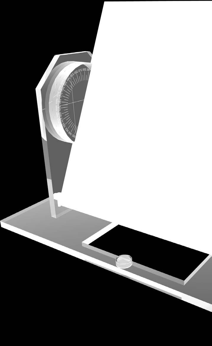
24 RADIOTHERAPY Pro-RT IsoBeam off-the-table measurement option overall dimensions: 203 x 457 x 330 mm screen 305 x 305 mm 2 opaque PMMA plates space for 25 x 30 cm film inscribed with lines precisely defining corners, edges, and centers of the screen field sizes: 2 x 2 mm, 50 x 50 mm, 100 x 100 mm, 150 x 150 mm and 200 x 200 mm center lines scribed with short lines 10 mm apart tungsten markers 2 mm in diameter in the center and corners of the field can rotate 360 in 45 increments base with three leveling non-slip screws and bubble level optional off-the-table measurement possibility (07-202) This is a multifunctional, precision quality assurance tool for daily, weekly or monthly quality assessments of all mechanical and geometrical treatment parameters of linear accelerators or teletherapy units. It is designed to easily and accurately check collimator isocentricity, gantry isocentricity, table isocentricity, collimator field size accuracy, radiation/light field congruence, isocenter rotational stability, ODI accuracy and laser light alignments. IAEA Technical Reports (Series No. 398) IAEA Technical Reports (Series No. 430) IAEA-TECDOC-1540, Specification and Acceptance Testing of Radiotherapy Treatment Planning Systems the Manual provides detailed guidelines for carrying out
 and spatial linearity (SL) patterns 50 mm This phantom is designed for quality control of a CT simulator, which usually consists of a CT scanner, virtual simulator software and a")

25 RADIOTHERAPY Pro-RT CTsim overall dimensions: 150 x 150 x 150 mm made of PMMA test object placed on four faces of the cube Top Face contains contrast details objects (CD), MTF, ray line divergence (RLD) and spatial linearity (SL) patterns 50 mm This phantom is designed for quality control of a CT simulator, which usually consists of a CT scanner, virtual simulator software and a laser marking device (marking of the centre of target volume). Since geometrical planning is the core of CT simulation, periodic quality control is essential for maintaining optimum image quality and patient care. The phantom allows performing detailed geometry and table movement tests as well as image quality assessment - low contrast resolution and high contrast detectability. 15 mm 15 mm cut out 20 mm Diameter Depth First Side Face contains MTF and spatial linearity (SL) patterns lp/mm lp/mm 10 mm lp/mm 10 mm lp/mm lp/mm Second and Third Side Faces contain ray line divergence (RLD) pattern 15 mm 53.8 mm 62.5 mm 75 mm 15 mm cut out 15 mm 20 mm optional carrying case IEC IEC manual provides detailed guidelines for carrying out

26 RADIOTHERAPY Pro-RT AlignCube A simple to use and economic tool to perform true spatial alignment and coincidence tests on IGRT systems. Pro-RT AlignCube contains engraved markings on the exterior and radiographic markers inside. The isocenters of both the OBI and the EPID can be checked for true spatial alignment and coincidence with that of the treatment beam. The phantom ensures the accuracy of linacs image-guidance systems, including CBCT, x-ray volumetric imaging (XVI) and on-board imaging (OBI). It can be used to test: 3D Cone Beam Registration kv and MV System Coincidence kv and MV Projection Images Laser and Light Field Coincidence Weekly Remote Table Adjustments (can be modified to customer specifications): Dimensions: 120 x 120 x 120 mm Made of material similar to water Markings for 100 x 100mm and 40x40mm radiation field check One central fiducial ceramic sphere 6.35mm diameter Two magnification-check fiducial ceramic spheres 6.35mm diameter One offset ceramic sphere target 6.35mm diameter Carrying case the manual provides detailed guidelines for carrying out
27


28
Conflicts of Interest Nuclear Medicine and PET physics reviewer for the ACR Accreditation program
 James R Halama, PhD Loyola University Medical Center Conflicts of Interest Nuclear Medicine and PET physics reviewer for the ACR Accreditation program Learning Objectives 1. Be familiar with recommendations
C a t p h a n / T h e P h a n t o m L a b o r a t o r y
 C a t p h a n 5 0 0 / 6 0 0 T h e P h a n t o m L a b o r a t o r y C a t p h a n 5 0 0 / 6 0 0 Internationally recognized for measuring the maximum obtainable performance of axial, spiral and multi-slice
James R Halama, PhD Loyola University Medical Center
 James R Halama, PhD Loyola University Medical Center Conflicts of Interest Nuclear Medicine and PET physics reviewer for the ACR Accreditation program Learning Objectives Be familiar with the tests recommended
Introduction to Positron Emission Tomography
 Planar and SPECT Cameras Summary Introduction to Positron Emission Tomography, Ph.D. Nuclear Medicine Basic Science Lectures srbowen@uw.edu System components: Collimator Detector Electronics Collimator
SPECT QA and QC. Bruce McBride St. Vincent s Hospital Sydney.
 SPECT QA and QC Bruce McBride St. Vincent s Hospital Sydney. SPECT QA and QC What is needed? Why? How often? Who says? QA and QC in Nuclear Medicine QA - collective term for all the efforts made to produce
Automated Image Analysis Software for Quality Assurance of a Radiotherapy CT Simulator
 Automated Image Analysis Software for Quality Assurance of a Radiotherapy CT Simulator Andrew J Reilly Imaging Physicist Oncology Physics Edinburgh Cancer Centre Western General Hospital EDINBURGH EH4
ImPACT. Information Leaflet No. 1: CT Scanner Acceptance Testing
 ImPACT Information Leaflet No. 1: CT Scanner Acceptance Testing Version 1.02, 18/05/01 CONTENTS: 1. SCOPE OF LEAFLET 2. GENERAL PRINCIPLES OF ACCEPTANCE AND COMMISSIONING 2.1 PHANTOMS 2.2 EXPOSURE AND
Design and performance characteristics of a Cone Beam CT system for Leksell Gamma Knife Icon
 Design and performance characteristics of a Cone Beam CT system for Leksell Gamma Knife Icon WHITE PAPER Introduction Introducing an image guidance system based on Cone Beam CT (CBCT) and a mask immobilization
Medical Imaging BMEN Spring 2016
 Name Medical Imaging BMEN 420-501 Spring 2016 Homework #4 and Nuclear Medicine Notes All questions are from the introductory Powerpoint (based on Chapter 7) and text Medical Imaging Signals and Systems,
S. Guru Prasad, Ph.D., DABR
 PURPOSE S. Guru Prasad, Ph.D., DABR Director of Medical Physics IAEA Consultant NorthShore University Health System and University of Chicago, Pritzker School of Medicine Current TPS utilize more information
UNCOMPROMISING QUALITY
 ION CHAMBERS UNCOMPROMISING QUALITY Designed with over 30 years of scientific integrity for a broad range of dosimetry measurements in diverse radiation beams Farmer-type Chambers For absolute dosimetry
Implementation and evaluation of a fully 3D OS-MLEM reconstruction algorithm accounting for the PSF of the PET imaging system
 Implementation and evaluation of a fully 3D OS-MLEM reconstruction algorithm accounting for the PSF of the PET imaging system 3 rd October 2008 11 th Topical Seminar on Innovative Particle and Radiation
INTERNATIONAL STANDARD
 INTERNATIONAL STANDARD IEC 61675-2 First edition 1998-01 Radionuclide imaging devices Characteristics and test conditions Part 2: Single photon emission computed tomographs Dispositifs d imagerie par radionucléides
Validation of GEANT4 for Accurate Modeling of 111 In SPECT Acquisition
 Validation of GEANT4 for Accurate Modeling of 111 In SPECT Acquisition Bernd Schweizer, Andreas Goedicke Philips Technology Research Laboratories, Aachen, Germany bernd.schweizer@philips.com Abstract.
Quality control phantoms and protocol for a tomography system
 Quality control phantoms and protocol for a tomography system Lucía Franco 1 1 CT AIMEN, C/Relva 27A O Porriño Pontevedra, Spain, lfranco@aimen.es Abstract Tomography systems for non-destructive testing
3D SCANNER. 3D Scanning Comes Full Circle. Your Most Valuable QA and Dosimetry Tools
 3D SCANNER 3D Scanning Comes Full Circle Your Most Valuable QA and Dosimetry Tools BETTER OUTCOMES THROUGH TECHNOLOGY The 3D SCANNER is different by design. 3D SCANNER has been developed from the ground
MEDICAL EQUIPMENT: COMPUTED TOMOGRAPHY. Prof. Yasser Mostafa Kadah
 MEDICAL EQUIPMENT: COMPUTED TOMOGRAPHY Prof. Yasser Mostafa Kadah www.k-space.org Recommended Textbook X-Ray Computed Tomography in Biomedical Engineering, by Robert Cierniak, Springer, 211 Computed Tomography
Corso di laurea in Fisica A.A Fisica Medica 5 SPECT, PET
 Corso di laurea in Fisica A.A. 2007-2008 Fisica Medica 5 SPECT, PET Step 1: Inject Patient with Radioactive Drug Drug is labeled with positron (β + ) emitting radionuclide. Drug localizes
BME I5000: Biomedical Imaging
 1 Lucas Parra, CCNY BME I5000: Biomedical Imaging Lecture 4 Computed Tomography Lucas C. Parra, parra@ccny.cuny.edu some slides inspired by lecture notes of Andreas H. Hilscher at Columbia University.
7/31/2011. Learning Objective. Video Positioning. 3D Surface Imaging by VisionRT
 CLINICAL COMMISSIONING AND ACCEPTANCE TESTING OF A 3D SURFACE MATCHING SYSTEM Hania Al-Hallaq, Ph.D. Assistant Professor Radiation Oncology The University of Chicago Learning Objective Describe acceptance
Developments in Dimensional Metrology in X-ray Computed Tomography at NPL
 Developments in Dimensional Metrology in X-ray Computed Tomography at NPL Wenjuan Sun and Stephen Brown 10 th May 2016 1 Possible factors influencing XCT measurements Components Influencing variables Possible
RITtrend allows you to effectively manage all your physics QA data in one powerful and customizable package.
 So much is asked of medical physicists these days We Can Help Automated Radiation Therapy Phantom Analysis in Seconds QC for Therapy OBI s and CT simulators. Radia s Catphan/OBI module performs analysis
INTERNATIONAL STANDARD
 INTERNATIONAL STANDARD IEC 60601-2-44 2001 AMENDMENT 1 2002-09 Amendment 1 Medical electrical equipment Part 2-44: Particular requirements for the safety of X-ray equipment for computed tomography Amendement
Emission Computed Tomography Notes
 Noll (24) ECT Notes: Page 1 Emission Computed Tomography Notes Introduction Emission computed tomography (ECT) is the CT applied to nuclear medicine. There are two varieties of ECT: 1. SPECT single-photon
Fits you like no other
 Fits you like no other BrightView X and XCT specifications The new BrightView X system is a fully featured variableangle camera that is field-upgradeable to BrightView XCT without any increase in room
THE WIRELESS PHANTOM PERFORM ACCURATE PATIENT QA IN LESS TIME THAN EVER!
 THE WIRELESS PHANTOM PERFORM ACCURATE PATIENT QA IN LESS TIME THAN EVER! Confidence in complex treatments Modern radiation therapy uses complex plans with techniques such as IMRT, VMAT and Tomotherapy.
IAEA-TECDOC-1583 Commissioning of Radiotherapy Treatment Planning Systems: Testing for Typical External Beam Treatment Techniques
 IAEA-TECDOC-1583 Commissioning of Radiotherapy Treatment Planning Systems: Testing for Typical External Beam Treatment Techniques Report of the Coordinated Research Project (CRP) on Development of Procedures
Slide 1. Technical Aspects of Quality Control in Magnetic Resonance Imaging. Slide 2. Annual Compliance Testing. of MRI Systems.
 Slide 1 Technical Aspects of Quality Control in Magnetic Resonance Imaging Slide 2 Compliance Testing of MRI Systems, Ph.D. Department of Radiology Henry Ford Hospital, Detroit, MI Slide 3 Compliance Testing
Optimization of CT Simulation Imaging. Ingrid Reiser Dept. of Radiology The University of Chicago
 Optimization of CT Simulation Imaging Ingrid Reiser Dept. of Radiology The University of Chicago Optimization of CT imaging Goal: Achieve image quality that allows to perform the task at hand (diagnostic
INTRODUCTION TO MEDICAL IMAGING- 3D LOCALIZATION LAB MANUAL 1. Modifications for P551 Fall 2013 Medical Physics Laboratory
 INTRODUCTION TO MEDICAL IMAGING- 3D LOCALIZATION LAB MANUAL 1 Modifications for P551 Fall 2013 Medical Physics Laboratory Introduction Following the introductory lab 0, this lab exercise the student through
A study on image quality provided by a kilovoltage cone-beam computed tomography
 JOURNAL OF APPLIED CLINICAL MEDICAL PHYSICS, VOLUME 14, NUMBER 1, 2013 A study on image quality provided by a kilovoltage cone-beam computed tomography Julia Garayoa a and Pablo Castro Servicio de Radiofísica,
Ch. 4 Physical Principles of CT
 Ch. 4 Physical Principles of CT CLRS 408: Intro to CT Department of Radiation Sciences Review: Why CT? Solution for radiography/tomography limitations Superimposition of structures Distinguishing between
Spiral CT. Protocol Optimization & Quality Assurance. Ge Wang, Ph.D. Department of Radiology University of Iowa Iowa City, Iowa 52242, USA
 Spiral CT Protocol Optimization & Quality Assurance Ge Wang, Ph.D. Department of Radiology University of Iowa Iowa City, Iowa 52242, USA Spiral CT Protocol Optimization & Quality Assurance Protocol optimization
Workshop on Quantitative SPECT and PET Brain Studies January, 2013 PUCRS, Porto Alegre, Brasil Corrections in SPECT and PET
 Workshop on Quantitative SPECT and PET Brain Studies 14-16 January, 2013 PUCRS, Porto Alegre, Brasil Corrections in SPECT and PET Físico João Alfredo Borges, Me. Corrections in SPECT and PET SPECT and
Financial disclosure. Onboard imaging modality for IGRT
 Tetrahedron Beam Computed Tomography Based On Multi-Pixel X- Ray Source and Its Application in Image Guided Radiotherapy Tiezhi Zhang, Ph.D. Advanced X-ray imaging Lab Financial disclosure Patent royalty
Tomotherapy Physics. Machine Twinning and Quality Assurance. Emilie Soisson, MS
 Tomotherapy Physics Machine Twinning and Quality Assurance Emilie Soisson, MS Tomotherapy at UW- Madison Treating for nearly 5 years Up to ~45 patients a day on 2 tomo units Units twinned to facilitate
INSTRUCTIONS RPD INFORMATION RPD PRODUCT INFORMATION. Address 5218 Barthel Industrial Drive Albertville, MN Website
 Expect Service INSTRUCTIONS Radiation Products Design Inc RPD INFORMATION Address 5218 Barthel Industrial Drive Albertville, MN 55301 Website www.rpdinc.com Email sales@rpdinc.com Phone 763-497-2071 or
Fits you like no other
 Fits you like no other Philips BrightView X and XCT specifications The new BrightView X system is a fully featured variableangle camera that is field-upgradeable to BrightView XCT without any increase
Shadow casting. What is the problem? Cone Beam Computed Tomography THE OBJECTIVES OF DIAGNOSTIC IMAGING IDEAL DIAGNOSTIC IMAGING STUDY LIMITATIONS
 Cone Beam Computed Tomography THE OBJECTIVES OF DIAGNOSTIC IMAGING Reveal pathology Reveal the anatomic truth Steven R. Singer, DDS srs2@columbia.edu IDEAL DIAGNOSTIC IMAGING STUDY Provides desired diagnostic
Dosimetry and Medical Radiation Physics section Division of Human Health. Quality Controls in Nuclear Medicine. IAEA-NMQC Toolkit
 Dosimetry and Medical Radiation Physics section Division of Human Health Quality Controls in Nuclear Medicine IAEA-NMQC Toolkit For Fiji Application Version 1.00 User s Manual Version 1.00 October, 2017
Automated Quality Assurance for Image-Guided Radiation Therapy
 JOURNAL OF APPLIED CLINICAL MEDICAL PHYSICS, VOLUME 10, NUMBER 1, WINTER 2009 Automated Quality Assurance for Image-Guided Radiation Therapy Eduard Schreibmann, a Eric Elder, Tim Fox Department of Radiation
CLASS HOURS: 4 CREDIT HOURS: 4 LABORATORY HOURS: 0
 Revised 10/10 COURSE SYLLABUS TM 220 COMPUTED TOMOGRAPHY PHYSICS CLASS HOURS: 4 CREDIT HOURS: 4 LABORATORY HOURS: 0 CATALOG COURSE DESCRIPTION: This course is one of a three course set in whole body Computed
Mediso AnyScan S Single-Head and Dual-Head SPECT
 Mediso AnyScan S Single-Head and Dual-Head SPECT ANYSCAN S (SINGLE-HEAD & DUAL-HEAD SPECT) Low Cost of Ownership Small Footprint Absolute Imaging Solutions tm is the exclusive U.S. source for Mediso Medical
DAILY LINAC QA BEAM QA
 BEAM QA DAILY LINAC QA The QA BeamChecker Plus allows for fast, reliable, and uncomplicated daily QA of Varian, Elekta, Siemens, and Accuray Treatment Machines. The QA BeamChecker Plus is specifically
Quick Reference Datasheet For All RIT113 Packages
 Quick Reference Datasheet For All RIT113 Packages For Rotational Therapies, IMRT & TG142 Highlights and selected product information only. A complete TG142 brochure is available. For more information on
Constructing System Matrices for SPECT Simulations and Reconstructions
 Constructing System Matrices for SPECT Simulations and Reconstructions Nirantha Balagopal April 28th, 2017 M.S. Report The University of Arizona College of Optical Sciences 1 Acknowledgement I would like
ph fax Deming Way Middleton WI USA
 www.standardimaging.com 800-261-4446. ph 608-831-0025. fax 608-831-2202 3120 Deming Way Middleton WI 53562-1461 USA 2007 Standard Imaging, Inc. 1239-21 (03.07) beam qa LINAC AND TOMOTHERAPY QA The NEW
Performance Evaluation of the Philips Gemini PET/CT System
 Performance Evaluation of the Philips Gemini PET/CT System Rebecca Gregory, Mike Partridge, Maggie A. Flower Joint Department of Physics, Institute of Cancer Research, Royal Marsden HS Foundation Trust,
Basic Radiation Oncology Physics
 Basic Radiation Oncology Physics T. Ganesh, Ph.D., DABR Chief Medical Physicist Fortis Memorial Research Institute Gurgaon Acknowledgment: I gratefully acknowledge the IAEA resources of teaching slides
Identification of Shielding Material Configurations Using NMIS Imaging
 Identification of Shielding Material Configurations Using NMIS Imaging B. R. Grogan, J. T. Mihalczo, S. M. McConchie, and J. A. Mullens Oak Ridge National Laboratory, P.O. Box 2008, MS-6010, Oak Ridge,
Philips SPECT/CT Systems
 Philips SPECT/CT Systems Ling Shao, PhD Director, Imaging Physics & System Analysis Nuclear Medicine, Philips Healthcare June 14, 2008 *Presented SNM08 Categorical Seminar - Quantitative SPECT and PET
Micro-CT Methodology Hasan Alsaid, PhD
 Micro-CT Methodology Hasan Alsaid, PhD Preclinical & Translational Imaging LAS, PTS, GlaxoSmithKline 20 April 2015 Provide basic understanding of technical aspects of the micro-ct Statement: All procedures
Evaluation of the Elekta Synergy concept for patient positioning in image guided radiotherapy
 Master of Science Thesis Evaluation of the Elekta Synergy concept for patient positioning in image guided radiotherapy Johan Renström Supervisor: Per Nilsson, PhD and Tommy Knöös, PhD Medical Radiation
Technical aspects of SPECT and SPECT-CT. John Buscombe
 Technical aspects of SPECT and SPECT-CT John Buscombe What does the clinician need to know? For SPECT What factors affect SPECT How those factors should be sought Looking for artefacts For SPECT-CT Issues
PURE. ViSION Edition PET/CT. Patient Comfort Put First.
 PURE ViSION Edition PET/CT Patient Comfort Put First. 2 System features that put patient comfort and safety first. Oncology patients deserve the highest levels of safety and comfort during scans. Our Celesteion
MEDICAL IMAGING 2nd Part Computed Tomography
 MEDICAL IMAGING 2nd Part Computed Tomography Introduction 2 In the last 30 years X-ray Computed Tomography development produced a great change in the role of diagnostic imaging in medicine. In convetional
GafChromic Protocol Multi-Channel Film Dosimetry + Gamma Map Analysis
 GafChromic Protocol Multi-Channel Film Dosimetry + Gamma Map Analysis Micke A. Ashland Inc. Advanced Materials Ashland proprietary patented technology GafChromic Film for Dose Measurement Radiotherapy
Radiology. Marta Anguiano Millán. Departamento de Física Atómica, Molecular y Nuclear Facultad de Ciencias. Universidad de Granada
 Departamento de Física Atómica, Molecular y Nuclear Facultad de Ciencias. Universidad de Granada Overview Introduction Overview Introduction Tecniques of imaging in Overview Introduction Tecniques of imaging
Review of PET Physics. Timothy Turkington, Ph.D. Radiology and Medical Physics Duke University Durham, North Carolina, USA
 Review of PET Physics Timothy Turkington, Ph.D. Radiology and Medical Physics Duke University Durham, North Carolina, USA Chart of Nuclides Z (protons) N (number of neutrons) Nuclear Data Evaluation Lab.
USING cone-beam geometry with pinhole collimation,
 IEEE TRANSACTIONS ON NUCLEAR SCIENCE, VOL. 56, NO. 3, JUNE 2009 687 A Backprojection-Based Parameter Estimation Technique for Skew-Slit Collimation Jacob A. Piatt, Student Member, IEEE, and Gengsheng L.
Introduction to Biomedical Imaging
 Alejandro Frangi, PhD Computational Imaging Lab Department of Information & Communication Technology Pompeu Fabra University www.cilab.upf.edu X-ray Projection Imaging Computed Tomography Digital X-ray
Mediso AnyScan. Single-Head and Dual-Head SPECT
 Mediso AnyScan S Single-Head and Dual-Head SPECT ANYSCAN S (SINGLE-HEAD & DUAL-HEAD SPECT) Low Cost of Ownership Associated Imaging Services is the exclusive U.S. source in the Midwest for Mediso Medical
Continuation Format Page
 C.1 PET with submillimeter spatial resolution Figure 2 shows two views of the high resolution PET experimental setup used to acquire preliminary data [92]. The mechanics of the proposed system are similar
Evaluation of Computed Tomography Techniques for Material Identification in Low Level and Intermediate Level Waste
 Evaluation of Computed Tomography Techniques for Material Identification in Low Level and Intermediate Level Waste. 16648 Stephen Halliwell, VJ Technologies Inc, 89 Carlough Road, Bohemia, NY 11716, USA.
Lucy Phantom MR Grid Evaluation
 Lucy Phantom MR Grid Evaluation Anil Sethi, PhD Loyola University Medical Center, Maywood, IL 60153 November 2015 I. Introduction: The MR distortion grid, used as an insert with Lucy 3D QA phantom, is
Protocol. Technical evaluation of X-ray tomographic image-guided radiotherapy devices CEP10070
 Protocol Technical evaluation of X-ray tomographic image-guided radiotherapy devices CEP10070 March 2010 Contents 2 Introduction... 3 Protocol design and validation... 4 General information... 7 Technical
Accessories for RaySafe Diagnostic X-ray Equipment
 Accessories for RaySafe Diagnostic X-ray Equipment RaySafe Solo RaySafe Xi RaySafe X2 Solo RaySafe X2 CASES 1922014/1922016* 4559871/4559880* RAYSAFE Xi STORM CASE Heavy duty, waterproof case with customized
Version 5.6. Quick Start Guide
 WWW..COM Version 5.6 Quick Start Guide STANDARD IMAGING, INC. 3120 Deming Way Middleton, WI 53562-1461 Jul / 2018 2018 Standard Imaging, Inc. TEL 800.261.4446 TEL 608.831.0025 FAX 608.831.2202 DOC # 80714-05
3D Scanning Comes Full Circle
 3D SCANNER TM 3D Scanning Comes Full Circle Relative 3D Dosimetry offering the easiest setup, most objectivity, and best consistency available Cross-Plane Advanced Design Ring and diameter drive mechanism
Multi-Purpose Body Phantom
 Multi-Purpose Body Phantom Legal & Copyright Notice Information in this manual is subject to change without notice. Permission is granted to Owners of the to reproduce the Worksheets included in this manual
Characterization of a Time-of-Flight PET Scanner based on Lanthanum Bromide
 2005 IEEE Nuclear Science Symposium Conference Record M04-8 Characterization of a Time-of-Flight PET Scanner based on Lanthanum Bromide J. S. Karp, Senior Member, IEEE, A. Kuhn, Member, IEEE, A. E. Perkins,
CT vs. VolumeScope: image quality and dose comparison
 CT vs. VolumeScope: image quality and dose comparison V.N. Vasiliev *a, A.F. Gamaliy **b, M.Yu. Zaytsev b, K.V. Zaytseva ***b a Russian Sci. Center of Roentgenology & Radiology, 86, Profsoyuznaya, Moscow,
Symbia E and S System Specifications Answers for life.
 www.siemens.com/mi Symbia E and S System Specifications Answers for life. Symbia E: Work with confidence. What does it mean to work with confidence? It is clarity not only image clarity but the clarity
An Acquisition Geometry-Independent Calibration Tool for Industrial Computed Tomography
 4th International Symposium on NDT in Aerospace 2012 - Tu.3.A.3 An Acquisition Geometry-Independent Calibration Tool for Industrial Computed Tomography Jonathan HESS *, Patrick KUEHNLEIN *, Steven OECKL
Patient Set-ups and Tumor Localizations
 Patient Set-ups and Tumor Localizations Amy S. Harrison Patient Positioning Prior to starting any localization or simulation procedure patients need to be positioned and immobilized Patients disease location
Digital Image Processing
 Digital Image Processing SPECIAL TOPICS CT IMAGES Hamid R. Rabiee Fall 2015 What is an image? 2 Are images only about visual concepts? We ve already seen that there are other kinds of image. In this lecture
Digital phantoms for the evaluation of a software used for an automatic analysis of the Winston-Lutz test in image guided radiation therapy
 Author manuscript, published in "Medical Imaging 008: Physics of Medical Imaging, San Diego, CA, USA : United States (008)" DOI : 10.1117/1.768668 Digital phantoms for the evaluation of a software used
1. Learn to incorporate QA for surface imaging
 Hania Al-Hallaq, Ph.D. Assistant Professor Radiation Oncology The University of Chicago ***No disclosures*** 1. Learn to incorporate QA for surface imaging into current QA procedures for IGRT. 2. Understand
Introduction to Emission Tomography
 Introduction to Emission Tomography Gamma Camera Planar Imaging Robert Miyaoka, PhD University of Washington Department of Radiology rmiyaoka@u.washington.edu Gamma Camera: - collimator - detector (crystal
CHAPTER 11 NUCLEAR MEDICINE IMAGING DEVICES
 CHAPTER 11 M.A. LODGE, E.C. FREY Russell H. Morgan Department of Radiology and Radiological Sciences, Johns Hopkins University, Baltimore, Maryland, United States of America 11.1. INTRODUCTION Imaging
Evaluation report. X-ray tomographic image guided radiotherapy systems CEP10071
 Evaluation report X-ray tomographic image guided radiotherapy systems CEP10071 March 2010 Contents 2 Summary... 3 Introduction... 6 Product description... 9 Methods... 14 Technical performance... 23 Purchasing...
Position accuracy analysis of the stereotactic reference defined by the CBCT on Leksell Gamma Knife Icon
 Position accuracy analysis of the stereotactic reference defined by the CBCT on Leksell Gamma Knife Icon WHITE PAPER Introduction An image guidance system based on Cone Beam CT (CBCT) is included in Leksell
Clinical implementation of photon beam flatness measurements to verify beam quality
 JOURNAL OF APPLIED CLINICAL MEDICAL PHYSICS, VOLUME 16, NUMBER 6, 2015 Clinical implementation of photon beam flatness measurements to verify beam quality Simon Goodall, a Nicholas Harding, Jake Simpson,
Symbia T Series System Specifications Answers for life.
 www.siemens.com/mi System Specifications Answers for life. 2 Diagnostic Excellence Today s healthcare institutions have increasingly diverse patient needs, greater budget limitations and the need for higher
Table of Contents. About AQUA
 User Manual About AQUA Table of Contents 1 About AQUA... 6 1.1 Radiation Therapy... 6 1.1.1 Workflow manager... 6 1.1.2 Reports... 7 1.1.3 Tests... 7 1.1.4 Many Ways to Work... 7 1.2 Technology... 7 1.3
Machine and Physics Data Guide
 WWW..COM Machine and Physics Data Guide STANDARD IMAGING, INC. 3120 Deming Way Middleton, WI 53562-1461 May / 2008 2008 Standard Imaging, Inc. TEL 800.261.4446 TEL 608.831.0025 FAX 608.831.2202 www.standardimaging.com
8/3/2016. Image Guidance Technologies. Introduction. Outline
 8/3/26 Session: Image Guidance Technologies and Management Strategies Image Guidance Technologies Jenghwa Chang, Ph.D.,2 Department of Radiation Medicine, Northwell Health 2 Hofstra Northwell School of
Corso di laurea in Fisica A.A Fisica Medica 4 TC
 Corso di laurea in Fisica A.A. 2007-2008 Fisica Medica 4 TC Computed Tomography Principles 1. Projection measurement 2. Scanner systems 3. Scanning modes Basic Tomographic Principle The internal structure
HIGH RESOLUTION COMPUTED TOMOGRAPHY FOR METROLOGY
 HIGH RESOLUTION COMPUTED TOMOGRAPHY FOR METROLOGY David K. Lehmann 1, Kathleen Brockdorf 1 and Dirk Neuber 2 1 phoenix x-ray Systems + Services Inc. St. Petersburg, FL, USA 2 phoenix x-ray Systems + Services
Image Quality Assessment and Quality Assurance of Advanced Imaging Systems for IGRT. AAPM Penn-Ohio Chapter Sep 25, 2015 Soyoung Lee, PhD
 Image Quality Assessment and Quality Assurance of Advanced Imaging Systems for IGRT AAPM Penn-Ohio Chapter Sep 25, 2015 Soyoung Lee, PhD 1 Outline q Introduction q Imaging performances in 4D-CBCT Image
Lightsheet Z.1. Light Sheet Fluorescence Microscopy by Carl Zeiss. Fabrice Schmitt, Sales Manager Carl ZEISS France
 Lightsheet Z.1 Light Sheet Fluorescence Microscopy by Carl Zeiss Fabrice Schmitt, Sales Manager Carl ZEISS France 12.12.2012 Light Sheet Fluorescence Microscopy (LSFM) Principle The Principle of Light
IMSURE QA SOFTWARE FAST, PRECISE QA SOFTWARE
 QA SOFTWARE FAST, PRECISE Software for accurate and independent verification of monitor units, dose, and overall validity of standard, IMRT, VMAT, SRS and brachytherapy plans no film, no phantoms, no linac
Qalitätssicherung an Multileafkollimatoren. Dr. Lutz Müller Würzburg jan 2004
 Qalitätssicherung an Multileafkollimatoren Dr. Lutz Müller Würzburg jan 2004 IMRT Verification - present Target Volume Constraints Inverse Planning Algorithm Fluence Map Leaf Sequencer Leaf & Gantry sequence
Thank-You Members of TG147 TG 147: QA for nonradiographic
 Thank-You Members of TG147 TG 147: QA for nonradiographic localization and positioning systems Twyla Willoughby, M.S. Medical Physicist Clinical AAPM Meeting March 2013 Department of Radiation Oncology
8/2/2016. Measures the degradation/distortion of the acquired image (relative to an ideal image) using a quantitative figure-of-merit
 using a quantitative figure-of-merit") Ke Li Assistant Professor Department of Medical Physics and Department of Radiology School of Medicine and Public Health, University of Wisconsin-Madison This work is partially supported by an NIH Grant
Ke Li Assistant Professor Department of Medical Physics and Department of Radiology School of Medicine and Public Health, University of Wisconsin-Madison This work is partially supported by an NIH Grant
Initial Clinical Experience with 3D Surface Image Guidance
 Initial Clinical Experience with 3D Surface Image Guidance Amanda Havnen-Smith, Ph.D. Minneapolis Radiation Oncology Ridges Radiation Therapy Center Burnsville, MN April 20 th, 2012 Non-funded research
Facility Questionnaire PART I (General Information for 3DCRT and IMRT)
") Facility Questionnaire PART I (General Information for 3DCRT and IMRT) The following items are required before you can enter cases on any RTOG protocol that requires data submission to the Image-Guided
Facility Questionnaire PART I (General Information for 3DCRT and IMRT) The following items are required before you can enter cases on any RTOG protocol that requires data submission to the Image-Guided
Spectral analysis of non-stationary CT noise
 Spectral analysis of non-stationary CT noise Kenneth M. Hanson Los Alamos Scientific Laboratory Int. Symposium and Course on Computed Tomography, Las Vegas, April 7-11, 1980 This presentation available
18th World Conference on Nondestructive Testing, April 2012, Durban, South Africa
 18th World Conference on Nondestructive Testing, 16-20 April 2012, Durban, South Africa TOWARDS ESTABLISHMENT OF STANDARDIZED PRACTICE FOR ASSESSMENT OF SPATIAL RESOLUTION AND CONTRAST OF INTERNATIONAL
Three-Dimensional Laser Scanner. Field Evaluation Specifications
 Stanford University June 27, 2004 Stanford Linear Accelerator Center P.O. Box 20450 Stanford, California 94309, USA Three-Dimensional Laser Scanner Field Evaluation Specifications Metrology Department