Real-Time FMRI Tools & Automation in AFNI & SUMA
|
|
|
- Kathleen Cannon
- 5 years ago
- Views:
Transcription


1 Real-Time FMRI Tools & Automation in AFNI & SUMA SSCC / NIMH & NINDS / NIH / DHHS / USA / EARTH

2 Why bother? Image quality control Spikes, distortion, ghosting, noise, Amount of motion Operator error Functional localization Localizer prior to main FMRI experiment for BCI or high-res imaging Pre operative scanning As Q/A in clinical settings or difficult / rare subject population 'scan to criteria' Teaching Feedback and Biofeedback Reduce motion Alter/interfere brain function Control of task/ stimulus computer Classification/BCI Signals in vegetative state Yang, S. et al. 05 Owen AM et al 06 Cox, RW et al. 95, Cohen, MS et al. 98, Frank, J. et al 99, Voyvodic, J. 99 Weiskopf, N. et al 04 Yang, S. et al 08 Weiskopf, N et al decharms. RC. et al. 04 decharms. RC. et al. 05 Posse S. et al. 03 LaConte SM. et al. 07 Yoo S. et al. 04
3 Image Quality Control Image quality control Spikes, distortion, ghosting, noise, Amount of motion Cox, RW et al. 95, Cohen, MS et al. 98, Frank, J. et al 99, Voyvodic, J. 99 Weiskopf, N et al. 2007

4 Image Quality Control Image quality control Spikes, distortion, ghosting, noise, Amount of motion Cox, RW et al. 95, Cohen, MS et al. 98, Frank, J. et al 99, Voyvodic, J. 99 Weiskopf, N et al. 2007


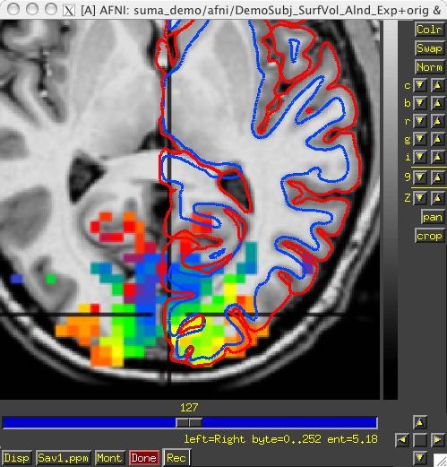
5 Image Quality Control Real-time Estimation of Functional Activation Real-time Estimation of subject movement
6 Reduce Motion with Feedback Feedback and Biofeedback Reduce motion Yang, S. et al. 08 Fig.6 from Yang, S. et al. Neuroimage 05 Fig.2 from Yang, S. et al. Neuroimage 05


7 Classification Classification maps high dimensional pattern into a set of classes This allows a complex brain activation pattern to be identified with a set of classes or brain states. Useful in to providing intuitive feedback from activation of multiple areas Useful for inferring brain state & $ $ $ $ % x x 1 2 x N #!!!! " x 1 " LaConte SM. 07" experiment x 2 " From LaConte S. FMRI Advanced Issues ISMRM 09 time


8 Single 2 second event From fast randomized event related FMRI" Figs.1 and 3 from Beauchamp, M.S. et al. HBM 09

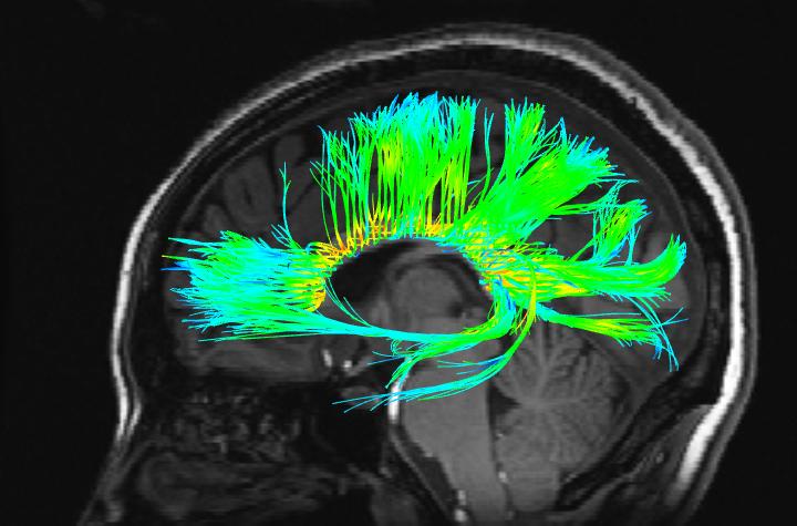
9 Brain Computer Interface Fig.1 Yoo S. et al. Neuroreport 04


10 The AFNI interface
11 The players Scanner" Real Time Setup" RT Plugin" Image Monitor" AFNI" Plugin" Real Time Receiver" Stimulus Display"

12 The players Scanner" Real Time Setup" Image Monitor" AFNI" Real Time Receiver" RT Plugin" Plugin" Stimulus Display" Scanner A user-supplied machine to acquire and reconstruct images in real time
13 The players Scanner" Real Time Setup" Image Monitor" AFNI" Real Time Receiver" RT Plugin" Plugin" Stimulus Display" Real Time Setup A user-supplied set of commands that tell AFNI what to do with incoming data Can be done from shell commands or from within C code Communicates with AFNI through TCP/IP socket Sets up ROIs for AFNI*
14 Setting up AFNI's RT plugin Manually Good for learning and demo

15 Setting up AFNI's RT plugin Via Environment Variables setenv AFNI_REALTIME_Registration setenv AFNI_REALTIME_Graph 3D:_realtime Realtime
16 Manually Environment variables Setting up AFNI See README.environment (~250 variables) Layout files Size and position windows just so Via plugout_drive Details will follow Via image_monitor module -drive options -drive_wait 'OPEN_WINDOW axialgraph keypress=a' -drive_afni 'CLOSE_WINDOW axialimage'
17 Demo time Get bootcamp data Motion monitoring cd AFNI_data6/realtime.demos tcsh demo.1.run1 illustrates real-time data acquisition and motion correction by AFNI Motion & function See demo.2.fback.0.readme for instructions Illustrates acquisition, motion correction and feedback Remove option -show_demo_gui yes from demo.2.fback. 1.receiver if it proves troublesome
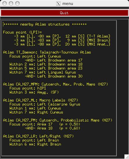
18 ROI selection options Standard atlases TT_Daemon : Created by tracing Talairach and Tournoux brain illustrations. Contibuted by Jack Lancaster and Peter Fox of RIC UTHSCSA CA_N27_MPM, CA_N27_ML, CA_N27_PM : Anatomy Toolbox's atlases, some created from cytoarchitectonic studies of 10 human post-mortem brains contributed by Simon Eickhoff, Katrin Amunts and Karl Zilles of IME, Julich, FreeSurfer, subject-based Functional localizer Etc.
19 Standard-space atlas ROI -region CA_N27_ML::Hip \!!!!-region CA_N27_ML::Amygda \!!!!-base TT_N27_r2+tlrc. \!!!!-anat doe_surfvol_alnd_exp+orig. \!!!!-roi_grid blur_vr_run1_motor_afb003+orig. \!!!!-prefix hip_amy -time!! less than 1min including skull stripping and xform to TLRC A couple of seconds for generating more ROIs

\" 3- Identify ROIs \" 4- Xform")
20 Atlas-based ROIs 1- Strip skull" 2- Find xform to atlas space" (about 40 secs, 2.5GhZ cpu)" 3- Identify ROIs " 4- Xform ROIs to native space" (about 2 seconds)"

21 Subject-based Anatomical ROIs From FreeSurfer's Parcellations" surfer.nmr.mgh.harvard.edu
22 The players Scanner" Real Time Setup" Image Monitor" AFNI" Stimulus Display" Real Time Receiver" RT Plugin" Plugin" Image Monitor An AFNI- or user- supplied program to wait for new images AFNI-supplied programs monitor files only: Imon (Monitors GE's old dreaded I files) Dimon (Monitors GE's DICOM images) RTfeedme (Breaks up timeseries dataset and sends it to AFNI) User-supplied programs usually interface with scanner software SIEMENS TRIO/ALLEGRA via functors (S. LaConte BCM, E. Stein NIDA) Often only program that runs on scanner computer Image Monitor sends new images or volumes to AFNI over TCP/IP socket
23 The players Scanner" Real Time Setup" Image Monitor" AFNI" Stimulus Display" Real Time Receiver" RT Plugin" Plugin" AFNI/RT plugin take incoming images/volumes and processes them per the setup instructions Assemble images/volumes into time series Perform image registration Perform (multi*) linear regression Send results to Real Time Receiver through TCP/IP socket Raw, volume registered, or residual volume* ROI based results Send raw or processed volumes to plugins registered to receive them Much faster than TCP/IP (just a data pointer is passed) Plugins can also communicate with Real Time Receiver
24 The players Scanner" Real Time Setup" Image Monitor" AFNI" Stimulus Display" Real Time Receiver" RT Plugin" Plugin" AFNI/RT plugin take incoming images/volumes and processes them per the setup instructions Assemble images/volumes into time series Perform image registration Perform (multi*) linear regression Send results to Real Time Receiver through TCP/IP socket Raw, volume registered, or residual volume* ROI based results Send raw or processed volumes to plugins registered to receive them Much faster than TCP/IP (just a data pointer is passed) Plugins can also communicate with Real Time Receiver
25 The players Scanner" Real Time Setup" Image Monitor" AFNI" Stimulus Display" Real Time Receiver" RT Plugin" Plugin" Real Time Receiver (e.g. serial_helper.c or realtime_receiver.py) AFNI- or User- supplied application that expects incoming data from AFNI and acts on it Motion parameters ROI-based data, all values or just average Entire volumes of raw, or preprocessed data Data from any RT plugin such as 3dsvm Process incoming data to your liking Optionally forward results to Stimulus Display either by serial connection, or TCP/IP*
26 Image Monitor (Dimon) Dimon: - monitor acquisition of Dicom or GE-Ifiles - optionally write to3d script for creation of AFNI datasets - optionally send volumes to afni's realtime plugin find first volume (wait forever, scanning may not have started) wait for volume: check every 2 seconds or every -sleep_init ms check slices to see if a volume is acquired once found: note grid, orientation, byte order, etc. if realtime: comm: open link try to open TCP channel to afni RT plugin check whether channel is ready for data comm: send control info send acquisition style (2D+zt), zorder, time pattern, TR, FOV, grid, datum, orientation, origin, etc. comm: send volume
27 Image Monitor (Dimon), part II set signal handlers, and note between-volume sleep time for each found volume while no new volume is yet found check whether the scanner has stalled (run cancelled?) sleep for one TR, or -sleep_vol ms, or -sleep_frac fraction of TR if this is a new run comm: send "end of (previous) run" message track volume statistics check orientation comm: if connection not yet established, send control info comm: send volume upon termination (ctrl-c or -quit and no more data) show run statistics possibly create to3d script comm: terminate connection
28 Plug_realtime plug_realtime: init: register work process with afni (to be called regularly) plugin main: sets plugin control variables main work process: asynchronously from main afni loop if new connection, initialize if data is bad or no new data after timeout write vol. to disk, plot final motion params, comm:close if new data: warn user and process process control info: TR, grid, orientation, DRIVE comds., etc. prepare to receive data from multiple channels setup new dataset if done with data: finish_dataset and cleanup while there is data to read store into images if we have a full volume add volume to dataset possibly register volume to base update registration graph possibly run regression comm: compute and send TR data to realtime receiver
29 Realtime_receiver.py set signal handlers to close all ports on exit open incoming socket and wait for connection... forever: process one run wait for the real-time plugin to talk to us check magic HELLO for type/amount of data to receive: only motion motion plus N ROI averages motion plus N voxel values (with coordinates, etc.) open outgoing serial port while no run termination, process one TR close data ports read incoming TCP data compute outgoing results write to serial port

30 Scanner" Image Monitor" Stimulus Display" RT SVM plugin* Real Time Setup" AFNI" Real Time Receiver" RT Plugin" Plugin" SVM plugin is being modified to accept RT data Given training models, classification is done in real-time Classification can go to text, or to Real Time Receiver

31 Real Time SVM* Scanner" Real Time Setup" Image Monitor" AFNI" Stimulus Display" Real Time Receiver" RT Plugin" Plugin" *Movie generated with Real Time setup in S. LaConte et al. HBM 2007"
32 Automation


 + No need to manage")

33 Automating Navigation Other applications can communicate with AFNI via a program which sends a series of commands for execution. + Program called via system function (shell invocation) + No need to manage sockets or format and transmit commands + User Interaction with GUI is uninterrupted GUI drivers Shell Script
34 Cycling trough 300 volumes while ($cnt < 300) plugout_drive -com "SWITCH_UNDERLAY A anat_${cnt}+orig" -com "SWITCH_OVERLAY A anat.ns_${cnt}+orig" -com 'OPEN_WINDOW A coronalimage opacity=0.5' -com 'OPEN_WINDOW A axialimage keypress=v opacity=0.4' -quit echo "Enter new number or hit enter for next brain:" set ans = $< if ( $ans == ) cnt ++ else set cnt = `expr $cnt + $ans` endif end
35 Cycling trough 300 volumes Loop over all volumes while ($cnt < 300) plugout_drive -com "SWITCH_UNDERLAY A anat_${cnt}+orig" -com "SWITCH_OVERLAY A anat.ns_${cnt}+orig" -com 'OPEN_WINDOW A coronalimage opacity=0.5' -com 'OPEN_WINDOW A axialimage keypress=v opacity=0.4' -quit echo "Enter new number or hit enter for next brain:" set ans = $< if ( $ans == ) cnt ++ else set cnt = `expr $cnt + $ans` endif end
36 Cycling trough 300 volumes Switch background volume while ($cnt < 300) plugout_drive -com "SWITCH_UNDERLAY A anat_${cnt}+orig" -com "SWITCH_OVERLAY A anat.ns_${cnt}+orig" -com 'OPEN_WINDOW A coronalimage opacity=0.5' -com 'OPEN_WINDOW A axialimage keypress=v opacity=0.4' -quit echo "Enter new number or hit enter for next brain:" set ans = $< if ( $ans == ) cnt ++ else set cnt = `expr $cnt + $ans` endif end
37 Cycling trough 300 volumes Switch foreground volume while ($cnt < 300) plugout_drive -com "SWITCH_UNDERLAY A anat_${cnt}+orig" -com "SWITCH_OVERLAY A anat.ns_${cnt}+orig" -com 'OPEN_WINDOW A coronalimage opacity=0.5' -com 'OPEN_WINDOW A axialimage keypress=v opacity=0.4' -quit echo "Enter new number or hit enter for next brain:" set ans = $< if ( $ans == ) cnt ++ else set cnt = `expr $cnt + $ans` endif end
38 Cycling trough 300 volumes Open coronal image with low opacity while ($cnt < 300) plugout_drive -com "SWITCH_UNDERLAY A anat_${cnt}+orig" -com "SWITCH_OVERLAY A anat.ns_${cnt}+orig" -com 'OPEN_WINDOW A coronalimage opacity=0.5' -com 'OPEN_WINDOW A axialimage keypress=v opacity=0.4' -quit echo "Enter new number or hit enter for next brain:" set ans = $< if ( $ans == ) cnt ++ else set cnt = `expr $cnt + $ans` endif end
39 Cycling trough 300 volumes Open axial image and start video mode while ($cnt < 300) plugout_drive -com "SWITCH_UNDERLAY A anat_${cnt}+orig" -com "SWITCH_OVERLAY A anat.ns_${cnt}+orig" -com 'OPEN_WINDOW A coronalimage opacity=0.5' -com 'OPEN_WINDOW A axialimage keypress=v opacity=0.4' -quit echo "Enter new number or hit enter for next brain:" set ans = $< if ( $ans == ) cnt ++ else set cnt = `expr $cnt + $ans` endif end

40 SUMA Movie Sample Use this link if viewing pdf. Video courtesy of Chunmao Wang."
41 "Help" sources Readme files README.driver README.environment README.realtime Demo material available on: Automation Demos Scripts in class data under:! AFNI_data6/realtime.demos/ Sample programs rtfeedme.c! Dimon.c! serial_helper.c! realtime_receiver.py Talk to us, we're interested in applications
AFNI Preprocessing: Outline, Recommendations, and New(ish) Stuff. Robert W Cox SSCC / NIMH & NINDS / NIH / DHHS / USA / EARTH
 Stuff. Robert W Cox SSCC / NIMH & NINDS / NIH / DHHS / USA / EARTH") AFNI Preprocessing: Outline, Recommendations, and New(ish) Stuff Robert W Cox SSCC / NIMH & NINDS / NIH / DHHS / USA / EARTH HBM 2016 As a work of a US Government official, this presentation is not copyrighted
AFNI Preprocessing: Outline, Recommendations, and New(ish) Stuff Robert W Cox SSCC / NIMH & NINDS / NIH / DHHS / USA / EARTH HBM 2016 As a work of a US Government official, this presentation is not copyrighted
Transforming Datasets to Talairach-Tournoux Coordinates
 -1- Transforming Datasets to Talairach-Tournoux Coordinates The original purpose of AFNI was to perform the transformation of datasets to Talairach-Tournoux (stereotaxic) coordinates The transformation
Measuring baseline whole-brain perfusion on GE 3.0T using arterial spin labeling (ASL) MRI
 MRI") Measuring baseline whole-brain perfusion on GE 3.0T using arterial spin labeling (ASL) MRI Revision date: 09/15/2008 Overview This document describes the procedure for measuring baseline whole-brain perfusion
Measuring baseline whole-brain perfusion on GE 3.0T using arterial spin labeling (ASL) MRI Revision date: 09/15/2008 Overview This document describes the procedure for measuring baseline whole-brain perfusion
Turbo-BrainVoyager. Setup guide
 Turbo-BrainVoyager Setup guide Turbo-BrainVoyager (TBV) is a highly optimized software package for real-time analysis and advanced visualization of functional and structural magnetic resonance imaging
Journal of Articles in Support of The Null Hypothesis
 Data Preprocessing Martin M. Monti, PhD UCLA Psychology NITP 2016 Typical (task-based) fmri analysis sequence Image Pre-processing Single Subject Analysis Group Analysis Journal of Articles in Support
Issues Regarding fmri Imaging Workflow and DICOM
 Issues Regarding fmri Imaging Workflow and DICOM Lawrence Tarbox, Ph.D. Fred Prior, Ph.D Mallinckrodt Institute of Radiology Washington University in St. Louis What is fmri fmri is used to localize functions
Function-Structure Integration in FreeSurfer
 Function-Structure Integration in FreeSurfer Outline Function-Structure Integration Function-Structure Registration in FreeSurfer fmri Analysis Preprocessing First-Level Analysis Higher-Level (Group) Analysis
Introduction to fmri. Pre-processing
 Introduction to fmri Pre-processing Tibor Auer Department of Psychology Research Fellow in MRI Data Types Anatomical data: T 1 -weighted, 3D, 1/subject or session - (ME)MPRAGE/FLASH sequence, undistorted
Overview of fmri Analysis Software. McConnell BIC Open Methods Meetup January 13th 2013
 Overview of fmri Analysis Software McConnell BIC Open Methods Meetup January 13th 2013 FSL Michael Ferreira Outline Introduction MRI scanners and fmri equipment FSL Image formats and conversion FSLView
Statistical Analysis of Neuroimaging Data. Phebe Kemmer BIOS 516 Sept 24, 2015
 Statistical Analysis of Neuroimaging Data Phebe Kemmer BIOS 516 Sept 24, 2015 Review from last time Structural Imaging modalities MRI, CAT, DTI (diffusion tensor imaging) Functional Imaging modalities
Slicer3 Tutorial: Registration Library Case 14. Intra-subject Brain PET-MRI fusion
 NA-MIC Slicer3 Tutorial: Registration Library Case 14 Intra-subject Brain PET-MRI fusion Dominik Meier, Ron Kikinis March 2010 Overview 1. Introduction 2. Prerequisites 3. Modules Used takes how long to
Automated MR Image Analysis Pipelines
 Automated MR Image Analysis Pipelines Andy Simmons Centre for Neuroimaging Sciences, Kings College London Institute of Psychiatry. NIHR Biomedical Research Centre for Mental Health at IoP & SLAM. Neuroimaging
Functional MRI data preprocessing. Cyril Pernet, PhD
 Functional MRI data preprocessing Cyril Pernet, PhD Data have been acquired, what s s next? time No matter the design, multiple volumes (made from multiple slices) have been acquired in time. Before getting
AFNI. h'p://afni.nimh.nih.gov/afni
 AFNI h'p://afni.nimh.nih.gov/afni AFNI Fundamentals Basic unit of data in AFNI is the dataset A collection of 1 or more 3D arrays of numbers o Each entry in the array is in a particular spatial location
FSL Pre-Processing Pipeline
 The Art and Pitfalls of fmri Preprocessing FSL Pre-Processing Pipeline Mark Jenkinson FMRIB Centre, University of Oxford FSL Pre-Processing Pipeline Standard pre-processing: Task fmri Resting-state fmri
The organization of the human cerebral cortex estimated by intrinsic functional connectivity
 1 The organization of the human cerebral cortex estimated by intrinsic functional connectivity Journal: Journal of Neurophysiology Author: B. T. Thomas Yeo, et al Link: https://www.ncbi.nlm.nih.gov/pubmed/21653723
FSL Pre-Processing Pipeline
 The Art and Pitfalls of fmri Preprocessing FSL Pre-Processing Pipeline Mark Jenkinson FMRIB Centre, University of Oxford FSL Pre-Processing Pipeline Standard pre-processing: Task fmri Resting-state fmri
Playing with data from lab
 Playing with data from lab Getting data off the scanner From the Patient Browser, select the folder for the study you want (or within that study, the set of images you want), and then from the Transfer
Fmri Spatial Processing
 Educational Course: Fmri Spatial Processing Ray Razlighi Jun. 8, 2014 Spatial Processing Spatial Re-alignment Geometric distortion correction Spatial Normalization Smoothing Why, When, How, Which Why is
MITK-DI. A new Diffusion Imaging Component for MITK. Klaus Fritzsche, Hans-Peter Meinzer
 MITK-DI A new Diffusion Imaging Component for MITK Klaus Fritzsche, Hans-Peter Meinzer Division of Medical and Biological Informatics, DKFZ Heidelberg k.fritzsche@dkfz-heidelberg.de Abstract. Diffusion-MRI
FMRI Pre-Processing and Model- Based Statistics
 FMRI Pre-Processing and Model- Based Statistics Brief intro to FMRI experiments and analysis FMRI pre-stats image processing Simple Single-Subject Statistics Multi-Level FMRI Analysis Advanced FMRI Analysis
2. Creating Field Maps Using the Field Map GUI (Version 2.0) in SPM5
 in SPM5") 1. Introduction This manual describes how to use the Field Map Toolbox Version 2.0 for creating unwrapped field maps that can be used to do geometric distortion correction of EPI images in SPM5. 1. 1.
1. Introduction This manual describes how to use the Field Map Toolbox Version 2.0 for creating unwrapped field maps that can be used to do geometric distortion correction of EPI images in SPM5. 1. 1.
Artifact Detection and Repair: Overview and Sample Outputs
 Artifact Detection and Repair: Overview and Sample Outputs Paul Mazaika February 2007 Programs originated in Gabrieli Neuroscience Laboratory, updated and enhanced at Center for Interdisciplinary Brain
Preprocessing of fmri data
 Preprocessing of fmri data Pierre Bellec CRIUGM, DIRO, UdM Flowchart of the NIAK fmri preprocessing pipeline fmri run 1 fmri run N individual datasets CIVET NUC, segmentation, spatial normalization slice
White Pixel Artifact. Caused by a noise spike during acquisition Spike in K-space <--> sinusoid in image space
 White Pixel Artifact Caused by a noise spike during acquisition Spike in K-space sinusoid in image space Susceptibility Artifacts Off-resonance artifacts caused by adjacent regions with different
Measuring baseline whole-brain perfusion on GE 3.0T using arterial spin labeling (ASL) MRI
 MRI") Measuring baseline whole-brain perfusion on GE 3.0T using arterial spin labeling (ASL) MRI Revision date: 11/20/2006 Overview This document describes the procedure for measuring baseline whole-brain perfusion
Measuring baseline whole-brain perfusion on GE 3.0T using arterial spin labeling (ASL) MRI Revision date: 11/20/2006 Overview This document describes the procedure for measuring baseline whole-brain perfusion
User s Guide Neuroimage Processing ToolKit (NPTK) Version 2.0 fmri Registration Software Pipeline for Functional Localization
 Version 2.0 fmri Registration Software Pipeline for Functional Localization") User s Guide Neuroimage Processing ToolKit (NPTK) Version 2.0 fmri Registration Software Pipeline for Functional Localization Software Written by Ali Gholipour SIP Lab, UTD, 2005-2010 Revision 2.0 February
User s Guide Neuroimage Processing ToolKit (NPTK) Version 2.0 fmri Registration Software Pipeline for Functional Localization Software Written by Ali Gholipour SIP Lab, UTD, 2005-2010 Revision 2.0 February
User s Guide Neuroimage Processing ToolKit (NPTK) Version.1.7 (beta) fmri Registration Software Pipeline for Functional Localization
 Version.1.7 (beta) fmri Registration Software Pipeline for Functional Localization") User s Guide Neuroimage Processing ToolKit (NPTK) Version.1.7 (beta) fmri Registration Software Pipeline for Functional Localization Software Written by Ali Gholipour SIP Lab, UTD, 2005-2007 Revision 1.7
User s Guide Neuroimage Processing ToolKit (NPTK) Version.1.7 (beta) fmri Registration Software Pipeline for Functional Localization Software Written by Ali Gholipour SIP Lab, UTD, 2005-2007 Revision 1.7
Single Subject Demo Data Instructions 1) click "New" and answer "No" to the "spatially preprocess" question.
 click New and answer No to the spatially preprocess question.") (1) conn - Functional connectivity toolbox v1.0 Single Subject Demo Data Instructions 1) click "New" and answer "No" to the "spatially preprocess" question. 2) in "Basic" enter "1" subject, "6" seconds
(1) conn - Functional connectivity toolbox v1.0 Single Subject Demo Data Instructions 1) click "New" and answer "No" to the "spatially preprocess" question. 2) in "Basic" enter "1" subject, "6" seconds
MITK-DI. A new Diffusion Imaging Component for MITK. Klaus Fritzsche, Hans-Peter Meinzer
 MITK-DI A new Diffusion Imaging Component for MITK Klaus Fritzsche, Hans-Peter Meinzer Division of Medical and Biological Informatics, DKFZ Heidelberg k.fritzsche@dkfz-heidelberg.de Abstract. Diffusion-MRI
Data Loading & 3D Visualization
 Neuroimage Analysis Center Data Loading & 3D Visualization Sonia Pujol, Ph.D. Surgical Planning Laboratory Harvard Medical School Leonardo da Vinci (1452-1519), Virgin and Child Alte Pinakothek, München
Slicer3 Tutorial. Manual Registration. NA-MIC National Alliance for Medical Image Computing Dominik Meier, Ron Kikinis
 NA-MIC Slicer3 Tutorial Manual Registration Dominik Meier, Ron Kikinis Overview Introduction takes how long to do? Prerequisites 3. Loading Example Dataset 10 sec 4. Creating New Transform 10 sec 5. Associate
fmri pre-processing Juergen Dukart
 fmri pre-processing Juergen Dukart Outline Why do we need pre-processing? fmri pre-processing Slice time correction Realignment Unwarping Coregistration Spatial normalisation Smoothing Overview fmri time-series
Classification of Subject Motion for Improved Reconstruction of Dynamic Magnetic Resonance Imaging
 1 CS 9 Final Project Classification of Subject Motion for Improved Reconstruction of Dynamic Magnetic Resonance Imaging Feiyu Chen Department of Electrical Engineering ABSTRACT Subject motion is a significant
I.e. Sex differences in child appetitive traits and Eating in the Absence of Hunger:
 Supplementary Materials I. Evidence of sex differences on eating behavior in children I.e. Sex differences in child appetitive traits and Eating in the Absence of Hunger: Table 2. Parent Report for Child
Atlas Registration & Label Merging
 NA-MIC Slicer3 Tutorial Atlas Registration & Label Merging Dominik Meier, Ron Kikinis February 2010 Overview 1. Introduction 2. Prerequisites 3. Modules Used takes how long to do? 4. Loading Example Dataset
0.1. Setting up the system path to allow use of BIAC XML headers (BXH). Depending on the computer(s), you may only have to do this once.
. Depending on the computer(s), you may only have to do this once.") Week 3 Exercises Last week you began working with MR data, both in the form of anatomical images and functional time series. This week we will discuss some concepts related to the idea of fmri data as
Week 3 Exercises Last week you began working with MR data, both in the form of anatomical images and functional time series. This week we will discuss some concepts related to the idea of fmri data as
Mapping of Hierarchical Activation in the Visual Cortex Suman Chakravartula, Denise Jones, Guillaume Leseur CS229 Final Project Report. Autumn 2008.
 Mapping of Hierarchical Activation in the Visual Cortex Suman Chakravartula, Denise Jones, Guillaume Leseur CS229 Final Project Report. Autumn 2008. Introduction There is much that is unknown regarding
Rat 2D EPSI Dual Band Variable Flip Angle 13 C Dynamic Spectroscopy
 Rat 2D EPSI Dual Band Variable Flip Angle 13 C Dynamic Spectroscopy In this example you will load a dynamic MRS animal data set acquired on a GE 3T scanner. This data was acquired with an EPSI sequence
Analysis of fmri data within Brainvisa Example with the Saccades database
 Analysis of fmri data within Brainvisa Example with the Saccades database 18/11/2009 Note : All the sentences in italic correspond to informations relative to the specific dataset under study TP participants
Brain Extraction, Registration & EPI Distortion Correction
 Brain Extraction, Registration & EPI Distortion Correction What use is Registration? Some common uses of registration: Combining across individuals in group studies: including fmri & diffusion Quantifying
Fiber Selection from Diffusion Tensor Data based on Boolean Operators
 Fiber Selection from Diffusion Tensor Data based on Boolean Operators D. Merhof 1, G. Greiner 2, M. Buchfelder 3, C. Nimsky 4 1 Visual Computing, University of Konstanz, Konstanz, Germany 2 Computer Graphics
SIVIC GUI Tutorial. HMTRC Workshop - March 23-24, 2017
 SIVIC GUI Tutorial HMTRC Workshop - March 23-24, 2017 Department of Radiology and Biomedical Imaging, UCSF Supported by NIBIB P41EB013598 Goal: The purpose of this tutorial is to introduce you to the SIVIC
GLIRT: Groupwise and Longitudinal Image Registration Toolbox
 Software Release (1.0.1) Last updated: March. 30, 2011. GLIRT: Groupwise and Longitudinal Image Registration Toolbox Guorong Wu 1, Qian Wang 1,2, Hongjun Jia 1, and Dinggang Shen 1 1 Image Display, Enhancement,
MRI Physics II: Gradients, Imaging
 MRI Physics II: Gradients, Imaging Douglas C., Ph.D. Dept. of Biomedical Engineering University of Michigan, Ann Arbor Magnetic Fields in MRI B 0 The main magnetic field. Always on (0.5-7 T) Magnetizes
Tutorial BOLD Module
 m a k i n g f u n c t i o n a l M R I e a s y n o r d i c B r a i n E x Tutorial BOLD Module Please note that this tutorial is for the latest released nordicbrainex. If you are using an older version please
QuickVol II Users Guide
 QuickVol II Users Guide Karl Schmidt (karl.schmidt@umassmed.edu) Working Draft 3/28/2006 This document is a working draft. If you find an error, please submit a bug on the Quickvol website. Thank you.
SPM8 for Basic and Clinical Investigators. Preprocessing. fmri Preprocessing
 SPM8 for Basic and Clinical Investigators Preprocessing fmri Preprocessing Slice timing correction Geometric distortion correction Head motion correction Temporal filtering Intensity normalization Spatial
DIFFUSION TENSOR IMAGING ANALYSIS. Using Analyze
 DIFFUSION TENSOR IMAGING ANALYSIS Using Analyze 2 Table of Contents 1. Introduction page 3 2. Loading DTI Data page 4 3. Computing DTI Maps page 5 4. Defining ROIs for Fiber Tracking page 6 5. Visualizing
icatvision Quick Reference
 icatvision Quick Reference Navigating the i-cat Interface This guide shows how to: View reconstructed images Use main features and tools to optimize an image. REMINDER Images are displayed as if you are
SIVIC Scripting Tutorial
 SIVIC Scripting Tutorial HMTRC Workshop - March 23-24, 2017 Department of Radiology and Biomedical Imaging, UCSF Supported by NIBIB P41EB013598 Goal: The purpose of this tutorial is to introduce you to
Artifact detection and repair in fmri
 Artifact detection and repair in fmri Paul K. Mazaika, Ph.D. Center for Interdisciplinary Brain Sciences Research (CIBSR) Division of Interdisciplinary Behavioral Sciences Stanford University School of
Basic fmri Design and Analysis. Preprocessing
 Basic fmri Design and Analysis Preprocessing fmri Preprocessing Slice timing correction Geometric distortion correction Head motion correction Temporal filtering Intensity normalization Spatial filtering
Update ASA 4.8. Expand your research potential with ASA 4.8. Highly advanced 3D display of single channel coherence
 Update ASA 4.8 Expand your research potential with ASA 4.8. The ASA 4.8 software has everything needed for a complete analysis of EEG / ERP and MEG data. From features like (pre)processing of data, co-registration
NeurAL Documentation. Release 1.0. Bill Gross
 NeurAL Documentation Release 1.0 Bill Gross Jul 10, 2018 Contents 1 Usage Example: 3 2 General Structure 5 3 Modules: 7 3.1 Wrapper Functions............................................ 7 3.2 Useful Utilities..............................................
Automatic segmentation of the cortical grey and white matter in MRI using a Region Growing approach based on anatomical knowledge
 Automatic segmentation of the cortical grey and white matter in MRI using a Region Growing approach based on anatomical knowledge Christian Wasserthal 1, Karin Engel 1, Karsten Rink 1 und André Brechmann
This exercise uses one anatomical data set (ANAT1) and two functional data sets (FUNC1 and FUNC2).
 and two functional data sets (FUNC1 and FUNC2).") Exploring Brain Anatomy This week s exercises will let you explore the anatomical organization of the brain to learn some of its basic properties, as well as the location of different structures. The human
Exploring Brain Anatomy This week s exercises will let you explore the anatomical organization of the brain to learn some of its basic properties, as well as the location of different structures. The human
HST.583 Functional Magnetic Resonance Imaging: Data Acquisition and Analysis Fall 2006
 MIT OpenCourseWare http://ocw.mit.edu HST.583 Functional Magnetic Resonance Imaging: Data Acquisition and Analysis Fall 2006 For information about citing these materials or our Terms of Use, visit: http://ocw.mit.edu/terms.
SPM Introduction. SPM : Overview. SPM: Preprocessing SPM! SPM: Preprocessing. Scott Peltier. FMRI Laboratory University of Michigan
 SPM Introduction Scott Peltier FMRI Laboratory University of Michigan! Slides adapted from T. Nichols SPM! SPM : Overview Library of MATLAB and C functions Graphical user interface Four main components:
Getting Started Guide
 Getting Started Guide Version 2.5 for BVQX 1.9 Rainer Goebel, Henk Jansma and Jochen Seitz Copyright 2007 Brain Innovation B.V. 2 Contents Preface...4 The Objects Tutorial...5 Scanning session information...5
SPM Introduction SPM! Scott Peltier. FMRI Laboratory University of Michigan. Software to perform computation, manipulation and display of imaging data
 SPM Introduction Scott Peltier FMRI Laboratory University of Michigan Slides adapted from T. Nichols SPM! Software to perform computation, manipulation and display of imaging data 1 1 SPM : Overview Library
Functional MRI in Clinical Research and Practice Preprocessing
 Functional MRI in Clinical Research and Practice Preprocessing fmri Preprocessing Slice timing correction Geometric distortion correction Head motion correction Temporal filtering Intensity normalization
Using Real-Time fmri to Control a Dynamical System by Brain Activity Classification
 Using Real-Time fmri to Control a Dynamical System by Brain Activity Classification Anders Eklund 1,2, Henrik Ohlsson 3, Mats Andersson 1,2, Joakim Rydell 1,2, Anders Ynnerman 4,2, and Hans Knutsson 1,2
Slicer3 Minute Tutorial
 Slicer3 Minute Tutorial Surgical Planning Laboratory Harvard Medical School Sonia Pujol, PhD Slicer3 Minute Tutorial This tutorial is a short introduction to the advanced 3D visualization capabilities
SIVIC GUI Overview. SIVIC GUI Layout Overview
 SIVIC GUI Overview SIVIC GUI Layout Overview At the top of the SIVIC GUI is a row of buttons called the Toolbar. It is a quick interface for loading datasets, controlling how the mouse manipulates the
GE Dynamic 13 C Acquisition
 GE Dynamic 13 C Acquisition In this example you will load a human prostate dynamic MRS data set (single slice, 24 time points) acquired on a GE 3T scanner. You will generate metabolite maps for pyruvate
Table of Contents. IntroLab < SPMLabs < Dynevor TWiki
 Table of Contents Lab 1: Introduction to SPM and data checking...1 Goals of this Lab...1 Prerequisites...1 An SPM Installation...1 SPM Defaults...2 L/R Brain Orientation...2 Memory Use for Data Processing...2
Slicer3 minute tutorial
 Slicer3 minute tutorial Sonia Pujol, Ph.D. Surgical Planning Laboratory Harvard Medical School -1- Slicer3 minute tutorial This tutorial is a short introduction to the advanced 3D visualization capabilities
A Study of Medical Image Analysis System
 Indian Journal of Science and Technology, Vol 8(25), DOI: 10.17485/ijst/2015/v8i25/80492, October 2015 ISSN (Print) : 0974-6846 ISSN (Online) : 0974-5645 A Study of Medical Image Analysis System Kim Tae-Eun
Supplementary Information
 Supplementary Information Magnetic resonance imaging reveals functional anatomy and biomechanics of a living dragon tree Linnea Hesse 1,2,*, Tom Masselter 1,2,3, Jochen Leupold 4, Nils Spengler 5, Thomas
This Time. fmri Data analysis
 This Time Reslice example Spatial Normalization Noise in fmri Methods for estimating and correcting for physiologic noise SPM Example Spatial Normalization: Remind ourselves what a typical functional image
3D Visualization of FreeSurfer Data
 3D Visualization of FreeSurfer Data Sonia Pujol, Ph.D. Silas Mann, B.Sc. Randy Gollub, MD., Ph.D. Surgical Planning Laboratory Athinoula A. Martinos Center Harvard University Acknowledgements NIH U54EB005149
User Manual Appointment System
 User Manual Appointment System Page 1 of 17 1.0 TABLE OF CONTENTS TABLE OF CONTENTS... 2 System Overview... 3 Menu Options... 3 Application Access... 3 Patient Registration... 6 Schedule Appointment...
SIVIC Scripting Tutorial
 SIVIC Scripting Tutorial HMTRC Workshop - March 23-24, 2017 Department of Radiology and Biomedical Imaging, UCSF Supported by NIBIB P41EB013598 Goal: The purpose of this tutorial is to introduce you to
NA-MIC National Alliance for Medical Image Computing fmri Data Analysis
 NA-MIC fmri Data Analysis Sonia Pujol, Ph.D. Wendy Plesniak, Ph.D. Randy Gollub, M.D., Ph.D. Acknowledgments NIH U54EB005149 Neuroimage Analysis Center NIH P41RR013218 FIRST Biomedical Informatics Research
Super-resolution Reconstruction of Fetal Brain MRI
 Super-resolution Reconstruction of Fetal Brain MRI Ali Gholipour and Simon K. Warfield Computational Radiology Laboratory Children s Hospital Boston, Harvard Medical School Worshop on Image Analysis for
CS 229 Final Project Report Learning to Decode Cognitive States of Rat using Functional Magnetic Resonance Imaging Time Series
 CS 229 Final Project Report Learning to Decode Cognitive States of Rat using Functional Magnetic Resonance Imaging Time Series Jingyuan Chen //Department of Electrical Engineering, cjy2010@stanford.edu//
A Novel Iterative Thresholding Algorithm for Compressed Sensing Reconstruction of Quantitative MRI Parameters from Insufficient Data
 A Novel Iterative Thresholding Algorithm for Compressed Sensing Reconstruction of Quantitative MRI Parameters from Insufficient Data Alexey Samsonov, Julia Velikina Departments of Radiology and Medical
Group (Level 2) fmri Data Analysis - Lab 4
 fmri Data Analysis - Lab 4") Group (Level 2) fmri Data Analysis - Lab 4 Index Goals of this Lab Before Getting Started The Chosen Ten Checking Data Quality Create a Mean Anatomical of the Group Group Analysis: One-Sample T-Test Examine
Group (Level 2) fmri Data Analysis - Lab 4 Index Goals of this Lab Before Getting Started The Chosen Ten Checking Data Quality Create a Mean Anatomical of the Group Group Analysis: One-Sample T-Test Examine
fmri/dti analysis using Dynasuite
 fmri/dti analysis using Dynasuite Contents 1 Logging in 2 Finding patient session 3 Viewing and adjusting images 4 Checking brain segmentation 5 Checking image registration 6 Seeing fmri results 7 Saving
HST.583 Functional Magnetic Resonance Imaging: Data Acquisition and Analysis Fall 2008
 MIT OpenCourseWare http://ocw.mit.edu HST.583 Functional Magnetic Resonance Imaging: Data Acquisition and Analysis Fall 2008 For information about citing these materials or our Terms of Use, visit: http://ocw.mit.edu/terms.
Computational Neuroanatomy
 Computational Neuroanatomy John Ashburner john@fil.ion.ucl.ac.uk Smoothing Motion Correction Between Modality Co-registration Spatial Normalisation Segmentation Morphometry Overview fmri time-series kernel
Event-related design efficiency and How to plot fmri time series. Sepideh Sadaghiani NeuroSpin Methods Meeting 15. Sept. 2008
 Event-related design efficiency and How to plot fmri time series Sepideh Sadaghiani NeuroSpin Methods Meeting 15. Sept. 2008 Event-related averaging or FIR? event-related fmri ability to average responses
fmri Preprocessing & Noise Modeling
 Translational Neuromodeling Unit fmri Preprocessing & Noise Modeling Lars Kasper September 25 th / October 17 th, 2015 MR-Technology Group & Translational Neuromodeling Unit An SPM Tutorial Institute for
BrainVoyager TM. Getting Started Guide. Version 3.0. for BV 21
 BrainVoyager TM Getting Started Guide Version 3.0 for BV 21 Rainer Goebel, Henk Jansma, Caroline Benjamins, Judith Eck, Hester Breman and Armin Heinecke Copyright 2018 Brain Innovation B.V. Contents About
Automatic Subthalamic Nucleus Targeting for Deep Brain Stimulation. A Validation Study
 Automatic Subthalamic Nucleus Targeting for Deep Brain Stimulation. A Validation Study F. Javier Sánchez Castro a, Claudio Pollo a,b, Jean-Guy Villemure b, Jean-Philippe Thiran a a École Polytechnique
fmri Basics: Spatial Pre-processing Workshop
 fmri Basics: Spatial Pre-processing Workshop Starting a VNC session: Most of your fmri analysis will be done on the central Linux machines accessed via a VNC (Virtual Network Computing) server. This is
EMPIRICALLY INVESTIGATING THE STATISTICAL VALIDITY OF SPM, FSL AND AFNI FOR SINGLE SUBJECT FMRI ANALYSIS
 EMPIRICALLY INVESTIGATING THE STATISTICAL VALIDITY OF SPM, FSL AND AFNI FOR SINGLE SUBJECT FMRI ANALYSIS Anders Eklund a,b,c, Thomas Nichols d, Mats Andersson a,c, Hans Knutsson a,c a Department of Biomedical
Estimating 3D Respiratory Motion from Orbiting Views
 Estimating 3D Respiratory Motion from Orbiting Views Rongping Zeng, Jeffrey A. Fessler, James M. Balter The University of Michigan Oct. 2005 Funding provided by NIH Grant P01 CA59827 Motivation Free-breathing
-Device. -Physical or virtual thing that does something -Software + hardware to operate a device (Controller runs port, Bus, device)
") Devices -Host -CPU -Device -Controller device) +memory +OS -Physical or virtual thing that does something -Software + hardware to operate a device (Controller runs port, Bus, Communication -Registers -Control
Devices -Host -CPU -Device -Controller device) +memory +OS -Physical or virtual thing that does something -Software + hardware to operate a device (Controller runs port, Bus, Communication -Registers -Control
ACQUIRING AND PROCESSING SUSCEPTIBILITY WEIGHTED IMAGING (SWI) DATA ON GE 3.0T
 DATA ON GE 3.0T") ACQUIRING AND PROCESSING SUSCEPTIBILITY WEIGHTED IMAGING (SWI) DATA ON GE 3.0T Revision date: 12/13/2010 Overview Susceptibility Weighted Imaging (SWI) is a relatively new data acquisition and processing
ACQUIRING AND PROCESSING SUSCEPTIBILITY WEIGHTED IMAGING (SWI) DATA ON GE 3.0T Revision date: 12/13/2010 Overview Susceptibility Weighted Imaging (SWI) is a relatively new data acquisition and processing
SPM8 for Basic and Clinical Investigators. Preprocessing
 SPM8 for Basic and Clinical Investigators Preprocessing fmri Preprocessing Slice timing correction Geometric distortion correction Head motion correction Temporal filtering Intensity normalization Spatial
MAGNETIC resonance (MR) imaging (MRI) is widely
 imaging (MRI) is widely") 2348 IEEE TRANSACTIONS ON MEDICAL IMAGING, VOL. 32, NO. 12, DECEMBER 2013 Magnetic Resonance Image Example-Based Contrast Synthesis Snehashis Roy, StudentMember,IEEE,AaronCarass*, Member, IEEE, and Jerry
2348 IEEE TRANSACTIONS ON MEDICAL IMAGING, VOL. 32, NO. 12, DECEMBER 2013 Magnetic Resonance Image Example-Based Contrast Synthesis Snehashis Roy, StudentMember,IEEE,AaronCarass*, Member, IEEE, and Jerry
EPI Data Are Acquired Serially. EPI Data Are Acquired Serially 10/23/2011. Functional Connectivity Preprocessing. fmri Preprocessing
 Functional Connectivity Preprocessing Geometric distortion Head motion Geometric distortion Head motion EPI Data Are Acquired Serially EPI Data Are Acquired Serially descending 1 EPI Data Are Acquired
BESA Research. CE certified software package for comprehensive, fast, and user-friendly analysis of EEG and MEG
 BESA Research CE certified software package for comprehensive, fast, and user-friendly analysis of EEG and MEG BESA Research choose the best analysis tool for your EEG and MEG data BESA Research is the
Basic Introduction to Data Analysis. Block Design Demonstration. Robert Savoy
 Basic Introduction to Data Analysis Block Design Demonstration Robert Savoy Sample Block Design Experiment Demonstration Use of Visual and Motor Task Separability of Responses Combined Visual and Motor
Multi-Input Cardiac Image Super-Resolution using Convolutional Neural Networks
 Multi-Input Cardiac Image Super-Resolution using Convolutional Neural Networks Ozan Oktay, Wenjia Bai, Matthew Lee, Ricardo Guerrero, Konstantinos Kamnitsas, Jose Caballero, Antonio de Marvao, Stuart Cook,
A User s Guide to Graphical-Model-based Multivariate Analysis
 A User s Guide to Graphical-Model-based Multivariate Analysis 1. Introduction Rong Chen December 2006 Graphical-Model-based Multivariate Analysis (GAMMA) is a Bayesian data mining software for structural
AFNI Introduction (Bob Cox)
") AFNI Introduction (Bob Cox) AFNI Analysis of FUnctional NeuroImages AFNI has become a standard largely because 1) it allows you to se all levels of the data and know what good/bad data looks like, and
AFNI Introduction (Bob Cox) AFNI Analysis of FUnctional NeuroImages AFNI has become a standard largely because 1) it allows you to se all levels of the data and know what good/bad data looks like, and
The alignment process uses two alignment methods: (1) the mrvista function, rxalign, for a rough alignment, and then (2) KNK's alignment for
 the mrvista function, rxalign, for a rough alignment, and then (2) KNK's alignment for") Alignment (Allegra) Download example alignment script here. Anatomical images are collected at much higher resolution (isotropic 1 mm voxels) than functional images (isotropic 2 mm voxels; sometimes 2.5).
Alignment (Allegra) Download example alignment script here. Anatomical images are collected at much higher resolution (isotropic 1 mm voxels) than functional images (isotropic 2 mm voxels; sometimes 2.5).