QIBA PET Amyloid BC March 11, Agenda
|
|
|
- Emmeline Stone
- 5 years ago
- Views:
Transcription
1 QIBA PET Amyloid BC March 11, Agenda 1. QIBA Round 6 Funding a. Deadlines b. What projects can be funded, what cannot c. Discussion of projects Mechanical phantom and DRO Paul & John? Any Profile gaps left to fill with a project? 2. QIBA Round 5 Project awarded to Dawn: subject motion 3. Status of Profile feedback a. Next steps
2 Round 6 Funding Details Project proposal due April 15 th Send to RSNA Staff: qiba@rsna.org Funding cannot support human studies Note new focus - for all Round-6 projects: All projects must support the NIBIB contract objectives Support the completion/advancement of a Profile and/or conformance procedures/checklists. BC leadership are charged with approving preliminary projects. Final selection in July
3 Project Ideas Continue work on DRO and Phantom (Paul and John) Current knowledge gaps in Profile Tracer uptake time differential between measurement time points Acceptable level of difference Ronald Boellaard will develop a draft project Dr. Vanderhayden will support, perhaps radiopharm vendors? Conformance testing project Sites, scanner vendors, analysis vendors Reader variability project Scanner/reconstruction harmonization project Ex: PET/CT scanner model is changed, is there a way to harmonize the SUVR values between the old and new scanners?

4 PET Amyloid BC QIBA Round 5 Funding Analyses to Support Amyloid Imaging Profile Development: Quantify the Effect of Misalignment and Subject Motion


5 Anne M. Smith, PhD Technical Support Siemens Molecular Imaging Dawn Matthews Principal Investigator ADMdx Quantitative Imaging Biomarker Alliance - QIBA
6 Profile Gaps We Want to Fill With This Work Characterize the effect of patient motion on SUVRs How significant is movement between CT and PET acquisitions How significant is movement during PET acquisitions Make recommendations for how much is too much motion Does the distribution of an 18-F amyloid tracer matter (If time) Effect of PET image reconstruction algorithm on SUVRs Determine if significant differences for these algorithms Reconstructed voxel size 1 mm x 1 mm x 2 mm (zoom=2) Project Workhorse OSEM3D (2i24s, 5 mm Gaussian) OSEM3D + TOF (2i21s, 5 mm Gaussian) OSEM3D + PSF (3i24s, 5 mm Gaussian) OSEM3D + TOF +PSF (2i21s, 5 mm Gaussian)

7 PET/CT Scanner Siemens mct 4 Ring Scanner 22.1 cm axial FOV 70 cm transaxial FOV 4.1 nsec coincidence window 12% FWHM Energy Resolution 540 psec TOF 33% scatter low act 10.2 cps/kbq Sensitivity NEMA Pt Source FWHM Resolution 400x400 cm Transaxial 4.5 mm 5.2 mm Axial 4.7 mm 6.1 mm
8 PET/CT Amyloid Data Avid Florbetapir Clinical Trial at University of Tennessee Medical Center Selected three datasets with minimal motion/misalignment Healthy Control, amnestic MCI, early AD 10 mci of Florbetapir injected with 50 min uptake time 120 kvp 50 mas non-diagnostic quality CT acquired, with Care Dose Used for PET attenuation and scatter corrections 30 cm transaxial FOV Subject s head secured with a bean bag Vac bag PET data Single bed position 10 minute listmode acquisition Reconstruction 400x400 matrix Matched axial slices of CT volume Reconstructed voxel size 1 mm x 1 mm x 2 mm (zoom=2) Multiple recon algorithms/parameters used (previous slide)
 Female")
 Male 78 years")
 Male 71 years old")
9 Topogram Patient Prep & Scan Planning Healthy Control (HC) Female 75 years old 73 kg 55 min uptake Amnestic MCI (amci) Male 78 years old 80 kg 52 min uptake Early Alzheimer s (ead) Male 71 years old 84 kg 54 min uptake

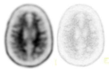
10 Assess for Subject Motion and CT-PET Misalignment HC

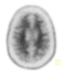
11 Assess for Subject Motion and CT-PET Misalignment amci

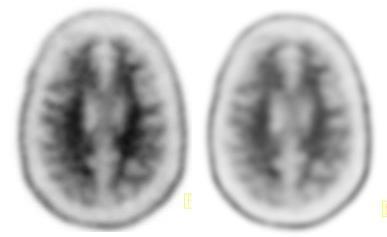
12 Assess for Subject Motion and CT-PET Misalignment ead


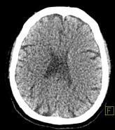
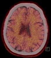
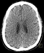
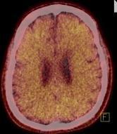
13 Static PET/CT Images HC amci ead

14 Test scans - ADMdx HC amci ead

15 Dynamic PET Images 1 minute frames 0-5 min HC 5-10 min 0-5 min amci 5-10 min 0-5 min ead 5-10 min
16 Effect of Reconstruction Algorithm OSEM3D +TOF +PSF +TOF+PSF +AllPass HC ead


17 Remove Head Holder From mu-map HC amci ead


18 Simulate Patient Motion and CT-PET Misalignment Misalign mu-map Recon static PET Simulates movement between CT and PET Misalign mu-maps Recon dyn PETs Simulates movement during PET and between CT and PET
19 Subject motion example from late MCI to mild AD scans SPM corrections needed to re-align images, using a neurological or right-handed coordinate system Average across all frames, referenced to frame 1 of each scan However, depending upon the site and disease severity, subject motion can be as great as 1 to 2 cm and/or many degrees. Study motion typically spans a greater range with greater disease severity (e.g. moderate AD, FTD).
20 Subject motion example from 140 late MCI to mild AD scans SPM corrections needed to re-align images, using a neurological or right-handed coordinate system Maximum absolute translation or rotation per scan to to to to to to 5.17 Depending upon the site and disease severity, subject motion can be as great as 1 to 2 cm and/or many degrees. Study motion typically spans a greater range with greater disease severity (e.g. moderate AD, FTD).


21 Frame of Reference and Technical Details for Project z y x +z +y +x y +z rot +x rot y z +y rot Radiological or Left- Handed Coordinate System (0,0,0) is the Image Center x z Transverse Sagittal Coronal Technical Details of transformations and reconstructions x Reverse PET Recon Order of Forward Transformations 1. translations 2. z-axis rotation 3. y-axis rotation 4. x-axis rotation Final PET image Forward


22 Severe subject motion example (ADNI 1) min min. Overlaid Motion during scan causes artifact due to: Sampling of blended/ incorrect tissue regions Attenuation over- or undercorrection due to misalignment with Tx scan Motion correction does not remove the embedded artifact, especially with severe movement Difference in position

23 Subject Motion: Impact on SUVR Scan of subject with minimal movement Target Region Reference region SUVR relatively constant throughout minute time window Scan of subject with severe motion Target Region Reference region SUVR at minutes = 1.5; SUVR at minutes = 1.0, a 50% difference In cases of severe motion, motion correction does not remove embedded artifact
24 Misalignment Parameters simulate patient movement X trans (mm) Y trans (mm) Z trans (mm) X rot (deg) Y rot (deg) Z rot (deg) Baseline Set Set Set Set Set Set Set Set Set


25 Analysis methods (two approaches of several) ADNI (Jagust Lab) PET image motion corrected, frames averaged, intensity normalized, smoothed PET coregistered to MRI Gray matter ROIs defined using Freesurfer Signal intensity measured Cortical average = frontal, AC, PC, lateral temporal, lateral parietal SUVRs calculated o Ref regions: Whole cer, brainstem, subcortical white matter, composite Avid (not on label) PET preprocessed PET spatially warped to PET template Probabilistic template ROIs applied Signal intensity measured SUVRs calculated o Ref regions: Whole cer, pons, subcortical white matter ADNI_UCBERKELEY_AV45_Methods_ pdf
26 Image Analysis For the Baseline and multiple Sets of images SUVRs calculated Will use ADMdx s PET Amyloid Analysis Package SUVR measures will be calculated = x 100%


27 QIBA Mission QIBA seeks to improve the value and practicality of quantitative imaging biomarkers by reducing variability across devices, patients, and time.
8/2/2017. Disclosure. Philips Healthcare (Cleveland, OH) provided the precommercial
 provided the precommercial") 8//0 AAPM0 Scientific Symposium: Emerging and New Generation PET: Instrumentation, Technology, Characteristics and Clinical Practice Aug Wednesday 0:4am :pm Solid State Digital Photon Counting PET/CT Instrumentation
8//0 AAPM0 Scientific Symposium: Emerging and New Generation PET: Instrumentation, Technology, Characteristics and Clinical Practice Aug Wednesday 0:4am :pm Solid State Digital Photon Counting PET/CT Instrumentation
Public Comment Form for QIBA Documents
 Public Comment Form for QIBA Documents Notes: 1. identify the commenter to facilitate clarification of the issue and/or communication of the resolution.. : ow. Typo or other minor correction that an editor
Deviceless respiratory motion correction in PET imaging exploring the potential of novel data driven strategies
 g Deviceless respiratory motion correction in PET imaging exploring the potential of novel data driven strategies Presented by Adam Kesner, Ph.D., DABR Assistant Professor, Division of Radiological Sciences,
3/27/2012 WHY SPECT / CT? SPECT / CT Basic Principles. Advantages of SPECT. Advantages of CT. Dr John C. Dickson, Principal Physicist UCLH
 3/27/212 Advantages of SPECT SPECT / CT Basic Principles Dr John C. Dickson, Principal Physicist UCLH Institute of Nuclear Medicine, University College London Hospitals and University College London john.dickson@uclh.nhs.uk
Review of PET Physics. Timothy Turkington, Ph.D. Radiology and Medical Physics Duke University Durham, North Carolina, USA
 Review of PET Physics Timothy Turkington, Ph.D. Radiology and Medical Physics Duke University Durham, North Carolina, USA Chart of Nuclides Z (protons) N (number of neutrons) Nuclear Data Evaluation Lab.
Philips SPECT/CT Systems
 Philips SPECT/CT Systems Ling Shao, PhD Director, Imaging Physics & System Analysis Nuclear Medicine, Philips Healthcare June 14, 2008 *Presented SNM08 Categorical Seminar - Quantitative SPECT and PET
Investigation of Motion Induced Errors in Scatter Correction for the HRRT Brain Scanner
 Investigation of Motion Induced Errors in Scatter Correction for the HRRT Brain Scanner Jose M Anton-Rodriguez 1, Merence Sibomana 2, Matthew D. Walker 1,4, Marc C. Huisman 3, Julian C. Matthews 1, Maria
Quantitative MRI of the Brain: Investigation of Cerebral Gray and White Matter Diseases
 Quantities Measured by MR - Quantitative MRI of the Brain: Investigation of Cerebral Gray and White Matter Diseases Static parameters (influenced by molecular environment): T, T* (transverse relaxation)
White Paper. EQ PET: Achieving NEMAreferenced. Technologies. Matthew Kelly, PhD, Siemens Healthcare
 White Paper EQ PET: Achieving NEMAreferenced SUV Across Technologies Matthew Kelly, PhD, Siemens Healthcare Table of Contents Introduction 1 Case Study 1 Cross-Scanner Response Assessment 2 Clinical Example
Introduction to Positron Emission Tomography
 Planar and SPECT Cameras Summary Introduction to Positron Emission Tomography, Ph.D. Nuclear Medicine Basic Science Lectures srbowen@uw.edu System components: Collimator Detector Electronics Collimator
Slide 1. Technical Aspects of Quality Control in Magnetic Resonance Imaging. Slide 2. Annual Compliance Testing. of MRI Systems.
 Slide 1 Technical Aspects of Quality Control in Magnetic Resonance Imaging Slide 2 Compliance Testing of MRI Systems, Ph.D. Department of Radiology Henry Ford Hospital, Detroit, MI Slide 3 Compliance Testing
Implementation and evaluation of a fully 3D OS-MLEM reconstruction algorithm accounting for the PSF of the PET imaging system
 Implementation and evaluation of a fully 3D OS-MLEM reconstruction algorithm accounting for the PSF of the PET imaging system 3 rd October 2008 11 th Topical Seminar on Innovative Particle and Radiation
V4.0 October 27, 2014
 ADNI 2 PET Technical Procedures Manual: Florbetapir As of May 9, 2014, no further FDG PET scans will be conducted under the ADNI 2 protocol. Additionally, enrollment for the ADNI 2 early frames add-on
Optimisation of Toshiba Aquilion ONE Volume Imaging
 Optimisation of Toshiba Aquilion ONE Volume Imaging Jane Edwards, RPRSG Royal Free London NHS Foundation Trust Dr Mufudzi Maviki, Plymouth Hospitals NHS Trust Background In 2011/12 Radiology at RFH was
Ch. 4 Physical Principles of CT
 Ch. 4 Physical Principles of CT CLRS 408: Intro to CT Department of Radiation Sciences Review: Why CT? Solution for radiography/tomography limitations Superimposition of structures Distinguishing between
Improvement of contrast using reconstruction of 3D Image by PET /CT combination system
 Available online at www.pelagiaresearchlibrary.com Advances in Applied Science Research, 2013, 4(1):285-290 ISSN: 0976-8610 CODEN (USA): AASRFC Improvement of contrast using reconstruction of 3D Image
Quantitative imaging for clinical dosimetry
 Quantitative imaging for clinical dosimetry Irène Buvat Laboratoire d Imagerie Fonctionnelle U678 INSERM - UPMC CHU Pitié-Salpêtrière, Paris buvat@imed.jussieu.fr http://www.guillemet.org/irene Methodology
Public Comment Form for QIBA Documents
 Public Comment Form for QIBA Documents Notes: 1. identify the commenter to facilitate clarification of the issue and/or communication of the resolution.. : ow. Typo or other minor correction that an editor
Micro-CT Methodology Hasan Alsaid, PhD
 Micro-CT Methodology Hasan Alsaid, PhD Preclinical & Translational Imaging LAS, PTS, GlaxoSmithKline 20 April 2015 Provide basic understanding of technical aspects of the micro-ct Statement: All procedures
CT NOISE POWER SPECTRUM FOR FILTERED BACKPROJECTION AND ITERATIVE RECONSTRUCTION
 CT NOISE POWER SPECTRUM FOR FILTERED BACKPROJECTION AND ITERATIVE RECONSTRUCTION Frank Dong, PhD, DABR Diagnostic Physicist, Imaging Institute Cleveland Clinic Foundation and Associate Professor of Radiology
NIH Public Access Author Manuscript J Nucl Med. Author manuscript; available in PMC 2010 February 9.
 NIH Public Access Author Manuscript Published in final edited form as: J Nucl Med. 2010 February ; 51(2): 237. doi:10.2967/jnumed.109.068098. An Assessment of the Impact of Incorporating Time-of-Flight
Technological Advances and Challenges: Experience with Time-Of-Flight PET Combined with 3T MRI. Floris Jansen, GE Healthcare July, 2015
 Technological Advances and Challenges: Experience with Time-Of-Flight PET Combined with 3T MRI Floris Jansen, GE Healthcare July, 2015 PET/MR 101 : challenges Thermal Workflow & Apps RF interactions?!!
Correlation between Model and Human Observer Performance on a Lesion Shape Discrimination Task in CT
 Correlation between Model and Human Observer Performance on a Lesion Shape Discrimination Task in CT Yi Zhang, Shuai Leng, Lifeng Yu and Cynthia McCollough Department of Radiology Mayo Clinic, Rochester
HST.583 Functional Magnetic Resonance Imaging: Data Acquisition and Analysis Fall 2008
 MIT OpenCourseWare http://ocw.mit.edu HST.583 Functional Magnetic Resonance Imaging: Data Acquisition and Analysis Fall 2008 For information about citing these materials or our Terms of Use, visit: http://ocw.mit.edu/terms.
Quantitative Analysis and Visualization with 3D Slicer
 Surgical Planning Laboratory Brigham and Women s Hospital Boston, Massachusetts USA a teaching affiliate of Harvard Medical School Quantitative Analysis and Visualization with 3D Slicer Jeffrey Yapp, PhD
Computational Neuroanatomy
 Computational Neuroanatomy John Ashburner john@fil.ion.ucl.ac.uk Smoothing Motion Correction Between Modality Co-registration Spatial Normalisation Segmentation Morphometry Overview fmri time-series kernel
Public Comment Form for QIBA Documents
 Public Comment Form for QIBA Documents Notes: 1. Initials identify the commenter to facilitate clarification of the issue and/or communication of the resolution. 2. Priority L: Low. Typo or other minor
The organization of the human cerebral cortex estimated by intrinsic functional connectivity
 1 The organization of the human cerebral cortex estimated by intrinsic functional connectivity Journal: Journal of Neurophysiology Author: B. T. Thomas Yeo, et al Link: https://www.ncbi.nlm.nih.gov/pubmed/21653723
Spiral CT. Protocol Optimization & Quality Assurance. Ge Wang, Ph.D. Department of Radiology University of Iowa Iowa City, Iowa 52242, USA
 Spiral CT Protocol Optimization & Quality Assurance Ge Wang, Ph.D. Department of Radiology University of Iowa Iowa City, Iowa 52242, USA Spiral CT Protocol Optimization & Quality Assurance Protocol optimization
Fast Timing and TOF in PET Medical Imaging
 Fast Timing and TOF in PET Medical Imaging William W. Moses Lawrence Berkeley National Laboratory October 15, 2008 Outline: Time-of-Flight PET History Present Status Future This work was supported in part
ADNI-GO PET Technical Procedures Manual AV-45 & FDG. V3.8.0 January 14, 2011
 ADNI-GO PET Technical Procedures Manual AV-45 & FDG V3.8.0 January 14, 2011 Page 1 of 36 Table of Contents General Information...3 Contact Information...3 Site Qualification...4 PET Scanners...4 Regulatory...4
NRM2018 PET Grand Challenge Dataset
 NRM2018 PET Grand Challenge Dataset An event part of London 2018 Neuroreceptor Mapping meeting (www.nrm2018.org) Contents Introduction... 2 Rationale... 2 Aims... 2 Description of the dataset content...
PET Quantification using STIR
 PET Quantification using STIR STIR User s Meeting Charalampos Tsoumpas, PhD King s College London Hammersmith Imanet 1 PET Quantification Image elements should correspond to concentration of the injected
Single Breath-hold Abdominal T 1 Mapping using 3-D Cartesian Sampling and Spatiotemporally Constrained Reconstruction
 Single Breath-hold Abdominal T 1 Mapping using 3-D Cartesian Sampling and Spatiotemporally Constrained Reconstruction Felix Lugauer 1,3, Jens Wetzl 1, Christoph Forman 2, Manuel Schneider 1, Berthold Kiefer
Performance Evaluation of the Philips Gemini PET/CT System
 Performance Evaluation of the Philips Gemini PET/CT System Rebecca Gregory, Mike Partridge, Maggie A. Flower Joint Department of Physics, Institute of Cancer Research, Royal Marsden HS Foundation Trust,
CT Basics Principles of Spiral CT Dose. Always Thinking Ahead.
 1 CT Basics Principles of Spiral CT Dose 2 Who invented CT? 1963 - Alan Cormack developed a mathematical method of reconstructing images from x-ray projections Sir Godfrey Hounsfield worked for the Central
Super-resolution Reconstruction of Fetal Brain MRI
 Super-resolution Reconstruction of Fetal Brain MRI Ali Gholipour and Simon K. Warfield Computational Radiology Laboratory Children s Hospital Boston, Harvard Medical School Worshop on Image Analysis for
PURE. ViSION Edition PET/CT. Patient Comfort Put First.
 PURE ViSION Edition PET/CT Patient Comfort Put First. 2 System features that put patient comfort and safety first. Oncology patients deserve the highest levels of safety and comfort during scans. Our Celesteion
664 IEEE TRANSACTIONS ON NUCLEAR SCIENCE, VOL. 52, NO. 3, JUNE 2005
 664 IEEE TRANSACTIONS ON NUCLEAR SCIENCE, VOL. 52, NO. 3, JUNE 2005 Attenuation Correction for the NIH ATLAS Small Animal PET Scanner Rutao Yao, Member, IEEE, Jürgen Seidel, Jeih-San Liow, Member, IEEE,
HST.583 Functional Magnetic Resonance Imaging: Data Acquisition and Analysis Fall 2008
 MIT OpenCourseWare http://ocw.mit.edu HST.583 Functional Magnetic Resonance Imaging: Data Acquisition and Analysis Fall 2008 For information about citing these materials or our Terms of Use, visit: http://ocw.mit.edu/terms.
Advancements in molecular medicine
 Advancements in molecular medicine Philips Ingenuity TF PET/MR attenuation correction Z. Hu, 1 S. Renisch, 2 B. Schweizer, 3 T. Blaffert, 2 N. Ojha, 1 T. Guo, 1 J. Tang, 1 C. Tung, 1 J. Kaste, 1 V. Schulz,
Estimating 3D Respiratory Motion from Orbiting Views
 Estimating 3D Respiratory Motion from Orbiting Views Rongping Zeng, Jeffrey A. Fessler, James M. Balter The University of Michigan Oct. 2005 Funding provided by NIH Grant P01 CA59827 Motivation Free-breathing
Factors influencing image quality and quantification: optimization of PET parameters
 Factors influencing image quality and quantification: optimization of PET parameters Ronald Boellaard Department of Radiology & Nuclear Medicine, VU University Medical Center, Amsterdam Department of Nuclear
Public Comment Form for QIBA Documents
 Public Comment Form for QIBA Documents Notes: 1. identify the commenter to facilitate clarification of the issue and/or communication of the resolution. 2. : ow. Typo or other minor correction that an
Conflicts of Interest Nuclear Medicine and PET physics reviewer for the ACR Accreditation program
 James R Halama, PhD Loyola University Medical Center Conflicts of Interest Nuclear Medicine and PET physics reviewer for the ACR Accreditation program Learning Objectives 1. Be familiar with recommendations
Agenda : Lung Density Breakout Session
 Agenda : Lung Density Breakout Session 1. : Mathew Fuld and Bernice Hoppel 2. Automatic Exposure Control (AEC) Evaluation : Sean Fain Round 4 Project 3. Dose reduction effects on emphysema metrics : Philip
Functional MRI data preprocessing. Cyril Pernet, PhD
 Functional MRI data preprocessing Cyril Pernet, PhD Data have been acquired, what s s next? time No matter the design, multiple volumes (made from multiple slices) have been acquired in time. Before getting
RADIOMICS: potential role in the clinics and challenges
 27 giugno 2018 Dipartimento di Fisica Università degli Studi di Milano RADIOMICS: potential role in the clinics and challenges Dr. Francesca Botta Medical Physicist Istituto Europeo di Oncologia (Milano)
Basic principles of MR image analysis. Basic principles of MR image analysis. Basic principles of MR image analysis
 Basic principles of MR image analysis Basic principles of MR image analysis Julien Milles Leiden University Medical Center Terminology of fmri Brain extraction Registration Linear registration Non-linear
Image Acquisition Systems
 Image Acquisition Systems Goals and Terminology Conventional Radiography Axial Tomography Computer Axial Tomography (CAT) Magnetic Resonance Imaging (MRI) PET, SPECT Ultrasound Microscopy Imaging ITCS
2005 IEEE Nuclear Science Symposium Conference Record M Influence of Depth of Interaction on Spatial Resolution and Image Quality for the HRRT
 2005 IEEE Nuclear Science Symposium Conference Record M03-271 Influence of Depth of Interaction on Spatial Resolution and Image Quality for the HRRT Stéphan Blinder, Marie-Laure Camborde, Ken R. Buckley,
Characterization of a Time-of-Flight PET Scanner based on Lanthanum Bromide
 2005 IEEE Nuclear Science Symposium Conference Record M04-8 Characterization of a Time-of-Flight PET Scanner based on Lanthanum Bromide J. S. Karp, Senior Member, IEEE, A. Kuhn, Member, IEEE, A. E. Perkins,
Preprocessing II: Between Subjects John Ashburner
 Preprocessing II: Between Subjects John Ashburner Pre-processing Overview Statistics or whatever fmri time-series Anatomical MRI Template Smoothed Estimate Spatial Norm Motion Correct Smooth Coregister
Fmri Spatial Processing
 Educational Course: Fmri Spatial Processing Ray Razlighi Jun. 8, 2014 Spatial Processing Spatial Re-alignment Geometric distortion correction Spatial Normalization Smoothing Why, When, How, Which Why is
Methods for data preprocessing
 Methods for data preprocessing John Ashburner Wellcome Trust Centre for Neuroimaging, 12 Queen Square, London, UK. Overview Voxel-Based Morphometry Morphometry in general Volumetrics VBM preprocessing
Help Guide. mm Copyright Mirada Medical Ltd, Mirada Medical RTx 1
 Help Guide mm3237-1.6-1 Copyright Mirada Medical Ltd, 2000-2014. Mirada Medical RTx 1 Contents Help Guide... 1 Contents... 2 Introduction to RTx... 4 Regulatory Statement... 6 Notes... 15 Data Supported...
Magnetic Resonance Elastography (MRE) of Liver Disease
 of Liver Disease") Magnetic Resonance Elastography (MRE) of Liver Disease Authored by: Jennifer Dolan Fox, PhD VirtualScopics Inc. jennifer_fox@virtualscopics.com 1-585-249-6231 1. Overview of MRE Imaging MRE is a magnetic
Magnetic Resonance Elastography (MRE) of Liver Disease Authored by: Jennifer Dolan Fox, PhD VirtualScopics Inc. jennifer_fox@virtualscopics.com 1-585-249-6231 1. Overview of MRE Imaging MRE is a magnetic
James R Halama, PhD Loyola University Medical Center
 James R Halama, PhD Loyola University Medical Center Conflicts of Interest Nuclear Medicine and PET physics reviewer for the ACR Accreditation program Learning Objectives Be familiar with the tests recommended
Automatic Generation of Training Data for Brain Tissue Classification from MRI
 Automatic Generation of Training Data for Brain Tissue Classification from MRI Chris A. COCOSCO, Alex P. ZIJDENBOS, and Alan C. EVANS http://www.bic.mni.mcgill.ca/users/crisco/ McConnell Brain Imaging
3-D PET Scatter Correction
 Investigation of Accelerated Monte Carlo Techniques for PET Simulation and 3-D PET Scatter Correction C.H. Holdsworth, Student Member, IEEE, C.S. Levin", Member, IEEE, T.H. Farquhar, Student Member, IEEE,
in PET Medical Imaging
 Fast Timing and TOF in PET Medical Imaging William W. Moses Lawrence Berkeley National Laboratory October 15, 2008 Outline: Time-of-Flight PET History Present Status Future This work was supported in part
Lucy Phantom MR Grid Evaluation
 Lucy Phantom MR Grid Evaluation Anil Sethi, PhD Loyola University Medical Center, Maywood, IL 60153 November 2015 I. Introduction: The MR distortion grid, used as an insert with Lucy 3D QA phantom, is
Symbia E and S System Specifications Answers for life.
 www.siemens.com/mi Symbia E and S System Specifications Answers for life. Symbia E: Work with confidence. What does it mean to work with confidence? It is clarity not only image clarity but the clarity
Shadow casting. What is the problem? Cone Beam Computed Tomography THE OBJECTIVES OF DIAGNOSTIC IMAGING IDEAL DIAGNOSTIC IMAGING STUDY LIMITATIONS
 Cone Beam Computed Tomography THE OBJECTIVES OF DIAGNOSTIC IMAGING Reveal pathology Reveal the anatomic truth Steven R. Singer, DDS srs2@columbia.edu IDEAL DIAGNOSTIC IMAGING STUDY Provides desired diagnostic
The Near Future in Cardiac CT Image Reconstruction
 SCCT 2010 The Near Future in Cardiac CT Image Reconstruction Marc Kachelrieß Institute of Medical Physics (IMP) Friedrich-Alexander Alexander-University Erlangen-Nürnberg rnberg www.imp.uni-erlangen.de
UNIVERSITY OF SOUTHAMPTON
 UNIVERSITY OF SOUTHAMPTON PHYS2007W1 SEMESTER 2 EXAMINATION 2014-2015 MEDICAL PHYSICS Duration: 120 MINS (2 hours) This paper contains 10 questions. Answer all questions in Section A and only two questions
Corso di laurea in Fisica A.A Fisica Medica 4 TC
 Corso di laurea in Fisica A.A. 2007-2008 Fisica Medica 4 TC Computed Tomography Principles 1. Projection measurement 2. Scanner systems 3. Scanning modes Basic Tomographic Principle The internal structure
Evaluation of multiple voxel-based morphometry approaches and applications in the analysis of white matter changes in temporal lobe epilepsy
 Evaluation of multiple voxel-based morphometry approaches and applications in the analysis of white matter changes in temporal lobe epilepsy Wenjing Li a, Huiguang He a, Jingjing Lu b, Bin Lv a, Meng Li
Journal of Articles in Support of The Null Hypothesis
 Data Preprocessing Martin M. Monti, PhD UCLA Psychology NITP 2016 Typical (task-based) fmri analysis sequence Image Pre-processing Single Subject Analysis Group Analysis Journal of Articles in Support
Semi-Quantitative Metrics in Positron Emission Tomography. Michael Adams. Department of Biomedical Engineering Duke University.
 Semi-Quantitative Metrics in Positron Emission Tomography by Michael Adams Department of Biomedical Engineering Duke University Date: Approved: Timothy G. Turkington, Supervisor Adam P. Wax Terence Z.
4DM Packages. 4DM Packages & License Types. Information to help you order the appropriate licenses for your site.
 4DM Packages 4DM Packages & License Types. Information to help you order the appropriate licenses for your site. Nuclear Cardiac Quantification, Review, and Reporting Select Your 4DM Package and corresponding
Effect of age and dementia on topology of brain functional networks. Paul McCarthy, Luba Benuskova, Liz Franz University of Otago, New Zealand
 Effect of age and dementia on topology of brain functional networks Paul McCarthy, Luba Benuskova, Liz Franz University of Otago, New Zealand 1 Structural changes in aging brain Age-related changes in
SUV reproducibility on different reporting platforms
 SUV reproducibility on different reporting platforms DJ Towey, Imperial College / Northampton General J Taylor, Sheffield teaching Hospital NHS Trust J Cullis, University Hospitals Coventry and Warwickshire
Fluorescence Tomography Source Reconstruction and Analysis
 TECHNICAL NOTE Pre-clinical in vivo imaging Fluorescence Tomography Source Reconstruction and Analysis Note: This Technical Note is part of a series for Fluorescence Imaging Tomography (FLIT). The user
UvA-DARE (Digital Academic Repository) Motion compensation for 4D PET/CT Kruis, M.F. Link to publication
 Motion compensation for 4D PET/CT Kruis, M.F. Link to publication") UvA-DARE (Digital Academic Repository) Motion compensation for 4D PET/CT Kruis, M.F. Link to publication Citation for published version (APA): Kruis, M. F. (2014). Motion compensation for 4D PET/CT General
UvA-DARE (Digital Academic Repository) Motion compensation for 4D PET/CT Kruis, M.F. Link to publication Citation for published version (APA): Kruis, M. F. (2014). Motion compensation for 4D PET/CT General
ADNI, ADNI_QH, SURVEY. Geometry. connection
 ADNI, ADNI_QH, SURVEY Geometry Coil selection = Head connection = d Multi coil Homogeneity correction ne FOV (mm) = 250.00 RFOV (%) = 100.00 Foldover suppression Matrix scan = 256 reconstruction = 256
Computer-Tomography II: Image reconstruction and applications
 Computer-Tomography II: Image reconstruction and applications Prof. Dr. U. Oelfke DKFZ Heidelberg Department of Medical Physics (E040) Im Neuenheimer Feld 280 69120 Heidelberg, Germany u.oelfke@dkfz.de
Development and anatomical details of Japanese adult male/female voxel models
 Development and anatomical details of Japanese adult male/female voxel models Tomoaki Nagaoka, Soichi Watanabe, *Etsuo kunieda, **Masao Taki and Yukio Yamanaka National Institute of Information and Communications
Statistical Analysis of Neuroimaging Data. Phebe Kemmer BIOS 516 Sept 24, 2015
 Statistical Analysis of Neuroimaging Data Phebe Kemmer BIOS 516 Sept 24, 2015 Review from last time Structural Imaging modalities MRI, CAT, DTI (diffusion tensor imaging) Functional Imaging modalities
Supplementary methods
 Supplementary methods This section provides additional technical details on the sample, the applied imaging and analysis steps and methods. Structural imaging Trained radiographers placed all participants
If it matters to you, it matters to us
 If it matters to you, it matters to us Philips clinical innovations in nuclear medicine Innovation with insight We understand that clinical innovations are only as valuable as the day-to-day difference
SISCOM (Subtraction Ictal SPECT CO-registered to MRI)
") SISCOM (Subtraction Ictal SPECT CO-registered to MRI) Introduction A method for advanced imaging of epilepsy patients has been developed with Analyze at the Mayo Foundation which uses a combination of
SISCOM (Subtraction Ictal SPECT CO-registered to MRI) Introduction A method for advanced imaging of epilepsy patients has been developed with Analyze at the Mayo Foundation which uses a combination of
Final Report: QIBA FDG-PET/CT Digital Reference Object Project
 Final Report: QIBA FDG-PET/CT Digital Reference Object Project Project team: Paul Kinahan, University of Washington Larry Pierce, University of Washington Brain Elston, University of Washington David Clunie,
Medical Image Analysis
 Computer assisted Image Analysis VT04 29 april 2004 Medical Image Analysis Lecture 10 (part 1) Xavier Tizon Medical Image Processing Medical imaging modalities XRay,, CT Ultrasound MRI PET, SPECT Generic
Image Registration + Other Stuff
 Image Registration + Other Stuff John Ashburner Pre-processing Overview fmri time-series Motion Correct Anatomical MRI Coregister m11 m 21 m 31 m12 m13 m14 m 22 m 23 m 24 m 32 m 33 m 34 1 Template Estimate
Multi-slice CT Image Reconstruction Jiang Hsieh, Ph.D.
 Multi-slice CT Image Reconstruction Jiang Hsieh, Ph.D. Applied Science Laboratory, GE Healthcare Technologies 1 Image Generation Reconstruction of images from projections. textbook reconstruction advanced
CHAPTER 9: Magnetic Susceptibility Effects in High Field MRI
 Figure 1. In the brain, the gray matter has substantially more blood vessels and capillaries than white matter. The magnified image on the right displays the rich vasculature in gray matter forming porous,
CT vs. VolumeScope: image quality and dose comparison
 CT vs. VolumeScope: image quality and dose comparison V.N. Vasiliev *a, A.F. Gamaliy **b, M.Yu. Zaytsev b, K.V. Zaytseva ***b a Russian Sci. Center of Roentgenology & Radiology, 86, Profsoyuznaya, Moscow,
Performance Evaluation of radionuclide imaging systems
 Performance Evaluation of radionuclide imaging systems Nicolas A. Karakatsanis STIR Users meeting IEEE Nuclear Science Symposium and Medical Imaging Conference 2009 Orlando, FL, USA Geant4 Application
SPECT QA and QC. Bruce McBride St. Vincent s Hospital Sydney.
 SPECT QA and QC Bruce McBride St. Vincent s Hospital Sydney. SPECT QA and QC What is needed? Why? How often? Who says? QA and QC in Nuclear Medicine QA - collective term for all the efforts made to produce
Improvement and Evaluation of a Time-of-Flight-based Patient Positioning System
 Improvement and Evaluation of a Time-of-Flight-based Patient Positioning System Simon Placht, Christian Schaller, Michael Balda, André Adelt, Christian Ulrich, Joachim Hornegger Pattern Recognition Lab,
RADIOLOGY AND DIAGNOSTIC IMAGING
 Day 2 part 2 RADIOLOGY AND DIAGNOSTIC IMAGING Dr hab. Zbigniew Serafin, MD, PhD serafin@cm.umk.pl 2 3 4 5 CT technique CT technique 6 CT system Kanal K: RSNA/AAPM web module: CT Systems & CT Image Quality
Inveon User Notes. Inveon Scanners and Inveon Acquisition Workplace 2.0
 Inveon User Notes Inveon Scanners and Inveon Acquisition Workplace 2.0 Legal Notice 2014 by Siemens Medical Solutions USA, Inc. All rights reserved. Siemens reserves the right to modify the design and
Design and Fabrication of Kidney Phantoms for Internal Radiation Dosimetry Using 3D Printing Technology
 Design and Fabrication of Kidney Phantoms for Internal Radiation Dosimetry Using 3D Printing Technology Johannes Tran-Gia, PhD NPL MEMPHYS Workshop Applications of 3D Printing for Medical Phantoms Klinik
Positron Emission Tomography
 Physics 656 Seminar on Physical Fundamentals of Medical Imaging Positron Emission Tomography Ahmed Qamesh Outline What is PET? PET mechanism Radionuclide and its synthesis Detection concept and Development
CLASS HOURS: 4 CREDIT HOURS: 4 LABORATORY HOURS: 0
 Revised 10/10 COURSE SYLLABUS TM 220 COMPUTED TOMOGRAPHY PHYSICS CLASS HOURS: 4 CREDIT HOURS: 4 LABORATORY HOURS: 0 CATALOG COURSE DESCRIPTION: This course is one of a three course set in whole body Computed
SPM Introduction. SPM : Overview. SPM: Preprocessing SPM! SPM: Preprocessing. Scott Peltier. FMRI Laboratory University of Michigan
 SPM Introduction Scott Peltier FMRI Laboratory University of Michigan! Slides adapted from T. Nichols SPM! SPM : Overview Library of MATLAB and C functions Graphical user interface Four main components:
Medical Imaging BMEN Spring 2016
 Name Medical Imaging BMEN 420-501 Spring 2016 Homework #4 and Nuclear Medicine Notes All questions are from the introductory Powerpoint (based on Chapter 7) and text Medical Imaging Signals and Systems,
AAPM Standard of Practice: CT Protocol Review Physicist
 AAPM Standard of Practice: CT Protocol Review Physicist Dianna Cody, Ph.D., DABR, FAAPM U.T.M.D. Anderson Cancer Center September 11, 2014 2014 Texas Radiation Regulatory Conference Goals Understand purpose
Evaluation of 1D, 2D and 3D nodule size estimation by radiologists for spherical and non-spherical nodules through CT thoracic phantom imaging
 Evaluation of 1D, 2D and 3D nodule size estimation by radiologists for spherical and non-spherical nodules through CT thoracic phantom imaging Nicholas Petrick, Hyun J. Grace Kim, David Clunie, Kristin