Introduction to. 3D Scanning Confocal Microscope with Spectrometer
|
|
|
- Brittney Griffith
- 6 years ago
- Views:
Transcription
1 Introduction to Nanofinder-S 3D Scanning Confocal Microscope with Spectrometer Alexei Kuzmin
2 Principle of Confocal Microscopy Laser X-Y Excitation Pinhole Excitation Filter Objective Z Emission Pinhole Detector (PMT) Emission Filter 3D Sample A type of light microscopy in which a point of illumination is projected or rastered over a specimen, and the reflected illumination is screened through an exit aperture in order to eliminate light from out-of-focus planes.
3 Nanofinder-S Before Now
4 Nanofinder-S Simultaneous / Multifunctional Analysis: Optical and Confocal Microscopy Raman Measurements Luminescence Measurements 0D, 1D, 2D & 3D High-speed Imaging and Spectroscopy
5 Main components: 1. Inverted microscope (bandpass nm) 2. CCD for microscope 3. Laser confocal microscope unit with photomultiplier tube (PMT) 4. Scanning unit with galvanometer mirror scanners (X and Y) 5. Opticomechanical unit 6. Monochromator-spectrograph 7. CCD for spectrograph 8. PMT for spectrograph 9. Reference PMT 10. Laser He-Cd: nm, 70 mw (up to 3 lasers possible) 11. Computer and electronics
6 Nanofinder-S modular optical layout
7 Inverted Nikon ECLIPSE TE2000-S microscope Works in Reflection & Transmission High-performance Objectives Plan Fluor 10X/0.30 Plan Fluor 40X/0.75 CF Plan Apo 100X/0.95 Coupled with color CCD camera Kappa DX 20 H SONY ICX 285 CCD Sensor 2/3" Interline, Progressive Scan 1384 x 1032 pixel Lux at 10 sec integration 12 bit digital Signal-to-noise ratio 63 db 10x 40x 100x Video Image size (µm) Confocal Image size (µm)
8 SCANNING UNITS 1. Scanning unit with galvanometer mirror scanners (X and Y) 110 μm 132 µm (with 100x objective) spacial optical resolution 200 nm 2. Piezo-scanner (Z) 0-80 µm (with 100x objective) spacial optical resolution 500 nm
9 OPTICOMECHANICAL UNIT (OMU) Optimized optics: nm Polarizers: Glan-Taylor prism (excitation and detection channels) Zoom beam expander: magnification factor Edge filters positioner: three-position Interference filters positioner: six-position Confocal pinhole: variable from 0 to 1.5 mm Laser beam attenuator: t VND filter
10 MONOCHROMATOR-SPECTROGRAPH MS5004i Configuration: Focal length: Ports: Flat field: Grating mounts: vertical 520 mm 1 input, 2 output (CCD & PMT) 28 mm x 10 mm 4-position turret Spatial resolution: µm Slit control: mm, step size 0.5 mm
11 MONOCHROMATOR-SPECTROGRAPH MS5004i Gratings (grooves/mm): Blaze wavelength (nm): Echelle Dispersion (nm/mm): Spectral resolution (nm): Wavelength accuracy (±nm): Wavelength repeatability (±nm):
12 Digital Slow Scan CCD Camera PROSCAN HS-101H for spectrograph Ahigh sensitive back-thinned CCD sensor pixels Spectral response range from 200 nm to 1100 nm Pixel size 24 x 24 µm Digitalization rate up to 1 MHz ADC 14 bit, correlative double sampling Peltier cooling with thermo stabilization & water cooling 10/100 Ethernet data transfer

 Luminous 2500 A/lm Radiant at 400nm 7.")
13 3 PHOTOMULTIPLIER TUBES (PMT) PMT for laser confocal microscope PMT for spectrograph Reference PMT Hamamatsu R928 Wide Spectral Response High Cathode Sensitivity Luminous Radiant at 400nm 185 to 900 nm 250 A/lm 74 ma/w High Anode Sensitivity (at 1000V) Luminous 2500 A/lm Radiant at 400nm A/W Low Drift and Hysteresis


14 LASER COMPUTER / ELECTRONICS He-Cd nm, 70 mw Pentium IV 3GHz, 1GB RAM, 3D Video card with 128 MB RAM,...
15 For more information please contact: SOLAR TII, LTD 15/2, Akademicheskaya str., Minsk , Republic of Belarus Tel: +375 (17) Fax: +375 (17) Internet:
16 EXAMPLES OF APPLICATIONS Imaging of Silicon Gratings for Scanning Probe Microscopy calibration 3D Confocal Microscopy optical tomography Optical Lithography information storage 2D Confocal Imaging and Raman Spectroscopy chemical phase mapping

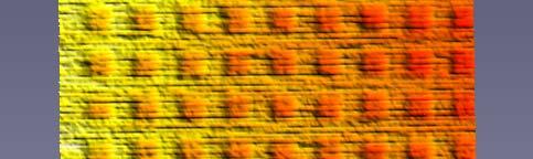
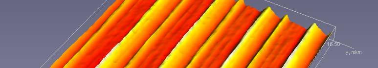
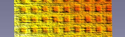
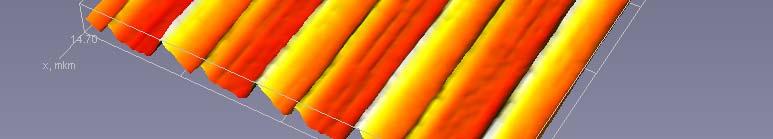
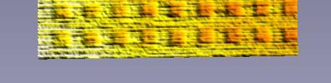


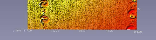
17 Calibrating Silicon Gratings for Scanning Probe Microscope 1.5 µm TGG1: image size µm TGQ1: image size µm


 of ZnO")


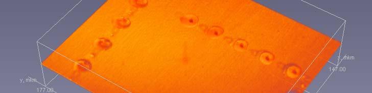
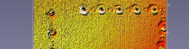
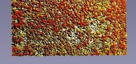
18 3D Confocal Microscopy Atmospheric pressure chemical vapour deposition (APCVD) of ZnO microcrystals on Si substrate 3D confocal images: 110 x 132 x 30 µm 2D optical image


19 Imaging and Raman Spectroscopy of ZnO needles 2E 1L Raman in ntensity E 2 E 1L ZnO powder Point 3 Point 2 c-si Point Wave number (cm -1 ) Point 2 Point 3 Point 1 29 µm 29 µm


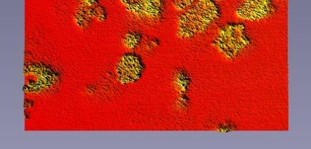
20 Materials for Optical Information Storage Video mode: transmission ZnO x thin film Confocal mode: volume Confocal mode: surface
21 Optical Data Storage CD-ROM CD-R CD-RW Track pitch = 1.6 μm Track pitch = 1.6 μm Images size: μm

 Objective 40 ; laser power: 70")
22 a-wo 3 cryst-wo 3 phase transition under laser irradiation an inten nsity (arb b. units) Ram thin film WO 3 /glass cryst-wo 3 a-wo Wave number (cm -1 ) Objective 40 ; laser power: 70 mw for writing, 12 mw for reading; detection ti by CCD.


 single-crystal CdWO 4")


23 Confocal Imaging and Raman Spectroscopy CdWO 4 : video mode µm Confocal mode: µm Raman intensity (a a.u.) single-crystal CdWO 4 polycrystalline t.f.-niwo nm excitation by He-Cd laser Wave number (cm -1 ) Confocal mode: µm


 Ra 300 Si")


 3D confocal")


24 2D Chemical Phase Mapping in Oxide Films man intensity (a a.u.) Ra 300 Si IrO x Si Wave number (cm -1 ) 3D confocal mode: µm IrO x Si 2D confocal mode: µm Raman mapping at 520 cm -1


 3D")


25 2D Chemical Phase Mapping in Oxide Films Rama an intensity (a a.u.) A 511: TaO x :ReO y B Wave number (cm -1 ) 3D confocal mode: µm A B 2D confocal mode: µm Raman mapping at 750 cm -1 Raman mapping at 1350 cm -1


26 2D Confocal Imaging and Raman Spectroscopy of Films 40x sity (a.u.) Raman inten 500 Si-Si Re-O Mixed thin film TaO x :ReO y /Si Wave number (cm -1 ) 40x


 Confocal m C")


27 Institute of Solid State Physics, University of Latvia NiO/MgO NiO/MgO irradiated by neutrons Ni0.98Co0.02O/MgO NiO powder 2-magnons 2 Raman intensity (a.u.) Confocal m C mode: 275 x 330 µm Confocal Imaging and Raman Spectroscopy of Magnons in Antiferromagnetic Compounds NiO foil NiO irradiated NiO O foil f NiO Ni0.98Co0.02O NiO powder Wave number (cm ) 2500 nano-nio


")
28 2D Confocal Imaging and Raman Spectroscopy of Glasses ) Raman inte ensity (a.u Microscope slide Wave number (cm -1 ) Confocal mode: µm


 Raman inte")
 A 1 (TO) E 1")


29 2D&3DI Imaging of ftechnological lprocess ZnO Gold contacts on silicon 40 3D nsity (a.u.) Raman inte E 2 (high) A 1 (TO) E 1 (LO) 2E 1 (LO) Contacts after heat treatment at 700 C and ZnO deposition µm ZnO column Wave number (cm -1 )
30 Thank you! Get more at
Confocal Raman Microscope RAMOS
 Confocal Raman Microscope RAMOS 1 future`s Confocal Raman Microscope RAMOS 2 Confocal Raman Microscope RAMOS Ostec Corporate Group produces and offers hi-tech innovative scientific and analytical equipment.
MonoVista CRS+ Raman Microscopes
 MonoVista CRS+ Benefits Deep UV to NIR wavelength range Up to 4 integrated multi-line lasers plus port for large external lasers Dual beam path for UV and VIS/NIR Motorized Laser selection Auto Alignment
Confocal Raman Systems SPECTROSCOPY GROUP
 Confocal Raman Systems SPECTROSCOPY GROUP MonoVista CRS Configuration Option PIXIS TE-Cooled CCD Camera Acton Spectrograph Micro-Raman Interface Witness Camera Optional Laser Micro/Macro Transfer Optics
SPECTRUM. The world s first fully automated Raman AFM. AFM - confocal Raman - SNOM - TERS AFM KPFM. Raman. AFM-Raman characterization of PS-PVAC
 Raman KPFM AFM AFM-Raman characterization of PS-PVAC polymer blend film SPECTRUM The world s first fully automated Raman AFM AFM - confocal Raman - SNOM - TERS The first fully integrated & automated AFM
Spectrograph overview:
 High performance measurement systems Monochromator Family Gilden Photonics offers a range of integrated optical wavelength solutions in customized designs, OEM design, manufacturing and value added resell
FLEX 2 NEW. Key points. 3D Confocal Raman, 2 lasers, fiber based, AFM combined
 FLEX 2 3D Confocal Raman, 2 lasers, fiber based, AFM combined NEW Key points Compact size 2 lasers, easily switchable 2 confocal operation modes, easily switchable : High Spatial Resolution 35 nm High
Versatile laser Raman Spectrometer. RMP-500 series
 Versatile laser Raman Spectrometer RMP-500 series About RMP-500 RMP-500 series is a compact and versatile laser Raman spectrometer consisting of a micro Raman probe connected through fiber optics to the
Depth analysis for laminated film by confocal Raman mapping
 Newest modular 3D Imaging Raman Microspectroscopy System Simple operation and low cost with all the basic features of our top of the line system. Structural images of transparent samples (plastic, film,
Imaging Spectrometers
 JOBIN YVON Imaging Spectrometers ihr Series Uniquely shaped for uniquely superior performance. ihr Series Imaging Spectrometers A Unique Shape for a Unique Spectrometer The difference between ihr spectrometers
Renishaw invia Raman Microscope (April 2006)
") Renishaw invia Raman Microscope (April 2006) I. Starting the System 1. The main system unit is ON all the time. 2. Switch on the Leica microscope and light source for reflective bright field (BF) imaging.
Renishaw invia Raman Microscope (April 2006) I. Starting the System 1. The main system unit is ON all the time. 2. Switch on the Leica microscope and light source for reflective bright field (BF) imaging.
phoenix Upgrade Packages for SLM Model 4800/48000/8000/8100 Spectrofluorometers
 Upgrade Packages for SLM Model 4800/48000/8000/8100 Spectrofluorometers Phoenix: The Upgrade Packages for SLM Model 4800/48000/8000/8100 Spectrofluorometers The Phoenix Upgrade Packages are designed to
Dr. Larry J. Paxton Johns Hopkins University Applied Physics Laboratory Laurel, MD (301) (301) fax
 (301) fax") Dr. Larry J. Paxton Johns Hopkins University Applied Physics Laboratory Laurel, MD 20723 (301) 953-6871 (301) 953-6670 fax Understand the instrument. Be able to convert measured counts/pixel on-orbit into
Dr. Larry J. Paxton Johns Hopkins University Applied Physics Laboratory Laurel, MD 20723 (301) 953-6871 (301) 953-6670 fax Understand the instrument. Be able to convert measured counts/pixel on-orbit into
Chemical Characterization of Diverse Pharmaceutical Samples by Confocal Raman Microscopy
 Whitepaper Chemical Characterization of Diverse Pharmaceutical Samples by Confocal Raman Microscopy WITec GmbH, Lise-Meitner-Str. 6, 89081 Ulm, Germany, www.witec.de Introduction The development and production
L. Pina, A. Fojtik, R. Havlikova, A. Jancarek, S.Palinek, M. Vrbova
 L. Pina, A. Fojtik, R. Havlikova, A. Jancarek, S.Palinek, M. Vrbova Faculty of Nuclear Sciences, Czech Technical University, Brehova 7, 115 19 Prague, Czech Republic CD EXPERIMENTAL ARRANGEMENT SPECTRAL
Spectrometers: Monochromators / Slits
 Spectrometers: Monochromators / Slits Monochromator Characteristics Dispersion: The separation, or wavelength selectivity, of a monochromator is dependent on its dispersion. Angular Dispersion: The change
Confocal Raman Imaging with WITec Sensitivity - Resolution - Speed. Always - Provable - Routinely
 Confocal Raman Imaging with WITec Sensitivity - Resolution - Speed Always - Provable - Routinely WITec GmbH, Ulm, Germany, info@witec.de, www.witec.de A modular microscope series An Example: FLIM optical
Certus Optic. NanoScanTechnology. Basic Datasheet. reasoned innovations. Integrated Optical and Scanning Probe Microscope
 NanoScanTechnology reasoned innovations Nano Scan Technology Ltd. Russia, 141700, Dolgoprudny, Zavodskaya St, 7 Phone: +7 (495) 642-40-68 +7 (495) 642-40-67 Skype: NanoScanTech E-mail: info@nanoscantech.ru
Certus Light. NanoScanTechnology. Basic Datasheet. reasoned innovations. Entry Level Scanning Probe Microscope. Scanning Probe Microscope
 NanoScanTechnology reasoned innovations Nano Scan Technology Ltd. Russia, 141700, Dolgoprudny, Zavodskaya St, 7 Phone: +7 (495) 642-40-68 +7 (495) 642-40-67 Skype: NanoScanTech E-mail: info@nanoscantech.ru
STANDARD SERIES MONOCHROMATOS FEATURES. Highly Customizable Modular Design. Two Configurable Input and Output Ports
 STANDARD SERIES MONOCHROMATOS FEATURES Highly Customizable Modular Design Two Configurable Input and Output Ports Configurable turret and Grating Options USB2.0 Communication A Full Line of Input and Output
Series Spectrometers PARTICLE CHARACTERIZATION ELEMENTAL ANALYSIS FLUORESCENCE GRATINGS & OEM SPECTROMETERS OPTICAL COMPONENTS RAMAN
 Series Spectrometers ELEMENTAL ANALYSIS FLUORESCENCE GRATINGS & OEM SPECTROMETERS OPTICAL COMPONENTS PARTICLE CHARACTERIZATION RAMAN SPECTROSCOPIC ELLIPSOMETRY SPR IMAGING ihr Series Imaging Spectrometers
picoemerald Tunable Two-Color ps Light Source Microscopy & Spectroscopy CARS SRS
 picoemerald Tunable Two-Color ps Light Source Microscopy & Spectroscopy CARS SRS 1 picoemerald Two Colors in One Box Microscopy and Spectroscopy with a Tunable Two-Color Source CARS and SRS microscopy
Modular Raman Spectrometers
 Modular Raman Flexible Raman from the Raman Experts horiba.com/scientific Flexible and Affordable Raman The new range of modular Raman spectrometers from HORIBA Scientific allows the user to have a flexible
LIFA. SPECIFICATIONs. Fluorescence Lifetime Attachment LIFA14001A02 25/02/2014
 LIFA Fluorescence Lifetime Attachment The LIFA is a dedicated system for Fluorescence Lifetime Imaging Microscopy (FLIM). It allows the generation of lifetime images on any widefield fluorescence microscope
Thermo Scientific DXR2 Raman Family
 MOLECULAR SPECTROSCOPY Thermo Scientific DXR2 Raman Family Focus on answers, not the technique Product Specifications Easily adapt to any sample challenge using the Thermo Scientific DXR 2 family of Raman
TABLE OF CONTENTS PRODUCT DESCRIPTION CINCAM CCD TECHNICAL DATA SENSOR RESPONSE DIMENSIONS CINCAM CCD LARGE FORMAT TECHNICAL DATA SENSOR RESPONSE
 TABLE OF CONTENTS PRODUCT DESCRIPTION CINCAM CCD TECHNICAL DATA SENSOR RESPONSE DIMENSIONS CINCAM CCD LARGE FORMAT TECHNICAL DATA SENSOR RESPONSE DIMENSIONS CINCAM CMOS TECHNICAL DATA SENSOR RESPONSE DIMENSIONS
Optical properties and characterization
 Optical properties and characterization Name Picture Description Site Responsible 1 Laser Nd:YAG MAPLE (Matrix Assisted Pulsed Laser Evaporation) system for biomaterials and polymeric thin film deposition
Leica TCS SPE. Spectacular Imaging! Technical Documentation
 Leica TCS SPE Spectacular Imaging! Technical Documentation Leica TCS SPE Spectacular Imaging Easy to Achieve A Reliable System Affordable Excellence The high resolution spectral confocal Leica TCS SPE
Spectroscopic equipment. Multispectral Imaging
 Spectroscopic equipment Multispectral Imaging Basic spectroscopic arrangement Source Sample Analyzer Detector Sun Lamps Lasers LEDs Synchrotron Plants Forests Tissue Cells Flames Chemical compounds etc.
LIFA KEY FEATURES APPLICATIONS. Fluorescence Lifetime Attachment LIFA 15001A02 16/03/2015
 LIFA Fluorescence Lifetime Attachment LIFA 151A2 16/3/215 The LIFA is a dedicated system for Fluorescence Lifetime Imaging Microscopy (FLIM). It allows the generation of lifetime images on any widefield
ICP-OES. By: Dr. Sarhan A. Salman
 ICP-OES By: Dr. Sarhan A. Salman AGENDA -- ICP-OES Overview - Module removal Optical components and gas control overview Module replacement --Continue module replacement Manual optical alignment --Precise
Modular Approach and Customized solutions (near field, UV-VIS, etc.)
") Compact & Flexible System Configuration High Resolution: < 0.3cm Measurement down to 10cm Confocal Optics for Microscope and Remote Probe Fully Automated 2D, 3D & 4D Raman Imaging Attachable to AFM, XRD,
Compact & Flexible System Configuration High Resolution: < 0.3cm Measurement down to 10cm Confocal Optics for Microscope and Remote Probe Fully Automated 2D, 3D & 4D Raman Imaging Attachable to AFM, XRD,
Optical Sectioning. Bo Huang. Pharmaceutical Chemistry
 Optical Sectioning Bo Huang Pharmaceutical Chemistry Approaches to 3D imaging Physical cutting Technical difficulty Highest resolution Highest sensitivity Optical sectioning Simple sample prep. No physical
NDD FLIM Systems for Leica SP2 MP and SP5 MP Multiphoton Microscopes
 NDD FLIM Systems for Leica SP2 MP and SP5 MP Multiphoton Microscopes bh FLIM systems for the confocal and the multiphoton versions of the Leica SP2 and SP5 microscopes are available since 2002 [4]. These
3D OPTICAL PROFILER MODEL 7503
 3D Optical Profiler MODEL 7503 Features: 3D OPTICAL PROFILER MODEL 7503 Chroma 7503 is a sub-nano 3D Optical Profiler developed using the technology of white light interference to measure and analyze the
Physics 625 Femtosecond laser Project
 Physics 625 Femtosecond laser Project The purpose of this project is for each person to gain experience in designing part of a femtosecond laser system for pump-probe experiments. The system diagram is
Introduction to Diffraction Gratings
 Introduction to Diffraction Diffraction (Ruled and Holographic) Diffraction gratings can be divided into two basic categories: holographic and ruled. A ruled grating is produced by physically forming grooves
WAVELENGTH MANAGEMENT
 Camera Accessories WAVELENGTH MANAGEMENT UV CONVERTERS UV Converters take advantage of a phenomenon called fluorescence to extend the performance range of the Beamage beam profiling camera to ultraviolet
Acton Research SpectruMM TM Complete Spectroscopic- Acquisition Systems
 Acton Research SpectruMM TM Complete Spectroscopic- Acquisition Systems A fully integrated spectroscopy lab is as easy as 1, 2, 3... 1. High-Resolution CCD Detectors 2. Industry-Standard Acton Research
WAVELENGTH MANAGEMENT
 BEAM DIAGNOS TICS SPECIAL PRODUCTS OEM DETECTORS THZ DETECTORS PHOTO DETECTORS HIGH POWER SOLUTIONS POWER DETECTORS ENERGY DETECTORS MONITORS Camera Accessories WAVELENGTH MANAGEMENT UV CONVERTERS UV Converters
WITec Raman Spectroscopy Solutions
 Confocal Raman Imaging WITec Raman Spectroscopy Solutions -40-20 0 20 CCD cts 500 1000 1500 2000 2500 3000 3000 relative wavenumbers (cm -1 ) www.witec.de WITec UHTS Ultra-High Throughput Spectrometers
PHYSICS 213 PRACTICE EXAM 3*
 PHYSICS 213 PRACTICE EXAM 3* *The actual exam will contain EIGHT multiple choice quiz-type questions covering concepts from lecture (16 points), ONE essay-type question covering an important fundamental
(Fiber-optic Reosc Echelle Spectrograph of Catania Observatory)
") (Fiber-optic Reosc Echelle Spectrograph of Catania Observatory) The echelle spectrograph delivered by REOSC (France), was designed to work at the F/15 cassegrain focus of the 91-cm telescope. The spectrograph
(Fiber-optic Reosc Echelle Spectrograph of Catania Observatory) The echelle spectrograph delivered by REOSC (France), was designed to work at the F/15 cassegrain focus of the 91-cm telescope. The spectrograph
Optical Ptychography Imaging
 Optical Ptychography Imaging Summer Project Annafee Azad Supervisors: Dr Fucai Zhang Prof Ian Robinson Summer 2014 23 October 2014 Optical Ptychography Imaging P a g e 2 Abstract This report details a
TABLE OF CONTENTS PRODUCT DESCRIPTION LASERDEC CL200 / CL200HP TECHNICAL DATA DIMENSIONS LASERDEC CL500 / CL500HP TECHNICAL DATA DIMENSIONS
 TABLE OF CONTENTS PRODUCT DESCRIPTION LASERDEC CL200 / CL200HP TECHNICAL DATA DIMENSIONS LASERDEC CL500 / CL500HP TECHNICAL DATA DIMENSIONS ACCESSORIES ATTENUATION UNIT 0 ATTENUATION UNIT 90 BEAM REDUCER
Section V: AD Series Detection Systems
 Section V: AD Series AD110B 0B Photobyte - P System AD131 Photodetector Module Cooled CCD Cameras AD202 AD205 AD206 Kestr estralspec Softwar are Data Acquisition 57 AD110B Photob obyte - P Photomultiplier
Optics Vac Work MT 2008
 Optics Vac Work MT 2008 1. Explain what is meant by the Fraunhofer condition for diffraction. [4] An aperture lies in the plane z = 0 and has amplitude transmission function T(y) independent of x. It is
Astronomical spectrographs. ASTR320 Wednesday February 20, 2019
 Astronomical spectrographs ASTR320 Wednesday February 20, 2019 Spectrographs A spectrograph is an instrument used to form a spectrum of an object Much higher spectral resolutions than possible with multiband
Attachable to other Advanced analytical tools (e.g AFM, XRD, SEM etc.) Detect and measure deposits in liquid as it is (Particle ID Detection)
 Detect and measure deposits in liquid as it is (Particle ID Detection)") Compact & Flexible System Configuration High Resolution: < 0.3cm Measurement down to 10cm Confocal Optics for Microscope and Remote Probe Fully Automated 2D, 3D & 4D Raman Imaging Attachable to other Advanced
Compact & Flexible System Configuration High Resolution: < 0.3cm Measurement down to 10cm Confocal Optics for Microscope and Remote Probe Fully Automated 2D, 3D & 4D Raman Imaging Attachable to other Advanced
ihr Series horiba.com/osd Research Grade Spectrometers Simply the best imaging spectrometers with no compromise
 ihr Series Research Grade Spectrometers Simply the best imaging spectrometers with no compromise horiba.com/osd Unmatched Flexibility in Applications HORIBA Scientific s Optical Spectroscopy Division
Ralf K. Heilmann CAT-GS: Critical-Angle Transmission Grating Spectrometer January 27,
 Ralf K. Heilmann CAT-GS: Critical-Angle Transmission Grating Spectrometer January 27, 2009 1 Overview of CAT-GS Mission requirements: Effective area > 1000 cm 2 (0.3 1 kev) Spectral resolution E/ΔE > 3000
Indiana Center for Biological Microscopy. BioRad MRC 1024 MP Confocal & Multi-Photon Microscope
 Indiana Center for Biological Microscopy BioRad MRC 1024 MP Confocal & Multi-Photon Microscope Microscope and the Attached Accessories A: B: C: D: E: F: G: H: Mercury Lamp Transmission Light Kr/Ar Laser
2011 Optical Science & Engineering PhD Qualifying Examination Optical Sciences Track: Advanced Optics Time allowed: 90 minutes
 2011 Optical Science & Engineering PhD Qualifying Examination Optical Sciences Track: Advanced Optics Time allowed: 90 minutes Answer all four questions. All questions count equally. 3(a) A linearly polarized
TissueFAXS SL Confocal high throughput configuration (actual appearance of the product may differ)
") TISSUEFAXS CONFOCAL TissueFAXS SL Confocal high throughput configuration (actual appearance of the product may differ) TissueFAXS Confocal provides a unique combination of digital slide scanning and laser
TISSUEFAXS CONFOCAL TissueFAXS SL Confocal high throughput configuration (actual appearance of the product may differ) TissueFAXS Confocal provides a unique combination of digital slide scanning and laser
Product Specifications
 Product Specifications The ksa RateRat Pro is a turnkey, real-time, in-situ optical reflectance probe designed for deposition monitoring of semi-absorbent thin films. The RateRat Pro measures deposition
Chemical Characterization of Pharmaceutical Samples by Confocal Raman Microscopy and Correlative Techniques
 APPLICATION NOTE Chemical Characterization of Pharmaceutical Samples by Confocal Raman Microscopy and Correlative Techniques WITec GmbH, Lise-Meitner-Str. 6, 89081 Ulm, Germany fon +49 (0) 731 140 700,
Certus Standard. NanoScanTechnology. Basic Datasheet. reasoned innovations. Basic Configuration of Scanning Probe Microscope
 NanoScanTechnology reasoned innovations Nano Scan Technology Ltd. Russia, 141700, Dolgoprudny, Zavodskaya St, 7 Phone: +7 (495) 642-40-68 +7 (495) 642-40-67 Skype: NanoScanTech E-mail: info@nanoscantech.ru
X-Ray fluorescence and Raman spectroscopy
 X-Ray fluorescence and Raman spectroscopy Advanced physics laboratory (nd part) 4CFU Catalini Letizia, De Angelis Giulia Vittoria, Piselli Verdiana Abstract In this paper we report about two different
Confocal Microscope Imaging of Single-Emitter Fluorescence and Hanbury Brown & Twiss Setup for Photon Antibunching. Edward Pei
 Confocal Microscope Imaging of Single-Emitter Fluorescence and Hanbury Brown & Twiss Setup for Photon Antibunching Edward Pei Abstract The purpose of these labs was to study single photon sources and measure
Coherent Gradient Sensing Microscopy: Microinterferometric Technique. for Quantitative Cell Detection
 Coherent Gradient Sensing Microscopy: Microinterferometric Technique for Quantitative Cell Detection Proceedings of the SEM Annual Conference June 7-10, 010 Indianapolis, Indiana USA 010 Society for Experimental
Leica TCS STED. The Fast Track to Superresolution Technical Documentation
 Leica TCS STED The Fast Track to Superresolution Technical Documentation 8 9 6 7 Inverted research microscope Leica DMI6000 CS Scan head Laser and power supply Computer table Air damped optical table 6
The Anfatec Level AFM a short description. Atomic Force Microscopy - approved devices for affordable prices
 The Anfatec Level AFM a short description Atomic Force Microscopy - approved devices for affordable prices Our system is complete for almost all typical applications. It provides all basic modes as: contact
DSU Start-Up instructions
 DSU Start-Up instructions Always: - start with the 10x objective - properly center the stage around the current objective before changing to another objective - when done, leave the 10x objective in standby
Voyage Confocal Raman Microscope. Raman Solution. True Confocality and True Flexibility. Features:
 Raman Solution MADAtec Srl ITALY WWW.MADATEC.COM Tel. +39 02-36542401 e-mail: sales@madatec.com Voyage Confocal Raman Microscope True Confocality and True Flexibility Features: Optimized True Confocal
Overview. Etalon-Based FSRS Setup APPLICATION NOTE
 2016 Princeton Instruments, Inc. All rights reserved. Advanced CCD Cameras and Imaging Spectrographs Facilitate Acquisition of Novel Femtosecond Stimulated Raman Spectroscopy Data To Improve SERS Biosensors
Minimizes reflection losses from UV - IR; Optional AR coatings & wedge windows are available.
 Now Powered by LightField PyLoN:100 1340 x 100 The PyLoN :100 is a controllerless, cryogenically-cooled CCD camera designed for quantitative scientific spectroscopy applications demanding the highest possible
MultiView 2000 TM. The First Tip and Sample Scanning Probe Microscope. The Next Evolution in SPM. The Next Evolution in SPM
 MultiView 2000 TM The First Tip and Sample Scanning Probe Microscope MultiView 2000 TM Using Two Award Winning Nanonics 3D FlatScan Stages MultiView 2000TM Top-View (Top) and open position (Bottom). The
Monochromator & Imaging Spectrograph
 & Imaging Spectrograph Series Selection Guide Features 1. Interface : USB, and RS232C 2. s : 150mm, 200mm, 320mm, 500mm, and 750mm 3. Speed : Fast switching illuminator(galvanometer type) Slow scan (Stepper
LS-785 High Throughput Lens Spectrograph User Manual
 15 Discovery Way, Acton, MA 01720 Phone: (978)263-3584, Fax: (978)263-5086 Web Site: www.piacton.com LS-785 High Throughput Lens Spectrograph User Manual Ver. 1.2 Table of Contents Chapter 1 -- General...5
Chemistry Instrumental Analysis Lecture 6. Chem 4631
 Chemistry 4631 Instrumental Analysis Lecture 6 UV to IR Components of Optical Basic components of spectroscopic instruments: stable source of radiant energy transparent container to hold sample device
Mag.x system 125 A new high end modular microscope. Dr. Ralf Großkloß QIOPTIQ
 Mag.x system 125 A new high end modular microscope Dr. Ralf Großkloß QIOPTIQ Mag.x system 125 A new high end modular microscope Dr. Ralf Großkloß QIOPTIQ Resolution Speed Sensitivity Qioptiq 2011 3 Optical
LLA Instruments GmbH. Multiplexed Near-Infrared-Spectrometer KUSTA 4004M. Product description
 LLA Instruments GmbH Multiplexed Near-Infrared-Spectrometer KUSTA 4004M Product description General information KUSTA 4004 series The Near- Infrared Spectrometer, in short NIR-Spectrometer, KUSTA 4004
TRiCAM APPLICATIONS KEY FEATURES. Time Resolved intensified CAMera. TRiCAM 13001A01 31/10/2013
 TRiCAM Time Resolved intensified CAMera The TRiCAM is a compact Intensified CCD camera for scientific and industrial applications that require 1) lowlight level imaging, 2) ultra-short exposures through
From Eye to Insight. Leica DMC2900. Digital microscope camera for easy, efficient documentation and presentation in industry and research
 From Eye to Insight Leica DMC2900 Digital microscope camera for easy, efficient documentation and presentation in industry and research 3 High Speed Imaging Leica DMC2900 with USB 3.0 interface for highest
ksa ICE - Integrated Control for Epitaxy
 Introduction The k-space Integrated Control for Epitaxy system (ksa ICE) is a modular in-situ metrology tool designed for today s MOCVD reactors. It combines proven ksa MOS, ksa BandiT, and ksa RateRat
WinCamD-LCM 1" CMOS Beam Profiling Camera, SuperSpeed USB 3.0, * nm * model-dependent
 Datasheet WinCamD-LCM 1" CMOS Beam Profiling Camera, SuperSpeed USB 3.0, 190 1610* nm * model-dependent With an 11.3 x 11.3 mm active area, 4.2 Mpixels, 5.5 x 5.5 μm pixels, optical and electronic triggering
AUTOFOCUS SENSORS & MICROSCOPY AUTOMATION IR LASER SCANNING CONFOCAL MICROSCOPE IRLC DEEP SEE. Now See Deeper than ever before
 AUTOFOCUS SENSORS & MICROSCOPY AUTOMATION IR LASER SCANNING CONFOCAL MICROSCOPE IRLC DEEP SEE Now See Deeper than ever before Review and inspection of non visible subsurface defects Non visible and subsurface
PInano 1x3 XYZ & XY Piezo Stage Systems
 New: Large Aperture for Slides, Petri Dishes, Heaters & Specimen Holders PInano 1x3 XYZ & XY Piezo Stage Systems Low-Profile, Low-Cost, Nanopositioning Systems for Super-Resolution Microscopy PInano series
Operating Procedure for Horiba Raman Microscope
 Operating Procedure for Horiba Raman Microscope SAFETY Be aware of Laser radiation at all times! Do not remove the covers of the instrument. Components are supplied with 110V electric source. Do not touch
Control of Light. Emmett Ientilucci Digital Imaging and Remote Sensing Laboratory Chester F. Carlson Center for Imaging Science 8 May 2007
 Control of Light Emmett Ientilucci Digital Imaging and Remote Sensing Laboratory Chester F. Carlson Center for Imaging Science 8 May 007 Spectro-radiometry Spectral Considerations Chromatic dispersion
195 mm Focal Length, nm, Echelle Spectrograph. Specifications. Wavelength range (nm) Focal length (mm) 195.
 Focal length (mm) 195.") Hg-Ar spectrum Acquired with Mechelle 5000, 104 x 104 pixel Gen Andor istar and 10 µm slit width Spectroscopy Features and Benefits Compact and robust design with no moving components Ideal for non-lab
Hg-Ar spectrum Acquired with Mechelle 5000, 104 x 104 pixel Gen Andor istar and 10 µm slit width Spectroscopy Features and Benefits Compact and robust design with no moving components Ideal for non-lab
Characterization of MEMS Devices
 MEMS: Characterization Characterization of MEMS Devices Prasanna S. Gandhi Assistant Professor, Department of Mechanical Engineering, Indian Institute of Technology, Bombay, Recap Fabrication of MEMS Conventional
For 3CCD/3CMOS/4CCD Line Scan Cameras. Designed to be suitable for PRISM based 3CCD/CMOS/4CCD line scan cameras
 BV-L series lenses For 3CCD/3CMOS/4CCD Line Scan Cameras Common Features Designed to be suitable for PRISM based 3CCD/CMOS/4CCD line scan cameras New optics design to improve the longitudinal chromatic
Spectrographs. C. A. Griffith, Class Notes, PTYS 521, 2016 Not for distribution.
 Spectrographs C A Griffith, Class Notes, PTYS 521, 2016 Not for distribution 1 Spectrographs and their characteristics A spectrograph is an instrument that disperses light into a frequency spectrum, which
Two slit interference - Prelab questions
 Two slit interference - Prelab questions 1. Show that the intensity distribution given in equation 3 leads to bright and dark fringes at y = mλd/a and y = (m + 1/2) λd/a respectively, where m is an integer.
Development of Ultrafast CXRS system in Heliotron J. Graduate School of Energy Science Kyoto University LU XIANGXUN 03/15/2016
 1 Development of Ultrafast CXRS system in Heliotron J Graduate School of Energy Science Kyoto University LU XIANGXUN 03/15/2016 2 Outline 1. Introduction 2. Charge exchange Recombination Spectroscopy (CXRS)
WORCESTER POLYTECHNIC INSTITUTE
 WORCESTER POLYTECHNIC INSTITUTE MECHANICAL ENGINEERING DEPARTMENT Optical Metrology and NDT ME-593L, C 2018 Introduction: Wave Optics January 2018 Wave optics: coherence Temporal coherence Review interference
Chapter 36. Image Formation
 Chapter 36 Image Formation Apr 22, 2012 Light from distant things We learn about a distant thing from the light it generates or redirects. The lenses in our eyes create images of objects our brains can
Leica TCS SPE. Spectacular Imaging! Technical Documentation
 Leica TCS SPE Spectacular Imaging! Technical Documentation Leica TCS SPE Spectacular Imaging Easy to Achieve Built-in Reliability Affordable Excellence The new high resolution spectral confocal Leica TCS
Progress of the Thomson Scattering Experiment on HSX
 Progress of the Thomson Scattering Experiment on HSX K. Zhai, F.S.B. Anderson, D.T. Anderson HSX Plasma Laboratory, UW-Madison Bill Mason PSL, UW-Madison, The Thomson scattering system being constructed
Mightex Simply Brighter. Polygon400 Multiwavelength Dynamic Spatial Illuminators (Part Numbers: DSI-x-xxxx-xxxx-xxx-xxx) PRODUCT DESCRIPTION FEATURES
 PRODUCT DESCRIPTION FEATURES") FEATURES Programmable image patterns Up to 4,000 frames per second 416K pixel DLP panel Wide range of available wavelengths Support LEDs or arc lamps Diffraction-limited projection High-throughput Etenduepreserving
FEATURES Programmable image patterns Up to 4,000 frames per second 416K pixel DLP panel Wide range of available wavelengths Support LEDs or arc lamps Diffraction-limited projection High-throughput Etenduepreserving
Samples Carolina sample slides (pollen, algae, ). Clean off oil with lens paper then OpticPad around lens (metal not glass) when done.
. Clean off oil with lens paper then OpticPad around lens (metal not glass) when done.") Bi/BE 227 Winter 2018 Assignment #1 Widefield and confocal laser scanning microscopy Schedule: Jan 3: Lecture Jan 3-12: Students get trained on how to use scopes, start on assignment Jan 3-17: Carrying
Bi/BE 227 Winter 2018 Assignment #1 Widefield and confocal laser scanning microscopy Schedule: Jan 3: Lecture Jan 3-12: Students get trained on how to use scopes, start on assignment Jan 3-17: Carrying
OPSE FINAL EXAM Fall CLOSED BOOK. Two pages (front/back of both pages) of equations are allowed.
 of equations are allowed.") CLOSED BOOK. Two pages (front/back of both pages) of equations are allowed. YOU MUST SHOW YOUR WORK. ANSWERS THAT ARE NOT JUSTIFIED WILL BE GIVEN ZERO CREDIT. ALL NUMERICAL ANSERS MUST HAVE UNITS INDICATED.
CLOSED BOOK. Two pages (front/back of both pages) of equations are allowed. YOU MUST SHOW YOUR WORK. ANSWERS THAT ARE NOT JUSTIFIED WILL BE GIVEN ZERO CREDIT. ALL NUMERICAL ANSERS MUST HAVE UNITS INDICATED.
Introduction to Microeletromechanical Systems (MEMS) Lecture 8 Topics. MEMS Overview
 Lecture 8 Topics. MEMS Overview") Introduction to Microeletromechanical Systems (MEMS) Lecture 8 Topics MicroOptoElectroMechanical Systems (MOEMS) Scanning D Micromirrors TI Digital Light Projection Device Basic Optics: Refraction and
Introduction to Microeletromechanical Systems (MEMS) Lecture 8 Topics MicroOptoElectroMechanical Systems (MOEMS) Scanning D Micromirrors TI Digital Light Projection Device Basic Optics: Refraction and
Durham Magneto Optics Ltd. NanoMOKE3
 Durham Magneto Optics Ltd NanoMOKE3 Specifications Marketed by Quantum Design 1 1. Introduction NanoMOKE3 is a new generation of ultra-high sensitivity magnetooptical magnetometer and Kerr microscope.
SYNCERITY TM 1024 x 256
 ELEMENTAL ANALYSIS FLUORESCENCE GRATINGS & OEM SPECTROMETERS OPTICAL COMPONENTS PARTICLE CHARACTERIZATION RAMAN SPECTROSCOPIC ELLIPSOMETRY SPR IMAGING SYNCERITY TM 1024 x 256 Open-Electrode TE-Cooled CCD
Introduction to Biomedical Imaging
 Alejandro Frangi, PhD Computational Imaging Lab Department of Information & Communication Technology Pompeu Fabra University www.cilab.upf.edu X-ray Projection Imaging Computed Tomography Digital X-ray
Development of InP Immersion Grating for the near to mid infrared wavelength
 Paper No.54 Development of InP Immersion Grating for the near to mid infrared wavelength Takashi. Sukegawa Y.Okura, T.Nakayasu Outline Introduction Immersion grating by CANON InP immersion grating Summary
Introductory Guide to Light Microscopy - Biomedical Confocal Microscopy
 Introductory Guide to Light Microscopy - Biomedical Confocal Microscopy 7 May 2007 Michael Hooker Microscopy Facility Michael Chua microscopy@unc.edu 843-3268 6007 Thurston Bowles Wendy Salmon wendy_salmon@med.unc.edu
Fluorescence Microscope with Spinning Disk Confocal and Total Internal Reflection Fluorescence Modules
 Open Tender Notification for the procurement of Fluorescence Microscope with Spinning Disk Confocal and Total Internal Reflection Fluorescence Modules at the Indian Institute of Science, Bangalore (Last
E x Direction of Propagation. y B y
 x E x Direction of Propagation k z z y B y An electromagnetic wave is a travelling wave which has time varying electric and magnetic fields which are perpendicular to each other and the direction of propagation,