Image analysis in IHC - overview, considerations and applications
|
|
|
- Cody Ryan
- 5 years ago
- Views:
Transcription
1 Image analysis in IHC - overview, considerations and applications Workshop in Diagnostic Immunohistochemistry Oud St. Jan/ Old St. John Brugge (Bruges), Belgium June 13th 15nd 2018 Rasmus Røge, MD, NordiQC scheme organizer


2 When? Time consuming repeatable tasks Standardizable Output are simple or quantifiable parameter: Count Length Area Volume Regions of Interest with specific characteristics Categorical
 interpretation Jobs that could easily be")
3 When not? We will just solve that by some image analysis Ready by Friday? Very complex setups that requires (human) interpretation Jobs that could easily be solved in another way
4 Theory Image analysis in IHC - overview, considerations and applications

")

5 Theory Digital Image numeric representation of twodimensional image Either Raster type: coordinate system of pixels, resolution-fixed (bmp, jpg, gif) Vector type: build from primitive geometrical shapes, not-resolutionfixed (pdf, ps, fonts) raster


6 Pixels

7 RGB colour model Additive colour model Red, green and blue light System to encode representation of colour

8

9
10

11
12 Digitization microscope / scanners Camera mounted on microscope Pro Area of interest Quick Con Time consuming Not standardizable Area of interest only Slide scanner Pro Standardizable Quality Con Price Time File size


13 Slide scanner Single or multi-slide scanner Whole experiment on same scanner! Whole experiment after calibration
14 Image analysis Selection of filters Preprocessing optimization of image to classification Noise filtering, enhancement Classification / Segmentation Post processing Report of quantitative results

 Manually or automatic")
15 Selection of relevant tissue TMA will often contain several irrelevant or less interesting areas Algorithm will analyse whole image or ROI (Region of interest) Manually or automatic detection of ROI?

16 Noise filtering


17 Edge Enhancement Standard deviation filter

18 Edge Enhancement Standard deviation filter > >
, modelfitting (K-means),")
19 Classification / segmentation Algorithms that group every pixels according to defined criteria Can be unsupervised or supervised Simple: based on threshold Complex: several thresholds, probabilistic (Bayesian), modelfitting (K-means), texture


20 Threshold
21 Bayesian
22 Bayesian
23 K-means Clustering algorithm Manually select number of categories (K) Randomly select K points (center of groups) Assign all point to category according to euclidian distance to center Calculate new center Repeat as needed
24 K-means

25 K-means


26 K-means

27 Post processing
28 Post processing Post-processing: Small green area, replaced by blue Small blue area, replaced by green

29 Report of quantitative results COUNT: Typical number or fraction of objects AREA: Area of each category
30 Image analysis example 1 Image analysis in IHC - overview, considerations and applications Ki67 & Virtual Double Staining
31 Ki67 why is it important? Breast cancer: Both a prognostic and predictive marker Cut-off points have been suggested Neuroendocrine tumours Grading

32 Digital Image Analysis Criteria Identify nuclei Distinguish Ki67 positive and negative nuclei Exclude non-tumour cells from analysis


33 Virtuel Double Staining: concept Cut serial sections (3µm): Slide stained for Ki67 Tissue Block Neighboring slide stained for pancytokeratin 33

34 Image analysis for identification of tumor Ki67 Pancytokeratin 34
 Ki67 Pancytokeratin")
35 Image analysis for identification of biomarker (Ki67) Ki67 Pancytokeratin 35
36 Validation of VDS + Ki67 counting Validation of the Nuclear detection and segmentation (number of positive and negative nuclei) Validation of the alignment algorithm Overlap/agreement between slides Sensitivity to distance between slides
37 Method 3 TMAs containing more than 100 cores of breast carcinomas 2 slides were cut from each block, one stained for PCK, one for Ki67 Areas were sampled from each core using SURS (systematic uniform randomized sampling) for manual counting Only a small percentage of total number of cells were counted ( )
38 Systematic Random Sampling

39 Systematic Random Sampling Grid of frames randomly placed on core Positive and negative tumour cells counted manually in each frame Each frame extracted as an image for Virtual Double Staining

40 Stereological counting

41

42 Bland-Altman
43 Image analysis example 2 Image analysis in IHC - overview, considerations and applications Ki67 clone comparison


44 Ki67 why staining quality is important


45 Ki67 - NordiQC Performance in 4 NordiQC runs Participants Sufficient 71% 73% 77% 89% Performance marks in Run B13 (2012) Optimal Good Borderline Poor Total Proportion 72% 17% 8% 3%


46 Antibody clone comparison
 Ready-To-Use format and concentrated format (In-House optimized protocol) Stained on all major staining platforms Parallel slide")
47 Experimental setup TMA with 40 breast cancers Stained using most commonly used mab: Mib1, SP6, 30.9, MM1 Stained using both (if available) Ready-To-Use format and concentrated format (In-House optimized protocol) Stained on all major staining platforms Parallel slide stained for PCK Proliferation Index calculated using Virtual Double Staining
48 Results


49 SP6 concentrate, Ventana platform Proliferation Index: 38 % MM1 RTU, Leica platform Proliferation Index: 12 %
50 Image analysis example 3 Image analysis in IHC - overview, considerations and applications HER2 connectivity and cell lines

51 Control material for HER2 IHC: performace control / consistency Histology: 3+ tumour Cell lines: tumour 2+ Applicable for DIA & ref data comparing run-to-run Courtesy of S. Nielsen
52 Control material for HER2 IHC: performace control / consistency Histocyte cell lines HER2: PATHWAY IHC Cell line 1 3+ Cell line 2 2+ Cell line 3 1+ Cell line 4 0 Courtesy of S. Nielsen


53 Control material for HER2 IHC: performace control / consistency Histocyte cell lines HER2: PATHWAY IHC Cell line 1 3+ Cell line 2 2+ Cell line 3 1+ Cell line 4 0 Courtesy of S. Nielsen
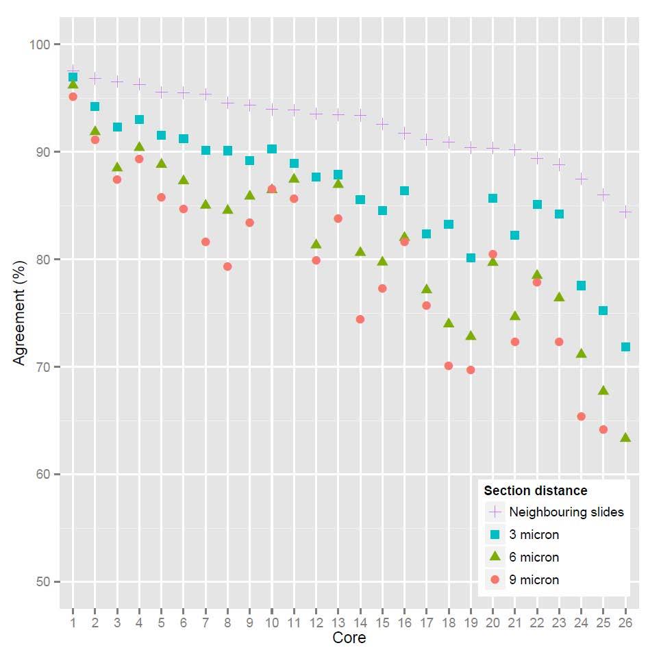
54 Software Image analysis in IHC - overview, considerations and applications
55 Software ImageJ ( ): Open-source, FREE, platformindependent, large community, Requires programming-skills VIS ( fully developed apps, expensive, database-handling of data and images, scanner independent Definiens INCA Aperio (Leica) PathXL / Philips Matlab
56 Thank you for your attention! Collaborators Søren Nielsen Rikke Riber-Hansen Alex Skovsbo Jørgensen Lasse Riis Østergaard Mogens Vyberg
57 Pitfalls Image analysis in IHC - overview, considerations and applications

58 Pitfalls - artefacts


59 Counter staining

60 Unspecific / Background staining

61 Staining of other cells

62 Scanning - background
63 Validation of alignment Digital Image Analysis Ki67


64 Validation of alignment

65

66


67 Five parallel slides of PCK
68 PCK-Alignment 5 parallel slides from TMA containing 40 breast cancers All stained for PCK TMA Only 26 (of 40) cores were usable Exclusion were due to Missing cores in one or more slides Damaged cores
69 PCK-Alignment Algorithm was developed that segmented 2 slides based on PCK expression Four categories based on PCK status in slide 1 and slide 2: + / + : PCK positive in both slides - / - : PCK negative in both slides + / - or - / +: PCK positive in only one slide

70

71

72

73 Overlap/agreement (%) Calculated as: PCK positive area in both slides + PCK negative area in both slides Divided by total area + + +


74 Good agreement (>90 %)


75 Less good agreement

76
77 Image analysis advanced algorithms Image analysis in IHC - overview, considerations and applications


78 Advanced algoritms More complex algorithms Successive application of several algorithms Not only thresholds Texture-based Architecture-based Feature-based training Feature may be selected statistically and unsupervised


79 Advanced algorithms architectural and texture


80 Advanced algoritms - texture Kather, J. N., Weis, C. A., Bianconi, F., Melchers, S. M., Schad, L. R., Gaiser, T.,... & Zöllner, F. G. (2016). Multi-class texture analysis in colorectal cancer histology. Scientific reports, 6,


81 Advanced algorithms cell nuclei texture Jørgensen, A. S., Rasmussen, A. M., Andersen, N. K. M., Andersen, S. K., Emborg, J., Røge, R., & Østergaard, L. R. (2017). Using cell nuclei features to detect colon cancer tissue in hematoxylin and eosin stained slides. Cytometry Part A, 91(8),

82 AI Djuric, Ugljesa, et al. "Precision histology: how deep learning is poised to revitalize histomorphology for personalized cancer care." npj Precision Oncology 1.1 (2017): 22.
Pathology Image Informatics Platform (PathIIP) Year 1 Update
 Year 1 Update") Pathology Image Informatics Platform (PathIIP) Year 1 Update PIIP Project sites Specific Aims Aim 1: Development of an improved plugin framework for the existing Sedeen viewer; Aim 2: Incorporate and evaluate
Pathology Image Informatics Platform (PathIIP) Year 1 Update PIIP Project sites Specific Aims Aim 1: Development of an improved plugin framework for the existing Sedeen viewer; Aim 2: Incorporate and evaluate
Colocalization Algorithm. User s Guide
 Colocalization Algorithm User s Guide Copyright 2008 Aperio Technologies, Inc. Part Number/Revision: MAN 0082, Revision A Date: March 7, 2008 This document applies to software versions Release 9.0 and
Algorithm User Guide:
 Algorithm User Guide: Membrane Quantification Use the Aperio algorithms to adjust (tune) the parameters until the quantitative results are sufficiently accurate for the purpose for which you intend to
Definiens. Tissue Studio 4.2. Tutorial 3: Metadata Import, Manual ROI Selection and Vessel Detection
 Definiens Tissue Studio 4.2 Tutorial 3: Metadata Import, Manual ROI Selection and Vessel Detection Tutorial 3: Metadata Import, Manual ROI Selection and Vessel Detection Imprint and Version Copyright 2015
The Pathology Company. Cytoplasm Algorithm. User s Guide
 The Pathology Company Cytoplasm Algorithm User s Guide MAN-0220, Revision C 1 December 2014 Cytoplasm Algorithm User s Guide This document applies to eslide Manager Release 12.2 and later. Copyright Notice
Out-of-sample extension of diffusion maps in a computer-aided diagnosis system. Application to breast cancer virtual slide images.
 Out-of-sample extension of diffusion maps in a computer-aided diagnosis system. Application to breast cancer virtual slide images. Philippe BELHOMME Myriam OGER Jean-Jacques MICHELS Benoit PLANCOULAINE
AUTOMATED DETECTION AND CLASSIFICATION OF CANCER METASTASES IN WHOLE-SLIDE HISTOPATHOLOGY IMAGES USING DEEP LEARNING
 AUTOMATED DETECTION AND CLASSIFICATION OF CANCER METASTASES IN WHOLE-SLIDE HISTOPATHOLOGY IMAGES USING DEEP LEARNING F. Ghazvinian Zanjani, S. Zinger, P. H. N. de With Electrical Engineering Department,
Color Deconvolution Algorithm. User s Guide
 Color Deconvolution Algorithm User s Guide Copyright 2007 Aperio Technologies, Inc. Part number/revision: MAN-0023, Revision A Date: February 26, 2007 This document applies to software versions Release
Definiens. Tissue Studio Release Notes
 Definiens Tissue Studio 3.0.1 Release Notes Definiens Documentation: Definiens Tissue Studio 3.0.1 Release Notes Imprint 2012 Definiens AG. All rights reserved. This document may be copied and printed
Solution resides in the details. Digital histology solutions from 3DHISTECH
 Solution resides in the details Digital histology solutions from 3DHISTECH Digital histology solutions from 3DHISTECH Paraffin block Routine immunohistochemistry, Fluorescent and serial sections, Citology
Algorithm User Guide:
 Algorithm User Guide: Microvessel Analysis Use the Aperio algorithms to adjust (tune) the parameters until the quantitative results are sufficiently accurate for the purpose for which you intend to use
Definiens. Tissue Studio 4.2. User Guide
 Definiens Tissue Studio 4.2 User Guide Definiens Documentation: Tissue Studio 4.2 User Guide Imprint 2015 Definiens AG. All rights reserved. This document may be copied and printed only in accordance with
Instructions for Ki67 Reproducibility Study Phase 3: Core Biopsies
 Instructions for Ki67 Reproducibility Study Phase 3: Core Biopsies Version: July 17, 2014 NOTE: It may be easier you for you to read these instructions in their entirety first. Although they appear long,
BioImaging facility update: from multi-photon in vivo imaging to highcontent high-throughput image-based screening. Alex Laude The BioImaging Unit
 BioImaging facility update: from multi-photon in vivo imaging to highcontent high-throughput image-based screening Alex Laude The BioImaging Unit Multi-dimensional, multi-modal imaging at the sub-cellular
Automated Diagnosis of Lymphoma with Digital Pathology Images Using Deep Learning
 Automated Diagnosis of Lymphoma with Digital Pathology Images Using Deep Learning Andy Nguyen, M.D., M.S. Medical Director, Hematopathology, Hematology and Coagulation Laboratory, Memorial Hermann Laboratory
Nuclei Segmentation of Whole Slide Images in Digital Pathology
 Nuclei Segmentation of Whole Slide Images in Digital Pathology Dennis Ai Department of Electrical Engineering Stanford University Stanford, CA dennisai@stanford.edu Abstract Pathology is the study of the
Analysis of Nuclei Detection with Stain Normalization in Histopathology Images
 Indian Journal of Science and Technology, Vol 8(23), DOI: 10.17485/ijst/2015/v8i23/85321, September 2015 ISSN (Print) : 0974-6846 ISSN (Online) : 0974-5645 Analysis of Nuclei Detection with Stain Normalization
Using Machine Learning for Classification of Cancer Cells
 Using Machine Learning for Classification of Cancer Cells Camille Biscarrat University of California, Berkeley I Introduction Cell screening is a commonly used technique in the development of new drugs.
Aperio epathology Image Analysis
 The Pathology Company Aperio epathology Image Analysis User s Guide MAN-0013, Revision F 1 December 2014 Aperio epathology Image Analysis User s Guide This document applies to eslide Manager Release 12.2
[7.3, EA], [9.1, CMB]
![[7.3, EA], [9.1, CMB]](/thumbs/76/73876791.jpg "[7.3, EA], [9.1, CMB]") K-means Clustering Ke Chen Reading: [7.3, EA], [9.1, CMB] Outline Introduction K-means Algorithm Example How K-means partitions? K-means Demo Relevant Issues Application: Cell Neulei Detection Summary
K-means Clustering Ke Chen Reading: [7.3, EA], [9.1, CMB] Outline Introduction K-means Algorithm Example How K-means partitions? K-means Demo Relevant Issues Application: Cell Neulei Detection Summary
CHAPTER-1 INTRODUCTION
 CHAPTER-1 INTRODUCTION 1.1 Fuzzy concept, digital image processing and application in medicine With the advancement of digital computers, it has become easy to store large amount of data and carry out
Towards Improved Epilepsia Diagnosis by Unsupervised Segmentation of Neuropathology Tissue Sections using Ripley s-ˆl Features
 Towards Improved Epilepsia Diagnosis by Unsupervised Segmentation of Neuropathology Tissue Sections using Ripley s-ˆl Features Timm Schoening 1, Volkmar H. Hans 2, Tim W. Nattkemper 1 1 Biodata Mining
Definiens. Tissue Studio 4.4. Tutorial 4: Manual ROI Selection and Marker Area Detection
 Definiens Tissue Studio 4.4 Tutorial 4: Manual ROI Selection and Marker Area Detection Tutorial 4: Manual ROI Selection and Marker Area Detection Imprint and Version Copyright 2017 Definiens AG. All rights
The Automated Counting Of Cancer Cells Using Computer Vision. Ben Carter. BSc Computing with Artificial Intelligence (Industry) 2005/2006
 2005/2006") The Automated Counting Of Cancer Cells Using Computer Vision Ben Carter BSc Computing with Artificial Intelligence (Industry) 2005/2006 The candidate confirms that the work submitted is their own and the
The Automated Counting Of Cancer Cells Using Computer Vision Ben Carter BSc Computing with Artificial Intelligence (Industry) 2005/2006 The candidate confirms that the work submitted is their own and the
Machine Learning for Medical Image Analysis. A. Criminisi
 Machine Learning for Medical Image Analysis A. Criminisi Overview Introduction to machine learning Decision forests Applications in medical image analysis Anatomy localization in CT Scans Spine Detection
Immunohistochemical stainers Overview Pros and Cons. Søren Nielsen Project coordinator & Scheme Manager NordiQC Aalborg University Hospital, Denmark
 Immunohistochemical stainers Overview Pros and Cons Søren Nielsen Project coordinator & Scheme Manager NordiQC Aalborg University Hospital, Denmark This lecture is meant to be a basis for an open discussion
COLOCALISATION. Alexia Loynton-Ferrand. Imaging Core Facility Biozentrum Basel
 COLOCALISATION Alexia Loynton-Ferrand Imaging Core Facility Biozentrum Basel OUTLINE Introduction How to best prepare your samples for colocalisation How to acquire the images for colocalisation How to
Statistical Analysis of Metabolomics Data. Xiuxia Du Department of Bioinformatics & Genomics University of North Carolina at Charlotte
 Statistical Analysis of Metabolomics Data Xiuxia Du Department of Bioinformatics & Genomics University of North Carolina at Charlotte Outline Introduction Data pre-treatment 1. Normalization 2. Centering,
RADIOMICS: potential role in the clinics and challenges
 27 giugno 2018 Dipartimento di Fisica Università degli Studi di Milano RADIOMICS: potential role in the clinics and challenges Dr. Francesca Botta Medical Physicist Istituto Europeo di Oncologia (Milano)
Pathology Image Informatics Platform (PathIIP)
") Pathology Image Informatics Platform (PathIIP) PIs: Anant Madabhushi (CWRU), Metin Gurcan (OSU), Anne Martel (UToronto) Year 2 Update Develop a digital pathology platform to facilitate wider adoption of
Pathology Image Informatics Platform (PathIIP) PIs: Anant Madabhushi (CWRU), Metin Gurcan (OSU), Anne Martel (UToronto) Year 2 Update Develop a digital pathology platform to facilitate wider adoption of
Quantitative analysis of Alzheimer plaques in mice using virtual microscopy
 Quantitative analysis of Alzheimer plaques in mice using virtual microscopy Monika Bieri, André Wethmar and Norbert Wey Technical- and Mediasupport Department of Pathology University Hospital Zurich, Switzerland
Figure 1: Workflow of object-based classification
 Technical Specifications Object Analyst Object Analyst is an add-on package for Geomatica that provides tools for segmentation, classification, and feature extraction. Object Analyst includes an all-in-one
Definiens Tissue Studio Action Library 4.2. Release Notes
 Definiens Tissue Studio 1.1.0 Action Library 4.2 Release Notes Imprint and Version Document Version Tissue Studio 1.1.0 AL 4.2 Copyright 2009 Definiens AG. All rights reserved. This document may be copied
Adversarial Stain Transfer for Histopathology Image Analysis
 1 Adversarial Stain Transfer for Histopathology Image Analysis Aı cha BenTaieb and Ghassan Hamarneh Medical Image Analysis Lab, School of Computing Science, Simon Fraser University, Burnaby, Canada Index
IN recent years, deep learning has shown great promise
 1 Unsupervised Feature Extraction via Deep Learning for Histopathological Classification of Colon Tissue Images Can Taylan Sari and Cigdem Gunduz-Demir*, Member, IEEE Abstract Histopathological examination
I understand that failure to attribute material which is obtained from another source may be considered as plagiarism. (Signature of student)
") A Computer Vision System for the Detection of Cancer in Barretts Oesophagus James Swainston BSc Computer Science with Artificial Intelligence 2007/2008 The candidate confirms that the work submitted is
A Computer Vision System for the Detection of Cancer in Barretts Oesophagus James Swainston BSc Computer Science with Artificial Intelligence 2007/2008 The candidate confirms that the work submitted is
INCREASING CLASSIFICATION QUALITY BY USING FUZZY LOGIC
 JOURNAL OF APPLIED ENGINEERING SCIENCES VOL. 1(14), issue 4_2011 ISSN 2247-3769 ISSN-L 2247-3769 (Print) / e-issn:2284-7197 INCREASING CLASSIFICATION QUALITY BY USING FUZZY LOGIC DROJ Gabriela, University
Blood Microscopic Image Analysis for Acute Leukemia Detection
 I J C T A, 9(9), 2016, pp. 3731-3735 International Science Press Blood Microscopic Image Analysis for Acute Leukemia Detection V. Renuga, J. Sivaraman, S. Vinuraj Kumar, S. Sathish, P. Padmapriya and R.
[project name]_[biomarker name]_[block letter][block number(s)]_[tma block version]_[slice/batch number]
![[project name]_[biomarker name]_[block letter][block number(s)]_[tma block version]_[slice/batch number]](/thumbs/74/71176963.jpg "[project name]_[biomarker name]_[block letter][block number(s)]_[tma block version]_[slice/batch number]") GPEC Digital TMA Image User Guide Below is a brief description regarding how to access and capture GPEC s digital images scanned by the Bliss system (Olympus, originally Bacus Lab: http://www.olympusamerica.com/seg_section/seg_vm.asp).
GPEC Digital TMA Image User Guide Below is a brief description regarding how to access and capture GPEC s digital images scanned by the Bliss system (Olympus, originally Bacus Lab: http://www.olympusamerica.com/seg_section/seg_vm.asp).
Available Online through
 Available Online through www.ijptonline.com ISSN: 0975-766X CODEN: IJPTFI Research Article ANALYSIS OF CT LIVER IMAGES FOR TUMOUR DIAGNOSIS BASED ON CLUSTERING TECHNIQUE AND TEXTURE FEATURES M.Krithika
Aperio Image Analysis. User s Guide
 Aperio Image Analysis User s Guide Copyright 2008 Aperio Technologies, Inc. Part Number/Revision: MAN 0013, Revision C Date: September 4, 2009 This document applies to software versions Release 10.1 and
Tutorial for cell nucleus intensity clustering on histochemically stained tissue microarrays.
 Tutorial for cell nucleus intensity clustering on histochemically stained tissue microarrays. http://www.nexus.ethz.ch/ -> Software -> TMARKER The plugin Intensity Clustering starts on step 7. Steps 1-6
Detection of Ductus in Mammary Gland Tissue by Computer Vision
 Detection of Ductus in Mammary Gland Tissue by Computer Vision Katarina Mele and Aleš Leonardis Computer Vision Laboratory Faculty of Computer and Information Science University of Ljubljana Tržaška 25,
Automatic Grayscale Classification using Histogram Clustering for Active Contour Models
 Research Article International Journal of Current Engineering and Technology ISSN 2277-4106 2013 INPRESSCO. All Rights Reserved. Available at http://inpressco.com/category/ijcet Automatic Grayscale Classification
10. Clustering. Introduction to Bioinformatics Jarkko Salojärvi. Based on lecture slides by Samuel Kaski
 10. Clustering Introduction to Bioinformatics 30.9.2008 Jarkko Salojärvi Based on lecture slides by Samuel Kaski Definition of a cluster Typically either 1. A group of mutually similar samples, or 2. A
Building an Ancillary System for Cancer Registries from SDC CAP Templates
 Building an Ancillary System for Cancer Registries from SDC CAP Templates Jennifer Seiffert, MLIS, CTR, Northrop Grumman, under contract to CDC s NPCR Sanjeev Baral, Northrop Grumman, under contract to
Automatic Segmentation of Cell Nuclei in Breast Histopathology Images and Classification Using Feed Forward Neural Network
 RESEARCH ARTICLE OPEN ACCESS Automatic Segmentation of Cell Nuclei in Breast Histopathology Images and Classification Using Feed Forward Neural Network Shraddha R. Raut #, Dr. S. S. Salankar *, Prof. V.
Detection of Leukemia in Blood Microscope Images
 I J C T A, 9(5), 2016, pp. 63-67 International Science Press Detection of Leukemia in Blood Microscope Images Indira P.*, Ganesh Babu T. R.**, Vidhya K.*** ABSTRACT Leukemia is a cancer of the blood and
Automated Tissue Image Analysis Using Pattern Recognition
 Digital Comprehensive Summaries of Uppsala Dissertations from the Faculty of Science and Technology 1175 Automated Tissue Image Analysis Using Pattern Recognition JIMMY AZAR ACTA UNIVERSITATIS UPSALIENSIS
InsituNet User Documentation
 InsituNet 1.0.4 User Documentation John Salamon, Xiaoyan Qian, Mats Nilsson, David J. Lynn 25 January 2018 Contact: John.Salamon@sahmri.com Abstract In situ sequencing is a novel method to generate spatially-resolved,
Quantitative Image Analysis and 3-D Digital Reconstruction of Aortic Valve Leaflet
 Quantitative Image Analysis and 3-D Digital Reconstruction of Aortic Valve Leaflet Chi Zheng 1 Mentors: John A. Stella 2 and Michael S. Sacks, Ph. D. 2 1 Bioengineering and Bioinformatics Summer Institute,
IMAGE PROCESSING IN DIGITAL PATHOLOGY: AN OPPORTUNITY TO SOLVE INTER-BATCH VARIABILITY OF IMMUNOHISTOCHEMICAL STAINING
 IMAGE PROCESSING IN DIGITAL PATHOLOGY: AN OPPORTUNITY TO SOLVE INTER-BATCH VARIABILITY OF IMMUNOHISTOCHEMICAL STAINING Yves-Rémi Van Eycke, Justine Allard, Isabelle Salmon, Olivier Debeir, Christine Decaestecker
Fast and accurate automated cell boundary determination for fluorescence microscopy
 Fast and accurate automated cell boundary determination for fluorescence microscopy Stephen Hugo Arce, Pei-Hsun Wu &, and Yiider Tseng Department of Chemical Engineering, University of Florida and National
COLOR BASED REMOTE SENSING IMAGE SEGMENTATION USING FUZZY C-MEANS AND IMPROVED SOBEL EDGE DETECTION ALGORITHM
 COLOR BASED REMOTE SENSING IMAGE SEGMENTATION USING FUZZY C-MEANS AND IMPROVED SOBEL EDGE DETECTION ALGORITHM Ms. B.SasiPrabha, Mrs.R.uma, MCA,M.Phil,M.Ed, Research scholar, Asst. professor, Department
TUMOR DETECTION IN MRI IMAGES
 TUMOR DETECTION IN MRI IMAGES Prof. Pravin P. Adivarekar, 2 Priyanka P. Khatate, 3 Punam N. Pawar Prof. Pravin P. Adivarekar, 2 Priyanka P. Khatate, 3 Punam N. Pawar Asst. Professor, 2,3 BE Student,,2,3
doi: /
 Yiting Xie ; Anthony P. Reeves; Single 3D cell segmentation from optical CT microscope images. Proc. SPIE 934, Medical Imaging 214: Image Processing, 9343B (March 21, 214); doi:1.1117/12.243852. (214)
Rare Event Detection Algorithm. User s Guide
 Rare Event Detection Algorithm User s Guide Copyright 2008 Aperio Technologies, Inc. Part Number/Revision: MAN 0123, Revision A Date: September 2, 2008 This document applies to software versions Release
Digital Media IA. EXAM INFORMATION Items. Points. Prerequisites. Grade Level. Course Length. Career Cluster. Performance Standards
 EXAM INFORMATION Items 37 Points 49 Prerequisites KEYBOARD PROFICIENCY COMPUTER TECHNOLOGY Grade Level 10-12 Course Length ONE SEMESTER DESCRIPTION Digital Media is the process of analyzing, designing
Introduction to Medical Image Processing
 Introduction to Medical Image Processing Δ Essential environments of a medical imaging system Subject Image Analysis Energy Imaging System Images Image Processing Feature Images Image processing may be
Microvessel Analysis Algorithm. User s Guide
 Microvessel Analysis Algorithm User s Guide Copyright 2008 Aperio Technologies, Inc. Part number/revision: MAN 0092, Revision A Date: March 10, 2008 This document applies to software versions Release 9.1
Nuclear Algorithm User s Guide
 The Pathology Company Nuclear Algorithm User s Guide Research Use Only MAN-0338, Revision A 5 August 2015 Nuclear Algorithm User s Guide (RUO) This document applies to eslide Manager Release 12.3 and later.
COLOCALISATION. Alexia Ferrand. Imaging Core Facility Biozentrum Basel
 COLOCALISATION Alexia Ferrand Imaging Core Facility Biozentrum Basel OUTLINE Introduction How to best prepare your samples for colocalisation How to acquire the images for colocalisation How to analyse
Pathological Lymph Node Classification
 Pathological Lymph Node Classification Jonathan Booher, Michael Mariscal and Ashwini Ramamoorthy SUNet ID: { jaustinb, mgm248, ashwinir } @stanford.edu Abstract Machine learning algorithms have the potential
Empowering Multiple Instance Histopathology Cancer Diagnosis by Cell Graphs
 Empowering Multiple Instance Histopathology Cancer Diagnosis by Cell Graphs Anonymous Authors No Institute Given Abstract. We introduce a probabilistic classifier that combines multiple instance learning
HETEROGENEITY ASSESSMENT OF HISTOLOGICAL TISSUE SECTIONS IN WHOLE SLIDE IMAGES
 12th European Congress on Digital Pathology HETEROGENEITY ASSESSMENT OF HISTOLOGICAL TISSUE SECTIONS IN WHOLE SLIDE IMAGES BELHOMME Philippe 1, TORALBA Simon 1,3, PLANCOULAINE Benoît 1, OGER Myriam 1,2,
8/3/2017. Contour Assessment for Quality Assurance and Data Mining. Objective. Outline. Tom Purdie, PhD, MCCPM
 Contour Assessment for Quality Assurance and Data Mining Tom Purdie, PhD, MCCPM Objective Understand the state-of-the-art in contour assessment for quality assurance including data mining-based techniques
Fast segmentation for texture-based cartography of Whole Slide Images
 Fast segmentation for texture-based cartography of Whole Slide Images Gregory Apou 1, Benoît Naegel 1, Germain Forestier 2, Friedrich Feuerhake 3 and Cédric Wemmert 1 1 ICube, University of Strasbourg,
Image Analysis and Morphometry
 Image Analysis and Morphometry Lukas Schärer Evolutionary Biology Zoological Institute University of Basel 1 13. /15.3.2013 Zoology & Evolution Block Course Summary Quantifying morphology why do we need
Tumor Detection and classification of Medical MRI UsingAdvance ROIPropANN Algorithm
 International Journal of Engineering Research and Advanced Technology (IJERAT) DOI:http://dx.doi.org/10.31695/IJERAT.2018.3273 E-ISSN : 2454-6135 Volume.4, Issue 6 June -2018 Tumor Detection and classification
Lecture on Modeling Tools for Clustering & Regression
 Lecture on Modeling Tools for Clustering & Regression CS 590.21 Analysis and Modeling of Brain Networks Department of Computer Science University of Crete Data Clustering Overview Organizing data into
A Computer Vision System for Graphical Pattern Recognition and Semantic Object Detection
 A Computer Vision System for Graphical Pattern Recognition and Semantic Object Detection Tudor Barbu Institute of Computer Science, Iaşi, Romania Abstract We have focused on a set of problems related to
CHAPTER 6 MODIFIED FUZZY TECHNIQUES BASED IMAGE SEGMENTATION
 CHAPTER 6 MODIFIED FUZZY TECHNIQUES BASED IMAGE SEGMENTATION 6.1 INTRODUCTION Fuzzy logic based computational techniques are becoming increasingly important in the medical image analysis arena. The significant
AnalySIS Tutorial part 2
 AnalySIS Tutorial part 2 Sveinung Lillehaug Neural Systems and Graphics Computing Laboratory Department of Anatomy University of Oslo N-0317 Oslo Norway www.nesys.uio.no Using AnalySIS to automatically
Clustering analysis of gene expression data
 Clustering analysis of gene expression data Chapter 11 in Jonathan Pevsner, Bioinformatics and Functional Genomics, 3 rd edition (Chapter 9 in 2 nd edition) Human T cell expression data The matrix contains
jslic: superpixels in ImageJ
 19 th Computer Vision Winter Workshop Zuzana Kúkelová and Jan Heller (eds.) Křtiny, Czech Republic, February 3 5, 2014 jlic: superpixels in ImageJ Jiří Borovec and Jan Kybic Faculty of Electrical Engineering,
Paceamker Basic Math. Correlated to. Alaska Math Grade Level Expectations For Eighth Grade
 Paceamker Basic Math Alaska Math Grade Level Expectations 1 Numeration Performance Standards M1.3.1 Read, write, model, and order real numbers, explaining scientific notation, exponents, and percents.
Colin Paul Updated 14 November Preparation of publication-quality videos using ImageJ
 Preparation of publication-quality videos using ImageJ Statements made in scientific papers are often much easier to understand if they are supplemented by representative videos. In the best case, these
THREE-DIMENSIONA L ELECTRON MICROSCOP Y OF MACROMOLECULAR ASSEMBLIE S. Visualization of Biological Molecules in Their Native Stat e.
 THREE-DIMENSIONA L ELECTRON MICROSCOP Y OF MACROMOLECULAR ASSEMBLIE S Visualization of Biological Molecules in Their Native Stat e Joachim Frank CHAPTER 1 Introduction 1 1 The Electron Microscope and
VC 11/12 T14 Visual Feature Extraction
 VC 11/12 T14 Visual Feature Extraction Mestrado em Ciência de Computadores Mestrado Integrado em Engenharia de Redes e Sistemas Informáticos Miguel Tavares Coimbra Outline Feature Vectors Colour Texture
Texture Classification by Combining Local Binary Pattern Features and a Self-Organizing Map
 Texture Classification by Combining Local Binary Pattern Features and a Self-Organizing Map Markus Turtinen, Topi Mäenpää, and Matti Pietikäinen Machine Vision Group, P.O.Box 4500, FIN-90014 University
Tomasz Markiewicz 1,2, Robert Koktysz 2, Stanisław Osowski 1, Michał Muszyński 1, Wojciech Kozłowski 2
 Tomasz Markiewicz 1,, Robert Koktysz, Stanisław Osowski 1, Michał Muszyński 1, Wojciech Kozłowski 1 Warsaw University of Technology, Dept. of Electrical Eng. and Military Institute of Medicine, Department
Computer Assisted Image Analysis TF 3p and MN1 5p Lecture 1, (GW 1, )
") Centre for Image Analysis Computer Assisted Image Analysis TF p and MN 5p Lecture, 422 (GW, 2.-2.4) 2.4) 2 Why put the image into a computer? A digital image of a rat. A magnification of the rat s nose.
Centre for Image Analysis Computer Assisted Image Analysis TF p and MN 5p Lecture, 422 (GW, 2.-2.4) 2.4) 2 Why put the image into a computer? A digital image of a rat. A magnification of the rat s nose.
Epithelium and Stroma Identification in Histopathological Images Using Unsupervised and Semi-Supervised Superpixel-Based Segmentation
 Journal of Imaging Article Epithelium and Stroma Identification in Histopathological Images Using Unsupervised and Semi-Supervised Superpixel-Based Segmentation Shereen Fouad 1, *, David Randell 1, Antony
Computer Aided Diagnosis Based on Medical Image Processing and Artificial Intelligence Methods
 International Journal of Information and Computation Technology. ISSN 0974-2239 Volume 3, Number 9 (2013), pp. 887-892 International Research Publications House http://www. irphouse.com /ijict.htm Computer
Presented at the FIG Congress 2018, May 6-11, 2018 in Istanbul, Turkey
 Presented at the FIG Congress 2018, May 6-11, 2018 in Istanbul, Turkey Evangelos MALTEZOS, Charalabos IOANNIDIS, Anastasios DOULAMIS and Nikolaos DOULAMIS Laboratory of Photogrammetry, School of Rural
Bayes Risk. Classifiers for Recognition Reading: Chapter 22 (skip 22.3) Discriminative vs Generative Models. Loss functions in classifiers
 Discriminative vs Generative Models. Loss functions in classifiers") Classifiers for Recognition Reading: Chapter 22 (skip 22.3) Examine each window of an image Classify object class within each window based on a training set images Example: A Classification Problem Categorize
Classifiers for Recognition Reading: Chapter 22 (skip 22.3) Examine each window of an image Classify object class within each window based on a training set images Example: A Classification Problem Categorize
CP467 Image Processing and Pattern Recognition
 CP467 Image Processing and Pattern Recognition Instructor: Hongbing Fan Introduction About DIP & PR About this course Lecture 1: an overview of DIP DIP&PR show What is Digital Image? We use digital image
SuRVoS Workbench. Super-Region Volume Segmentation. Imanol Luengo
 SuRVoS Workbench Super-Region Volume Segmentation Imanol Luengo Index - The project - What is SuRVoS - SuRVoS Overview - What can it do - Overview of the internals - Current state & Limitations - Future
Introduction. Welcome. Machine Learning
 Introduction Welcome Machine Learning SPAM Machine Learning -Grew out of work in AI -New capability for computers Examples: -Database mining Large datasets from growth of automation/web. E.g., Web click
DIS: Design and imaging software
 Using IT productivity tools and applications This is the ability to use a software application designed to create, modify and layout artwork or images for display in print or on a screen (eg vector graphics
Intelligent Image and Graphics Processing
 Intelligent Image and Graphics Processing 智能图像图形处理图形处理 布树辉 bushuhui@nwpu.edu.cn http://www.adv-ci.com Clustering Clustering Attach label to each observation or data points in a set You can say this unsupervised
Automated Image Analysis Of Estrogen Receptor Immunohistochemistry In. Breast Cancer
 Automated Image Analysis Of Estrogen Receptor Immunohistochemistry In Breast Cancer by c Joel Mercer, B.Eng, M.D. A thesis submitted to the School of Graduate Studies in partial fulfillment of the requirements
For questions please contact: Lei-Ann Arceneaux, 4/13/2011
 TMALab TMALab is a web-based function in Spectrum Plus, working for Tissue Microarrays (TMAs). TMAs allow researchers to validate new biomarkers or discover and dissect molecular pathways in hundreds of
Data mining for neuroimaging data. John Ashburner
 Data mining for neuroimaging data John Ashburner MODELLING The Scientific Process MacKay, David JC. Bayesian interpolation. Neural computation 4, no. 3 (1992): 415-447. Model Selection Search for the best
Surveyor Autocalibration Guide Objective Imaging Ltd.
 Surveyor Autocalibration Guide Objective Imaging Ltd. 1 Overview The calibration in Surveyor defines a correlation between the pixel size of the camera to the samesized area of the stage measured in microns,
CHAPTER 4 DETECTION OF DISEASES IN PLANT LEAF USING IMAGE SEGMENTATION
 CHAPTER 4 DETECTION OF DISEASES IN PLANT LEAF USING IMAGE SEGMENTATION 4.1. Introduction Indian economy is highly dependent of agricultural productivity. Therefore, in field of agriculture, detection of
DIGITAL IMAGE ANALYSIS. Image Classification: Object-based Classification
 DIGITAL IMAGE ANALYSIS Image Classification: Object-based Classification Image classification Quantitative analysis used to automate the identification of features Spectral pattern recognition Unsupervised
Classifiers for Recognition Reading: Chapter 22 (skip 22.3)
") Classifiers for Recognition Reading: Chapter 22 (skip 22.3) Examine each window of an image Classify object class within each window based on a training set images Slide credits for this chapter: Frank
Classifiers for Recognition Reading: Chapter 22 (skip 22.3) Examine each window of an image Classify object class within each window based on a training set images Slide credits for this chapter: Frank
CHAPTER 6 DETECTION OF MASS USING NOVEL SEGMENTATION, GLCM AND NEURAL NETWORKS
 130 CHAPTER 6 DETECTION OF MASS USING NOVEL SEGMENTATION, GLCM AND NEURAL NETWORKS A mass is defined as a space-occupying lesion seen in more than one projection and it is described by its shapes and margin
Exploring high dimensional data with Butterfly: a novel classification algorithm based on discrete dynamical systems
 Exploring high dimensional data with Butterfly: a novel classification algorithm based on discrete dynamical systems J o s e p h G e r a c i, M o y e z D h a r s e e, P a u l o N u i n, A l e x a n d r