Quantitative Image Analysis and 3-D Digital Reconstruction of Aortic Valve Leaflet
|
|
|
- Amanda Rice
- 5 years ago
- Views:
Transcription
1 Quantitative Image Analysis and 3-D Digital Reconstruction of Aortic Valve Leaflet Chi Zheng 1 Mentors: John A. Stella 2 and Michael S. Sacks, Ph. D. 2 1 Bioengineering and Bioinformatics Summer Institute, Department of Computational Biology, University of Pittsburgh 2 McGowan Institute for Regenerative Medicine and Department of Bioengineering, University of Pittsburgh

2 Introduction Aortic valve (AV): most commonly replaced valve Tissue engineering (TE) valve offers customized availability, growth potential, durability, and biocompatibility. Must first understand structure of the valve to replicate its function by TE
 Ventricularis : Collagen and")
3 Background Each valve is composed of three leaflets (right-coronary, left coronary, non-coronary) Each leaflet is composed of 3 cell layers Fibrosa: Collagen and little elastin Spongiosa: Glucosaminoglycans (GAGs) Ventricularis : Collagen and elastin
4 Objectives Original objective: Construct 3D representation of a porcine right coronary AV leaflet containing Cell count and distribution Layer thickness variations Track collagen and elastin fibers Technological limitations bifurcated the objective Quantification 3D reconstruction


5 Status 15 mm 35 mm Total histological sections: ~ 300 slides Imaged, quantified, and 3D reconstructed 50 slides
6 Histology and Image Acquisition 5 μm thick circumferential slices fixed in formalin stained with Movat s pentachrome Collagen = Yellow GAGs = Blue Nuclei and Elastin = Dark Purple Acquisition Quantification: 17 slices, spaced 90 μm apart, were digitally captured using bright field microscopy at 20x and montaged 3D Reconstruction: 50 slices, spaced μm apart were scanned individually using slide scanner
7 Principles of Image Analysis Contrast 256 shades of gray in a 8-bit monochrome image Defines edges/borders of objects Threshold Enhances contrast by dividing image into two category Objects within the image (subsets of image data) defined by contiguous pixels similar in intensity Automatic threshold selection uses mode pixel intensity


8 Color Separation
9 Layer Separation V S F
10 Cytometry Cell nuclei counting completed with particle analysis function in NIH s Image J RGB image converted to monochrome on the green channel Threshold is user defined Definition of a cell size: pixel 2 circularity:


11 Particle Analysis of Nuclei


12 Collagen and Elastin Content
13 Layer Thickness Measurement Scale bar added during image acquisition used to establish calibration between pixels and μm in Metamorph TM Take representative samples, spaced apart by 500 μm, along the radial length of each slice Results indicate Average layer thickness Topographic representation Local variations


14 Measuring Thickness
15 Quantification Results Distribution of Cells Ventricularis Spongiosa Fibrosa 100% 80% 60% 40% 20% 0% Circumferential Distance (mm) Percent of Cells
16 Quantification Results (cont d) Collagen and Elastin Content Fibrosa: % area occupied by collagen Ventricularis: % collagen % elastin Need for comparision with literature values (entire tissue composition: 13% elastin and 50% collagen by dry weight) Average thickness Fibrosa: μm (~ μm) Spongiosa: μm (~ μm) Ventricularis: μm (~ μm)

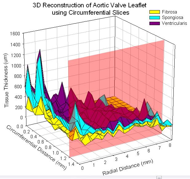
17 Topography of Layer Thickness

18 Regional Variation Basal Attachment Belly Coaptation
19 3D Reconstruction Images of 50 slices, spaced μm apart, were digitally aligned by morphology and stacked to construct a 3D representation Software designed for fluorescent microscopy so image colors had to be inverted Collagen = Blue GAGs= Red Nuclei and Elastin = Yellow 3D representation allows user to visualize the leaflet better than topography

20 Inversion

21 Ooo.
22 Ahh
23 Conclusion and Future Work Preliminary results calls for completion of the entire leaflet Statistical validation of quantification upon completion Explore other imaging techniques (fluorescent microscopy, X-ray, ultrasound, acoustic microscopy, SEM) Construct a 3D representation containing quantitative information Use 3D reconstruction to simulate and visualize dynamic response to applied load
24 Acknowledgements Engineered Tissue Mechanics Laboratory, Department of Bioengineering, University of Pittsburgh (Dr. Michael Sacks, John Stella, et al.) Center for Biologic Imaging, University of Pittsburgh (Dr. Simon Watkins, Jason Devlin, Stuart Shand, et al.) Department of Computational Biology, University of Pittsburgh Developers of NIH s Image J
25 References Scott, M. and Vesely, I. Aortic valve cusp microstructure: the role of elastin. Ann. Thorac. Surg :S Lacefield et al. Three-dimensional visualization and thickness estimation of aortic valve cusps using high-frequency ultrasound. Physiol. Meas :27-36 Boughne et al. A precise radiographic technique for the measurement of dimensionalchanges in heart valve biomaterials following fixation. Journal of Biomechanics : Lu et al. Evaluation of progression in nonrheumatic aortic valvular stenosis by scanning acoustic microscopy. Ultrasound in Medicine & Biology :4: P. Chieco, A. Jonker and C.J.F. van Noorden. Image Cytometry. 1st edition. Springer
26 Questions?
Algorithm User Guide:
 Algorithm User Guide: Membrane Quantification Use the Aperio algorithms to adjust (tune) the parameters until the quantitative results are sufficiently accurate for the purpose for which you intend to
BioImaging facility update: from multi-photon in vivo imaging to highcontent high-throughput image-based screening. Alex Laude The BioImaging Unit
 BioImaging facility update: from multi-photon in vivo imaging to highcontent high-throughput image-based screening Alex Laude The BioImaging Unit Multi-dimensional, multi-modal imaging at the sub-cellular
InVivo Analyzer Suite
 InVivo Analyzer Suite Live Cell Imaging Software for Fast Acquisition and Complete Analysis The Complete Live Cell Imaging Solution Events happen fast in live cell experimentation. InVivo InVivo Analyzer
Monday, Tuesday, Wednesday, and Thursday, 1 pm to 3 or 4 pm. (See Course Schedule for details)
") Anatomy 6201 Course Director: Dr. Ernesto Phone: (303) 724-3430 Office: RC1 South Rm 11124 Office Hours: by appointment Email: ernesto.salcedo@ucdenver Location ED 2 South Room 2206.! Course Hours Monday,
Anatomy 6201 Course Director: Dr. Ernesto Phone: (303) 724-3430 Office: RC1 South Rm 11124 Office Hours: by appointment Email: ernesto.salcedo@ucdenver Location ED 2 South Room 2206.! Course Hours Monday,
Colocalization Algorithm. User s Guide
 Colocalization Algorithm User s Guide Copyright 2008 Aperio Technologies, Inc. Part Number/Revision: MAN 0082, Revision A Date: March 7, 2008 This document applies to software versions Release 9.0 and
Bosch Institute Advanced Microscopy Facility Workshops Summer/Autumn 2016
 Bosch Institute Advanced Microscopy Facility Workshops Summer/Autumn 2016 Presented by Dr Louise Cole and Dr Cathy Payne, Advanced Microscopy Facility, Bosch Institute, School of Medical Sciences, The
Developments & validation of innovative techniques for the skin study in vivo
 Developments & validation of innovative techniques for the skin study in vivo Jean-Christophe Pittet, ORION Concept COSM O Lab., Tours, France Introduction New techniques why? Interest of new technologies
Rare Event Detection Algorithm. User s Guide
 Rare Event Detection Algorithm User s Guide Copyright 2008 Aperio Technologies, Inc. Part Number/Revision: MAN 0123, Revision A Date: September 2, 2008 This document applies to software versions Release
Algorithm User Guide:
 Algorithm User Guide: Microvessel Analysis Use the Aperio algorithms to adjust (tune) the parameters until the quantitative results are sufficiently accurate for the purpose for which you intend to use
Introduction. Loading Images
 Introduction CellProfiler is a free Open Source software for automated image analysis. Versions for Mac, Windows and Linux are available and can be downloaded at: http://www.cellprofiler.org/. CellProfiler
Anatomic measurement accuracy: CT parameters and 3D rendering effects
 Anatomic measurement accuracy: CT parameters and 3D rendering effects Brian J Whyms a, E Michael Schimek a, Houri K Vorperian a, Lindell R Gentry b, and Edward T Bersu c University of Wisconsin-Madison
EyeTech. Particle Size Particle Shape Particle concentration Analyzer ANKERSMID
 EyeTech Particle Size Particle Shape Particle concentration Analyzer A new technology for measuring particle size in combination with particle shape and concentration. COMBINED LASERTECHNOLOGY & DIA Content
Counting Particles or Cells Using IMAQ Vision
 Application Note 107 Counting Particles or Cells Using IMAQ Vision John Hanks Introduction To count objects, you use a common image processing technique called particle analysis, often referred to as blob
Quantitative analysis of Alzheimer plaques in mice using virtual microscopy
 Quantitative analysis of Alzheimer plaques in mice using virtual microscopy Monika Bieri, André Wethmar and Norbert Wey Technical- and Mediasupport Department of Pathology University Hospital Zurich, Switzerland
Tomography. Introduction to Tomography TEM Tilt-Series Tomography in Life Science STEM Tomography in Materials Science
 Tomography Introduction to Tomography TEM Tilt-Series Tomography in Life Science STEM Tomography in Materials Science Introduction to Tomography Tomography is imaging by sections or sectioning. A device
STEM electron tomography in the Scanning Electron Microscope
 Journal of Physics: Conference Series PAPER OPEN ACCESS STEM electron tomography in the Scanning Electron Microscope To cite this article: M Ferroni et al 2015 J. Phys.: Conf. Ser. 644 012012 Recent citations
APPROACHES IN QUANTITATIVE, MULTI-DIMENSIONAL MICROSCOPY
 APPROACHES IN QUANTITATIVE, MULTI-DIMENSIONAL MICROSCOPY Profs. Zvi Kam and Benjamin Geiger Department of Molecular Cell Biology The Weizmann Institute of Science Rehovot, Israel In this presentation we
A Transmission Line Matrix Model for Shielding Effects in Stents
 A Transmission Line Matrix Model for hielding Effects in tents Razvan Ciocan (1), Nathan Ida (2) (1) Clemson University Physics and Astronomy Department Clemson University, C 29634-0978 ciocan@clemon.edu
Clinical Importance. Aortic Stenosis. Aortic Regurgitation. Ultrasound vs. MRI. Carotid Artery Stenosis
 Clinical Importance Rapid cardiovascular flow quantitation using sliceselective Fourier velocity encoding with spiral readouts Valve disease affects 10% of patients with heart disease in the U.S. Most
S93-8 Page 1 TOMOGRAPHIC APPROACHES TO NONWOVENS STRUCTURE DEFINITION
 S93-8 Page 1 TOMOGRAPHIC APPROACHES TO NONWOVENS STRUCTURE DEFINITION PIs: T. Gilmore, H. Davis, Z. Mi, North Carolina State University Code: S93-8 Date of Report: 9/94 RELEVANCE TO NTC MISSION AND GOALS:
Particle Size & Shape Analyzer
 Particle Size & Shape Analyzer Slide 2/30 Table of Content Laser Channel.... 3-7 Video Channel...... 8-15 General Application...16-17 S/W Features. 18-29 Summery...... 30 Slide 3/30 DIPA-2000 Measuring
Pathobin Product Catalogue 2018
 Pathobin Product Catalogue 2018 Download a purchase order form www.pathobin.com Pathobin 3D System... 3 Pathobin C-mount Camera... 5 Pathobin Mosaic software... 7 PathobinTools software... 9 2 Pathobin
Voxel-based Registration Methods in in vivo Imaging
 Vladimír Ulman Laboratory of Optical Microscopy, FI MU 14th April, 2005 Informatics seminar, FI MU Outline Introduction to bioinformatics Introduction to in vivo imaging Point-based registration Optical
Image Acquisition Systems
 Image Acquisition Systems Goals and Terminology Conventional Radiography Axial Tomography Computer Axial Tomography (CAT) Magnetic Resonance Imaging (MRI) PET, SPECT Ultrasound Microscopy Imaging ITCS
Bosch Institute Advanced Microscopy Facility Workshops Summer/Autumn 2017
 Bosch Institute Advanced Microscopy Facility Workshops Summer/Autumn 2017 Presented by Dr Louise Cole, Advanced Microscopy Facility, Bosch Institute, School of Medical Sciences, The University of Sydney.
Medical Imaging Projects
 NSF REU MedIX Summer 2006 Medical Imaging Projects Daniela Stan Raicu, PhD http://facweb.cs.depaul.edu/research draicu@cs.depaul.edu Outline Medical Informatics Imaging Modalities Computed Tomography Medical
Supplementary Figure 1
 Supplementary Figure 1 BOLD and CBV functional maps showing EPI versus line-scanning FLASH fmri. A. Colored BOLD and CBV functional maps are shown in the highlighted window (green frame) of the raw EPI
Pilot study of semiautomated localization of the dermal/epidermal junction in reflectance confocal microscopy images of skin
 Pilot study of semiautomated localization of the dermal/epidermal junction in reflectance confocal microscopy images of skin Sila Kurugol Jennifer G. Dy Dana H. Brooks Milind Rajadhyaksha Journal of Biomedical
Orientations of collagen fibers in aortic histological section
 BULLETIN OF APPLIED MECHANICS 6(22), 25-29 (2010) 25 Orientations of collagen fibers in aortic histological section L. Horny, J. Kronek, H. Chlup, R. Zitny, J. Vesely, M. Hulan? Abstract Histological sections
Digital Image Processing
 Digital Image Processing SPECIAL TOPICS CT IMAGES Hamid R. Rabiee Fall 2015 What is an image? 2 Are images only about visual concepts? We ve already seen that there are other kinds of image. In this lecture
Fluorescence Tomography Source Reconstruction and Analysis
 TECHNICAL NOTE Pre-clinical in vivo imaging Fluorescence Tomography Source Reconstruction and Analysis Note: This Technical Note is part of a series for Fluorescence Imaging Tomography (FLIT). The user
WCIF COLOCALIS ATION PLUGINS
 WCIF COLOCALIS ATION PLUGINS Colocalisation Test Background When a coefficient is calculated for two images, it is often unclear quite what this means, in particular for intermediate values. This raises
Dynamic pre-processing software for the hyperviscoelastic modeling of complex anisotropic biological tissue materials
 Advances in Engineering Software 37 (2006) 609 623 www.elsevier.com/locate/advengsoft Dynamic pre-processing software for the hyperviscoelastic modeling of complex anisotropic biological tissue materials
Single Cell Peptide Heterogeneity of Rat Islets of Langerhans
 Single Cell Peptide Heterogeneity of Rat Islets of Langerhans Erik T. Jansson, Troy J. Comi, Stanislav S. Rubakhin, and Jonathan V. Sweedler* Department of Chemistry and the Beckman Institute for Advanced
Three-dimensional impedance map analysis of rabbit liver
 Three-dimensional impedance map analysis of rabbit liver Alexander D. Pawlicki, Alexander J. Dapore, Sandhya Sarwate, and William D. O Brien Jr. Department of Electrical and Computer Engineering, University
From Eye to Insight. Leica DMC2900. Digital microscope camera for easy, efficient documentation and presentation in industry and research
 From Eye to Insight Leica DMC2900 Digital microscope camera for easy, efficient documentation and presentation in industry and research 3 High Speed Imaging Leica DMC2900 with USB 3.0 interface for highest
Color Deconvolution Algorithm. User s Guide
 Color Deconvolution Algorithm User s Guide Copyright 2007 Aperio Technologies, Inc. Part number/revision: MAN-0023, Revision A Date: February 26, 2007 This document applies to software versions Release
Digital Volume Correlation for Materials Characterization
 19 th World Conference on Non-Destructive Testing 2016 Digital Volume Correlation for Materials Characterization Enrico QUINTANA, Phillip REU, Edward JIMENEZ, Kyle THOMPSON, Sharlotte KRAMER Sandia National
Leica Microsystems Intelligent Structured Illumination Microscopy
 Widefield Mouse kidney section. Maximum projection of a stack containing 65 planes. Green: glomeruli and convoluted tubules (wheat germ agglutinin Alexa Fluor 488) Blue: Nuclei (DAPI). Structured Illumination
The Pathology Company. Cytoplasm Algorithm. User s Guide
 The Pathology Company Cytoplasm Algorithm User s Guide MAN-0220, Revision C 1 December 2014 Cytoplasm Algorithm User s Guide This document applies to eslide Manager Release 12.2 and later. Copyright Notice
A Generic Lie Group Model for Computer Vision
 A Generic Lie Group Model for Computer Vision Within this research track we follow a generic Lie group approach to computer vision based on recent physiological research on how the primary visual cortex
Fast Z-stacking 3D Microscopy Extended Depth of Field Autofocus Z Depth Measurement 3D Surface Analysis
 MICROSCOPE 3D ADD-ON FAST PRECISE AFFORDABLE 3D ADD-ON FOR MICROSCOPY Fast Z-stacking 3D Microscopy Extended Depth of Field Autofocus Z Depth Measurement 3D Surface Analysis Compatible With Transmitted
Image analysis in IHC - overview, considerations and applications
 Image analysis in IHC - overview, considerations and applications Workshop in Diagnostic Immunohistochemistry Oud St. Jan/ Old St. John Brugge (Bruges), Belgium June 13th 15nd 2018 Rasmus Røge, MD, NordiQC
Quantitative IntraVascular UltraSound (QCU)
") Quantitative IntraVascular UltraSound (QCU) Authors: Jouke Dijkstra, Ph.D. and Johan H.C. Reiber, Ph.D., Leiden University Medical Center, Dept of Radiology, Leiden, The Netherlands Introduction: For decades,
Quantitative IntraVascular UltraSound (QCU) Authors: Jouke Dijkstra, Ph.D. and Johan H.C. Reiber, Ph.D., Leiden University Medical Center, Dept of Radiology, Leiden, The Netherlands Introduction: For decades,
RADIOMICS: potential role in the clinics and challenges
 27 giugno 2018 Dipartimento di Fisica Università degli Studi di Milano RADIOMICS: potential role in the clinics and challenges Dr. Francesca Botta Medical Physicist Istituto Europeo di Oncologia (Milano)
Display. Introduction page 67 2D Images page 68. All Orientations page 69 Single Image page 70 3D Images page 71
 Display Introduction page 67 2D Images page 68 All Orientations page 69 Single Image page 70 3D Images page 71 Intersecting Sections page 71 Cube Sections page 72 Render page 73 1. Tissue Maps page 77
Introduction to Medical Image Processing
 Introduction to Medical Image Processing Δ Essential environments of a medical imaging system Subject Image Analysis Energy Imaging System Images Image Processing Feature Images Image processing may be
Motion artifact detection in four-dimensional computed tomography images
 Motion artifact detection in four-dimensional computed tomography images G Bouilhol 1,, M Ayadi, R Pinho, S Rit 1, and D Sarrut 1, 1 University of Lyon, CREATIS; CNRS UMR 5; Inserm U144; INSA-Lyon; University
Characteristic Quantities of Microvascular Structures in CLSM Volume Datasets K. Winter¹, L. H.-W. Metz, J.-P. Kuska², B. Frerich³
 Characteristic Quantities of Microvascular Structures in CLSM Volume Datasets K. Winter¹, L. H.-W. Metz, J.-P. Kuska², B. Frerich³ ¹Translational Centre for Regenerative Medicine (TRM-Leipzig), University
Dragonfly Pro. Visual Pathway to Quantitative Answers ORS. Exclusive to ZEISS OBJECT RESEARCH SYSTEMS
 Dragonfly Pro Exclusive to ZEISS Visual Pathway to Quantitative Answers ORS OBJECT RESEARCH SYSTEMS Visualization and analysis software without bounds Dragonfly Pro by Object Research Systems (ORS) is
Cell Segmentation and Tracking in Phase Contrast Images using Graph Cut with Asymmetric Boundary Costs
 Cell Segmentation and Tracking in Phase Contrast Images using Graph Cut with Asymmetric Boundary Costs Robert Bensch and Olaf Ronneberger Computer Science Department and BIOSS Centre for Biological Signalling
1. Editorial. N 10 February Content
 N 10 February 2012 Content 1. Editorial 2. In vivo micro CT Imaging Technology Available at CIF Epalinges 3. Zeiss LSM 700 Confocal Microscope at CIF Dorigny 4. New Powerful Image Processing Setups 5.
Image Analysis and Morphometry
 Image Analysis and Morphometry Lukas Schärer Evolutionary Biology Zoological Institute University of Basel 1 13. /15.3.2013 Zoology & Evolution Block Course Summary Quantifying morphology why do we need
Using X-Ray Micro Tomography to assess the quality of Packaging Foil
 Using X-Ray Micro Tomography to assess the quality of Packaging Foil Martin Koster 1, Gerard van Dalen 2, Robert Hoeve 3 1 Unilever Research 2 Unilever Research 3 Unilever Research, Olivier van Noortlaaan
Ch. 4 Physical Principles of CT
 Ch. 4 Physical Principles of CT CLRS 408: Intro to CT Department of Radiation Sciences Review: Why CT? Solution for radiography/tomography limitations Superimposition of structures Distinguishing between
3/27/2012 WHY SPECT / CT? SPECT / CT Basic Principles. Advantages of SPECT. Advantages of CT. Dr John C. Dickson, Principal Physicist UCLH
 3/27/212 Advantages of SPECT SPECT / CT Basic Principles Dr John C. Dickson, Principal Physicist UCLH Institute of Nuclear Medicine, University College London Hospitals and University College London john.dickson@uclh.nhs.uk
Computer Graphics and Image Processing Introduction
 Image Processing Computer Graphics and Image Processing Introduction Part 3 Image Processing Lecture 1 1 Lecturers: Patrice Delmas (303.389 Contact details: p.delmas@auckland.ac.nz Office: 303-391 (3 rd
ND Processing Tools in NIS-Elements
 ND Processing Tools in NIS-Elements Overview This technical note describes basic uses of the ND processing tools available in NIS-Elements. These tools are specifically designed for arithmetic functions
Polarized light spatial frequency domain imaging for non-destructive quantification of soft tissue fibrous structures
 Polarized light spatial frequency domain imaging for non-destructive quantification of soft tissue fibrous structures Bin Yang, 1 John Lesicko, 2 Manu Sharma, 1 Michael Hill, 2 Michael S. Sacks, 2 and
Advanced materials research using the Real-Time 3D Analytical FIB-SEM 'NX9000'
 SCIENTIFIC INSTRUMENT NEWS 2017 Vol. 9 SEPTEMBER Technical magazine of Electron Microscope and Analytical Instruments. Technical Explanation Advanced materials research using the Real-Time 3D Analytical
Independent Resolution Test of
 Independent Resolution Test of as conducted and published by Dr. Adam Puche, PhD University of Maryland June 2005 as presented by (formerly Thales Optem Inc.) www.qioptiqimaging.com Independent Resolution
COLOCALISATION. Alexia Ferrand. Imaging Core Facility Biozentrum Basel
 COLOCALISATION Alexia Ferrand Imaging Core Facility Biozentrum Basel OUTLINE Introduction How to best prepare your samples for colocalisation How to acquire the images for colocalisation How to analyse
Volocity ver (2013) Standard Operation Protocol
 Standard Operation Protocol") Faculty Core Facility Volocity 6.3.0 (2013) SOP A-1 Volocity ver. 6.3.0 (2013) Standard Operation Protocol Faculty Core Facility Volocity 6.3.0 (2013) SOP A-2 A. Content Overview. 3 Start up. 3 Change
Faculty Core Facility Volocity 6.3.0 (2013) SOP A-1 Volocity ver. 6.3.0 (2013) Standard Operation Protocol Faculty Core Facility Volocity 6.3.0 (2013) SOP A-2 A. Content Overview. 3 Start up. 3 Change
SUPPLEMENTARY FILE S1: 3D AIRWAY TUBE RECONSTRUCTION AND CELL-BASED MECHANICAL MODEL. RELATED TO FIGURE 1, FIGURE 7, AND STAR METHODS.
 SUPPLEMENTARY FILE S1: 3D AIRWAY TUBE RECONSTRUCTION AND CELL-BASED MECHANICAL MODEL. RELATED TO FIGURE 1, FIGURE 7, AND STAR METHODS. 1. 3D AIRWAY TUBE RECONSTRUCTION. RELATED TO FIGURE 1 AND STAR METHODS
A Study of Medical Image Analysis System
 Indian Journal of Science and Technology, Vol 8(25), DOI: 10.17485/ijst/2015/v8i25/80492, October 2015 ISSN (Print) : 0974-6846 ISSN (Online) : 0974-5645 A Study of Medical Image Analysis System Kim Tae-Eun
CHAPTER-1 INTRODUCTION
 CHAPTER-1 INTRODUCTION 1.1 Fuzzy concept, digital image processing and application in medicine With the advancement of digital computers, it has become easy to store large amount of data and carry out
Supporting Information for Critical Factors of the 3D Microstructural Formation. in Hybrid Conductive Adhesive Materials by X-ray Nano-tomography
 Electronic Supplementary Material (ESI) for Nanoscale. This journal is The Royal Society of Chemistry 2014 Supporting Information for Critical Factors of the 3D Microstructural Formation in Hybrid Conductive
INDUSTRIAL SYSTEM DEVELOPMENT FOR VOLUMETRIC INTEGRITY
 INDUSTRIAL SYSTEM DEVELOPMENT FOR VOLUMETRIC INTEGRITY VERIFICATION AND ANALYSIS M. L. Hsiao and J. W. Eberhard CR&D General Electric Company Schenectady, NY 12301 J. B. Ross Aircraft Engine - QTC General
SEM topography. Live quantitative surface topography in SEM
 SEM topography Live quantitative surface topography in SEM 2 Measure surface topography with SEM. n Use conventional segmented BSE signals. n Get immediate feedback with automated topographic reconstruction.
Radiology. Marta Anguiano Millán. Departamento de Física Atómica, Molecular y Nuclear Facultad de Ciencias. Universidad de Granada
 Departamento de Física Atómica, Molecular y Nuclear Facultad de Ciencias. Universidad de Granada Overview Introduction Overview Introduction Tecniques of imaging in Overview Introduction Tecniques of imaging
Characterization of Powder Injection Molded Components Using X-Ray CT. David P. Harding, Zhigang Zak Fang, C.L. Lin, Jan D. Miller
 Characterization of Powder Injection Molded Components Using X-Ray CT David P. Harding, Zhigang Zak Fang, C.L. Lin, Jan D. Miller Department of Metallurgical Engineering University of Utah 135 South 1450
Three-dimensional volume analysis of vasculature in engineered tissues
 Best Student Paper Three-dimensional volume analysis of vasculature in engineered tissues Mohammed YousefHussien* a, Kelley Garvin c, Diane Dalecki c, Eli Saber a,b, and María Helguera* b a Department
3D Energy Dispersive Spectroscopy Elemental Tomography in the Scanning Transmission Electron Microscope
 3D Energy Dispersive Spectroscopy Elemental Tomography in the Scanning Transmission Electron Microscope Brian Van Devener Topics 1.Introduction to EDS in the STEM 2.Extending EDS into three dimensions
Colin Paul Updated 14 November Preparation of publication-quality videos using ImageJ
 Preparation of publication-quality videos using ImageJ Statements made in scientific papers are often much easier to understand if they are supplemented by representative videos. In the best case, these
MetaMorph Standard Operation Protocol Basic Application
 MetaMorph Standard Operation Protocol Basic Application Contents Basic Navigation and Image Handling... 2 Opening Images... 2 Separating Multichannel Images... 2 Cropping an Image... 3 Changing an 8 bit
Nuclei Segmentation of Whole Slide Images in Digital Pathology
 Nuclei Segmentation of Whole Slide Images in Digital Pathology Dennis Ai Department of Electrical Engineering Stanford University Stanford, CA dennisai@stanford.edu Abstract Pathology is the study of the
In this lecture. Background. Background. Background. PAM3012 Digital Image Processing for Radiographers
 PAM3012 Digital Image Processing for Radiographers Image Enhancement in the Spatial Domain (Part I) In this lecture Image Enhancement Introduction to spatial domain Information Greyscale transformations
Image Analysis & Cell Phenotyping
 Image Analysis & Cell Phenotyping Biological Image Processing Programs Feature ImageJ CellProfiler MetaMorph Definiens Matlab Neurolucida Price Free Free $$ $$$$ $ $$ Flexibility ++++ + ++ Optimized for
OBCOL. (Organelle Based CO-Localisation) Users Guide
 Users Guide") OBCOL (Organelle Based CO-Localisation) Users Guide INTRODUCTION OBCOL is an ImageJ plugin designed to autonomously detect objects within an image (or image stack) and analyse them separately as individual
OBCOL (Organelle Based CO-Localisation) Users Guide INTRODUCTION OBCOL is an ImageJ plugin designed to autonomously detect objects within an image (or image stack) and analyse them separately as individual
Whole Body MRI Intensity Standardization
 Whole Body MRI Intensity Standardization Florian Jäger 1, László Nyúl 1, Bernd Frericks 2, Frank Wacker 2 and Joachim Hornegger 1 1 Institute of Pattern Recognition, University of Erlangen, {jaeger,nyul,hornegger}@informatik.uni-erlangen.de
AnalySIS Tutorial part 2
 AnalySIS Tutorial part 2 Sveinung Lillehaug Neural Systems and Graphics Computing Laboratory Department of Anatomy University of Oslo N-0317 Oslo Norway www.nesys.uio.no Using AnalySIS to automatically
Image Analysis Image Segmentation (Basic Methods)
") Image Analysis Image Segmentation (Basic Methods) Christophoros Nikou cnikou@cs.uoi.gr Images taken from: R. Gonzalez and R. Woods. Digital Image Processing, Prentice Hall, 2008. Computer Vision course
Image Analysis Image Segmentation (Basic Methods) Christophoros Nikou cnikou@cs.uoi.gr Images taken from: R. Gonzalez and R. Woods. Digital Image Processing, Prentice Hall, 2008. Computer Vision course
Using Virtual Slides in Medical Education with the Virtual Slice System. Jack Glaser, President MicroBrightField, Inc.
 Using Virtual Slides in Medical Education with the Virtual Slice System Jack Glaser, President MicroBrightField, Inc. Advantages of Virtual Slides Overview 0.08x Single section 0.63x Entire 2 x3 inch
IN COSMETICS 2009 INNOVATIVE IMAGING OF THE SKIN METHOD DEVELOPMENTS - APPLICATIONS &VALIDATION FOR RESEARCH AND SKIN CARE PRODUCTS EFFICACY
 IN COSMETICS 2009 INNOVATIVE IMAGING OF THE SKIN METHOD DEVELOPMENTS - APPLICATIONS &VALIDATION FOR RESEARCH AND SKIN CARE PRODUCTS EFFICACY See to understand Show to convince Environment Digital Imaging
COLOCALISATION. Alexia Loynton-Ferrand. Imaging Core Facility Biozentrum Basel
 COLOCALISATION Alexia Loynton-Ferrand Imaging Core Facility Biozentrum Basel OUTLINE Introduction How to best prepare your samples for colocalisation How to acquire the images for colocalisation How to
Intensity Transformation and Spatial Filtering
 Intensity Transformation and Spatial Filtering Outline of the Lecture Introduction. Intensity Transformation Functions. Piecewise-Linear Transformation Functions. Introduction Definition: Image enhancement
TN425: A study of fluorescence standards confirms that OptiGrid confocal images are suitable for quantitative microscopy
 TN425: A study of fluorescence standards confirms that OptiGrid confocal images are suitable for quantitative microscopy Introduction The OptiGrid converts the illumination system of a conventional wide
Constrained Reconstruction of Sparse Cardiac MR DTI Data
 Constrained Reconstruction of Sparse Cardiac MR DTI Data Ganesh Adluru 1,3, Edward Hsu, and Edward V.R. DiBella,3 1 Electrical and Computer Engineering department, 50 S. Central Campus Dr., MEB, University
Cell Image Analyzer - A visual scripting interface for ImageJ and its usage at the microscopy facility Montpellier RIO Imaging
 Cell Image Analyzer - A visual scripting interface for ImageJ and its usage at the microscopy facility Montpellier RIO Imaging Volker Baecker a and Pierre Travo a a Montpellier RIO Imaging, CNRS, 1919,
Towards Improved Epilepsia Diagnosis by Unsupervised Segmentation of Neuropathology Tissue Sections using Ripley s-ˆl Features
 Towards Improved Epilepsia Diagnosis by Unsupervised Segmentation of Neuropathology Tissue Sections using Ripley s-ˆl Features Timm Schoening 1, Volkmar H. Hans 2, Tim W. Nattkemper 1 1 Biodata Mining
Introduction to grayscale image processing by mathematical morphology
 Introduction to grayscale image processing by mathematical morphology Jean Cousty MorphoGraph and Imagery 2011 J. Cousty : Morpho, graphes et imagerie 3D 1/15 Outline of the lecture 1 Grayscale images
Automated Quantification of Biological Microstructures Using Unbiased Stereology
 University of South Florida Scholar Commons Graduate Theses and Dissertations Graduate School 2011 Automated Quantification of Biological Microstructures Using Unbiased Stereology Om Pavithra Bonam University
All-In-One. digital inverted microscope. High-quality Imaging Has Never Been Easier. evos-ca.com
 All-In-One digital inverted microscope High-quality Imaging Has Never Been Easier evos-ca.com Fluorescence Imaging Is Now Easier Than Ever ALL-IN-ONE, digital inverted fluorescence microscope The easiest
Microvessel Analysis Algorithm. User s Guide
 Microvessel Analysis Algorithm User s Guide Copyright 2008 Aperio Technologies, Inc. Part number/revision: MAN 0092, Revision A Date: March 10, 2008 This document applies to software versions Release 9.1
RKUniversity, India. Key Words Digital image processing, Image enhancement, FPGA, Hardware design languages, Verilog.
 Volume 4, Issue 2, February 2014 ISSN: 2277 128X International Journal of Advanced Research in Computer Science and Software Engineering Research Paper Available online at: www.ijarcsse.com Image Enhancement
Chemical Characterization of Pharmaceutical Samples by Confocal Raman Microscopy and Correlative Techniques
 APPLICATION NOTE Chemical Characterization of Pharmaceutical Samples by Confocal Raman Microscopy and Correlative Techniques WITec GmbH, Lise-Meitner-Str. 6, 89081 Ulm, Germany fon +49 (0) 731 140 700,
User s Guide to the LMD Laser Micro-dissection. System
 User s Guide to the LMD-6000 Laser Micro-dissection System Page 1 Glen MacDonald October 31, 2018 Start-up Procedure for Leica LMD-6000. 1. Turn on mercury lamp by pressing the rocker switch; a. beige
P L U S. Powerful and Customizable Image Processing and Analysis Software for Life Sciences
 L I F E S C I E N C E S P L U S Powerful and Customizable Image Processing and Analysis Software for Life Sciences Image Analysis Software for Life Science Research combines the latest tools for scientific
Fast and accurate automated cell boundary determination for fluorescence microscopy
 Fast and accurate automated cell boundary determination for fluorescence microscopy Stephen Hugo Arce, Pei-Hsun Wu &, and Yiider Tseng Department of Chemical Engineering, University of Florida and National
THREE-DIMENSIONA L ELECTRON MICROSCOP Y OF MACROMOLECULAR ASSEMBLIE S. Visualization of Biological Molecules in Their Native Stat e.
 THREE-DIMENSIONA L ELECTRON MICROSCOP Y OF MACROMOLECULAR ASSEMBLIE S Visualization of Biological Molecules in Their Native Stat e Joachim Frank CHAPTER 1 Introduction 1 1 The Electron Microscope and