Machine Learning for Medical Image Analysis. A. Criminisi
|
|
|
- Isaac Benson
- 5 years ago
- Views:
Transcription

1 Machine Learning for Medical Image Analysis A. Criminisi

2 Overview Introduction to machine learning Decision forests Applications in medical image analysis Anatomy localization in CT Scans Spine Detection in CT Scans Brain Tumour Segmentation in MR Scans
3 Machine Learning Training phase Lots of labelled data Training algorithm A predictor (e.g. a classifier) Test phase Previously unseen data Predictor Predicted label
 & associated class labels structure & parameters (colors used to show")
4 Supervised Machine Learning (classification) Training phase (usually offline) Training data set Learned model Training algorithm measurements (features) & associated class labels structure & parameters (colors used to show class labels)
5 Supervised Machine Learning (classification) Test phase (run time, online) Input test data point Learned model Output measurements (features) only structure + parameters predicted class label
6 Data representation, feature vectors and data points Features in 2D space Data point = Feature vector Features in 3D space

7 Data representation, feature vectors and data points Features in 2D space


8 Application: Kinect body part recognition Task: assigning body part labels to each pixel in Kinect depth images Input test depth image Body part segmentation image measurements made relative to pixel classifier per-pixel prediction of class label e.g. depth, color, neighbors
9 Overview Introduction to machine learning Decision forests Applications in medical image analysis Anatomy localization in CT Scans Spine Detection in CT Scans Brain Tumour Segmentation in MR Scans


10 Decision trees A general (binary) tree structure A decision tree 0 root node Is top part blue? internal (split) node 1 2 Is bottom part green? Is bottom part blue? terminal (leaf) node


11 Decision forests Forest prediction is an aggregate of the predictions across all trees (e.g. average probability)
12 Decision forests: key concepts Forest is an ensemble (collection) of trees The output of a forest aggregates the outputs of multiple trees e.g. average Number of trees will depend on application with lots of data you can get away with fewer, deeper trees (e.g. Kinect) less data probably requires more trees

13 Decision trees: test time prediction test input data prediction

14 D=13 D=5 Effect of tree depth and randomness Weak learner: axis aligned Weak learner: oriented line Weak learner: conic section Parameters: T=400 predictor model = prob.
15 Overview Introduction to machine learning Decision forests Applications in medical image analysis Anatomy localization in CT Scans Spine Detection in CT Scans Brain Tumour Segmentation in MR Scans


16 Anatomy Localization in 3D Computed Tomography Scans - Direct mapping of voxels to organ bounding boxes. - No search, no sliding window. - No atlas registration. Input CT scan Output anatomy localization Key idea: each voxel votes (probabilistically) for the position of each organ s bounding box.


17 Organ labelling: why is it hard? spleen liver gall bladder left kidney High variability in appearance, shape, location, resolution, noise, pathologies
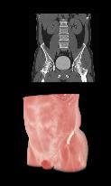


18 Organ labelling: the ground-truth database Different image cropping, noise, contrast/no-contrast, resolution, scanners, body shapes/sizes, patient position


")
 (mean over displaced 3D boxes) Error in model fit (weighted")
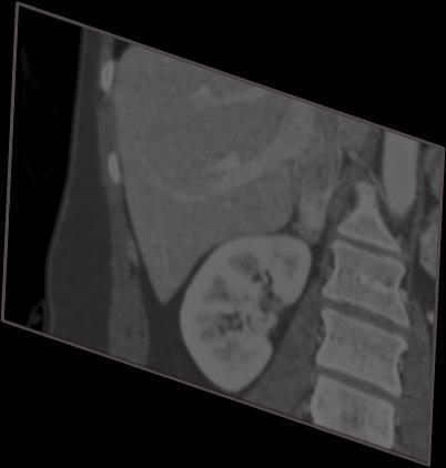
19 Organ labelling: regression forest Each voxel in the volume votes for the position of the 6 box sides We wish to learn a set of discriminative points (landmarks, clusters) which can predict the kidney position with high confidence. Input data point Output Multiple organs Node split function Node optimization Node training Feature response (voxel position in volume) (bound. box continuous pos.) (mean over displaced 3D boxes) Error in model fit (weighted uncertainty for all organs) (relative displacement) (Gaussian repres. of distribs) Regressing an n-d piece-wise constant model


20 Organ labelling: context-rich visual features Possible visual features Computing the feature response Capturing spatial context


21 Organ labelling: automatic landmark discovery Discovery of landmark regions Here the system is trained to detect left and right kidneys. The system learns to use bottom of lung and top of pelvis to localize kidneys with highest confidence. Input CT scan and detected landmark regions
22 Overview Introduction to machine learning Decision forests Applications in medical image analysis Anatomy localization in CT Scans Spine Detection in CT Scans Brain Tumour Segmentation in MR Scans

23 Vertebrae Detection and Classification


24 Where? Which? Name of this vertebra?


25 Challenges


26 Challenges Repetitive nature of structures Variability of normal anatomy Presence of pathologies Varying image acquisition (FOV, noise level, resolution, )

27 Clinical motivation Patient-specific coordinate system Guided visualization/navigation in diagnostic tools Impact on Clinical Routine! Longitudinal assessment after surgical Intervention Shape/population analysis for disease modelling Impact on Clinical Research!

28 Some results


29 Some results

30 Some results
31 Overview Introduction to machine learning Decision forests Applications in medical image analysis Anatomy localization in CT Scans Spine Detection in CT Scans Brain Tumour Segmentation in MR Scans


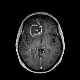
32 Automatic Segmentation of Brain Tumour Segmentation of tumorous tissues: T1-gad T1 T2 DTI-p 3D MRI input data FLAIR DTI-q ---- Active cells ---- Necrotic core ---- Edema ---- Background


33 Training a Pixel-Wise Forest Classifier Tumour Tissue Classification
34 Testing the Pixel-Wise Forest Classifier New Patient, previously unseen Tumour Tissue Classification

35 Building the Training Database of Patients Images 1 st Step: Obtain Expert Segmentation
36 Building the Training Database of Patients Images 1 st Step: Obtain Expert Segmentation
37 Building the Training Database of Patients Images 1 st Step: Obtain Expert Segmentation

38 Glioblastoma Segmentation


39 Glioblastoma Segmentation
40 Machine learning can have a huge impact on medicine!
MEDICAL IMAGE COMPUTING (CAP 5937) LECTURE 20: Machine Learning in Medical Imaging II (deep learning and decision forests)
 LECTURE 20: Machine Learning in Medical Imaging II (deep learning and decision forests)") SPRING 2016 1 MEDICAL IMAGE COMPUTING (CAP 5937) LECTURE 20: Machine Learning in Medical Imaging II (deep learning and decision forests) Dr. Ulas Bagci HEC 221, Center for Research in Computer Vision (CRCV),
SPRING 2016 1 MEDICAL IMAGE COMPUTING (CAP 5937) LECTURE 20: Machine Learning in Medical Imaging II (deep learning and decision forests) Dr. Ulas Bagci HEC 221, Center for Research in Computer Vision (CRCV),
Supervised Learning for Image Segmentation
 Supervised Learning for Image Segmentation Raphael Meier 06.10.2016 Raphael Meier MIA 2016 06.10.2016 1 / 52 References A. Ng, Machine Learning lecture, Stanford University. A. Criminisi, J. Shotton, E.
Context-sensitive Classification Forests for Segmentation of Brain Tumor Tissues
 Context-sensitive Classification Forests for Segmentation of Brain Tumor Tissues D. Zikic, B. Glocker, E. Konukoglu, J. Shotton, A. Criminisi, D. H. Ye, C. Demiralp 3, O. M. Thomas 4,5, T. Das 4, R. Jena
Hierarchical Multi structure Segmentation Guided by Anatomical Correlations
 Hierarchical Multi structure Segmentation Guided by Anatomical Correlations Oscar Alfonso Jiménez del Toro oscar.jimenez@hevs.ch Henning Müller henningmueller@hevs.ch University of Applied Sciences Western
NIH Public Access Author Manuscript Proc IEEE Int Symp Biomed Imaging. Author manuscript; available in PMC 2014 November 15.
 NIH Public Access Author Manuscript Published in final edited form as: Proc IEEE Int Symp Biomed Imaging. 2013 April ; 2013: 748 751. doi:10.1109/isbi.2013.6556583. BRAIN TUMOR SEGMENTATION WITH SYMMETRIC
Regression Forests for Efficient Anatomy Detection and Localization in Computed Tomography Scans
 Regression Forests for Efficient Anatomy Detection and Localization in Computed Tomography Scans A. Criminisi, D. Robertson, E. Konukoglu, J. Shotton, S. Pathak, S. White, and K. Siddiqui Microsoft Research
Object Identification in Ultrasound Scans
 Object Identification in Ultrasound Scans Wits University Dec 05, 2012 Roadmap Introduction to the problem Motivation Related Work Our approach Expected Results Introduction Nowadays, imaging devices like
Ulas Bagci
 CAP5415-Computer Vision Lecture 14-Decision Forests for Computer Vision Ulas Bagci bagci@ucf.edu 1 Readings Slide Credits: Criminisi and Shotton Z. Tu R.Cipolla 2 Common Terminologies Randomized Decision
MR IMAGE SEGMENTATION
 MR IMAGE SEGMENTATION Prepared by : Monil Shah What is Segmentation? Partitioning a region or regions of interest in images such that each region corresponds to one or more anatomic structures Classification
Using Probability Maps for Multi organ Automatic Segmentation
 Using Probability Maps for Multi organ Automatic Segmentation Ranveer Joyseeree 1,2, Óscar Jiménez del Toro1, and Henning Müller 1,3 1 University of Applied Sciences Western Switzerland (HES SO), Sierre,
Joint CI-JAI advanced accelerator lecture series Imaging and detectors for medical physics Lecture 1: Medical imaging
 Joint CI-JAI advanced accelerator lecture series Imaging and detectors for medical physics Lecture 1: Medical imaging Dr Barbara Camanzi barbara.camanzi@stfc.ac.uk Course layout Day AM 09.30 11.00 PM 15.30
Knowledge-Based Organ Identification from CT Images. Masahara Kobashi and Linda Shapiro Best-Paper Prize in Pattern Recognition Vol. 28, No.
 Knowledge-Based Organ Identification from CT Images Masahara Kobashi and Linda Shapiro Best-Paper Prize in Pattern Recognition Vol. 28, No. 4 1995 1 Motivation The extraction of structure from CT volumes
Medical Image Segmentation
 Medical Image Segmentation Xin Yang, HUST *Collaborated with UCLA Medical School and UCSB Segmentation to Contouring ROI Aorta & Kidney 3D Brain MR Image 3D Abdominal CT Image Liver & Spleen Caudate Nucleus
VALIDATION OF DIR. Raj Varadhan, PhD, DABMP Minneapolis Radiation Oncology
 VALIDATION OF DIR Raj Varadhan, PhD, DABMP Minneapolis Radiation Oncology Overview Basics: Registration Framework, Theory Discuss Validation techniques Using Synthetic CT data & Phantoms What metrics to
Classification of Subject Motion for Improved Reconstruction of Dynamic Magnetic Resonance Imaging
 1 CS 9 Final Project Classification of Subject Motion for Improved Reconstruction of Dynamic Magnetic Resonance Imaging Feiyu Chen Department of Electrical Engineering ABSTRACT Subject motion is a significant
Norbert Schuff VA Medical Center and UCSF
 Norbert Schuff Medical Center and UCSF Norbert.schuff@ucsf.edu Medical Imaging Informatics N.Schuff Course # 170.03 Slide 1/67 Objective Learn the principle segmentation techniques Understand the role
Available Online through
 Available Online through www.ijptonline.com ISSN: 0975-766X CODEN: IJPTFI Research Article ANALYSIS OF CT LIVER IMAGES FOR TUMOUR DIAGNOSIS BASED ON CLUSTERING TECHNIQUE AND TEXTURE FEATURES M.Krithika
Applied Statistics for Neuroscientists Part IIa: Machine Learning
 Applied Statistics for Neuroscientists Part IIa: Machine Learning Dr. Seyed-Ahmad Ahmadi 04.04.2017 16.11.2017 Outline Machine Learning Difference between statistics and machine learning Modeling the problem
Deformable Segmentation using Sparse Shape Representation. Shaoting Zhang
 Deformable Segmentation using Sparse Shape Representation Shaoting Zhang Introduction Outline Our methods Segmentation framework Sparse shape representation Applications 2D lung localization in X-ray 3D
Segmenting Glioma in Multi-Modal Images using a Generative-Discriminative Model for Brain Lesion Segmentation
 Segmenting Glioma in Multi-Modal Images using a Generative-Discriminative Model for Brain Lesion Segmentation Bjoern H. Menze 1,2, Ezequiel Geremia 2, Nicholas Ayache 2, and Gabor Szekely 1 1 Computer
Methodological progress in image registration for ventilation estimation, segmentation propagation and multi-modal fusion
 Methodological progress in image registration for ventilation estimation, segmentation propagation and multi-modal fusion Mattias P. Heinrich Julia A. Schnabel, Mark Jenkinson, Sir Michael Brady 2 Clinical
Applying Supervised Learning
 Applying Supervised Learning When to Consider Supervised Learning A supervised learning algorithm takes a known set of input data (the training set) and known responses to the data (output), and trains
Supervoxel Classification Forests for Estimating Pairwise Image Correspondences
 Supervoxel Classification Forests for Estimating Pairwise Image Correspondences Fahdi Kanavati 1, Tong Tong 1, Kazunari Misawa 2, Michitaka Fujiwara 3, Kensaku Mori 4, Daniel Rueckert 1, and Ben Glocker
Semantic Context Forests for Learning- Based Knee Cartilage Segmentation in 3D MR Images
 Semantic Context Forests for Learning- Based Knee Cartilage Segmentation in 3D MR Images MICCAI 2013: Workshop on Medical Computer Vision Authors: Quan Wang, Dijia Wu, Le Lu, Meizhu Liu, Kim L. Boyer,
Medical Image Registration
 Medical Image Registration Submitted by NAREN BALRAJ SINGH SB ID# 105299299 Introduction Medical images are increasingly being used within healthcare for diagnosis, planning treatment, guiding treatment
Joint Classification-Regression Forests for Spatially Structured Multi-Object Segmentation
 Joint Classification-Regression Forests for Spatially Structured Multi-Object Segmentation Ben Glocker 1, Olivier Pauly 2,3, Ender Konukoglu 1, Antonio Criminisi 1 1 Microsoft Research, Cambridge, UK 2
Automatic Vertebrae Localization in Spine CT using Decision Forests
 Automatic Vertebrae Localization in Spine CT using Decision Forests 1, Angel Alberich-Bayarri 1,2, Belén Fos-Guarinos 1, Fabio García-Castro 1, Luis Martí-Bonmatí 1,3 1 QUIBIM S.L., Valencia, Spain 2 La
Prostate Detection Using Principal Component Analysis
 Prostate Detection Using Principal Component Analysis Aamir Virani (avirani@stanford.edu) CS 229 Machine Learning Stanford University 16 December 2005 Introduction During the past two decades, computed
Robust Linear Registration of CT images using Random Regression Forests
 Robust Linear Registration of CT images using Random Regression Forests Ender Konukoglu a Antonio Criminisi a Sayan Pathak b Duncan Robertson a Steve White b David Haynor c Khan Siddiqui b a Microsoft
8/3/2017. Contour Assessment for Quality Assurance and Data Mining. Objective. Outline. Tom Purdie, PhD, MCCPM
 Contour Assessment for Quality Assurance and Data Mining Tom Purdie, PhD, MCCPM Objective Understand the state-of-the-art in contour assessment for quality assurance including data mining-based techniques
Advanced Visual Medicine: Techniques for Visual Exploration & Analysis
 Advanced Visual Medicine: Techniques for Visual Exploration & Analysis Interactive Visualization of Multimodal Volume Data for Neurosurgical Planning Felix Ritter, MeVis Research Bremen Multimodal Neurosurgical
Introduction to Neuroimaging Janaina Mourao-Miranda
 Introduction to Neuroimaging Janaina Mourao-Miranda Neuroimaging techniques have changed the way neuroscientists address questions about functional anatomy, especially in relation to behavior and clinical
Automatic Vertebrae Localization in Pathological Spine CT using Decision Forests
 Automatic Vertebrae Localization in Pathological Spine CT using Decision Forests Ana Jiménez-Pastor 1, Esther Tomás-González 1, Ángel Alberich-Bayarri 1,2, Fabio García-Castro 1, David García-Juan 1, Luis
REAL-TIME ADAPTIVITY IN HEAD-AND-NECK AND LUNG CANCER RADIOTHERAPY IN A GPU ENVIRONMENT
 REAL-TIME ADAPTIVITY IN HEAD-AND-NECK AND LUNG CANCER RADIOTHERAPY IN A GPU ENVIRONMENT Anand P Santhanam Assistant Professor, Department of Radiation Oncology OUTLINE Adaptive radiotherapy for head and
Whole Body MRI Intensity Standardization
 Whole Body MRI Intensity Standardization Florian Jäger 1, László Nyúl 1, Bernd Frericks 2, Frank Wacker 2 and Joachim Hornegger 1 1 Institute of Pattern Recognition, University of Erlangen, {jaeger,nyul,hornegger}@informatik.uni-erlangen.de
COSC160: Detection and Classification. Jeremy Bolton, PhD Assistant Teaching Professor
 COSC160: Detection and Classification Jeremy Bolton, PhD Assistant Teaching Professor Outline I. Problem I. Strategies II. Features for training III. Using spatial information? IV. Reducing dimensionality
Prototype of Silver Corpus Merging Framework
 www.visceral.eu Prototype of Silver Corpus Merging Framework Deliverable number D3.3 Dissemination level Public Delivery data 30.4.2014 Status Authors Final Markus Krenn, Allan Hanbury, Georg Langs This
Accurate Intervertebral Disc Localisation and Segmentation in MRI using Vantage Point Hough Forests and Multi-Atlas Fusion
 Accurate Intervertebral Disc Localisation and Segmentation in MRI using Vantage Point Hough Forests and Multi-Atlas Fusion Mattias P. Heinrich 1 and Ozan Oktay 2 1 Institute of Medical Informatics, University
Advanced Video Content Analysis and Video Compression (5LSH0), Module 8B
, Module 8B") Advanced Video Content Analysis and Video Compression (5LSH0), Module 8B 1 Supervised learning Catogarized / labeled data Objects in a picture: chair, desk, person, 2 Classification Fons van der Sommen
Advanced Video Content Analysis and Video Compression (5LSH0), Module 8B 1 Supervised learning Catogarized / labeled data Objects in a picture: chair, desk, person, 2 Classification Fons van der Sommen
arxiv: v1 [cs.cv] 17 May 2017
![arxiv: v1 [cs.cv] 17 May 2017](/thumbs/80/81663121.jpg "arxiv: v1 [cs.cv] 17 May 2017") Automatic Vertebra Labeling in Large-Scale 3D CT using Deep Image-to-Image Network with Message Passing and Sparsity Regularization arxiv:1705.05998v1 [cs.cv] 17 May 2017 Dong Yang 1, Tao Xiong 2, Daguang
Automatic Vertebra Labeling in Large-Scale 3D CT using Deep Image-to-Image Network with Message Passing and Sparsity Regularization arxiv:1705.05998v1 [cs.cv] 17 May 2017 Dong Yang 1, Tao Xiong 2, Daguang
CS 559: Machine Learning Fundamentals and Applications 10 th Set of Notes
 1 CS 559: Machine Learning Fundamentals and Applications 10 th Set of Notes Instructor: Philippos Mordohai Webpage: www.cs.stevens.edu/~mordohai E-mail: Philippos.Mordohai@stevens.edu Office: Lieb 215
RADIOMICS: potential role in the clinics and challenges
 27 giugno 2018 Dipartimento di Fisica Università degli Studi di Milano RADIOMICS: potential role in the clinics and challenges Dr. Francesca Botta Medical Physicist Istituto Europeo di Oncologia (Milano)
7. Boosting and Bagging Bagging
 Group Prof. Daniel Cremers 7. Boosting and Bagging Bagging Bagging So far: Boosting as an ensemble learning method, i.e.: a combination of (weak) learners A different way to combine classifiers is known
GE Healthcare. AppsLinq* remote courses catalogue
 GE Healthcare AppsLinq* remote courses catalogue AppsLinq * remote training for MR Magnetic Resonance AppsLinq* a GE Training in Partnership (TiP) program, revolutionizes applications training with live,
A New GPU-Based Level Set Method for Medical Image Segmentation
 A New GPU-Based Level Set Method for Medical Image Segmentation Wenzhe Xue Research Assistant Radiology Department Mayo Clinic, Scottsdale, AZ Ph.D. Student Biomedical Informatics Arizona State University,
Fully Automatic Multi-organ Segmentation based on Multi-boost Learning and Statistical Shape Model Search
 Fully Automatic Multi-organ Segmentation based on Multi-boost Learning and Statistical Shape Model Search Baochun He, Cheng Huang, Fucang Jia Shenzhen Institutes of Advanced Technology, Chinese Academy
Normalization for clinical data
 Normalization for clinical data Christopher Rorden, Leonardo Bonilha, Julius Fridriksson, Benjamin Bender, Hans-Otto Karnath (2012) Agespecific CT and MRI templates for spatial normalization. NeuroImage
Tomographic Reconstruction
 Tomographic Reconstruction 3D Image Processing Torsten Möller Reading Gonzales + Woods, Chapter 5.11 2 Overview Physics History Reconstruction basic idea Radon transform Fourier-Slice theorem (Parallel-beam)
Skull Segmentation of MR images based on texture features for attenuation correction in PET/MR
 Skull Segmentation of MR images based on texture features for attenuation correction in PET/MR CHAIBI HASSEN, NOURINE RACHID ITIO Laboratory, Oran University Algeriachaibih@yahoo.fr, nourine@yahoo.com
arxiv: v1 [cs.cv] 6 Jun 2017
![arxiv: v1 [cs.cv] 6 Jun 2017](/thumbs/96/128307097.jpg "arxiv: v1 [cs.cv] 6 Jun 2017") Volume Calculation of CT lung Lesions based on Halton Low-discrepancy Sequences Liansheng Wang a, Shusheng Li a, and Shuo Li b a Department of Computer Science, Xiamen University, Xiamen, China b Dept.
Volume Calculation of CT lung Lesions based on Halton Low-discrepancy Sequences Liansheng Wang a, Shusheng Li a, and Shuo Li b a Department of Computer Science, Xiamen University, Xiamen, China b Dept.
ISSN: X Impact factor: 4.295
 ISSN: 2454-132X Impact factor: 4.295 (Volume3, Issue1) Available online at: www.ijariit.com Performance Analysis of Image Clustering Algorithm Applied to Brain MRI Kalyani R.Mandlik 1, Dr. Suresh S. Salankar
Sampling-Based Ensemble Segmentation against Inter-operator Variability
 Sampling-Based Ensemble Segmentation against Inter-operator Variability Jing Huo 1, Kazunori Okada, Whitney Pope 1, Matthew Brown 1 1 Center for Computer vision and Imaging Biomarkers, Department of Radiological
Computational Medical Imaging Analysis Chapter 4: Image Visualization
 Computational Medical Imaging Analysis Chapter 4: Image Visualization Jun Zhang Laboratory for Computational Medical Imaging & Data Analysis Department of Computer Science University of Kentucky Lexington,
Joint Tumor Segmentation and Dense Deformable Registration of Brain MR Images
 Joint Tumor Segmentation and Dense Deformable Registration of Brain MR Images Sarah Parisot 1,2,3, Hugues Duffau 4, Stéphane Chemouny 3, Nikos Paragios 1,2 1. Center for Visual Computing, Ecole Centrale
CHAPTER 3 TUMOR DETECTION BASED ON NEURO-FUZZY TECHNIQUE
 32 CHAPTER 3 TUMOR DETECTION BASED ON NEURO-FUZZY TECHNIQUE 3.1 INTRODUCTION In this chapter we present the real time implementation of an artificial neural network based on fuzzy segmentation process
Computer Vision Group Prof. Daniel Cremers. 8. Boosting and Bagging
 Prof. Daniel Cremers 8. Boosting and Bagging Repetition: Regression We start with a set of basis functions (x) =( 0 (x), 1(x),..., M 1(x)) x 2 í d The goal is to fit a model into the data y(x, w) =w T
Automatic Detection and Segmentation of Kidneys in Magnetic Resonance Images Using Image Processing Techniques
 Biomedical Statistics and Informatics 2017; 2(1): 22-26 http://www.sciencepublishinggroup.com/j/bsi doi: 10.11648/j.bsi.20170201.15 Automatic Detection and Segmentation of Kidneys in Magnetic Resonance
Bioimage Informatics
 Bioimage Informatics Lecture 13, Spring 2012 Bioimage Data Analysis (IV) Image Segmentation (part 2) Lecture 13 February 29, 2012 1 Outline Review: Steger s line/curve detection algorithm Intensity thresholding
Atlas-Based Segmentation of Abdominal Organs in 3D Ultrasound, and its Application in Automated Kidney Segmentation
 University of Toronto Atlas-Based Segmentation of Abdominal Organs in 3D Ultrasound, and its Application in Automated Kidney Segmentation Authors: M. Marsousi, K. N. Plataniotis, S. Stergiopoulos Presenter:
Big Data Methods. Chapter 5: Machine learning. Big Data Methods, Chapter 5, Slide 1
 Big Data Methods Chapter 5: Machine learning Big Data Methods, Chapter 5, Slide 1 5.1 Introduction to machine learning What is machine learning? Concerned with the study and development of algorithms that
Automatic Quantification of DTI Parameters along Fiber Bundles
 Automatic Quantification of DTI Parameters along Fiber Bundles Jan Klein 1, Simon Hermann 1, Olaf Konrad 1, Horst K. Hahn 1, and Heinz-Otto Peitgen 1 1 MeVis Research, 28359 Bremen Email: klein@mevis.de
Segmenting Lesions in Multiple Sclerosis Patients James Chen, Jason Su
 Segmenting Lesions in Multiple Sclerosis Patients James Chen, Jason Su Radiologists and researchers spend countless hours tediously segmenting white matter lesions to diagnose and study brain diseases.
Medical Image Synthesis Methods and Applications
 MR Intensity Scale is Arbitrary This causes problems in most postprocessing methods Inconsistency or algorithm failure 11/5/2015 2 Joint Histogram 1.5 T GE SPGR 3 T Philips MPRAGE 11/5/2015 3 Problem With
Multi-Sectional Views Textural Based SVM for MS Lesion Segmentation in Multi-Channels MRIs
 56 The Open Biomedical Engineering Journal, 2012, 6, 56-72 Open Access Multi-Sectional Views Textural Based SVM for MS Lesion Segmentation in Multi-Channels MRIs Bassem A. Abdullah*, Akmal A. Younis and
Automated segmentation methods for liver analysis in oncology applications
 University of Szeged Department of Image Processing and Computer Graphics Automated segmentation methods for liver analysis in oncology applications Ph. D. Thesis László Ruskó Thesis Advisor Dr. Antal
Entangled Decision Forests and their Application for Semantic Segmentation of CT Images
 Entangled Decision Forests and their Application for Semantic Segmentation of CT Images Albert Montillo,2, Jamie Shotton 2, John Winn 2, Juan Eugenio Iglesias 2,3, Dimitri Metaxas 4, and Antonio Criminisi
MEDICAL IMAGE COMPUTING (CAP 5937) LECTURE 10: Medical Image Segmentation as an Energy Minimization Problem
 LECTURE 10: Medical Image Segmentation as an Energy Minimization Problem") SPRING 07 MEDICAL IMAGE COMPUTING (CAP 97) LECTURE 0: Medical Image Segmentation as an Energy Minimization Problem Dr. Ulas Bagci HEC, Center for Research in Computer Vision (CRCV), University of Central
SPRING 07 MEDICAL IMAGE COMPUTING (CAP 97) LECTURE 0: Medical Image Segmentation as an Energy Minimization Problem Dr. Ulas Bagci HEC, Center for Research in Computer Vision (CRCV), University of Central
Basic principles of MR image analysis. Basic principles of MR image analysis. Basic principles of MR image analysis
 Basic principles of MR image analysis Basic principles of MR image analysis Julien Milles Leiden University Medical Center Terminology of fmri Brain extraction Registration Linear registration Non-linear
Assessing Accuracy Factors in Deformable 2D/3D Medical Image Registration Using a Statistical Pelvis Model
 Assessing Accuracy Factors in Deformable 2D/3D Medical Image Registration Using a Statistical Pelvis Model Jianhua Yao National Institute of Health Bethesda, MD USA jyao@cc.nih.gov Russell Taylor The Johns
Image Segmentation and Registration
 Image Segmentation and Registration Dr. Christine Tanner (tanner@vision.ee.ethz.ch) Computer Vision Laboratory, ETH Zürich Dr. Verena Kaynig, Machine Learning Laboratory, ETH Zürich Outline Segmentation
IEEE TRANSACTIONS ON IMAGE PROCESSING, VOL. 26, NO. 10, OCTOBER
 IEEE TRANSACTIONS ON IMAGE PROCESSING, VOL. 26, NO. 10, OCTOBER 2017 4753 Detecting Anatomical Landmarks From Limited Medical Imaging Data Using Two-Stage Task-Oriented Deep Neural Networks Jun Zhang,
On non-linear characterization of tissue abnormality by constructing disease manifolds
 On non-linear characterization of tissue abnormality by constructing disease manifolds Nematollah Batmanghelich Ragini Verma Section of Biomedical Image Analysis, Department of Radiology, University of
Tumor Detection and classification of Medical MRI UsingAdvance ROIPropANN Algorithm
 International Journal of Engineering Research and Advanced Technology (IJERAT) DOI:http://dx.doi.org/10.31695/IJERAT.2018.3273 E-ISSN : 2454-6135 Volume.4, Issue 6 June -2018 Tumor Detection and classification
White Matter Lesion Segmentation (WMLS) Manual
 Manual") White Matter Lesion Segmentation (WMLS) Manual 1. Introduction White matter lesions (WMLs) are brain abnormalities that appear in different brain diseases, such as multiple sclerosis (MS), head injury,
White Matter Lesion Segmentation (WMLS) Manual 1. Introduction White matter lesions (WMLs) are brain abnormalities that appear in different brain diseases, such as multiple sclerosis (MS), head injury,
Random Forest A. Fornaser
 Random Forest A. Fornaser alberto.fornaser@unitn.it Sources Lecture 15: decision trees, information theory and random forests, Dr. Richard E. Turner Trees and Random Forests, Adele Cutler, Utah State University
3 Object Detection. BVM 2018 Tutorial: Advanced Deep Learning Methods. Paul F. Jaeger, Division of Medical Image Computing
 3 Object Detection BVM 2018 Tutorial: Advanced Deep Learning Methods Paul F. Jaeger, of Medical Image Computing What is object detection? classification segmentation obj. detection (1 label per pixel)
Seeing the Big Picture
 Seeing the Big Picture Segmenting Images to Create Data 15.071x The Analytics Edge Image Segmentation Divide up digital images to salient regions/clusters corresponding to individual surfaces, objects,
A Workflow for Improving Medical Visualization of Semantically Annotated CT-Images
 A Workflow for Improving Medical Visualization of Semantically Annotated CT-Images Alexander Baranya 1,2, Luis Landaeta 1,2, Alexandra La Cruz 1, and Maria-Esther Vidal 2 1 Biophysic and Bioengeneering
Segmenting Glioma in Multi-Modal Images using a Generative Model for Brain Lesion Segmentation
 Segmenting Glioma in Multi-Modal Images using a Generative Model for Brain Lesion Segmentation Bjoern H. Menze 1,2, Koen Van Leemput 3, Danial Lashkari 4 Marc-André Weber 5, Nicholas Ayache 2, and Polina
Is deformable image registration a solved problem?
 Is deformable image registration a solved problem? Marcel van Herk On behalf of the imaging group of the RT department of NKI/AVL Amsterdam, the Netherlands DIR 1 Image registration Find translation.deformation
Stroke Quantification Tool (Sonia) Ver User Manual
 Ver User Manual") Stroke Quantification Tool (Sonia) Ver. 1.0 User Manual English. 12/2016 Rev. 1.0 www.wakeup-stroke.eu 1 Table of Contents 1. Introduction...3 2. Installation...4 3. Data Import...5 4. Registration...7
Stroke Quantification Tool (Sonia) Ver. 1.0 User Manual English. 12/2016 Rev. 1.0 www.wakeup-stroke.eu 1 Table of Contents 1. Introduction...3 2. Installation...4 3. Data Import...5 4. Registration...7
Learning-based Neuroimage Registration
 Learning-based Neuroimage Registration Leonid Teverovskiy and Yanxi Liu 1 October 2004 CMU-CALD-04-108, CMU-RI-TR-04-59 School of Computer Science Carnegie Mellon University Pittsburgh, PA 15213 Abstract
Computational Medical Imaging Analysis Chapter 5: Processing and Analysis
 Computational Medical Imaging Analysis Chapter 5: Processing and Analysis Jun Zhang Laboratory for Computational Medical Imaging & Data Analysis Department of Computer Science University of Kentucky Lexington,
A Generation Methodology for Numerical Phantoms with Statistically Relevant Variability of Geometric and Physical Properties
 A Generation Methodology for Numerical Phantoms with Statistically Relevant Variability of Geometric and Physical Properties Steven Dolly 1, Eric Ehler 1, Yang Lou 2, Mark Anastasio 2, Hua Li 2 (1) University
Learn Image Segmentation Basics with Hands-on Introduction to ITK-SNAP. RSNA 2016 Courses RCB22 and RCB54
 Learn Image Segmentation Basics with Hands-on Introduction to ITK-SNAP RSNA 2016 Courses RCB22 and RCB54 RCB22 Mon, Nov 28 10:30-12:00 PM, Room S401CD RCB54 Thu, Dec 1 2:30-4:30 PM, Room S401CD Presenters:
A Study of Medical Image Analysis System
 Indian Journal of Science and Technology, Vol 8(25), DOI: 10.17485/ijst/2015/v8i25/80492, October 2015 ISSN (Print) : 0974-6846 ISSN (Online) : 0974-5645 A Study of Medical Image Analysis System Kim Tae-Eun
Deviceless respiratory motion correction in PET imaging exploring the potential of novel data driven strategies
 g Deviceless respiratory motion correction in PET imaging exploring the potential of novel data driven strategies Presented by Adam Kesner, Ph.D., DABR Assistant Professor, Division of Radiological Sciences,
CT Basics Principles of Spiral CT Dose. Always Thinking Ahead.
 1 CT Basics Principles of Spiral CT Dose 2 Who invented CT? 1963 - Alan Cormack developed a mathematical method of reconstructing images from x-ray projections Sir Godfrey Hounsfield worked for the Central
Trade-offs in Explanatory
 1 Trade-offs in Explanatory 21 st of February 2012 Model Learning Data Analysis Project Madalina Fiterau DAP Committee Artur Dubrawski Jeff Schneider Geoff Gordon 2 Outline Motivation: need for interpretable
Automatic Detection of Multiple Organs Using Convolutional Neural Networks
 Automatic Detection of Multiple Organs Using Convolutional Neural Networks Elizabeth Cole University of Massachusetts Amherst Amherst, MA ekcole@umass.edu Sarfaraz Hussein University of Central Florida
Fluorescence Tomography Source Reconstruction and Analysis
 TECHNICAL NOTE Pre-clinical in vivo imaging Fluorescence Tomography Source Reconstruction and Analysis Note: This Technical Note is part of a series for Fluorescence Imaging Tomography (FLIT). The user
Use of MRI in Radiotherapy: Technical Consideration
 Use of MRI in Radiotherapy: Technical Consideration Yanle Hu, PhD Department of Radiation Oncology, Mayo Clinic Arizona 04/07/2018 2015 MFMER slide-1 Conflict of Interest: None 2015 MFMER slide-2 Objectives
Ischemic Stroke Lesion Segmentation Proceedings 5th October 2015 Munich, Germany
 0111010001110001101000100101010111100111011100100011011101110101101012 Ischemic Stroke Lesion Segmentation www.isles-challenge.org Proceedings 5th October 2015 Munich, Germany Preface Stroke is the second
An Introduction To Automatic Tissue Classification Of Brain MRI. Colm Elliott Mar 2014
 An Introduction To Automatic Tissue Classification Of Brain MRI Colm Elliott Mar 2014 Tissue Classification Tissue classification is part of many processing pipelines. We often want to classify each voxel
Chapter 3 Set Redundancy in Magnetic Resonance Brain Images
 16 Chapter 3 Set Redundancy in Magnetic Resonance Brain Images 3.1 MRI (magnetic resonance imaging) MRI is a technique of measuring physical structure within the human anatomy. Our proposed research focuses
UNIVERSITY OF SOUTHAMPTON
 UNIVERSITY OF SOUTHAMPTON PHYS2007W1 SEMESTER 2 EXAMINATION 2014-2015 MEDICAL PHYSICS Duration: 120 MINS (2 hours) This paper contains 10 questions. Answer all questions in Section A and only two questions
AN essential part of any computer-aided surgery is planning
 1 A Model Based Validation Scheme for Organ Segmentation in CT Scan Volumes Hossein Badakhshannoory, Student Member, IEEE, and Parvaneh Saeedi, Member, IEEE Abstract In this work, we propose a novel approach
Visual Perception for Robots
 Visual Perception for Robots Sven Behnke Computer Science Institute VI Autonomous Intelligent Systems Our Cognitive Robots Complete systems for example scenarios Equipped with rich sensors Flying robot
Automated Brain Tumor Segmentation Using Hsom
 IOSR Journal of Electronics and Communication Engineering (IOSR-JECE) e-issn: 2278-2834, p- ISSN: 2278-8735. Volume 5, Issue 2 (Mar. - Apr. 2013), PP 28-33 Automated Brain Tumor Segmentation Using Hsom
Manifold Learning-based Data Sampling for Model Training
 Manifold Learning-based Data Sampling for Model Training Shuqing Chen 1, Sabrina Dorn 2, Michael Lell 3, Marc Kachelrieß 2,Andreas Maier 1 1 Pattern Recognition Lab, FAU Erlangen-Nürnberg 2 German Cancer