Joint Tumor Segmentation and Dense Deformable Registration of Brain MR Images
|
|
|
- Aron Tucker
- 5 years ago
- Views:
Transcription
1 Joint Tumor Segmentation and Dense Deformable Registration of Brain MR Images Sarah Parisot 1,2,3, Hugues Duffau 4, Stéphane Chemouny 3, Nikos Paragios 1,2 1. Center for Visual Computing, Ecole Centrale Paris 2. Equipe GALEN, INRIA Saclay-Ile de France 3. Intrasense SAS, Montpellier 4. Département de Neurochirurgie, Hôpital Gui de Chauliac, Montpellier


2 Introduction Brain Tumor Segmentation and Registration from healthy to pathological subject treated separately Fuzzy boundaries inhomogeneous appearances Various shapes intensity overlap with healthy tissue Methods Classification techniques + pairwise smoothing Lee et al. MICCAI 2008 Atlas based segmentation: dependent on registration quality Prastawa et al. Academic Radiology 2003 No correspondences in the tumor area: use of common methods impossible Methods Growth models: computationally expensive/user interaction Cuadra et al. Comput. meth. prog. bio Zacharaki et al. Neuroimage 2009 Masking the pathology: dependent on segmentation quality Stefanescu et al. MICCAI 2004
3 Introduction Brain Tumor Segmentation and Registration from healthy to pathological subject treated separately Fuzzy boundaries inhomogeneous appearances Various shapes intensity overlap with healthy tissue Methods Classification techniques + pairwise smoothing Lee et al. MICCAI 2008 Atlas based segmentation: dependent on registration quality Prastawa et al. Academic Radiology 2003 No correspondences in the tumor area: use of common methods impossible Methods Growth models: computationally expensive/user interaction Cuadra et al. Comput. meth. prog. bio Zacharaki et al. Neuroimage 2009 Masking the pathology: dependent on segmentation quality Stefanescu et al. MICCAI 2004
4 Method Overview Simultaneously register a healthy subject to a diseased subject and find the tumor s segmentation map Tumor segmentation Healthy to diseased registration 3
5 Method Overview Simultaneously register a healthy subject to a diseased subject and find the tumor s segmentation map Tumor segmentation Healthy to diseased registration Find correspondences Similarity criterion between the images glocker et al. Medical Image Analysis,
6 Method Overview Simultaneously register a healthy subject to a diseased subject and find the tumor s segmentation map Tumor segmentation Healthy to diseased registration High score Learned classifier Friedman et al. The Annals of Statistics 2000 Find correspondences Similarity criterion between the images glocker et al. Medical Image Analysis,
7 Method Overview Simultaneously register a healthy subject to a diseased subject and find the tumor s segmentation map Tumor segmentation Healthy to diseased registration High score Learned classifier Friedman et al. The Annals of Statistics 2000 Detect tumor: Areas without correspondences (low similarity Find correspondences Similarity criterion between the images glocker et al. Medical Image Analysis,
8 Method Overview Simultaneously register a healthy subject to a diseased subject and find the tumor s segmentation map Tumor segmentation Deformation in the tumor: interpolation Healthy to diseased registration High score Learned classifier Friedman et al. The Annals of Statistics 2000 Detect tumor: Areas without correspondences (low similarity Find correspondences Similarity criterion between the images glocker et al. Medical Image Analysis,
9 Method Overview Simultaneously register a healthy subject to a diseased subject and find the tumor s segmentation map Tumor segmentation Deformation in the tumor: interpolation Healthy to diseased registration High score Learned classifier Friedman et al. The Annals of Statistics 2000 Detect tumor: Areas without correspondences (low similarity Find correspondences Similarity criterion between the images glocker et al. Medical Image Analysis, 2008 Discrete Markov Random Field Formulation 3
10 Parametrization A I 4
11 Parametrization A Deformation field T (x A T (x = I(x I 4
12 Parametrization A Deformation field T (x A T (x = I(x I S(x Segmentation map 4
13 Parametrization A Deformation field T (x A T (x = I(x I S(x Segmentation map Free form deformation approach: Deformation and Segmentation estimated on a grid G superimposed to the images Evaluation on the whole image by interpolation 4

14 Parametrization A Deformation field T (x A T (x = I(x I S(x Segmentation map Free form deformation approach: Deformation and Segmentation estimated on a grid G superimposed to the images Evaluation on the whole image by interpolation 4
15 Markov Random Field Model Assign to each grid node p a label l p corresponding to a pair segmentation (s l / displacement (d l l p = {s l p,d l p } {0,1} {d 1,...,d k } 5
16 Markov Random Field Model Assign to each grid node p a label l p corresponding to a pair segmentation (s l / displacement (d l l p = {s l p,d l p } {0,1} {d 1,...,d k } Discrete displacement set 5
17 Markov Random Field Model Assign to each grid node p a label l p corresponding to a pair segmentation (s l / displacement (d l l p = {s l p,d l p } {0,1} {d 1,...,d k } Discrete displacement set Find the optimal labeling: l opt = argmin l E def,seg (l Evaluate the optimal segmentation and displacement in a one shot optimization 5
18 Markov Random Field Model Assign to each grid node p a label l p corresponding to a pair segmentation (s l / displacement (d l l p = {s l p,d l p } {0,1} {d 1,...,d k } Discrete displacement set Find the optimal labeling: l opt = argmin l E def,seg (l Evaluate the optimal segmentation and displacement in a one shot optimization Markov Random Field Energy E def,seg (l = 1 G V p + pg V pq,l q pg qn ( p 5
19 Markov Random Field Model Assign to each grid node p a label l p corresponding to a pair segmentation (s l / displacement (d l l p = {s l p,d l p } {0,1} {d 1,...,d k } Discrete displacement set Find the optimal labeling: l opt = argmin l E def,seg (l Evaluate the optimal segmentation and displacement in a one shot optimization Markov Random Field Energy E def,seg (l = 1 G V p + pg V pq,l q pg qn ( p Pairwise term with neighbor nodes Local consistency of the segmentation Registration regularization 5

20 Markov Random Field Model Assign to each grid node p a label l p corresponding to a pair segmentation (s l / displacement (d l l p = {s l p,d l p } {0,1} {d 1,...,d k } Discrete displacement set Find the optimal labeling: l opt = argmin l E def,seg (l Evaluate the optimal segmentation and displacement in a one shot optimization Markov Random Field Energy E def,seg (l = 1 G Unary term V p + pg V p = V def + (1 V seg V pq,l q pg qn ( p Pairwise term with neighbor nodes Local consistency of the segmentation Registration regularization 5


21 Unary Term: Segmentation V p = V def + (1 V seg Learning of a tumor vs background classifier Features extracted from images Gentle Adaboost algorithm Construction of a strong classifier as a combination of weak classifiers (decision stumps Any classification technique can be used Nodes with high classification probability of being tumor should be labeled accordingly Boosting probability map 6
22 Unary Term: Registration V p = V def + (1 V seg ( if s l p = 0, Background Sim I(x, A(x + d l p C tm if s l p = 1, Tumor 7
23 Unary Term: Registration V p = V def + (1 V seg Sim I(x, A(x + d l p C tm if s l p = 1, Tumor Outside the tumor area: Find correspondences ( if s l p = 0, Background Compute the similarity measure for all possible displacements A I 7
24 Unary Term: Registration V p = V def + (1 V seg Sim I(x, A(x + d l p C tm if s l p = 1, Tumor Outside the tumor area: Find correspondences ( if s l p = 0, Background Compute the similarity measure for all possible displacements A I 7
25 Unary Term: Registration V p = V def + (1 V seg Sim I(x, A(x + d l p C tm if s l p = 1, Tumor Outside the tumor area: Find correspondences ( if s l p = 0, Background Compute the similarity measure for all possible displacements A I 7
26 Unary Term: Registration V p = V def + (1 V seg Sim I(x, A(x + d l p C tm if s l p = 1, Tumor Outside the tumor area: Find correspondences ( if s l p = 0, Background Compute the similarity measure for all possible displacements A I 7
27 Unary Term: Registration V p = V def + (1 V seg Sim I(x, A(x + d l p C tm if s l p = 1, Tumor Outside the tumor area: Find correspondences ( if s l p = 0, Background Compute the similarity measure for all possible displacements A I 7
28 Unary Term: Registration V p = V def + (1 V seg Sim I(x, A(x + d l p C tm if s l p = 1, Tumor Outside the tumor area: Find correspondences ( if s l p = 0, Background Compute the similarity measure for all possible displacements A Similarity measure Sim(I(x, A(T (x I 7
29 Unary Term: Registration V p = V def + (1 V seg Any kind of similarity criterion Sim I(x, A(x + d l p C tm if s l p = 1, Tumor Outside the tumor area: Find correspondences ( if s l p = 0, Background Compute the similarity measure for all possible displacements A Similarity measure Sim(I(x, A(T (x I 7


30 Unary Term: Registration V p = V def + (1 V seg Sim I(x, A(x + d l p C tm if s l p = 1, Tumor In the tumor area: No correspondences Constant cost independent of the displacement ( if s l p = 0, Background A Constant Cost I 8

31 Implementation Incremental Approach 3 image levels, 4 grid resolutions Increasing influence of the segmentation (progressive diminution of value Optimization Linear programming (Komodakis et al. CVIU, 2008 Overall run time: 6 min (matlab implementation 9
32 Experimental Validation Our method Boosting Database: 97 T2 FLAIR volumes Data likelihood learned on 40 volumes Evaluation on 57 volumes Segmentation Evaluated w.r.t manual segmentations Compared with boosting classification with added pairwise smoothing (right on boxplots Median Dice: 77 to 80%, False positives: 30 to 20%, Mean Absolute Distance (MAD: 4.8 to 4.2mm Registration Qualitative evaluation Dice Score False positive rate Compared with Glocker et al. 2008, with masked pathology MAD 10
33 Experimental Validation Our method Boosting Database: 97 T2 FLAIR volumes Data likelihood learned on 40 volumes Evaluation on 57 volumes Segmentation Evaluated w.r.t manual segmentations Compared with boosting classification with added pairwise smoothing (right on boxplots Median Dice: 77 to 80%, False positives: 30 to 20%, Mean Absolute Distance (MAD: 4.8 to 4.2mm Registration Qualitative evaluation Dice Score False positive rate Compared with Glocker et al. 2008, with masked pathology MAD 10


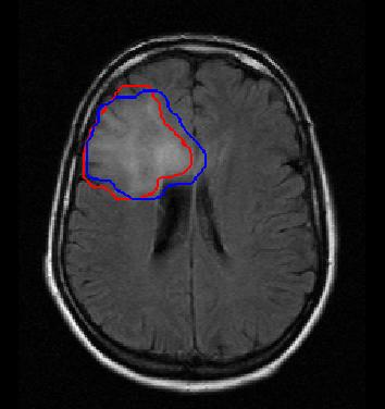

34 Red: Ground truth Blue: Automatic segmentation 11


35 Original image Glocker et al Deformed image Our method Deformed image Left: Glocker 08 Deformation field Right Our method Deformation field 12
36 Conclusion Simultaneous registration and segmentation method Modular w.r.t image modality, similarity criterion and classification technique Can be adapted to any clinical context Fast and efficient optimization (ongoing work to reduce the run time to a few seconds State of the art results Future work Local spatial position prior information Registration uncertainties Adaptation to registration/segmentation before and during surgery with tumor resection 13
37 Poster Th-1-AG-14 Thursday 13:30-15:00 Thank you for your attention Questions? 14
Graph-based Detection, Segmentation & Characterization of Brain Tumors
 Graph-based Detection, Segmentation & Characterization of Brain Tumors Sarah Parisot 1,2,4, Hugues Duffau 5,Stéphane Chemouny 4, Nikos Paragios 1,2,3 1 Center for Visual Computing, Ecole Centrale de Paris,
Biomedical Image Analysis Using Markov Random Fields & Efficient Linear Programing
 Biomedical Image Analysis Using Markov Random Fields & Efficient Linear Programing Nikos Komodakis Ahmed Besbes Ben Glocker Nikos Paragios Abstract Computer-aided diagnosis through biomedical image analysis
Non-rigid Registration using Discrete MRFs: Application to Thoracic CT Images
 Non-rigid Registration using Discrete MRFs: Application to Thoracic CT Images Ben Glocker 1, Nikos Komodakis 2, Nikos Paragios 3,4, and Nassir Navab 1 1 Computer Aided Medical Procedures (CAMP), TU Mï
Inter and Intra-Modal Deformable Registration:
 Inter and Intra-Modal Deformable Registration: Continuous Deformations Meet Efficient Optimal Linear Programming Ben Glocker 1,2, Nikos Komodakis 1,3, Nikos Paragios 1, Georgios Tziritas 3, Nassir Navab
Methodological progress in image registration for ventilation estimation, segmentation propagation and multi-modal fusion
 Methodological progress in image registration for ventilation estimation, segmentation propagation and multi-modal fusion Mattias P. Heinrich Julia A. Schnabel, Mark Jenkinson, Sir Michael Brady 2 Clinical
A Nonparametric Model for Brain Tumor Segmentation and Volumetry in Longitudinal MR Sequences
 A Nonparametric Model for Brain Tumor Segmentation and Volumetry in Longitudinal MR Sequences Esther Alberts 1,2,6, Guillaume Charpiat 3, Yuliya Tarabalka 4, Thomas Huber 1, Marc-André Weber 5, Jan Bauer
Machine Learning for Medical Image Analysis. A. Criminisi
 Machine Learning for Medical Image Analysis A. Criminisi Overview Introduction to machine learning Decision forests Applications in medical image analysis Anatomy localization in CT Scans Spine Detection
MEDICAL IMAGE COMPUTING (CAP 5937) LECTURE 10: Medical Image Segmentation as an Energy Minimization Problem
 LECTURE 10: Medical Image Segmentation as an Energy Minimization Problem") SPRING 07 MEDICAL IMAGE COMPUTING (CAP 97) LECTURE 0: Medical Image Segmentation as an Energy Minimization Problem Dr. Ulas Bagci HEC, Center for Research in Computer Vision (CRCV), University of Central
SPRING 07 MEDICAL IMAGE COMPUTING (CAP 97) LECTURE 0: Medical Image Segmentation as an Energy Minimization Problem Dr. Ulas Bagci HEC, Center for Research in Computer Vision (CRCV), University of Central
Manifold Learning: Applications in Neuroimaging
 Your own logo here Manifold Learning: Applications in Neuroimaging Robin Wolz 23/09/2011 Overview Manifold learning for Atlas Propagation Multi-atlas segmentation Challenges LEAP Manifold learning for
1 Introduction Motivation and Aims Functional Imaging Computational Neuroanatomy... 12
 Contents 1 Introduction 10 1.1 Motivation and Aims....... 10 1.1.1 Functional Imaging.... 10 1.1.2 Computational Neuroanatomy... 12 1.2 Overview of Chapters... 14 2 Rigid Body Registration 18 2.1 Introduction.....
Context-sensitive Classification Forests for Segmentation of Brain Tumor Tissues
 Context-sensitive Classification Forests for Segmentation of Brain Tumor Tissues D. Zikic, B. Glocker, E. Konukoglu, J. Shotton, A. Criminisi, D. H. Ye, C. Demiralp 3, O. M. Thomas 4,5, T. Das 4, R. Jena
Automatized & Interactive. Muscle tissues characterization using. Na MRI
 Automatized & Interactive Human Skeletal Muscle Segmentation Muscle tissues characterization using 23 Na MRI Noura Azzabou 30 April 2013 What is muscle segmentation? Axial slice of the thigh of a healthy
Semantic Context Forests for Learning- Based Knee Cartilage Segmentation in 3D MR Images
 Semantic Context Forests for Learning- Based Knee Cartilage Segmentation in 3D MR Images MICCAI 2013: Workshop on Medical Computer Vision Authors: Quan Wang, Dijia Wu, Le Lu, Meizhu Liu, Kim L. Boyer,
Deformable Medical Image Registration: Methods
 Deformable Medical Image Registration 1 Deformable Medical Image Registration: Setting The State Of The Art With Discrete Methods Ben Glocker Microsoft Research, Cambridge, UK Aristeidis Sotiras Ecole
A Registration-Based Atlas Propagation Framework for Automatic Whole Heart Segmentation
 A Registration-Based Atlas Propagation Framework for Automatic Whole Heart Segmentation Xiahai Zhuang (PhD) Centre for Medical Image Computing University College London Fields-MITACS Conference on Mathematics
Graph-based Deformable Image Registration
 Graph-based Deformable Image Registration Aristeidis Sotiras, Yangming Ou, Nikos Paragios and Christos Davatzikos Abstract Deformable image registration is a field that has received considerable attention
Segmenting Glioma in Multi-Modal Images using a Generative Model for Brain Lesion Segmentation
 Segmenting Glioma in Multi-Modal Images using a Generative Model for Brain Lesion Segmentation Bjoern H. Menze 1,2, Koen Van Leemput 3, Danial Lashkari 4 Marc-André Weber 5, Nicholas Ayache 2, and Polina
ANALYSIS OF PULMONARY FIBROSIS IN MRI, USING AN ELASTIC REGISTRATION TECHNIQUE IN A MODEL OF FIBROSIS: Scleroderma
 ANALYSIS OF PULMONARY FIBROSIS IN MRI, USING AN ELASTIC REGISTRATION TECHNIQUE IN A MODEL OF FIBROSIS: Scleroderma ORAL DEFENSE 8 th of September 2017 Charlotte MARTIN Supervisor: Pr. MP REVEL M2 Bio Medical
The NeuroLOG Platform Federating multi-centric neuroscience resources
 Software technologies for integration of process and data in medical imaging The Platform Federating multi-centric neuroscience resources Johan MONTAGNAT Franck MICHEL Vilnius, Apr. 13 th 2011 ANR-06-TLOG-024
NIH Public Access Author Manuscript Proc IEEE Int Symp Biomed Imaging. Author manuscript; available in PMC 2014 November 15.
 NIH Public Access Author Manuscript Published in final edited form as: Proc IEEE Int Symp Biomed Imaging. 2013 April ; 2013: 748 751. doi:10.1109/isbi.2013.6556583. BRAIN TUMOR SEGMENTATION WITH SYMMETRIC
Segmenting Glioma in Multi-Modal Images using a Generative-Discriminative Model for Brain Lesion Segmentation
 Segmenting Glioma in Multi-Modal Images using a Generative-Discriminative Model for Brain Lesion Segmentation Bjoern H. Menze 1,2, Ezequiel Geremia 2, Nicholas Ayache 2, and Gabor Szekely 1 1 Computer
Automated segmentation methods for liver analysis in oncology applications
 University of Szeged Department of Image Processing and Computer Graphics Automated segmentation methods for liver analysis in oncology applications Ph. D. Thesis László Ruskó Thesis Advisor Dr. Antal
Incorporating Statistical Measures of Anatomical Variability in Atlas-to-Subject Registration for Conformal Brain Radiotherapy
 Incorporating Statistical Measures of Anatomical Variability in Atlas-to-Subject Registration for Conformal Brain Radiotherapy Olivier Commowick 1,2, Radu Stefanescu 1, Pierre Fillard 1, Vincent Arsigny
Ischemic Stroke Lesion Segmentation Proceedings 5th October 2015 Munich, Germany
 0111010001110001101000100101010111100111011100100011011101110101101012 Ischemic Stroke Lesion Segmentation www.isles-challenge.org Proceedings 5th October 2015 Munich, Germany Preface Stroke is the second
CS395T paper review. Indoor Segmentation and Support Inference from RGBD Images. Chao Jia Sep
 CS395T paper review Indoor Segmentation and Support Inference from RGBD Images Chao Jia Sep 28 2012 Introduction What do we want -- Indoor scene parsing Segmentation and labeling Support relationships
Automatic MS Lesion Segmentation by Outlier Detection and Information Theoretic Region Partitioning Release 0.00
 Automatic MS Lesion Segmentation by Outlier Detection and Information Theoretic Region Partitioning Release 0.00 Marcel Prastawa 1 and Guido Gerig 1 Abstract July 17, 2008 1 Scientific Computing and Imaging
Deformable Segmentation using Sparse Shape Representation. Shaoting Zhang
 Deformable Segmentation using Sparse Shape Representation Shaoting Zhang Introduction Outline Our methods Segmentation framework Sparse shape representation Applications 2D lung localization in X-ray 3D
An Introduction To Automatic Tissue Classification Of Brain MRI. Colm Elliott Mar 2014
 An Introduction To Automatic Tissue Classification Of Brain MRI Colm Elliott Mar 2014 Tissue Classification Tissue classification is part of many processing pipelines. We often want to classify each voxel
Multiple Sclerosis Brain MRI Segmentation Workflow deployment on the EGEE grid
 Multiple Sclerosis Brain MRI Segmentation Workflow deployment on the EGEE grid Erik Pernod 1, Jean-Christophe Souplet 1, Javier Rojas Balderrama 2, Diane Lingrand 2, Xavier Pennec 1 Speaker: Grégoire Malandain
Supervised Learning for Image Segmentation
 Supervised Learning for Image Segmentation Raphael Meier 06.10.2016 Raphael Meier MIA 2016 06.10.2016 1 / 52 References A. Ng, Machine Learning lecture, Stanford University. A. Criminisi, J. Shotton, E.
Regional Manifold Learning for Deformable Registration of Brain MR Images
 Regional Manifold Learning for Deformable Registration of Brain MR Images Dong Hye Ye, Jihun Hamm, Dongjin Kwon, Christos Davatzikos, and Kilian M. Pohl Department of Radiology, University of Pennsylvania,
Registration Techniques
 EMBO Practical Course on Light Sheet Microscopy Junior-Prof. Dr. Olaf Ronneberger Computer Science Department and BIOSS Centre for Biological Signalling Studies University of Freiburg Germany O. Ronneberger,
Introduction to Medical Image Processing
 Introduction to Medical Image Processing Δ Essential environments of a medical imaging system Subject Image Analysis Energy Imaging System Images Image Processing Feature Images Image processing may be
MARS: Multiple Atlases Robust Segmentation
 Software Release (1.0.1) Last updated: April 30, 2014. MARS: Multiple Atlases Robust Segmentation Guorong Wu, Minjeong Kim, Gerard Sanroma, and Dinggang Shen {grwu, mjkim, gerard_sanroma, dgshen}@med.unc.edu
MEDICAL IMAGE COMPUTING (CAP 5937) LECTURE 10: Medical Image Segmentation as an Energy Minimization Problem
 LECTURE 10: Medical Image Segmentation as an Energy Minimization Problem") SPRING 06 MEDICAL IMAGE COMPUTING (CAP 97) LECTURE 0: Medical Image Segmentation as an Energy Minimization Problem Dr. Ulas Bagci HEC, Center for Research in Computer Vision (CRCV), University of Central
SPRING 06 MEDICAL IMAGE COMPUTING (CAP 97) LECTURE 0: Medical Image Segmentation as an Energy Minimization Problem Dr. Ulas Bagci HEC, Center for Research in Computer Vision (CRCV), University of Central
MARS: Multiple Atlases Robust Segmentation
 Software Release (1.0.1) Last updated: April 30, 2014. MARS: Multiple Atlases Robust Segmentation Guorong Wu, Minjeong Kim, Gerard Sanroma, and Dinggang Shen {grwu, mjkim, gerard_sanroma, dgshen}@med.unc.edu
Dense Image Registration through MRFs and Efficient Linear Programming
 Revised Manuscript Dense Image Registration through MRFs and Efficient Linear Programming Ben Glocker a,b,, Nikos Komodakis a,c, Georgios Tziritas c, Nassir Navab b, Nikos Paragios a a GALEN Group, Laboratoire
Fuzzy Multi-channel Clustering with Individualized Spatial Priors for Segmenting Brain Lesions and Infarcts
 Fuzzy Multi-channel Clustering with Individualized Spatial Priors for Segmenting Brain Lesions and Infarcts Evangelia Zacharaki, Guray Erus, Anastasios Bezerianos, Christos Davatzikos To cite this version:
Learning-based Neuroimage Registration
 Learning-based Neuroimage Registration Leonid Teverovskiy and Yanxi Liu 1 October 2004 CMU-CALD-04-108, CMU-RI-TR-04-59 School of Computer Science Carnegie Mellon University Pittsburgh, PA 15213 Abstract
Tumor Detection and classification of Medical MRI UsingAdvance ROIPropANN Algorithm
 International Journal of Engineering Research and Advanced Technology (IJERAT) DOI:http://dx.doi.org/10.31695/IJERAT.2018.3273 E-ISSN : 2454-6135 Volume.4, Issue 6 June -2018 Tumor Detection and classification
Available Online through
 Available Online through www.ijptonline.com ISSN: 0975-766X CODEN: IJPTFI Research Article ANALYSIS OF CT LIVER IMAGES FOR TUMOUR DIAGNOSIS BASED ON CLUSTERING TECHNIQUE AND TEXTURE FEATURES M.Krithika
Discrete Multi Atlas Segmentation using Agreement Constraints
 Discrete Multi Atlas Segmentation using Agreement Constraints Stavros Alchatzidis, Aristeidis Sotiras, Nikos Paragios To cite this version: Stavros Alchatzidis, Aristeidis Sotiras, Nikos Paragios. Discrete
Norbert Schuff Professor of Radiology VA Medical Center and UCSF
 Norbert Schuff Professor of Radiology Medical Center and UCSF Norbert.schuff@ucsf.edu 2010, N.Schuff Slide 1/67 Overview Definitions Role of Segmentation Segmentation methods Intensity based Shape based
REAL-TIME ADAPTIVITY IN HEAD-AND-NECK AND LUNG CANCER RADIOTHERAPY IN A GPU ENVIRONMENT
 REAL-TIME ADAPTIVITY IN HEAD-AND-NECK AND LUNG CANCER RADIOTHERAPY IN A GPU ENVIRONMENT Anand P Santhanam Assistant Professor, Department of Radiation Oncology OUTLINE Adaptive radiotherapy for head and
Automatic Generation of Training Data for Brain Tissue Classification from MRI
 MICCAI-2002 1 Automatic Generation of Training Data for Brain Tissue Classification from MRI Chris A. Cocosco, Alex P. Zijdenbos, and Alan C. Evans McConnell Brain Imaging Centre, Montreal Neurological
Applying Supervised Learning
 Applying Supervised Learning When to Consider Supervised Learning A supervised learning algorithm takes a known set of input data (the training set) and known responses to the data (output), and trains
White Matter Lesion Segmentation (WMLS) Manual
 Manual") White Matter Lesion Segmentation (WMLS) Manual 1. Introduction White matter lesions (WMLs) are brain abnormalities that appear in different brain diseases, such as multiple sclerosis (MS), head injury,
White Matter Lesion Segmentation (WMLS) Manual 1. Introduction White matter lesions (WMLs) are brain abnormalities that appear in different brain diseases, such as multiple sclerosis (MS), head injury,
CHAPTER 3 TUMOR DETECTION BASED ON NEURO-FUZZY TECHNIQUE
 32 CHAPTER 3 TUMOR DETECTION BASED ON NEURO-FUZZY TECHNIQUE 3.1 INTRODUCTION In this chapter we present the real time implementation of an artificial neural network based on fuzzy segmentation process
GLIRT: Groupwise and Longitudinal Image Registration Toolbox
 Software Release (1.0.1) Last updated: March. 30, 2011. GLIRT: Groupwise and Longitudinal Image Registration Toolbox Guorong Wu 1, Qian Wang 1,2, Hongjun Jia 1, and Dinggang Shen 1 1 Image Display, Enhancement,
Where are we now? Structural MRI processing and analysis
 Where are we now? Structural MRI processing and analysis Pierre-Louis Bazin bazin@cbs.mpg.de Leipzig, Germany Structural MRI processing: why bother? Just use the standards? SPM FreeSurfer FSL However:
A Clustering-Based Method for. Brain Tumor Segmentation
 Contemporary Engineering Sciences, Vol. 9, 2016, no. 15, 743-754 HIKARI Ltd, www.m-hikari.com http://dx.doi.org/10.12988/ces.2016.6564 A Clustering-Based Method for Brain Tumor Segmentation Idanis Diaz
Neuroimaging and mathematical modelling Lesson 2: Voxel Based Morphometry
 Neuroimaging and mathematical modelling Lesson 2: Voxel Based Morphometry Nivedita Agarwal, MD Nivedita.agarwal@apss.tn.it Nivedita.agarwal@unitn.it Volume and surface morphometry Brain volume White matter
Vertebrae Segmentation in 3D CT Images based on a Variational Framework
 Vertebrae Segmentation in 3D CT Images based on a Variational Framework Kerstin Hammernik, Thomas Ebner, Darko Stern, Martin Urschler, and Thomas Pock Abstract Automatic segmentation of 3D vertebrae is
Comparison Study of Clinical 3D MRI Brain Segmentation Evaluation
 Comparison Study of Clinical 3D MRI Brain Segmentation Evaluation Ting Song 1, Elsa D. Angelini 2, Brett D. Mensh 3, Andrew Laine 1 1 Heffner Biomedical Imaging Laboratory Department of Biomedical Engineering,
Sparsity Based Spectral Embedding: Application to Multi-Atlas Echocardiography Segmentation!
 Sparsity Based Spectral Embedding: Application to Multi-Atlas Echocardiography Segmentation Ozan Oktay, Wenzhe Shi, Jose Caballero, Kevin Keraudren, and Daniel Rueckert Department of Compu.ng Imperial
Accounting for Large Geometric Changes During Radiotherapy. Disclosures. Current Generation DIR in RT 8/3/2016
 Accounting for Large Geometric Changes During Radiotherapy Geoff Hugo, Ph.D. Department of Radiation Oncology Virginia Commonwealth University, Richmond, Virginia, USA Disclosures Research support: Philips
Weakly-Supervised Learning of Metric Aggregations for Deformable Image Registration
 IEEE JOURNAL OF BIOMEDICAL AND HEALTH INFORMATICS 1 Weakly-Supervised Learning of Metric Aggregations for Deformable Image Registration Enzo Ferrante, Puneet K. Dokania, Rafael Marini Silva, Nikos Paragios
ISSN: X Impact factor: 4.295
 ISSN: 2454-132X Impact factor: 4.295 (Volume3, Issue1) Available online at: www.ijariit.com Performance Analysis of Image Clustering Algorithm Applied to Brain MRI Kalyani R.Mandlik 1, Dr. Suresh S. Salankar
Computational Radiology Lab, Children s Hospital, Harvard Medical School, Boston, MA.
 Shape prior integration in discrete optimization segmentation algorithms M. Freiman Computational Radiology Lab, Children s Hospital, Harvard Medical School, Boston, MA. Email: moti.freiman@childrens.harvard.edu
A Review on Label Image Constrained Multiatlas Selection
 A Review on Label Image Constrained Multiatlas Selection Ms. VAIBHAVI NANDKUMAR JAGTAP 1, Mr. SANTOSH D. KALE 2 1PG Scholar, Department of Electronics and Telecommunication, SVPM College of Engineering,
The Anatomical Equivalence Class Formulation and its Application to Shape-based Computational Neuroanatomy
 The Anatomical Equivalence Class Formulation and its Application to Shape-based Computational Neuroanatomy Sokratis K. Makrogiannis, PhD From post-doctoral research at SBIA lab, Department of Radiology,
Modern Medical Image Analysis 8DC00 Exam
 Parts of answers are inside square brackets [... ]. These parts are optional. Answers can be written in Dutch or in English, as you prefer. You can use drawings and diagrams to support your textual answers.
Modeling 4D Changes in Pathological Anatomy using Domain Adaptation: Analysis of TBI Imaging using a Tumor Database
 Modeling 4D Changes in Pathological Anatomy using Domain Adaptation: Analysis of TBI Imaging using a Tumor Database Bo Wang 1,2,, Marcel Prastawa 1,2, Avishek Saha 1,2, Suyash P. Awate 1,2, Andrei Irimia
Contextual Classification with Functional Max-Margin Markov Networks
 Contextual Classification with Functional Max-Margin Markov Networks Dan Munoz Nicolas Vandapel Drew Bagnell Martial Hebert Geometry Estimation (Hoiem et al.) Sky Problem 3-D Point Cloud Classification
Geometric Metamorphosis
 Geometric Metamorphosis Marc Niethammer 12, Gabriel L. Hart 3, Danielle F. Pace 3, Paul M. Vespa 5, Andrei Irimia 4, John D. Van Horn 4, and Stephen R. Aylward 3 1 University of North Carolina (UNC), Chapel
Algorithms for medical image registration and segmentation
 Algorithms for medical image registration and segmentation Multi-atlas methods ernst.schwartz@meduniwien.ac.at www.cir.meduniwien.ac.at Overview Medical imaging hands-on Data formats: DICOM, NifTI Software:
EMSegmenter Tutorial (Advanced Mode)
") EMSegmenter Tutorial (Advanced Mode) Dominique Belhachemi Section of Biomedical Image Analysis Department of Radiology University of Pennsylvania 1/65 Overview The goal of this tutorial is to apply the
EMSegmenter Tutorial (Advanced Mode) Dominique Belhachemi Section of Biomedical Image Analysis Department of Radiology University of Pennsylvania 1/65 Overview The goal of this tutorial is to apply the
Image Segmentation and Registration
 Image Segmentation and Registration Dr. Christine Tanner (tanner@vision.ee.ethz.ch) Computer Vision Laboratory, ETH Zürich Dr. Verena Kaynig, Machine Learning Laboratory, ETH Zürich Outline Segmentation
arxiv: v1 [cs.cv] 13 Sep 2018
![arxiv: v1 [cs.cv] 13 Sep 2018](/thumbs/96/128954874.jpg "arxiv: v1 [cs.cv] 13 Sep 2018") Linear and Deformable Image Registration with 3D Convolutional Neural Networks Christodoulidis Stergios 1, Sahasrabudhe Mihir 2, Vakalopoulou Maria 2, Chassagnon Guillaume 2,3, Revel Marie-Pierre 3, Mougiakakou
Linear and Deformable Image Registration with 3D Convolutional Neural Networks Christodoulidis Stergios 1, Sahasrabudhe Mihir 2, Vakalopoulou Maria 2, Chassagnon Guillaume 2,3, Revel Marie-Pierre 3, Mougiakakou
18 October, 2013 MVA ENS Cachan. Lecture 6: Introduction to graphical models Iasonas Kokkinos
 Machine Learning for Computer Vision 1 18 October, 2013 MVA ENS Cachan Lecture 6: Introduction to graphical models Iasonas Kokkinos Iasonas.kokkinos@ecp.fr Center for Visual Computing Ecole Centrale Paris
MEDICAL IMAGE COMPUTING (CAP 5937) LECTURE 20: Machine Learning in Medical Imaging II (deep learning and decision forests)
 LECTURE 20: Machine Learning in Medical Imaging II (deep learning and decision forests)") SPRING 2016 1 MEDICAL IMAGE COMPUTING (CAP 5937) LECTURE 20: Machine Learning in Medical Imaging II (deep learning and decision forests) Dr. Ulas Bagci HEC 221, Center for Research in Computer Vision (CRCV),
SPRING 2016 1 MEDICAL IMAGE COMPUTING (CAP 5937) LECTURE 20: Machine Learning in Medical Imaging II (deep learning and decision forests) Dr. Ulas Bagci HEC 221, Center for Research in Computer Vision (CRCV),
Adaptive Dictionary Learning For Competitive Classification Of Multiple Sclerosis Lesions
 Adaptive Dictionary Learning For Competitive Classification Of Multiple Sclerosis Lesions Hrishikesh Deshpande, Pierre Maurel, Christian Barillot To cite this version: Hrishikesh Deshpande, Pierre Maurel,
Non-rigid Image Registration
 Overview Non-rigid Image Registration Introduction to image registration - he goal of image registration - Motivation for medical image registration - Classification of image registration - Nonrigid registration
Auxiliary Anatomical Labels for Joint Segmentation and Atlas Registration
 Auxiliary Anatomical Labels for Joint Segmentation and Atlas Registration Tobias Gass, Gabor Szekely and Orcun Goksel Computer Vision Lab, ETH Zurich, Switzerland. {gasst, szekely, ogoksel}@vision.ee.ethz.ch
On non-linear characterization of tissue abnormality by constructing disease manifolds
 On non-linear characterization of tissue abnormality by constructing disease manifolds Nematollah Batmanghelich Ragini Verma Section of Biomedical Image Analysis, Department of Radiology, University of
STIC AmSud Project. Graph cut based segmentation of cardiac ventricles in MRI: a shape-prior based approach
 STIC AmSud Project Graph cut based segmentation of cardiac ventricles in MRI: a shape-prior based approach Caroline Petitjean A joint work with Damien Grosgeorge, Pr Su Ruan, Pr JN Dacher, MD October 22,
PROSTATE CANCER DETECTION USING LABEL IMAGE CONSTRAINED MULTIATLAS SELECTION
 PROSTATE CANCER DETECTION USING LABEL IMAGE CONSTRAINED MULTIATLAS SELECTION Ms. Vaibhavi Nandkumar Jagtap 1, Mr. Santosh D. Kale 2 1 PG Scholar, 2 Assistant Professor, Department of Electronics and Telecommunication,
Non-rigid Image Registration using Electric Current Flow
 Non-rigid Image Registration using Electric Current Flow Shu Liao, Max W. K. Law and Albert C. S. Chung Lo Kwee-Seong Medical Image Analysis Laboratory, Department of Computer Science and Engineering,
Automatic Subcortical Segmentation Using a Contextual Model
 Automatic Subcortical Segmentation Using a Contextual Model Jonathan H. Morra 1, Zhuowen Tu 1, Liana G. Apostolova 1,2, Amity E. Green 1,2, Arthur W. Toga 1, and Paul M. Thompson 1 1 Laboratory of Neuro
MEDICAL IMAGE COMPUTING (CAP 5937) LECTURE 9: Medical Image Segmentation (III) (Fuzzy Connected Image Segmentation)
 LECTURE 9: Medical Image Segmentation (III) (Fuzzy Connected Image Segmentation)") SPRING 2017 1 MEDICAL IMAGE COMPUTING (CAP 5937) LECTURE 9: Medical Image Segmentation (III) (Fuzzy Connected Image Segmentation) Dr. Ulas Bagci HEC 221, Center for Research in Computer Vision (CRCV),
SPRING 2017 1 MEDICAL IMAGE COMPUTING (CAP 5937) LECTURE 9: Medical Image Segmentation (III) (Fuzzy Connected Image Segmentation) Dr. Ulas Bagci HEC 221, Center for Research in Computer Vision (CRCV),
Learning and Inferring Depth from Monocular Images. Jiyan Pan April 1, 2009
 Learning and Inferring Depth from Monocular Images Jiyan Pan April 1, 2009 Traditional ways of inferring depth Binocular disparity Structure from motion Defocus Given a single monocular image, how to infer
Automatic Registration-Based Segmentation for Neonatal Brains Using ANTs and Atropos
 Automatic Registration-Based Segmentation for Neonatal Brains Using ANTs and Atropos Jue Wu and Brian Avants Penn Image Computing and Science Lab, University of Pennsylvania, Philadelphia, USA Abstract.
CHAPTER 2. Morphometry on rodent brains. A.E.H. Scheenstra J. Dijkstra L. van der Weerd
 CHAPTER 2 Morphometry on rodent brains A.E.H. Scheenstra J. Dijkstra L. van der Weerd This chapter was adapted from: Volumetry and other quantitative measurements to assess the rodent brain, In vivo NMR
Using K-means Clustering and MI for Non-rigid Registration of MRI and CT
 Using K-means Clustering and MI for Non-rigid Registration of MRI and CT Yixun Liu 1,2 and Nikos Chrisochoides 2 1 Department of Computer Science, College of William and Mary, enjoywm@cs.wm.edu 2 Department
A New GPU-Based Level Set Method for Medical Image Segmentation
 A New GPU-Based Level Set Method for Medical Image Segmentation Wenzhe Xue Research Assistant Radiology Department Mayo Clinic, Scottsdale, AZ Ph.D. Student Biomedical Informatics Arizona State University,
Landmark-based 3D Elastic Registration of Pre- and Postoperative Liver CT Data
 Landmark-based 3D Elastic Registration of Pre- and Postoperative Liver CT Data An Experimental Comparison Thomas Lange 1, Stefan Wörz 2, Karl Rohr 2, Peter M. Schlag 3 1 Experimental and Clinical Research
Undirected Graphical Models. Raul Queiroz Feitosa
 Undirected Graphical Models Raul Queiroz Feitosa Pros and Cons Advantages of UGMs over DGMs UGMs are more natural for some domains (e.g. context-dependent entities) Discriminative UGMs (CRF) are better
Nearest Neighbor 3D Segmentation with Context Features
 Paper 10574-21 Session 4: Machine Learning, 3:30 PM - 5:30 PM, Salon B Nearest Neighbor 3D Segmentation with Context Features Evelin Hristova, Heinrich Schulz, Tom Brosch, Mattias P. Heinrich, Hannes Nickisch
RIGID IMAGE REGISTRATION
 RIGID IMAGE REGISTRATION Duygu Tosun-Turgut, Ph.D. Center for Imaging of Neurodegenerative Diseases Department of Radiology and Biomedical Imaging duygu.tosun@ucsf.edu What is registration? Image registration
Norbert Schuff Professor of Radiology VA Medical Center and UCSF
 Norbert Schuff Professor of Radiology Medical Center and UCSF Norbert.schuff@ucsf.edu Slide 1/67 Overview Definitions Role of Segmentation Segmentation methods Intensity based Shape based Texture based
The Insight Toolkit. Image Registration Algorithms & Frameworks
 The Insight Toolkit Image Registration Algorithms & Frameworks Registration in ITK Image Registration Framework Multi Resolution Registration Framework Components PDE Based Registration FEM Based Registration
Automatic Rapid Segmentation of Human Lung from 2D Chest X-Ray Images
 Automatic Rapid Segmentation of Human Lung from 2D Chest X-Ray Images Abstract. In this paper, we propose a complete framework that segments lungs from 2D Chest X-Ray (CXR) images automatically and rapidly.
Fuzzy k-c-means Clustering Algorithm for Medical Image. Segmentation
 Fuzzy k-c-means Clustering Algorithm for Medical Image Segmentation Ajala Funmilola A*, Oke O.A, Adedeji T.O, Alade O.M, Adewusi E.A Department of Computer Science and Engineering, LAUTECH Ogbomoso, Oyo
CHAPTER 6 MODIFIED FUZZY TECHNIQUES BASED IMAGE SEGMENTATION
 CHAPTER 6 MODIFIED FUZZY TECHNIQUES BASED IMAGE SEGMENTATION 6.1 INTRODUCTION Fuzzy logic based computational techniques are becoming increasingly important in the medical image analysis arena. The significant
Automatic Segmentation of Multiple Sclerosis Lesions in Brain MR Images
 JOURNAL OF BIOMEDICAL ENGINEERING AND MEDICAL IMAGING SOCIETY FOR SCIENCE AND EDUCATION UNITED KINGDOM VOLUME 2 ISSUE 5 ISSN: 2055-1266 Automatic Segmentation of Multiple Sclerosis Lesions in Brain MR
Discriminative, Semantic Segmentation of Brain Tissue in MR Images
 Discriminative, Semantic Segmentation of Brain Tissue in MR Images Zhao Yi 1, Antonio Criminisi 2, Jamie Shotton 2, and Andrew Blake 2 1 University of California, Los Angeles, USA. zyi@ucla.edu. 2 Microsoft
Hierarchical Multi structure Segmentation Guided by Anatomical Correlations
 Hierarchical Multi structure Segmentation Guided by Anatomical Correlations Oscar Alfonso Jiménez del Toro oscar.jimenez@hevs.ch Henning Müller henningmueller@hevs.ch University of Applied Sciences Western
Medical Image Registration by Maximization of Mutual Information
 Medical Image Registration by Maximization of Mutual Information EE 591 Introduction to Information Theory Instructor Dr. Donald Adjeroh Submitted by Senthil.P.Ramamurthy Damodaraswamy, Umamaheswari Introduction
Multilevel Segmentation and Integrated Bayesian Model Classification with an Application to Brain Tumor Segmentation
 To appear in MICCAI 2006. Multilevel Segmentation and Integrated Bayesian Model Classification with an Application to Brain Tumor Segmentation Jason J. Corso, Eitan Sharon 2, and Alan Yuille 2 Medical
Dept of CSE, CIT Gubbi, Tumkur, Mysore, India
 Volume 5, Issue 6, June 2015 ISSN: 2277 128X International Journal of Advanced Research in Computer Science and Software Engineering Research Paper Available online at: www.ijarcsse.com MRI Tissue Segmentation