Where are we now? Structural MRI processing and analysis
|
|
|
- Neil McDowell
- 5 years ago
- Views:
Transcription

1 Where are we now? Structural MRI processing and analysis Pierre-Louis Bazin Leipzig, Germany
2 Structural MRI processing: why bother? Just use the standards? SPM FreeSurfer FSL However: - Software are application-specific - Medical image computing research is continuously active - Every method has limitations or biases - Multiple analysis approaches help validate measurements - Customized processing enhance analysis capabilities
3 Structural MRI processing applications fmri localization Group statistics Anatomy-guided signal averaging Morphometry Lesion and tumor quantification Plasticity Brain atrophy monitoring Surgical planning Micro-anatomy parcellation Anatomical atlases...
4 Overview Images Cortical mapping Segmentation Registration Dealing with pathology


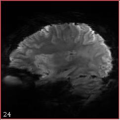
5 Images: can you name this organ?
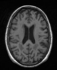
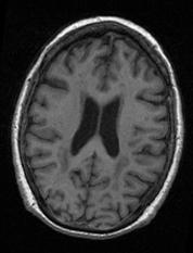
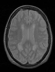
6 Images: can you name these modalities?

7 Images: basic definitions superior Sagittal 3D image Coronal posterior Axial anterior 3D Voxel inferior right left Resolution: voxel dimension A. Vesalius, on the Fabric of the Human Body
 = card ({")
![x I ( x) ] I ε, I + ε [ } ) Multi-channel](/docs-images/86/93360892/images/8-3.jpg "images Intensity histogram Longitudinal")
8 Images: basic definitions density 3D image volume intensity H ( I ) = card ({ x I ( x) ] I ε, I + ε [ } ) Multi-channel images Intensity histogram Longitudinal images
9 Structural image processing 1. Segmentation Applications: labeling, morphometry, cortical thickness, shape analysis, lesion detection


10 Structural image processing 2. Registration Applications: group statistics, atlasing, longitudinal processing
11 Structural image processing 3. Cortical mapping Applications: fmri processing, area parcellation, tissue quantification, cortical atrophy


12 Structural image processing Special cases: pathology
13 Image Segmentation Goal: label automatically brain structures Many methods depending on structures, applications
14 Image Segmentation: tissue classification Each voxel is assigned to WM, GM or CSF Basic assumption: tissue types cluster in separable intensity distributions Methods: unsupervised (EM, FCM, etc) or supervised (knn, SVM, etc)
15 Segmentation method: binary threshold Algorithm: 1. Choose a threshold value 2. Classify voxels above and below threshold to different classes

16 Segmentation method: K-means Algorithm: 1. Choose K mean values 2. Classify voxels to belong to the closest mean value 3. Recompute the means from the classified voxels

17 Segmentation method: Fuzzy C-means Algorithm: 1. Choose K mean values 2. Give a fuzzy membership to voxels based on distance to all means 3. Recompute the means from a weighted average of fuzzy voxels
18 Segmentation method: EM Algorithm Algorithm: 1. Choose K mean values 2. Give a membership probability to voxels based on distance 3. Recompute the means from the expectation on voxels

19 So, which to choose? Image slice K-means FCM EM Threshold: depends on how the threshold is determined K-means: very sensitive to initialization, oscillatory behavior FCM: stable convergence, lowest sensitivity EM: stable, probabilistic interpretation, sensitive to initialization
20 Problems: noise Pre-filtering to remove noise (anisotropic diffusion, total variation..) Post-filtering to smooth the classification Smoothness penalty functions/markov Random Fields


21 More problems: image artifacts Segmentation algorithms must also deal with inhomogeneities, partial volume effects
22 Intensity Modeling Model of choice: Mixture of Gaussian WM GM CSF Reality: Not so nice T1 T1 WM GM Vasculature Dura mater CSF


23 Spatial Structure Modeling vs. Statistical atlas prior Multi-atlas segmentation Important challenge: image registration


24 Higher-dimensional data Multi-modal segmentation DWI: segmentation of white matter tracts fmri: network identification


25 Recent trends in segmentation Multi-atlas segmentation Patch-based segmentation Surface evolution with boundary learning
26 How to compare? Validation Manual delineations Automated segmentations Overlap Distances Dice & Jaccard coefficients 2 A B A+ B A B A B Sensitivity, specificity,. Average surface distance Signed surface distance Hausdorff distance
 -")


 -")
27 How to compare? Validation Brain phantoms (Brainweb) - simulated MRI - ground truth anatomy Manual delineation databases - real MRI data - specific delineation goals Scan-rescan experiments - real data, ideal comparison - only measures robustness Grand Challenges (MICCAI) - real data, blind processing - direct comparison of mehods
28 Segmentation summary Segmentation extracts regions of interest from brain MRI - There are many segmentation methods & approaches with different assumptions and biases - Anatomical complexity, noise, inhomogeneities, pathology, require elaborate prior models for automated segmentation - Recent trends include non-linear registration, patch matching, machine learning, data fusion, - Validation performance is important but task-specific - Pathology makes many generic methods misinterpret data
29 Image Registration Registration is the application of a geometric transformation to the coordinate system of an image to bring it into correspondence with a second image. T(A) -1 T (B) Image A Image B
30 Image Registration Example ORIGINAL IMAGES
31 Image Registration Example ORIGINAL IMAGES REGISTERED IMAGES
32 Image Registration Basic Steps 1. Define class of transformations (rigid body, affine, nonlinear) 2. Define similarity index (homologous features, intensity, other more complex measures) 3. Optimize transformation parameters to maximize similarity index (a.k.a minimize disparity index, cost function) 4. Transform the images with interpolation How do we decide on the class of transformation or similarity index to be used?
33 Spatial transformation examples Rotation Shearing Translation Perspective Scaling Non-linear
34 Similarity index examples Intensity difference d ( A, B)= I A ( X ) I B (D ( X )) Landmark matching d ( A, B)= X L, A D ( X L, B ) 2 2 X L Normalized cross correlation (I A ( X ) I A)( I B (D( X )) I B ) s (A, B)= σ Aσ B X Normalized mutual information H ( A)+ H (B) s (A, B)= = H (A, B) p( I A )ln p( I A )+ p ( I B ) ln p ( I B ) I I I p (I A, I B ) ln p ( I A, I B )

35 Intramodal / Intersubject How do we decide on the class of transformation or similarity index to be used?
36 Intermodal / Intrasubject MRI PET How do we decide on the class of transformation or similarity index to be used?

37 Intramodal / Intrasubject Time 1 Time 2 2 years later How do we decide on the class of transformation or similarity index to be used?


38 Template registration How do we decide on the class of transformation or similarity index to be used?
39 Image Interpolation Interpolation the model-based recovery of continuous data from discrete data within a known range of abscissas f(x)?? x 1-D 2-D 3-D
40 Interpolation: Nearest Neighbors Choose the value from the closest sample point Advantages: - simple and fast - never modifies input values Disadvantages: - coarsest possible method - blocking artifact
41 Interpolation: Linear Choose a weighted average from the closest 2dim points Advantages: - still simple and fast - maintains intensity bounds Disadvantages: - still coarse - severe blurring artifact
42 Interpolation: Windowed Sinc Convolve the data with a sinc kernel Advantages: - ideal case for band-limited signals Disadvantages: - requires windowing (Lanczos) - ringing artifact at edges - somewhat slow

43 Interpolation artifacts - Ringing Oscillations occur at sharp boundaries

44 Interpolation artifacts - Aliasing Occurs when downsampling to a resolution that does not sufficiently represent structure

45 Interpolation artifacts - Blocking Occurs mainly with nearest-neighbor interpolation

46 Interpolation artifacts - Blurring Occurs with repeated interpolations Can be minimized by composing transformations

47 Non-linear Transformations How do we represent non-linear transformations? What is a diffeomorphic transformation? What is a symmetric transformation? Are there artifacts?

")
48 Registration Validation? Groupwise average quality manual delineations (NIREP, MindBoggle) Application-specific metrics?
49 Image Registration Trends ANTs / SyN Demons algorithm Symmetric diffeomorphism - conservative deformations - best on evaluation experiments - estimates both deformations - almost perfect? Fast registration - many variants: symmetric, diffeomorphic, spherical, diffusion... - easy to implement Are we done with registration research??


50 Image Registration Challenges Correspondences? Partial coverage Complex shapes
51 Registration Summary Registration bring brain images in correspondence - assumptions of meaningful correspondences - variable results for inter- intra- subject / modality - interpolation may introduce artifacts - best methods highly accurate, but still limited - validation tools are becoming available - challenges arise with pathologies, slab imaging, high resolution imaging of complex shapes


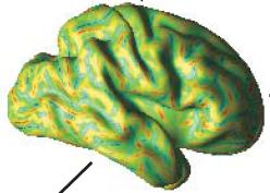

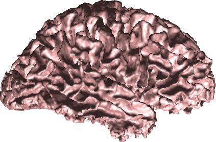
52 Cortical Mapping Segmentation of the cortex: cortical surfaces Inflated representations:


53 How are surfaces generated? Given a segmentation map: j, ps j [ 0,1] ps j 0.5 ps j < 0.5 inside the object outside the object Implicit representation: X Surface ps ( X ) = 0.5
54 How are surfaces generated? Contours defined by linear interpolation between voxels Marching squares Implicit resolution 6 or 18-C Marching cubes 26-C Voxel connectivity matters

55 Cortical Meshes & Resolution Mesh resolution ~ number of points [10, ,000]
56 Cortical Mapping Mapping of surface geometry & surface anatomy Curvedness Shape index Mapping of cortical thickness Mapping of volumetric data mean Quantitative T1 profiles
57 Cortical Thickness How to measure cortical thickness? [Yezzi & Prince, 2003]
58 Cortical Layer Geometry? CSF WM?

59 Cortical Layer Geometry CSF WM The bending of cortical layer changes their relative thickness in order to preserve their respective local volume [Bok, Z. ges. Neurol. Psychiat., 1929]

60 Cortical Layer Geometry 2 mm

61 Cortical Layer Mapping Incorrect layering model: curvature artifacts


62 Cortical Layer Mapping CSF T1 profiles oc r t i c la d e p ht WM
63 Cortical Mapping Summary Cortical mapping projects MR data onto the cortical sheet - Surface inflation enhances cortex visualization - Geometric shape information available, not very useful - Cortical thickness hard to define everywhere consistently - Defining cortical depth brings back the 3rd dimension, outlines laminar structure of the cortex - Cortical layer geometry impacts intra-cortical mapping
64 Pathologies For segmentation: - undefined tissue types - lower SNR - differ from priors For registration: - unusual shapes - non-matching lesions Dedicated methods?


65 Pathologies For segmentation: - undefined tissue types - lower SNR - differ from priors For registration: - unusual shapes - non-matching lesions Dedicated methods?
66 How to choose an algorithm? SPM The standards are good, but newer methods have improved upon them FreeSurfer FSL Where to find these new methods? NITRC.org MICCAI Grand Challenges How to evaluate these methods? Check validation experiments Evaluate relevance to your study How do I combine algorithms? Pipeline environments (LONI, JIST, Nipype)

67 NITRC: NeuroInformatics Tools & Resources


68 MICCAI Grand Challenges Principle: - a manually segmented database is created - competitors receive most of the data but only a subset of labels - some new data must be processed 'live' Since 2007 Many Grand Challenges are used afterwards for validation of new methods


69 Software developed at the Institute
70 Conclusions Structural image processing enables: - segmentation, measurement of brain structures - detection, quantification of pathology - grouping of multi-modal, multi-subject data Many methods are available: - search for what you want to do, not what's customary - interrogate validation & challenges - use integrative, modular systems to combine tools
Surface-based Analysis: Inter-subject Registration and Smoothing
 Surface-based Analysis: Inter-subject Registration and Smoothing Outline Exploratory Spatial Analysis Coordinate Systems 3D (Volumetric) 2D (Surface-based) Inter-subject registration Volume-based Surface-based
An Introduction To Automatic Tissue Classification Of Brain MRI. Colm Elliott Mar 2014
 An Introduction To Automatic Tissue Classification Of Brain MRI Colm Elliott Mar 2014 Tissue Classification Tissue classification is part of many processing pipelines. We often want to classify each voxel
Norbert Schuff VA Medical Center and UCSF
 Norbert Schuff Medical Center and UCSF Norbert.schuff@ucsf.edu Medical Imaging Informatics N.Schuff Course # 170.03 Slide 1/67 Objective Learn the principle segmentation techniques Understand the role
1 Introduction Motivation and Aims Functional Imaging Computational Neuroanatomy... 12
 Contents 1 Introduction 10 1.1 Motivation and Aims....... 10 1.1.1 Functional Imaging.... 10 1.1.2 Computational Neuroanatomy... 12 1.2 Overview of Chapters... 14 2 Rigid Body Registration 18 2.1 Introduction.....
MR IMAGE SEGMENTATION
 MR IMAGE SEGMENTATION Prepared by : Monil Shah What is Segmentation? Partitioning a region or regions of interest in images such that each region corresponds to one or more anatomic structures Classification
Automatic Registration-Based Segmentation for Neonatal Brains Using ANTs and Atropos
 Automatic Registration-Based Segmentation for Neonatal Brains Using ANTs and Atropos Jue Wu and Brian Avants Penn Image Computing and Science Lab, University of Pennsylvania, Philadelphia, USA Abstract.
Image Registration + Other Stuff
 Image Registration + Other Stuff John Ashburner Pre-processing Overview fmri time-series Motion Correct Anatomical MRI Coregister m11 m 21 m 31 m12 m13 m14 m 22 m 23 m 24 m 32 m 33 m 34 1 Template Estimate
Automatic Generation of Training Data for Brain Tissue Classification from MRI
 Automatic Generation of Training Data for Brain Tissue Classification from MRI Chris A. COCOSCO, Alex P. ZIJDENBOS, and Alan C. EVANS http://www.bic.mni.mcgill.ca/users/crisco/ McConnell Brain Imaging
CHAPTER 2. Morphometry on rodent brains. A.E.H. Scheenstra J. Dijkstra L. van der Weerd
 CHAPTER 2 Morphometry on rodent brains A.E.H. Scheenstra J. Dijkstra L. van der Weerd This chapter was adapted from: Volumetry and other quantitative measurements to assess the rodent brain, In vivo NMR
Preprocessing II: Between Subjects John Ashburner
 Preprocessing II: Between Subjects John Ashburner Pre-processing Overview Statistics or whatever fmri time-series Anatomical MRI Template Smoothed Estimate Spatial Norm Motion Correct Smooth Coregister
The organization of the human cerebral cortex estimated by intrinsic functional connectivity
 1 The organization of the human cerebral cortex estimated by intrinsic functional connectivity Journal: Journal of Neurophysiology Author: B. T. Thomas Yeo, et al Link: https://www.ncbi.nlm.nih.gov/pubmed/21653723
Computational Neuroanatomy
 Computational Neuroanatomy John Ashburner john@fil.ion.ucl.ac.uk Smoothing Motion Correction Between Modality Co-registration Spatial Normalisation Segmentation Morphometry Overview fmri time-series kernel
Norbert Schuff Professor of Radiology VA Medical Center and UCSF
 Norbert Schuff Professor of Radiology Medical Center and UCSF Norbert.schuff@ucsf.edu 2010, N.Schuff Slide 1/67 Overview Definitions Role of Segmentation Segmentation methods Intensity based Shape based
Methods for data preprocessing
 Methods for data preprocessing John Ashburner Wellcome Trust Centre for Neuroimaging, 12 Queen Square, London, UK. Overview Voxel-Based Morphometry Morphometry in general Volumetrics VBM preprocessing
Automatic segmentation of the cortical grey and white matter in MRI using a Region Growing approach based on anatomical knowledge
 Automatic segmentation of the cortical grey and white matter in MRI using a Region Growing approach based on anatomical knowledge Christian Wasserthal 1, Karin Engel 1, Karsten Rink 1 und André Brechmann
Spatio-Temporal Registration of Biomedical Images by Computational Methods
 Spatio-Temporal Registration of Biomedical Images by Computational Methods Francisco P. M. Oliveira, João Manuel R. S. Tavares tavares@fe.up.pt, www.fe.up.pt/~tavares Outline 1. Introduction 2. Spatial
Subvoxel Segmentation and Representation of Brain Cortex Using Fuzzy Clustering and Gradient Vector Diffusion
 Subvoxel Segmentation and Representation of Brain Cortex Using Fuzzy Clustering and Gradient Vector Diffusion Ming-Ching Chang Xiaodong Tao GE Global Research Center {changm, taox} @ research.ge.com SPIE
Methodological progress in image registration for ventilation estimation, segmentation propagation and multi-modal fusion
 Methodological progress in image registration for ventilation estimation, segmentation propagation and multi-modal fusion Mattias P. Heinrich Julia A. Schnabel, Mark Jenkinson, Sir Michael Brady 2 Clinical
The Insight Toolkit. Image Registration Algorithms & Frameworks
 The Insight Toolkit Image Registration Algorithms & Frameworks Registration in ITK Image Registration Framework Multi Resolution Registration Framework Components PDE Based Registration FEM Based Registration
ADAPTIVE GRAPH CUTS WITH TISSUE PRIORS FOR BRAIN MRI SEGMENTATION
 ADAPTIVE GRAPH CUTS WITH TISSUE PRIORS FOR BRAIN MRI SEGMENTATION Abstract: MIP Project Report Spring 2013 Gaurav Mittal 201232644 This is a detailed report about the course project, which was to implement
Neuroimaging and mathematical modelling Lesson 2: Voxel Based Morphometry
 Neuroimaging and mathematical modelling Lesson 2: Voxel Based Morphometry Nivedita Agarwal, MD Nivedita.agarwal@apss.tn.it Nivedita.agarwal@unitn.it Volume and surface morphometry Brain volume White matter
Image Registration I
 Image Registration I Comp 254 Spring 2002 Guido Gerig Image Registration: Motivation Motivation for Image Registration Combine images from different modalities (multi-modality registration), e.g. CT&MRI,
Norbert Schuff Professor of Radiology VA Medical Center and UCSF
 Norbert Schuff Professor of Radiology Medical Center and UCSF Norbert.schuff@ucsf.edu Slide 1/67 Overview Definitions Role of Segmentation Segmentation methods Intensity based Shape based Texture based
Comparison Study of Clinical 3D MRI Brain Segmentation Evaluation
 Comparison Study of Clinical 3D MRI Brain Segmentation Evaluation Ting Song 1, Elsa D. Angelini 2, Brett D. Mensh 3, Andrew Laine 1 1 Heffner Biomedical Imaging Laboratory Department of Biomedical Engineering,
Supplementary methods
 Supplementary methods This section provides additional technical details on the sample, the applied imaging and analysis steps and methods. Structural imaging Trained radiographers placed all participants
Automated MR Image Analysis Pipelines
 Automated MR Image Analysis Pipelines Andy Simmons Centre for Neuroimaging Sciences, Kings College London Institute of Psychiatry. NIHR Biomedical Research Centre for Mental Health at IoP & SLAM. Neuroimaging
Medical Image Synthesis Methods and Applications
 MR Intensity Scale is Arbitrary This causes problems in most postprocessing methods Inconsistency or algorithm failure 11/5/2015 2 Joint Histogram 1.5 T GE SPGR 3 T Philips MPRAGE 11/5/2015 3 Problem With
A Design Toolbox to Generate Complex Phantoms for the Evaluation of Medical Image Processing Algorithms
 A Design Toolbox to Generate Complex Phantoms for the Evaluation of Medical Image Processing Algorithms Omar Hamo, Georg Nelles, Gudrun Wagenknecht Central Institute for Electronics, Research Center Juelich,
Magnetic resonance image tissue classification using an automatic method
 Yazdani et al. Diagnostic Pathology (2014) 9:207 DOI 10.1186/s13000-014-0207-7 METHODOLOGY Open Access Magnetic resonance image tissue classification using an automatic method Sepideh Yazdani 1, Rubiyah
Functional MRI data preprocessing. Cyril Pernet, PhD
 Functional MRI data preprocessing Cyril Pernet, PhD Data have been acquired, what s s next? time No matter the design, multiple volumes (made from multiple slices) have been acquired in time. Before getting
Math in image processing
 Math in image processing Math in image processing Nyquist theorem Math in image processing Discrete Fourier Transformation Math in image processing Image enhancement: scaling Math in image processing Image
Automatic Generation of Training Data for Brain Tissue Classification from MRI
 MICCAI-2002 1 Automatic Generation of Training Data for Brain Tissue Classification from MRI Chris A. Cocosco, Alex P. Zijdenbos, and Alan C. Evans McConnell Brain Imaging Centre, Montreal Neurological
Quantitative MRI of the Brain: Investigation of Cerebral Gray and White Matter Diseases
 Quantities Measured by MR - Quantitative MRI of the Brain: Investigation of Cerebral Gray and White Matter Diseases Static parameters (influenced by molecular environment): T, T* (transverse relaxation)
Performance Evaluation of the TINA Medical Image Segmentation Algorithm on Brainweb Simulated Images
 Tina Memo No. 2008-003 Internal Memo Performance Evaluation of the TINA Medical Image Segmentation Algorithm on Brainweb Simulated Images P. A. Bromiley Last updated 20 / 12 / 2007 Imaging Science and
Structural MRI analysis
 Structural MRI analysis volumetry and voxel-based morphometry cortical thickness measurements structural covariance network mapping Boris Bernhardt, PhD Department of Social Neuroscience, MPI-CBS bernhardt@cbs.mpg.de
Image Segmentation and Registration
 Image Segmentation and Registration Dr. Christine Tanner (tanner@vision.ee.ethz.ch) Computer Vision Laboratory, ETH Zürich Dr. Verena Kaynig, Machine Learning Laboratory, ETH Zürich Outline Segmentation
Biomedical Image Analysis based on Computational Registration Methods. João Manuel R. S. Tavares
 Biomedical Image Analysis based on Computational Registration Methods João Manuel R. S. Tavares tavares@fe.up.pt, www.fe.up.pt/~tavares Outline 1. Introduction 2. Methods a) Spatial Registration of (2D
Automatic Optimization of Segmentation Algorithms Through Simultaneous Truth and Performance Level Estimation (STAPLE)
") Automatic Optimization of Segmentation Algorithms Through Simultaneous Truth and Performance Level Estimation (STAPLE) Mahnaz Maddah, Kelly H. Zou, William M. Wells, Ron Kikinis, and Simon K. Warfield
Automatic Optimization of Segmentation Algorithms Through Simultaneous Truth and Performance Level Estimation (STAPLE) Mahnaz Maddah, Kelly H. Zou, William M. Wells, Ron Kikinis, and Simon K. Warfield
The Anatomical Equivalence Class Formulation and its Application to Shape-based Computational Neuroanatomy
 The Anatomical Equivalence Class Formulation and its Application to Shape-based Computational Neuroanatomy Sokratis K. Makrogiannis, PhD From post-doctoral research at SBIA lab, Department of Radiology,
Basic principles of MR image analysis. Basic principles of MR image analysis. Basic principles of MR image analysis
 Basic principles of MR image analysis Basic principles of MR image analysis Julien Milles Leiden University Medical Center Terminology of fmri Brain extraction Registration Linear registration Non-linear
Introduction to Medical Image Processing
 Introduction to Medical Image Processing Δ Essential environments of a medical imaging system Subject Image Analysis Energy Imaging System Images Image Processing Feature Images Image processing may be
Learning-based Neuroimage Registration
 Learning-based Neuroimage Registration Leonid Teverovskiy and Yanxi Liu 1 October 2004 CMU-CALD-04-108, CMU-RI-TR-04-59 School of Computer Science Carnegie Mellon University Pittsburgh, PA 15213 Abstract
Knowledge-Based Segmentation of Brain MRI Scans Using the Insight Toolkit
 Knowledge-Based Segmentation of Brain MRI Scans Using the Insight Toolkit John Melonakos 1, Ramsey Al-Hakim 1, James Fallon 2 and Allen Tannenbaum 1 1 Georgia Institute of Technology, Atlanta GA 30332,
Fmri Spatial Processing
 Educational Course: Fmri Spatial Processing Ray Razlighi Jun. 8, 2014 Spatial Processing Spatial Re-alignment Geometric distortion correction Spatial Normalization Smoothing Why, When, How, Which Why is
Structural Segmentation
 Structural Segmentation FAST tissue-type segmentation FIRST sub-cortical structure segmentation FSL-VBM voxelwise grey-matter density analysis SIENA atrophy analysis FAST FMRIB s Automated Segmentation
Whole Body MRI Intensity Standardization
 Whole Body MRI Intensity Standardization Florian Jäger 1, László Nyúl 1, Bernd Frericks 2, Frank Wacker 2 and Joachim Hornegger 1 1 Institute of Pattern Recognition, University of Erlangen, {jaeger,nyul,hornegger}@informatik.uni-erlangen.de
CHAPTER 6 MODIFIED FUZZY TECHNIQUES BASED IMAGE SEGMENTATION
 CHAPTER 6 MODIFIED FUZZY TECHNIQUES BASED IMAGE SEGMENTATION 6.1 INTRODUCTION Fuzzy logic based computational techniques are becoming increasingly important in the medical image analysis arena. The significant
A Multiple-Layer Flexible Mesh Template Matching Method for Nonrigid Registration between a Pelvis Model and CT Images
 A Multiple-Layer Flexible Mesh Template Matching Method for Nonrigid Registration between a Pelvis Model and CT Images Jianhua Yao 1, Russell Taylor 2 1. Diagnostic Radiology Department, Clinical Center,
Analysis of Functional MRI Timeseries Data Using Signal Processing Techniques
 Analysis of Functional MRI Timeseries Data Using Signal Processing Techniques Sea Chen Department of Biomedical Engineering Advisors: Dr. Charles A. Bouman and Dr. Mark J. Lowe S. Chen Final Exam October
Neuroimage Processing
 Neuroimage Processing Instructor: Moo K. Chung mkchung@wisc.edu Lecture 2-3. General Linear Models (GLM) Voxel-based Morphometry (VBM) September 11, 2009 What is GLM The general linear model (GLM) is a
A Registration-Based Atlas Propagation Framework for Automatic Whole Heart Segmentation
 A Registration-Based Atlas Propagation Framework for Automatic Whole Heart Segmentation Xiahai Zhuang (PhD) Centre for Medical Image Computing University College London Fields-MITACS Conference on Mathematics
8/3/2017. Contour Assessment for Quality Assurance and Data Mining. Objective. Outline. Tom Purdie, PhD, MCCPM
 Contour Assessment for Quality Assurance and Data Mining Tom Purdie, PhD, MCCPM Objective Understand the state-of-the-art in contour assessment for quality assurance including data mining-based techniques
A Model-Independent, Multi-Image Approach to MR Inhomogeneity Correction
 Tina Memo No. 2007-003 Published in Proc. MIUA 2007 A Model-Independent, Multi-Image Approach to MR Inhomogeneity Correction P. A. Bromiley and N.A. Thacker Last updated 13 / 4 / 2007 Imaging Science and
Lilla Zöllei A.A. Martinos Center, MGH; Boston, MA
 Lilla Zöllei lzollei@nmr.mgh.harvard.edu A.A. Martinos Center, MGH; Boston, MA Bruce Fischl Gheorghe Postelnicu Jean Augustinack Anastasia Yendiki Allison Stevens Kristen Huber Sita Kakonoori + the FreeSurfer
Statistical Analysis of Neuroimaging Data. Phebe Kemmer BIOS 516 Sept 24, 2015
 Statistical Analysis of Neuroimaging Data Phebe Kemmer BIOS 516 Sept 24, 2015 Review from last time Structural Imaging modalities MRI, CAT, DTI (diffusion tensor imaging) Functional Imaging modalities
MEDICAL IMAGE COMPUTING (CAP 5937) LECTURE 4: Pre-Processing Medical Images (II)
 LECTURE 4: Pre-Processing Medical Images (II)") SPRING 2016 1 MEDICAL IMAGE COMPUTING (CAP 5937) LECTURE 4: Pre-Processing Medical Images (II) Dr. Ulas Bagci HEC 221, Center for Research in Computer Vision (CRCV), University of Central Florida (UCF),
SPRING 2016 1 MEDICAL IMAGE COMPUTING (CAP 5937) LECTURE 4: Pre-Processing Medical Images (II) Dr. Ulas Bagci HEC 221, Center for Research in Computer Vision (CRCV), University of Central Florida (UCF),
Introduction to fmri. Pre-processing
 Introduction to fmri Pre-processing Tibor Auer Department of Psychology Research Fellow in MRI Data Types Anatomical data: T 1 -weighted, 3D, 1/subject or session - (ME)MPRAGE/FLASH sequence, undistorted
Structural Segmentation
 Structural Segmentation FAST tissue-type segmentation FIRST sub-cortical structure segmentation FSL-VBM voxelwise grey-matter density analysis SIENA atrophy analysis FAST FMRIB s Automated Segmentation
Image Segmentation. Ross Whitaker SCI Institute, School of Computing University of Utah
 Image Segmentation Ross Whitaker SCI Institute, School of Computing University of Utah What is Segmentation? Partitioning images/volumes into meaningful pieces Partitioning problem Labels Isolating a specific
Manifold Learning: Applications in Neuroimaging
 Your own logo here Manifold Learning: Applications in Neuroimaging Robin Wolz 23/09/2011 Overview Manifold learning for Atlas Propagation Multi-atlas segmentation Challenges LEAP Manifold learning for
Multi-atlas labeling with population-specific template and non-local patch-based label fusion
 Multi-atlas labeling with population-specific template and non-local patch-based label fusion Vladimir Fonov, Pierrick Coupé, Simon Eskildsen, Jose Manjon, Louis Collins To cite this version: Vladimir
FROM IMAGE RECONSTRUCTION TO CONNECTIVITY ANALYSIS: A JOURNEY THROUGH THE BRAIN'S WIRING. Francesca Pizzorni Ferrarese
 FROM IMAGE RECONSTRUCTION TO CONNECTIVITY ANALYSIS: A JOURNEY THROUGH THE BRAIN'S WIRING Francesca Pizzorni Ferrarese Pipeline overview WM and GM Segmentation Registration Data reconstruction Tractography
Topology Preserving Brain Tissue Segmentation Using Graph Cuts
 Topology Preserving Brain Tissue Segmentation Using Graph Cuts Xinyang Liu 1, Pierre-Louis Bazin 2, Aaron Carass 3, and Jerry Prince 3 1 Brigham and Women s Hospital, Boston, MA 1 xinyang@bwh.harvard.edu
Knowledge-Based Segmentation of Brain MRI Scans Using the Insight Toolkit
 Knowledge-Based Segmentation of Brain MRI Scans Using the Insight Toolkit John Melonakos 1, Ramsey Al-Hakim 1, James Fallon 2 and Allen Tannenbaum 1 1 Georgia Institute of Technology, Atlanta GA 30332,
Ischemic Stroke Lesion Segmentation Proceedings 5th October 2015 Munich, Germany
 0111010001110001101000100101010111100111011100100011011101110101101012 Ischemic Stroke Lesion Segmentation www.isles-challenge.org Proceedings 5th October 2015 Munich, Germany Preface Stroke is the second
Image Segmentation. Ross Whitaker SCI Institute, School of Computing University of Utah
 Image Segmentation Ross Whitaker SCI Institute, School of Computing University of Utah What is Segmentation? Partitioning images/volumes into meaningful pieces Partitioning problem Labels Isolating a specific
SPM Introduction. SPM : Overview. SPM: Preprocessing SPM! SPM: Preprocessing. Scott Peltier. FMRI Laboratory University of Michigan
 SPM Introduction Scott Peltier FMRI Laboratory University of Michigan! Slides adapted from T. Nichols SPM! SPM : Overview Library of MATLAB and C functions Graphical user interface Four main components:
Automatic Segmentation of Parotids from CT Scans Using Multiple Atlases
 Automatic Segmentation of Parotids from CT Scans Using Multiple Atlases Jinzhong Yang, Yongbin Zhang, Lifei Zhang, and Lei Dong Department of Radiation Physics, University of Texas MD Anderson Cancer Center
STATISTICAL ATLAS-BASED SUB-VOXEL SEGMENTATION OF 3D BRAIN MRI
 STATISTICA ATAS-BASED SUB-VOXE SEGMENTATION OF 3D BRAIN MRI Marcel Bosc 1,2, Fabrice Heitz 1, Jean-Paul Armspach 2 (1) SIIT UMR-7005 CNRS / Strasbourg I University, 67400 Illkirch, France (2) IPB UMR-7004
Histograms. h(r k ) = n k. p(r k )= n k /NM. Histogram: number of times intensity level rk appears in the image
 = n k. p(r k )= n k /NM. Histogram: number of times intensity level rk appears in the image") Histograms h(r k ) = n k Histogram: number of times intensity level rk appears in the image p(r k )= n k /NM normalized histogram also a probability of occurence 1 Histogram of Image Intensities Create
Histograms h(r k ) = n k Histogram: number of times intensity level rk appears in the image p(r k )= n k /NM normalized histogram also a probability of occurence 1 Histogram of Image Intensities Create
SPM Introduction SPM! Scott Peltier. FMRI Laboratory University of Michigan. Software to perform computation, manipulation and display of imaging data
 SPM Introduction Scott Peltier FMRI Laboratory University of Michigan Slides adapted from T. Nichols SPM! Software to perform computation, manipulation and display of imaging data 1 1 SPM : Overview Library
This Time. fmri Data analysis
 This Time Reslice example Spatial Normalization Noise in fmri Methods for estimating and correcting for physiologic noise SPM Example Spatial Normalization: Remind ourselves what a typical functional image
Advanced Visual Medicine: Techniques for Visual Exploration & Analysis
 Advanced Visual Medicine: Techniques for Visual Exploration & Analysis Interactive Visualization of Multimodal Volume Data for Neurosurgical Planning Felix Ritter, MeVis Research Bremen Multimodal Neurosurgical
Measuring longitudinal brain changes in humans and small animal models. Christos Davatzikos
 Measuring longitudinal brain changes in humans and small animal models Christos Davatzikos Section of Biomedical Image Analysis University of Pennsylvania (Radiology) http://www.rad.upenn.edu/sbia Computational
An ITK Filter for Bayesian Segmentation: itkbayesianclassifierimagefilter
 An ITK Filter for Bayesian Segmentation: itkbayesianclassifierimagefilter John Melonakos 1, Karthik Krishnan 2 and Allen Tannenbaum 1 1 Georgia Institute of Technology, Atlanta GA 30332, USA {jmelonak,
Machine Learning for Medical Image Analysis. A. Criminisi
 Machine Learning for Medical Image Analysis A. Criminisi Overview Introduction to machine learning Decision forests Applications in medical image analysis Anatomy localization in CT Scans Spine Detection
Basic fmri Design and Analysis. Preprocessing
 Basic fmri Design and Analysis Preprocessing fmri Preprocessing Slice timing correction Geometric distortion correction Head motion correction Temporal filtering Intensity normalization Spatial filtering
CHAPTER 3 TUMOR DETECTION BASED ON NEURO-FUZZY TECHNIQUE
 32 CHAPTER 3 TUMOR DETECTION BASED ON NEURO-FUZZY TECHNIQUE 3.1 INTRODUCTION In this chapter we present the real time implementation of an artificial neural network based on fuzzy segmentation process
Automatized & Interactive. Muscle tissues characterization using. Na MRI
 Automatized & Interactive Human Skeletal Muscle Segmentation Muscle tissues characterization using 23 Na MRI Noura Azzabou 30 April 2013 What is muscle segmentation? Axial slice of the thigh of a healthy
Multi-Sectional Views Textural Based SVM for MS Lesion Segmentation in Multi-Channels MRIs
 56 The Open Biomedical Engineering Journal, 2012, 6, 56-72 Open Access Multi-Sectional Views Textural Based SVM for MS Lesion Segmentation in Multi-Channels MRIs Bassem A. Abdullah*, Akmal A. Younis and
A Generic Lie Group Model for Computer Vision
 A Generic Lie Group Model for Computer Vision Within this research track we follow a generic Lie group approach to computer vision based on recent physiological research on how the primary visual cortex
RIGID IMAGE REGISTRATION
 RIGID IMAGE REGISTRATION Duygu Tosun-Turgut, Ph.D. Center for Imaging of Neurodegenerative Diseases Department of Radiology and Biomedical Imaging duygu.tosun@ucsf.edu What is registration? Image registration
Computational Medical Imaging Analysis Chapter 5: Processing and Analysis
 Computational Medical Imaging Analysis Chapter 5: Processing and Analysis Jun Zhang Laboratory for Computational Medical Imaging & Data Analysis Department of Computer Science University of Kentucky Lexington,
腦部結構影像 標準化 組織分割 體素型態 本週課程內容. Analysis Softwares. A Course of MRI
 本週課程內容 腦部結構影像 A Course of MRI 盧家鋒助理教授國立陽明大學物理治療暨輔助科技學系 alvin4016@ym.edu.tw 腦部結構影像 空間標準化 (Spatial normalization) 均勻度校正 (Bias correction) 組織分割 (Segmentation) 體素形態學分析 (Voxel-based morphometry, VBM) 影像平滑化
MARS: Multiple Atlases Robust Segmentation
 Software Release (1.0.1) Last updated: April 30, 2014. MARS: Multiple Atlases Robust Segmentation Guorong Wu, Minjeong Kim, Gerard Sanroma, and Dinggang Shen {grwu, mjkim, gerard_sanroma, dgshen}@med.unc.edu
Biomedical Imaging Registration Trends and Applications. Francisco P. M. Oliveira, João Manuel R. S. Tavares
 Biomedical Imaging Registration Trends and Applications Francisco P. M. Oliveira, João Manuel R. S. Tavares tavares@fe.up.pt, www.fe.up.pt/~tavares Outline 1. Introduction 2. Spatial Registration of (2D
Semantic Context Forests for Learning- Based Knee Cartilage Segmentation in 3D MR Images
 Semantic Context Forests for Learning- Based Knee Cartilage Segmentation in 3D MR Images MICCAI 2013: Workshop on Medical Computer Vision Authors: Quan Wang, Dijia Wu, Le Lu, Meizhu Liu, Kim L. Boyer,
White Pixel Artifact. Caused by a noise spike during acquisition Spike in K-space <--> sinusoid in image space
 White Pixel Artifact Caused by a noise spike during acquisition Spike in K-space sinusoid in image space Susceptibility Artifacts Off-resonance artifacts caused by adjacent regions with different
Classification of Subject Motion for Improved Reconstruction of Dynamic Magnetic Resonance Imaging
 1 CS 9 Final Project Classification of Subject Motion for Improved Reconstruction of Dynamic Magnetic Resonance Imaging Feiyu Chen Department of Electrical Engineering ABSTRACT Subject motion is a significant
Automatic MS Lesion Segmentation by Outlier Detection and Information Theoretic Region Partitioning Release 0.00
 Automatic MS Lesion Segmentation by Outlier Detection and Information Theoretic Region Partitioning Release 0.00 Marcel Prastawa 1 and Guido Gerig 1 Abstract July 17, 2008 1 Scientific Computing and Imaging
Shape-based Diffeomorphic Registration on Hippocampal Surfaces Using Beltrami Holomorphic Flow
 Shape-based Diffeomorphic Registration on Hippocampal Surfaces Using Beltrami Holomorphic Flow Abstract. Finding meaningful 1-1 correspondences between hippocampal (HP) surfaces is an important but difficult
Slicer3 Training Tutorial Using EM Segmenter with Non- Human Primate Images
 Slicer3 Training Compendium Slicer3 Training Tutorial Using EM Segmenter with Non- Human Primate Images Vidya Rajagopalan Christopher Wyatt BioImaging Systems Lab Dept. of Electrical Engineering Virginia
Distance Transforms in Multi Channel MR Image Registration
 Distance Transforms in Multi Channel MR Image Registration Min Chen 1, Aaron Carass 1, John Bogovic 1, Pierre-Louis Bazin 2 and Jerry L. Prince 1 1 Image Analysis and Communications Laboratory, 2 The Laboratory
Diffusion-MRI processing for group analysis
 Diffusion-MRI processing for group analysis Felix Renard IRMaGe: Inserm US 17 / CNRS UMS 3552 University Hospital of Grenoble - France 25/09/2015 felixrenard@gmail.com 1 Diffusion-MRI processing for group
Nonrigid Registration using Free-Form Deformations
 Nonrigid Registration using Free-Form Deformations Hongchang Peng April 20th Paper Presented: Rueckert et al., TMI 1999: Nonrigid registration using freeform deformations: Application to breast MR images
Voxel-Based Morphometry & DARTEL. Ged Ridgway, London With thanks to John Ashburner and the FIL Methods Group
 Zurich SPM Course 2012 Voxel-Based Morphometry & DARTEL Ged Ridgway, London With thanks to John Ashburner and the FIL Methods Group Aims of computational neuroanatomy * Many interesting and clinically
A Generation Methodology for Numerical Phantoms with Statistically Relevant Variability of Geometric and Physical Properties
 A Generation Methodology for Numerical Phantoms with Statistically Relevant Variability of Geometric and Physical Properties Steven Dolly 1, Eric Ehler 1, Yang Lou 2, Mark Anastasio 2, Hua Li 2 (1) University
MEDICAL IMAGE ANALYSIS
 SECOND EDITION MEDICAL IMAGE ANALYSIS ATAM P. DHAWAN g, A B IEEE Engineering in Medicine and Biology Society, Sponsor IEEE Press Series in Biomedical Engineering Metin Akay, Series Editor +IEEE IEEE PRESS
Auto-Segmentation Using Deformable Image Registration. Disclosure. Objectives 8/4/2011
 Auto-Segmentation Using Deformable Image Registration Lei Dong, Ph.D. Dept. of Radiation Physics University of Texas MD Anderson Cancer Center, Houston, Texas AAPM Therapy Educational Course Aug. 4th 2011
Efficient population registration of 3D data
 Efficient population registration of 3D data Lilla Zöllei 1, Erik Learned-Miller 2, Eric Grimson 1, William Wells 1,3 1 Computer Science and Artificial Intelligence Lab, MIT; 2 Dept. of Computer Science,