Pre-processing of ASL data T CT
|
|
|
- Roger Harrell
- 5 years ago
- Views:
Transcription

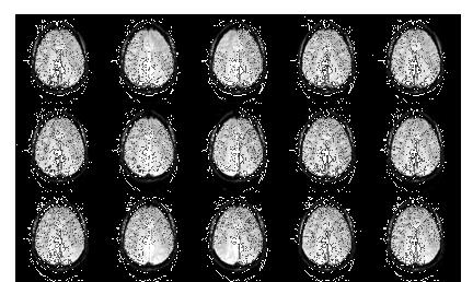
1 Wed October 2, 2013 Image Processing Pre-processing: motion correction, denoising, outlier detection Alessandra Bertoldo Pre-processing of ASL data T CT C T C Single TI ASL T T T T C CCC average Pre-processing of ASL data T CT C T C Single TI ASL T T T T C CCC average 1
2 Pre-processing of ASL data T CT C T C Single TI ASL T T T T C CCC average Pre-processing of ASL data: SUMMARY 1) Outlier detection/ Intra-scan quality control 2) Motion correction 3) Image denoising-deblurring 4) Smoothing Single TI ASL 5) Coregistration 6) EPI distortion correction Outlier Detection TAG: 30 repetitions CONTROL: 30 repetitions 2

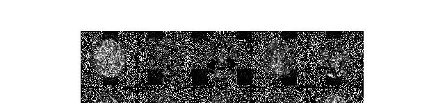
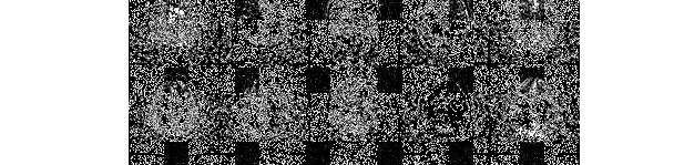


3 Outlier Detection TAG: 30 repetitions CONTROL: 30 repetitions Blood (3%-5%) Signal Tissue (95%-97%) Outlier Detection TAG - CONTROL Outlier Detection TAG - CONTROL 3


. average & SD")


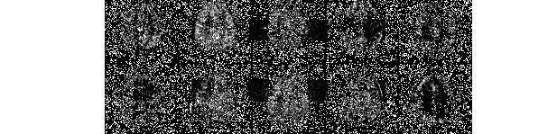
4 Outlier Detection TAG - CONTROL Coregistration: Multi TI ASL QUASAR Tag-control (24). average & SD TIs Coregistration: Multi TI 3rd Deep blue = low correlation value 23rd 24th Map of the 2D correlation values between the 24 averages Discard lower values 4
 PASL 2. Maumet C., Maurel P., Ferré J.C., Barillotan C.")
 before being averaged.")
5 Coregistration: Multi TI ASL QUASAR Tag-control (24). average & SD TIs Outlier Detection Currently only few attempts: 1. Tan H., Maldjian J.A., Pollock J.M., Burdette J.H., Yang L.Y., Deibler A.R., Kraft R.A. «A fast, effective filtering method for improving clinical pulsed arterial spin labeling MRI» Journal of MagneticResonanceImaging 29, (2009) PASL 2. Maumet C., Maurel P., Ferré J.C., Barillotan C. «Robust Cerebral Blood Flow Map Estimation in Arterial Spin Labeling» Lecture Notes in Computer Science 7509, (2012) PICORE Q2TIPS + simulated data Outlier Detection A mean and standard deviation-based filter was implemented to remove outliers in the set of perfusionweighted images (control label) before being averaged. the filtering procedure was performed on both a volume-by-volume basis and a slice-by-slice basis (Tan et al., 2009) m=1.5 n=2.5 Tan et al., J Magn Reson Imaging May; 29(5):


6 Outlier Detection Tan et al., J Magn Reson Imaging May; 29(5): Outlier Detection Maumet et al. proposed to quantify CBF values by using the Huber s Maximum likelihoodtype-estimator since it is less influenced by outliers than other estimators <<In all configuration Huber s M-estimators is either better or as good as z-thresholding to estimate robust CBF maps. In the presence of outliers, Huber s M-estimator is always more accurate than the sample average>> Maumet et al., Lecture Notes in Computer Science 7509, (2012) Motion correction 6
7 Motion correction Head motion results in severe image artifacts especially in ASL since it requires identical positioning of label and control images Currently it is possible to retrieve only few contributions to solve this issue hardware software e.g. systems tracking the movement e.g. algorithms for motion correction Motion correction: basic strategy T CT C T C Before averaging, motion correction is performed through 3D rigid body registration of the images. e.g. FSL mc-flirt -all tag & ctrl -meanvol Common Reference Often six-parameter based rigid body transformation is used. It minimizes the distance between each volume and the reference volume (for instance, the first image of the ASL series) Motion correction: basic strategy T CT C T C BUT: e.g. FSL mc-flirt -all tag & ctrl -meanvol Common Reference differences can be interpreted as motion but generated by the change of the ASL perfusion signal between ctrl & tag (zig-zagged spin labeling paradigm) 7
8 Motion correction Wang Z. «Improving cerebral blood flow quantification for arterial spin labeled perfusion MRI by removing residual motion artifacts and global signal fluctuations» Magn Reson Imaging 30: (2012) 1) all ASL images, tag+ctrlstandard strategy (six-parameter based rigid body trasformation) 2) the zig-zagged label-control patterns Motion correction Motion correction Wang Z. «Improving cerebral blood flow quantification for arterial spin labeled perfusion MRI by removing residual motion artifacts and global signal fluctuations» Magn Reson Imaging 30: (2012) 1) all ASL images, tag+ctrlstandard strategy (six-parameter based rigid body trasformation) 2) the zig-zagged label-control patterns regressed out from those motion time courses through simple regressions. DenoMng label by N 1 and control by 1, the zig-zagged label-control paradigm is a binary numerical series consisting of oscillating -1 and 1. 3) Third, the cleaned motion parameters then used for real motion correction (SPM ASLtbx) 8

: 91 105 (2010) PROspective Motion (PROMO) is a navigator based module developed to be inserted into various MR pulse sequences for real-time 3D motion estimation and correction.")
9 Motion correction mc-flirt -in tag -meanvol flsmaths tag_mcf -Tmean flsmaths ctrl_mcf -Tmean mc-flirt -in ctrl -meanvol mc-flirt -in tag_mcf -ref C_R Common Reference mc-flirt -in ctrl_mcf -ref C_R Suggested by the study of: Di Cataldo et al., 2011 IEEE International Conference on Bioinformatics and Biomedicine Motion correction: software White N., Roddey C., Shankaranarayanan A., Han E., Rettmann D., Santos J., Kuperman J., Dale A. «PROMO Realtime Prospective Motion Correction in MRI using Image-based Tracking» Magn Reson Med January; 63(1): (2010) PROspective Motion (PROMO) is a navigator based module developed to be inserted into various MR pulse sequences for real-time 3D motion estimation and correction. The PROMO approach utilizes three orthogonal 2D spiral navigator acquisitions (SP-Navs) along with a flexible image-based tracking method based on the Extended Kalman Filter algorithm for real-time motion measurement. PROMO has been integrated into PCASL Motion correction: software two healthy young males 9
 A real-time head motion tracking system is evaluated")
 Possible solutions: - Average of a high number of volumes (40-100) - Increase the magnetic field strength - Improve the coil")

10 Motion correction: hardware Hoßbach M., Gregori J., Wesarg S., Günther M. «Head Motion Compensation for Arterial Spin Labeling Using Optical Motion Tracking» Lecture Notes in Computer Science 7761: 1-8 (2013) A real-time head motion tracking system is evaluated using MR-compatible cameras attached to the head coil, tracking blue dot markers on the forehead Motion correction: hardware Denoising Method 1 Signal-to-noise (SNR) ratio is a critical issue in ASL data (especially PASL QUIPSS II or Q2TIPS) Possible solutions: - Average of a high number of volumes (40-100) - Increase the magnetic field strength - Improve the coil efficiency - Employ a denoising algorithm in the post-processing Gaussian Smoothing Image convolved with a Gaussian Function (blur of the pixel) Voxel size = 3.75 x 3.75 x 6.3 mm 3 Filter = two-dimensional gaussian kernel FWHM = 8 mm and FWHM = 5.6 mm Wavelet Denoising W = wavelet-transform matrix X = measured image S = true object signal N = noise (gaussian) Y = wavelet coefficient Bibic A et al., Denoising of ASL data: wavelet domain filtering compared with gaussian smoothing, MAGMA 2010, 23:
 GS (FWHM = 5.")

11 Denoising Method 1 Ground Truth Difference Maps 300 averages 20 averages Bibic A et al., Denoising of ASL data: wavelet domain filtering compared with gaussian smoothing, MAGMA 2010, 23: WV Denoising Method 1 GS (FWHM = 8 mm) GS (FWHM = 5.6 mm) Bias Maps Bias = CBF true CBF filtered CBF true = 300 averages 5 averages 20 averages Bibic A et al., Denoising of ASL data: wavelet domain filtering compared with gaussian smoothing, MAGMA 2010, 23: Ground Truth Denoising Method 2 Wiener Filter AD Filter Difference map obtained from the average of 64 control/label pairs 3D GRASE pcaslacquisition, single PLD, 2 =Meanandvariancewithina3x3 kernelcenteredineachpixel C=pixelvalue 2 =noisevariance b=filteredpixel MAD=MedianAbsoluteDeviation!I=imageintensitygradient K=scaleparameter Gaussian Filter M = S /G Wavelet Filter: Harr wavelet ICA filter S=originalimage G=gaussiankernel (sd=0.5voxel,kernelsize=3x3) M=resultingimage v = noise variance y = number of pixels thr = optimal threshold (soft and hard) a = independent signal components S = weigth coefficients C = total signal Wells JA et al., Reduction of Errors in ASL Cerebral Perfusion and Arterial Transit Time Maps Using Image De-Noising, MRM 2010,64:

12 Denoising Method 2 Difference Maps MAD= Median Absolute Deviation!I= image intensity gradient K= scale parameter CNR = contrast to noise ratio in the two ROIs Contrast = mean signal ROI 1 mean signal ROI 2 Wells JA et al., Reduction of Errors in ASL Cerebral Perfusion and Arterial Transit Time Maps Using Image De-Noising, MRM 2010,64: SMOOTHING Denoising of arterial spin labeling data can be done also using smoothing. Two different levels: 1) Smoothing the raw ASL images (usually by a conventional Gaussian filter) 2) Smoothing after CBF calculation and spatial normalization (usually by a conventional Gaussian filter) Partial volume effect Signal to noise ratio Coregistration M0 T1w Template T1w space MNI space BET (FSL) T1w space M0 space FLIRT(FSL) T1w space FNIRT (FSL) M0 space FLIRT (FSL) T1w space Adapted from: Thomas W Okell, Michael A Chappell, Michael E Kelly and Peter Jezzard, Journal of Cerebral Blood Flow & Metabolism 2013; doi: /jcbfm


 for each slice needs to be corrected for the slice acquisition time, i.")

 The most successful approach to minimizing EPI distortions makes use of")

13 Method 1 Coregistration of ASL data to Structural Image Coregistration CBF Computation in Structural Space CBF maps in Structural Space Method 2 Coregistration of ASL data to Structural Image CBF Computation in ASL Space Use parameters from coregistration to obtain CBF maps in Structural Space Coregistration Considerations M 0b : if we use an external calibration scan and a voxelwise approach for M 0t estimation, it is necessary to coregister also this image to the Structural image. This increases the possibility of bias in the CBF estimations. 2D readout: in this case, the acquisition time (inversion time TI or post-label delay PLD) for each slice needs to be corrected for the slice acquisition time, i.e. TI i = TI + (i-1)*slice scan_time This correction results to be not completely precise if you use Method 1. EPI distortion correction Addressed for fmri and DTI studies but still open issue in ASL 1) The most successful approach to minimizing EPI distortions makes use of acquired magnetic "field maps" that can be used for subsequent dewarping of the associated EPI images (a capability that is included in the FSL and SPM software packages) 2) DTI TORTOISE software package based on the use of SD maps of the DTI quantities of interest for datasets with different phase-encoding directions 13
Functional MRI in Clinical Research and Practice Preprocessing
 Functional MRI in Clinical Research and Practice Preprocessing fmri Preprocessing Slice timing correction Geometric distortion correction Head motion correction Temporal filtering Intensity normalization
SPM8 for Basic and Clinical Investigators. Preprocessing. fmri Preprocessing
 SPM8 for Basic and Clinical Investigators Preprocessing fmri Preprocessing Slice timing correction Geometric distortion correction Head motion correction Temporal filtering Intensity normalization Spatial
A Spatio-temporal Denoising Approach based on Total Variation Regularization for Arterial Spin Labeling
 A Spatio-temporal Denoising Approach based on Total Variation Regularization for Arterial Spin Labeling Cagdas Ulas 1,2, Stephan Kaczmarz 3, Christine Preibisch 3, Jonathan I Sperl 2, Marion I Menzel 2,
SPM8 for Basic and Clinical Investigators. Preprocessing
 SPM8 for Basic and Clinical Investigators Preprocessing fmri Preprocessing Slice timing correction Geometric distortion correction Head motion correction Temporal filtering Intensity normalization Spatial
Basic fmri Design and Analysis. Preprocessing
 Basic fmri Design and Analysis Preprocessing fmri Preprocessing Slice timing correction Geometric distortion correction Head motion correction Temporal filtering Intensity normalization Spatial filtering
EPI Data Are Acquired Serially. EPI Data Are Acquired Serially 10/23/2011. Functional Connectivity Preprocessing. fmri Preprocessing
 Functional Connectivity Preprocessing Geometric distortion Head motion Geometric distortion Head motion EPI Data Are Acquired Serially EPI Data Are Acquired Serially descending 1 EPI Data Are Acquired
Existing Packages. Overview. Siemens. Michael A. Chappell
 Existing Packages Michael A. Chappell michael.chappell@eng.ox.ac.uk www.ibme.ox.ac.uk/qubic Institute of Biomedical Engineering & Oxford Centre for Functional MRI of the Brain University of Oxford. Scanner
Correction of Partial Volume Effects in Arterial Spin Labeling MRI
 Correction of Partial Volume Effects in Arterial Spin Labeling MRI By: Tracy Ssali Supervisors: Dr. Keith St. Lawrence and Udunna Anazodo Medical Biophysics 3970Z Six Week Project April 13 th 2012 Introduction
Politecnico di Torino. Porto Institutional Repository
 Politecnico di Torino Porto Institutional Repository [Proceeding] Motion artifact correction in ASL images: automated procedure an improved Original Citation: Di Cataldo S., Ficarra E., Acquaviva A., Macii
Journal of Articles in Support of The Null Hypothesis
 Data Preprocessing Martin M. Monti, PhD UCLA Psychology NITP 2016 Typical (task-based) fmri analysis sequence Image Pre-processing Single Subject Analysis Group Analysis Journal of Articles in Support
Functional MRI data preprocessing. Cyril Pernet, PhD
 Functional MRI data preprocessing Cyril Pernet, PhD Data have been acquired, what s s next? time No matter the design, multiple volumes (made from multiple slices) have been acquired in time. Before getting
ASAP_2.0 (Automatic Software for ASL Processing) USER S MANUAL
 USER S MANUAL") ASAP_2.0 (Automatic Software for ASL Processing) USER S MANUAL ASAP was developed as part of the COST Action "Arterial Spin Labelling Initiative in Dementia (AID)" by: Department of Neuroimaging, Institute
ASAP_2.0 (Automatic Software for ASL Processing) USER S MANUAL ASAP was developed as part of the COST Action "Arterial Spin Labelling Initiative in Dementia (AID)" by: Department of Neuroimaging, Institute
FMRI Pre-Processing and Model- Based Statistics
 FMRI Pre-Processing and Model- Based Statistics Brief intro to FMRI experiments and analysis FMRI pre-stats image processing Simple Single-Subject Statistics Multi-Level FMRI Analysis Advanced FMRI Analysis
White Pixel Artifact. Caused by a noise spike during acquisition Spike in K-space <--> sinusoid in image space
 White Pixel Artifact Caused by a noise spike during acquisition Spike in K-space sinusoid in image space Susceptibility Artifacts Off-resonance artifacts caused by adjacent regions with different
DeepASL: Kinetic Model Incorporated Loss for Denoising Arterial Spin Labeled MRI via Deep Residual Learning
 DeepASL: Kinetic Model Incorporated Loss for Denoising Arterial Spin Labeled MRI via Deep Residual Learning Cagdas Ulas 1, Giles Tetteh 1, Stephan Kaczmarz 2, Christine Preibisch 2, and Bjoern H. Menze
Fmri Spatial Processing
 Educational Course: Fmri Spatial Processing Ray Razlighi Jun. 8, 2014 Spatial Processing Spatial Re-alignment Geometric distortion correction Spatial Normalization Smoothing Why, When, How, Which Why is
Introduction to fmri. Pre-processing
 Introduction to fmri Pre-processing Tibor Auer Department of Psychology Research Fellow in MRI Data Types Anatomical data: T 1 -weighted, 3D, 1/subject or session - (ME)MPRAGE/FLASH sequence, undistorted
Analysis of fmri data within Brainvisa Example with the Saccades database
 Analysis of fmri data within Brainvisa Example with the Saccades database 18/11/2009 Note : All the sentences in italic correspond to informations relative to the specific dataset under study TP participants
MB-EPI PCASL. Release Notes for Version February 2015
 MB-EPI PCASL Release Notes for Version 1.0 20 February 2015 1 Background High-resolution arterial spin labeling (ASL) imaging is highly desirable in both neuroscience research and clinical applications
Enhao Gong, PhD Candidate, Electrical Engineering, Stanford University Dr. John Pauly, Professor in Electrical Engineering, Stanford University Dr.
 Enhao Gong, PhD Candidate, Electrical Engineering, Stanford University Dr. John Pauly, Professor in Electrical Engineering, Stanford University Dr. Greg Zaharchuk, Associate Professor in Radiology, Stanford
Supplementary Figure 1
 Supplementary Figure 1 BOLD and CBV functional maps showing EPI versus line-scanning FLASH fmri. A. Colored BOLD and CBV functional maps are shown in the highlighted window (green frame) of the raw EPI
The organization of the human cerebral cortex estimated by intrinsic functional connectivity
 1 The organization of the human cerebral cortex estimated by intrinsic functional connectivity Journal: Journal of Neurophysiology Author: B. T. Thomas Yeo, et al Link: https://www.ncbi.nlm.nih.gov/pubmed/21653723
Role of Parallel Imaging in High Field Functional MRI
 Role of Parallel Imaging in High Field Functional MRI Douglas C. Noll & Bradley P. Sutton Department of Biomedical Engineering, University of Michigan Supported by NIH Grant DA15410 & The Whitaker Foundation
SPM Introduction. SPM : Overview. SPM: Preprocessing SPM! SPM: Preprocessing. Scott Peltier. FMRI Laboratory University of Michigan
 SPM Introduction Scott Peltier FMRI Laboratory University of Michigan! Slides adapted from T. Nichols SPM! SPM : Overview Library of MATLAB and C functions Graphical user interface Four main components:
fmri pre-processing Juergen Dukart
 fmri pre-processing Juergen Dukart Outline Why do we need pre-processing? fmri pre-processing Slice time correction Realignment Unwarping Coregistration Spatial normalisation Smoothing Overview fmri time-series
SPM Introduction SPM! Scott Peltier. FMRI Laboratory University of Michigan. Software to perform computation, manipulation and display of imaging data
 SPM Introduction Scott Peltier FMRI Laboratory University of Michigan Slides adapted from T. Nichols SPM! Software to perform computation, manipulation and display of imaging data 1 1 SPM : Overview Library
Supplementary methods
 Supplementary methods This section provides additional technical details on the sample, the applied imaging and analysis steps and methods. Structural imaging Trained radiographers placed all participants
MRI Physics II: Gradients, Imaging
 MRI Physics II: Gradients, Imaging Douglas C., Ph.D. Dept. of Biomedical Engineering University of Michigan, Ann Arbor Magnetic Fields in MRI B 0 The main magnetic field. Always on (0.5-7 T) Magnetizes
Preprocessing of fmri data
 Preprocessing of fmri data Pierre Bellec CRIUGM, DIRO, UdM Flowchart of the NIAK fmri preprocessing pipeline fmri run 1 fmri run N individual datasets CIVET NUC, segmentation, spatial normalization slice
HST.583 Functional Magnetic Resonance Imaging: Data Acquisition and Analysis Fall 2008
 MIT OpenCourseWare http://ocw.mit.edu HST.583 Functional Magnetic Resonance Imaging: Data Acquisition and Analysis Fall 2008 For information about citing these materials or our Terms of Use, visit: http://ocw.mit.edu/terms.
Empirical analyses of null-hypothesis perfusion FMRI data at 1.5 and 4 T
 NeuroImage 19 (2003) 1449 1462 www.elsevier.com/locate/ynimg Empirical analyses of null-hypothesis perfusion FMRI data at 1.5 and 4 T Jiongjiong Wang, a,b, * Geoffrey K. Aguirre, a,c Daniel Y. Kimberg,
HST.583 Functional Magnetic Resonance Imaging: Data Acquisition and Analysis Fall 2006
 MIT OpenCourseWare http://ocw.mit.edu HST.583 Functional Magnetic Resonance Imaging: Data Acquisition and Analysis Fall 2006 For information about citing these materials or our Terms of Use, visit: http://ocw.mit.edu/terms.
Brain Extraction, Registration & EPI Distortion Correction
 Brain Extraction, Registration & EPI Distortion Correction What use is Registration? Some common uses of registration: Combining across individuals in group studies: including fmri & diffusion Quantifying
Classification of Subject Motion for Improved Reconstruction of Dynamic Magnetic Resonance Imaging
 1 CS 9 Final Project Classification of Subject Motion for Improved Reconstruction of Dynamic Magnetic Resonance Imaging Feiyu Chen Department of Electrical Engineering ABSTRACT Subject motion is a significant
Analysis of Functional MRI Timeseries Data Using Signal Processing Techniques
 Analysis of Functional MRI Timeseries Data Using Signal Processing Techniques Sea Chen Department of Biomedical Engineering Advisors: Dr. Charles A. Bouman and Dr. Mark J. Lowe S. Chen Final Exam October
Statistical Analysis of Neuroimaging Data. Phebe Kemmer BIOS 516 Sept 24, 2015
 Statistical Analysis of Neuroimaging Data Phebe Kemmer BIOS 516 Sept 24, 2015 Review from last time Structural Imaging modalities MRI, CAT, DTI (diffusion tensor imaging) Functional Imaging modalities
Head motion in diffusion MRI
 Head motion in diffusion MRI Anastasia Yendiki HMS/MGH/MIT Athinoula A. Martinos Center for Biomedical Imaging 11/06/13 Head motion in diffusion MRI 0/33 Diffusion contrast Basic principle of diffusion
XI Conference "Medical Informatics & Technologies" VALIDITY OF MRI BRAIN PERFUSION IMAGING METHOD
 XI Conference "Medical Informatics & Technologies" - 2006 medical imaging, MRI, brain perfusion Bartosz KARCZEWSKI 1, Jacek RUMIŃSKI 1 VALIDITY OF MRI BRAIN PERFUSION IMAGING METHOD Brain perfusion imaging
Nonrigid Motion Compensation of Free Breathing Acquired Myocardial Perfusion Data
 Nonrigid Motion Compensation of Free Breathing Acquired Myocardial Perfusion Data Gert Wollny 1, Peter Kellman 2, Andrés Santos 1,3, María-Jesus Ledesma 1,3 1 Biomedical Imaging Technologies, Department
6 credits. BMSC-GA Practical Magnetic Resonance Imaging II
 BMSC-GA 4428 - Practical Magnetic Resonance Imaging II 6 credits Course director: Ricardo Otazo, PhD Course description: This course is a practical introduction to image reconstruction, image analysis
Slide 1. Technical Aspects of Quality Control in Magnetic Resonance Imaging. Slide 2. Annual Compliance Testing. of MRI Systems.
 Slide 1 Technical Aspects of Quality Control in Magnetic Resonance Imaging Slide 2 Compliance Testing of MRI Systems, Ph.D. Department of Radiology Henry Ford Hospital, Detroit, MI Slide 3 Compliance Testing
This Time. fmri Data analysis
 This Time Reslice example Spatial Normalization Noise in fmri Methods for estimating and correcting for physiologic noise SPM Example Spatial Normalization: Remind ourselves what a typical functional image
n o r d i c B r a i n E x Tutorial DSC Module
 m a k i n g f u n c t i o n a l M R I e a s y n o r d i c B r a i n E x Tutorial DSC Module Please note that this tutorial is for the latest released nordicbrainex. If you are using an older version please
Bayesian Inference of Hemodynamic Changes in Functional Arterial Spin Labeling Data
 Bayesian Inference of Hemodynamic Changes in Functional Arterial Spin Labeling Data Mark W. Woolrich, 1, * Peter Chiarelli, 1 Daniel Gallichan, 1 Joanna Perthen, 2 and Thomas T. Liu 2 Magnetic Resonance
Deep Learning for Fast and Spatially- Constrained Tissue Quantification from Highly-Undersampled Data in Magnetic Resonance Fingerprinting (MRF)
") Deep Learning for Fast and Spatially- Constrained Tissue Quantification from Highly-Undersampled Data in Magnetic Resonance Fingerprinting (MRF) Zhenghan Fang 1, Yong Chen 1, Mingxia Liu 1, Yiqiang Zhan
Deep Learning for Fast and Spatially- Constrained Tissue Quantification from Highly-Undersampled Data in Magnetic Resonance Fingerprinting (MRF) Zhenghan Fang 1, Yong Chen 1, Mingxia Liu 1, Yiqiang Zhan
Patch-Based Super-Resolution of Arterial Spin Labeling Magnetic Resonance Images
 Patch-Based Super-Resolution of Arterial Spin Labeling Magnetic Resonance Images Cédric Meurée, Pierre Maurel, Jean-Christophe Ferré, Christian Barillot To cite this version: Cédric Meurée, Pierre Maurel,
Introduction to MRI data processing with FSL. Anna Blazejewska
 Introduction to MRI data processing with FSL Anna Blazejewska FSL = FMRIB Software Library FMRIB = Functional Magnetic Resonance Imaging of the Brain @ Oxford since 2000, last stable FSL 5.0, free! for
COBRE Scan Information
 COBRE Scan Information Below is more information on the directory structure for the COBRE imaging data. Also below are the imaging parameters for each series. Directory structure: var/www/html/dropbox/1139_anonymized/human:
Sources of Distortion in Functional MRI Data
 Human Brain Mapping 8:80 85(1999) Sources of Distortion in Functional MRI Data Peter Jezzard* and Stuart Clare FMRIB Centre, Department of Clinical Neurology, University of Oxford, Oxford, UK Abstract:
fmri Image Preprocessing
 fmri Image Preprocessing Rick Hoge, Ph.D. Laboratoire de neuroimagerie vasculaire (LINeV) Centre de recherche de l institut universitaire de gériatrie de Montréal, Université de Montréal Outline Motion
MEDICAL IMAGE ANALYSIS
 SECOND EDITION MEDICAL IMAGE ANALYSIS ATAM P. DHAWAN g, A B IEEE Engineering in Medicine and Biology Society, Sponsor IEEE Press Series in Biomedical Engineering Metin Akay, Series Editor +IEEE IEEE PRESS
High dynamic range magnetic resonance flow imaging in the abdomen
 High dynamic range magnetic resonance flow imaging in the abdomen Christopher M. Sandino EE 367 Project Proposal 1 Motivation Time-resolved, volumetric phase-contrast magnetic resonance imaging (also known
Sparse sampling in MRI: From basic theory to clinical application. R. Marc Lebel, PhD Department of Electrical Engineering Department of Radiology
 Sparse sampling in MRI: From basic theory to clinical application R. Marc Lebel, PhD Department of Electrical Engineering Department of Radiology Objective Provide an intuitive overview of compressed sensing
Module 4. K-Space Symmetry. Review. K-Space Review. K-Space Symmetry. Partial or Fractional Echo. Half or Partial Fourier HASTE
 MRES 7005 - Fast Imaging Techniques Module 4 K-Space Symmetry Review K-Space Review K-Space Symmetry Partial or Fractional Echo Half or Partial Fourier HASTE Conditions for successful reconstruction Interpolation
Diffusion MRI Acquisition. Karla Miller FMRIB Centre, University of Oxford
 Diffusion MRI Acquisition Karla Miller FMRIB Centre, University of Oxford karla@fmrib.ox.ac.uk Diffusion Imaging How is diffusion weighting achieved? How is the image acquired? What are the limitations,
Introduction to Neuroimaging Janaina Mourao-Miranda
 Introduction to Neuroimaging Janaina Mourao-Miranda Neuroimaging techniques have changed the way neuroscientists address questions about functional anatomy, especially in relation to behavior and clinical
HST.583 Functional Magnetic Resonance Imaging: Data Acquisition and Analysis Fall 2008
 MIT OpenCourseWare http://ocw.mit.edu HST.583 Functional Magnetic Resonance Imaging: Data Acquisition and Analysis Fall 2008 For information about citing these materials or our Terms of Use, visit: http://ocw.mit.edu/terms.
Noise and Artifacts in FMRI
 Noise and Artifacts in FMRI Instructor: Luis Hernandez-Garcia, Ph.D. Associate Research Professor FMRI Laboratory, Biomedical Engineering FMRI analysis - synopsis of what you ll do next week 1. Formulate
Functional Imaging With Turbo-CASL: Transit Time and Multislice Imaging Considerations
 Functional Imaging With Turbo-CASL: Transit Time and Multislice Imaging Considerations Gregory R. Lee,* Luis Hernandez-Garcia, and Douglas C. Noll Magnetic Resonance in Medicine 57:661 669 (2007) The optimal
FSL Pre-Processing Pipeline
 The Art and Pitfalls of fmri Preprocessing FSL Pre-Processing Pipeline Mark Jenkinson FMRIB Centre, University of Oxford FSL Pre-Processing Pipeline Standard pre-processing: Task fmri Resting-state fmri
Advanced MRI Techniques (and Applications)
") Advanced MRI Techniques (and Applications) Jeffry R. Alger, PhD Department of Neurology Ahmanson-Lovelace Brain Mapping Center Brain Research Institute Jonsson Comprehensive Cancer Center University of
Advanced MRI Techniques (and Applications) Jeffry R. Alger, PhD Department of Neurology Ahmanson-Lovelace Brain Mapping Center Brain Research Institute Jonsson Comprehensive Cancer Center University of
Locating Motion Artifacts in Parametric fmri Analysis
 Tina Memo No. 200-002 Presented at MICCAI 999 Locating Motion Artifacts in Parametric fmri Analysis A.J.Lacey, N.A.Thacker, E. Burton, and A.Jackson Last updated 2 / 02 / 2002 Imaging Science and Biomedical
Joint Reconstruction of Multi-contrast MR Images for Multiple Sclerosis Lesion Segmentation
 Joint Reconstruction of Multi-contrast MR Images for Multiple Sclerosis Lesion Segmentation Pedro A Gómez 1,2,3, Jonathan I Sperl 3, Tim Sprenger 2,3, Claudia Metzler-Baddeley 4, Derek K Jones 4, Philipp
MR IMAGE SEGMENTATION
 MR IMAGE SEGMENTATION Prepared by : Monil Shah What is Segmentation? Partitioning a region or regions of interest in images such that each region corresponds to one or more anatomic structures Classification
Motion Robust Magnetic Susceptibility and Field Inhomogeneity Estimation Using Regularized Image Restoration Techniques for fmri
 Motion Robust Magnetic Susceptibility and Field Inhomogeneity Estimation Using Regularized Image Restoration Techniques for fmri Desmond Tec Beng Yeo 1,, Jeffrey A. Fessler 1,, and Bolye Kim 1 1 Department
Chapter 3 Set Redundancy in Magnetic Resonance Brain Images
 16 Chapter 3 Set Redundancy in Magnetic Resonance Brain Images 3.1 MRI (magnetic resonance imaging) MRI is a technique of measuring physical structure within the human anatomy. Our proposed research focuses
Investigating White Matter Perfusion Using Optimal Sampling Strategy Arterial Spin Labeling at 7 Tesla
 IMAGING METHODOLOGY - Notes Magnetic Resonance in Medicine 73:2243 2248 (2015) Investigating White Matter Perfusion Using Optimal Sampling Strategy Arterial Spin Labeling at 7 Tesla Alexander G. Gardener
Deviceless respiratory motion correction in PET imaging exploring the potential of novel data driven strategies
 g Deviceless respiratory motion correction in PET imaging exploring the potential of novel data driven strategies Presented by Adam Kesner, Ph.D., DABR Assistant Professor, Division of Radiological Sciences,
Dynamic Autocalibrated Parallel Imaging Using Temporal GRAPPA (TGRAPPA)
") Magnetic Resonance in Medicine 53:981 985 (2005) Dynamic Autocalibrated Parallel Imaging Using Temporal GRAPPA (TGRAPPA) Felix A. Breuer, 1 * Peter Kellman, 2 Mark A. Griswold, 1 and Peter M. Jakob 1 Current
Magnetic Resonance in Medicine 53:981 985 (2005) Dynamic Autocalibrated Parallel Imaging Using Temporal GRAPPA (TGRAPPA) Felix A. Breuer, 1 * Peter Kellman, 2 Mark A. Griswold, 1 and Peter M. Jakob 1 Current
CP Generalize Concepts in Abstract Multi-dimensional Image Model Component Semantics. David Clunie.
 CP-1390 - Generalize Concepts in Abstract Multi-dimensional Image Model Semantics Page 1 STATUS Date of Last Update Person Assigned Submitter Name Submission Date Assigned 2014/06/09 David Clunie mailto:dclunie@dclunie.com
Measuring baseline whole-brain perfusion on GE 3.0T using arterial spin labeling (ASL) MRI
 MRI") Measuring baseline whole-brain perfusion on GE 3.0T using arterial spin labeling (ASL) MRI Revision date: 11/20/2006 Overview This document describes the procedure for measuring baseline whole-brain perfusion
Measuring baseline whole-brain perfusion on GE 3.0T using arterial spin labeling (ASL) MRI Revision date: 11/20/2006 Overview This document describes the procedure for measuring baseline whole-brain perfusion
Norbert Schuff VA Medical Center and UCSF
 Norbert Schuff Medical Center and UCSF Norbert.schuff@ucsf.edu Medical Imaging Informatics N.Schuff Course # 170.03 Slide 1/67 Objective Learn the principle segmentation techniques Understand the role
Constrained Reconstruction of Sparse Cardiac MR DTI Data
 Constrained Reconstruction of Sparse Cardiac MR DTI Data Ganesh Adluru 1,3, Edward Hsu, and Edward V.R. DiBella,3 1 Electrical and Computer Engineering department, 50 S. Central Campus Dr., MEB, University
Field Maps. 1 Field Map Acquisition. John Pauly. October 5, 2005
 Field Maps John Pauly October 5, 25 The acquisition and reconstruction of frequency, or field, maps is important for both the acquisition of MRI data, and for its reconstruction. Many of the imaging methods
MRI image formation 8/3/2016. Disclosure. Outlines. Chen Lin, PhD DABR 3. Indiana University School of Medicine and Indiana University Health
 MRI image formation Indiana University School of Medicine and Indiana University Health Disclosure No conflict of interest for this presentation 2 Outlines Data acquisition Spatial (Slice/Slab) selection
2. Creating Field Maps Using the Field Map GUI (Version 2.0) in SPM5
 in SPM5") 1. Introduction This manual describes how to use the Field Map Toolbox Version 2.0 for creating unwrapped field maps that can be used to do geometric distortion correction of EPI images in SPM5. 1. 1.
1. Introduction This manual describes how to use the Field Map Toolbox Version 2.0 for creating unwrapped field maps that can be used to do geometric distortion correction of EPI images in SPM5. 1. 1.
Artifact detection and repair in fmri
 Artifact detection and repair in fmri Paul K. Mazaika, Ph.D. Center for Interdisciplinary Brain Sciences Research (CIBSR) Division of Interdisciplinary Behavioral Sciences Stanford University School of
Surface-based Analysis: Inter-subject Registration and Smoothing
 Surface-based Analysis: Inter-subject Registration and Smoothing Outline Exploratory Spatial Analysis Coordinate Systems 3D (Volumetric) 2D (Surface-based) Inter-subject registration Volume-based Surface-based
Lab Location: MRI, B2, Cardinal Carter Wing, St. Michael s Hospital, 30 Bond Street
 Lab Location: MRI, B2, Cardinal Carter Wing, St. Michael s Hospital, 30 Bond Street MRI is located in the sub basement of CC wing. From Queen or Victoria, follow the baby blue arrows and ride the CC south
Methods for data preprocessing
 Methods for data preprocessing John Ashburner Wellcome Trust Centre for Neuroimaging, 12 Queen Square, London, UK. Overview Voxel-Based Morphometry Morphometry in general Volumetrics VBM preprocessing
The simulator can be applied in a number of diverse applications which span both
 Chapter 6 Simulator applications The simulator can be applied in a number of diverse applications which span both MRI and FMRI fields These applications include the simulation and removal of various imaging
Functional MRI. Jerry Allison, Ph. D. Medical College of Georgia
 Functional MRI Jerry Allison, Ph. D. Medical College of Georgia BOLD Imaging Technique Blood Oxygen Level Dependent contrast can be used to map brain function Right Hand Motor Task Outline fmri BOLD Contrast
K-Space Trajectories and Spiral Scan
 K-Space and Spiral Scan Presented by: Novena Rangwala nrangw2@uic.edu 1 Outline K-space Gridding Reconstruction Features of Spiral Sampling Pulse Sequences Mathematical Basis of Spiral Scanning Variations
A Model-Independent, Multi-Image Approach to MR Inhomogeneity Correction
 Tina Memo No. 2007-003 Published in Proc. MIUA 2007 A Model-Independent, Multi-Image Approach to MR Inhomogeneity Correction P. A. Bromiley and N.A. Thacker Last updated 13 / 4 / 2007 Imaging Science and
FSL Pre-Processing Pipeline
 The Art and Pitfalls of fmri Preprocessing FSL Pre-Processing Pipeline Mark Jenkinson FMRIB Centre, University of Oxford FSL Pre-Processing Pipeline Standard pre-processing: Task fmri Resting-state fmri
Image Acquisition Systems
 Image Acquisition Systems Goals and Terminology Conventional Radiography Axial Tomography Computer Axial Tomography (CAT) Magnetic Resonance Imaging (MRI) PET, SPECT Ultrasound Microscopy Imaging ITCS
Cocozza S., et al. : ALTERATIONS OF FUNCTIONAL CONNECTIVITY OF THE MOTOR CORTEX IN FABRY'S DISEASE: AN RS-FMRI STUDY
 ALTERATIONS OF FUNCTIONAL CONNECTIVITY OF THE MOTOR CORTEX IN FABRY'S DISEASE: AN RS-FMRI STUDY SUPPLEMENTARY MATERIALS Sirio Cocozza, MD 1*, Antonio Pisani, MD, PhD 2, Gaia Olivo, MD 1, Francesco Saccà,
Measuring baseline whole-brain perfusion on GE 3.0T using arterial spin labeling (ASL) MRI
 MRI") Measuring baseline whole-brain perfusion on GE 3.0T using arterial spin labeling (ASL) MRI Revision date: 09/15/2008 Overview This document describes the procedure for measuring baseline whole-brain perfusion
Measuring baseline whole-brain perfusion on GE 3.0T using arterial spin labeling (ASL) MRI Revision date: 09/15/2008 Overview This document describes the procedure for measuring baseline whole-brain perfusion
Effect of age and dementia on topology of brain functional networks. Paul McCarthy, Luba Benuskova, Liz Franz University of Otago, New Zealand
 Effect of age and dementia on topology of brain functional networks Paul McCarthy, Luba Benuskova, Liz Franz University of Otago, New Zealand 1 Structural changes in aging brain Age-related changes in
G Practical Magnetic Resonance Imaging II Sackler Institute of Biomedical Sciences New York University School of Medicine. Compressed Sensing
 G16.4428 Practical Magnetic Resonance Imaging II Sackler Institute of Biomedical Sciences New York University School of Medicine Compressed Sensing Ricardo Otazo, PhD ricardo.otazo@nyumc.org Compressed
Improved Spatial Localization in 3D MRSI with a Sequence Combining PSF-Choice, EPSI and a Resolution Enhancement Algorithm
 Improved Spatial Localization in 3D MRSI with a Sequence Combining PSF-Choice, EPSI and a Resolution Enhancement Algorithm L.P. Panych 1,3, B. Madore 1,3, W.S. Hoge 1,3, R.V. Mulkern 2,3 1 Brigham and
RADIOMICS: potential role in the clinics and challenges
 27 giugno 2018 Dipartimento di Fisica Università degli Studi di Milano RADIOMICS: potential role in the clinics and challenges Dr. Francesca Botta Medical Physicist Istituto Europeo di Oncologia (Milano)
HST.583 Functional Magnetic Resonance Imaging: Data Acquisition and Analysis Fall 2008
 MIT OpenCourseWare http://ocw.mit.edu HST.583 Functional Magnetic Resonance Imaging: Data Acquisition and Analysis Fall 2008 For information about citing these materials or our Terms of Use, visit: http://ocw.mit.edu/terms.
A System Identification Approach to Estimating a Dynamic Model of Head Motion for MRI Motion Correction
 A System Identification Approach to Estimating a Dynamic Model of Head Motion for MRI Motion Correction Burak Erem 1, Onur Afacan 1, Ali Gholipour 1, Sanjay P. Prabhu 2, and Simon K. Warfield 1 1 Computational
Characterization and Correction of Interpolation Effects in the Realignment of fmri Time Series
 NeuroImage 11, 49 57 (2000) doi:10.1006/nimg.1999.0515, available online at http://www.idealibrary.com on Characterization and Correction of Interpolation Effects in the Realignment of fmri Time Series
Motion Correction in fmri by Mapping Slice-to-Volume with Concurrent Field-Inhomogeneity Correction
 Motion Correction in fmri by Mapping Slice-to-Volume with Concurrent Field-Inhomogeneity Correction Desmond T.B. Yeo 1,2, Jeffery A. Fessler 2, and Boklye Kim 1 1 Department of Radiology, University of
Quantitative MRI of the Brain: Investigation of Cerebral Gray and White Matter Diseases
 Quantities Measured by MR - Quantitative MRI of the Brain: Investigation of Cerebral Gray and White Matter Diseases Static parameters (influenced by molecular environment): T, T* (transverse relaxation)
Artifact Detection and Repair: Overview and Sample Outputs
 Artifact Detection and Repair: Overview and Sample Outputs Paul Mazaika February 2007 Programs originated in Gabrieli Neuroscience Laboratory, updated and enhanced at Center for Interdisciplinary Brain
A novel noise removal using homomorphic normalization for multi-echo knee MRI
 A novel noise removal using homomorphic normalization for multi-echo knee MRI Xuenan Cui 1a),HakilKim 1b), Seongwook Hong 1c), and Kyu-Sung Kwack 2d) 1 School of Information and Communication Engineering,