Functional MRI. Jerry Allison, Ph. D. Medical College of Georgia
|
|
|
- Shavonne Lee
- 5 years ago
- Views:
Transcription
1 Functional MRI Jerry Allison, Ph. D. Medical College of Georgia
2 BOLD Imaging Technique Blood Oxygen Level Dependent contrast can be used to map brain function
3 Right Hand Motor Task
4 Outline fmri BOLD Contrast An fmri Exam Pulse Sequences for fmri fmri Acquisition Parameters fmri Artifacts
5 fmri BOLD Contrast Neuronal events (cerebral activation) O2 consumption increases (+5%) Cerebral blood flow increases (+50%) Oxygen extraction fraction decreases Oxygenation increases in venous blood
6 fmri BOLD Contrast Concentration of paramagnetic deoxyhemoglobin decreases Intravoxel dephasing decreases T2* increases T2* weighted image intensity increases (+ few %)
7 Latency: 5-8 seconds may elapse between neuronal activation and T2* changes

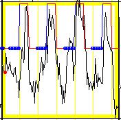
8 Idealized fmri Study Activate Control Activate Control Activate Control T2* Images Voxel Time Course (one voxel)
9 Voxel Time Course Voxel T ime Course Series Image Number Voxel Int ensit y 840
10 fmri Signal from each voxel is characterized by φ :Phase A : Amplitude ω : Frequency T2*
11 T2* has two components 1/T2* = γ B + 1/T2 T2 = nmr spin-spin dephasing γ B = magnetic field inhomogeneity The BOLD phenomenon changes γ B
12 Magnetic Field Inhomogeneity results from Inhomogeneity of the magnet s B0 field Variation in magnetic susceptibility of patient s tissues
13 Magnetic Field Inhomogeneity occurs Near the boundaries of tissues having disparate susceptibility tissue/ air tissue/ bone The sphenoid sinus causes magnetic susceptibility artifacts in EPI images Inferior frontal cortex Inferior lateral temporal cortex
14 Magnetic Field Inhomogeneity also results from The BOLD effect Differences in magnetic susceptibility around a paramagnetic deoxyhemoglobin molecule Changes in magnetic susceptibility around a small blood vessel (capillary, venule, small vein) that has an increased concentration/fraction of oxygenated hemoglobin
15 T2* weighted images are used in fmri to demonstrate changes in magnetic susceptibility associated with the BOLD effect
16 Signal in a T2* weighted BOLD image is affected by Blood volume (CBV) Blood flow (CBF) Arterial hemoglobin concentration Venous hemoglobin concentration Oxygen extraction rate Hematocrit
17 Signal in a T2* weighted BOLD image is thought to be inversely proportional to the number of deoxyhemoglobin molecules in the voxel
18 T2* Contrast is available via: Conventional gradient echo techniques with one RF transmission per phase encoded line of k-space FLASH GRASS FISP
19 T2* Contrast is available via: Single shot EPI techniques with a complete survey of k-space (and subsequent image reconstruction) for each RF transmission EPI - SE techniques EPI - GE techniques Multi-shot techniques (more than one RF transmission per image; more than one echo per RF transmission)
20 EPI Images EPI - GE Technique (T2* weighted)
21 To Map Brain Function Acquire T2* weighted images during a brain task Images will have slightly higher intensity in active brain regions Acquire T2* weighted images during a control state with the brain task suspended Statistically subtract control images from task images to map areas of brain activation associated with the task
22 An fmri Exam involves: Brain Activation Paradigm Task presentation systems Patient response monitoring Image acquisition Synchronization Data processing
23 A brain activation paradigm Control task Activation task: a task designed to produce brain activation (e.g. motor, sensory, language, memory) Try to avoid Habituation Learning Inattention
24 Event Related fmri fmri need not be constrained by task on/ task off block designs By measuring BOLD response to brief stimuli (typically presented at irregular intervals), it is possible to characterize the hemodynamic response function
25
26 Noun Verb Task


27 Right Hand Motor Task

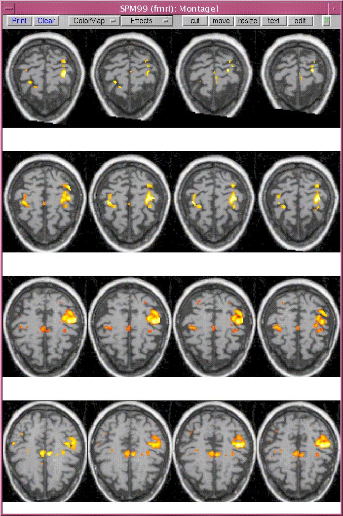
28 Left Hand Motor Task

29 Auditory Task
30 Task presentation systems Audio system w/ Input for external sound sources such as computer audio, VCR, stereo, microphone Attenuate gradient noise while enabling communication Non-pneumatic audio offers improved quality Electrical stimulation A dark room and dark magnet bore
31 Task presentation systems Visual presentation Slide projector LCD panel & overhead projector & rear screen projection Large screen LCD MRI projection systems Electronic goggles MRI compatible corrective lenses
32 fmri Projection System

33 fmri I/O Devices
34 Response monitoring In order to document whether the patient is doing the task Key pad Joystick Track ball

35 MRI Control Room
36 Synchronization Task presentation and patient response monitoring should be in synchrony with the acquisition of T2* weighted MRI images Ideally, all of this apparatus should be controlled by one host.
37 Signal conditioning Wires and tubes that pass in/out of the exam room must pass through RF filters or RF waveguides
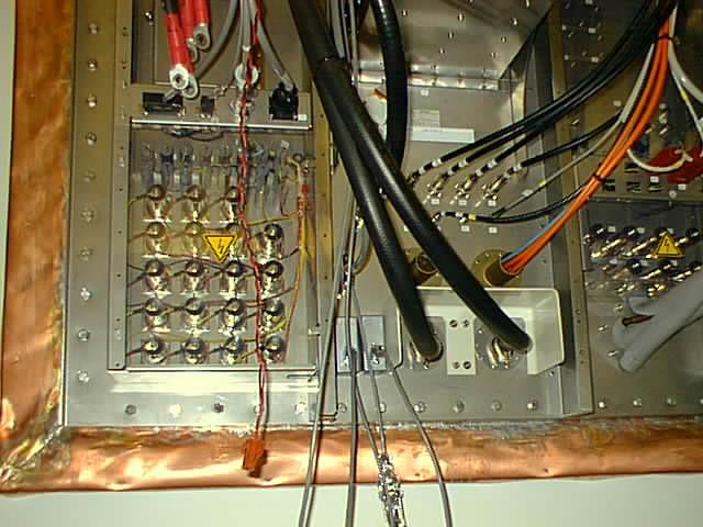
38 Penetration Panel
39 Image acquisition T2* weighted images are acquired while the patient alternates between periods having an activating task and periods of a control state.
40 Motor task Begin imaging Control state Rest for 30 seconds Activating task Finger tapping w/ Rt. hand for 30 seconds Alternate these activities for 6 minutes
41 Motor task Acquire 120 T2* weighted MRI image sets Each set: Transverse oblique 32 Slices Primary motor cortex to cerebellum
42 Motor task Thus for each brain voxel, we have temporal data (the voxel time course) having 120 data points (a sample every three seconds) There can be hundreds of thousands of brain voxels
43 Motor task 3840 total EPI images Some scanners limit the number of images in one study (512, 2048, etc.)

44 Sagittal Localizer
45 We use one of two pulse sequences ep2d_fid_66b1190_62.ekc ep2d_fid_60b2080_62_64.ekc
46 ep2d_fid_66b1190_62.ekc Gradient refocused single shot EPI technique Uses optional hybrid gradient overdrive amplifiers Matrix: 128x128
47 ep2d_fid_66b1190_62.ekc TE: 66 msec TR: 3 sec We acquire 22 slices every 5 seconds
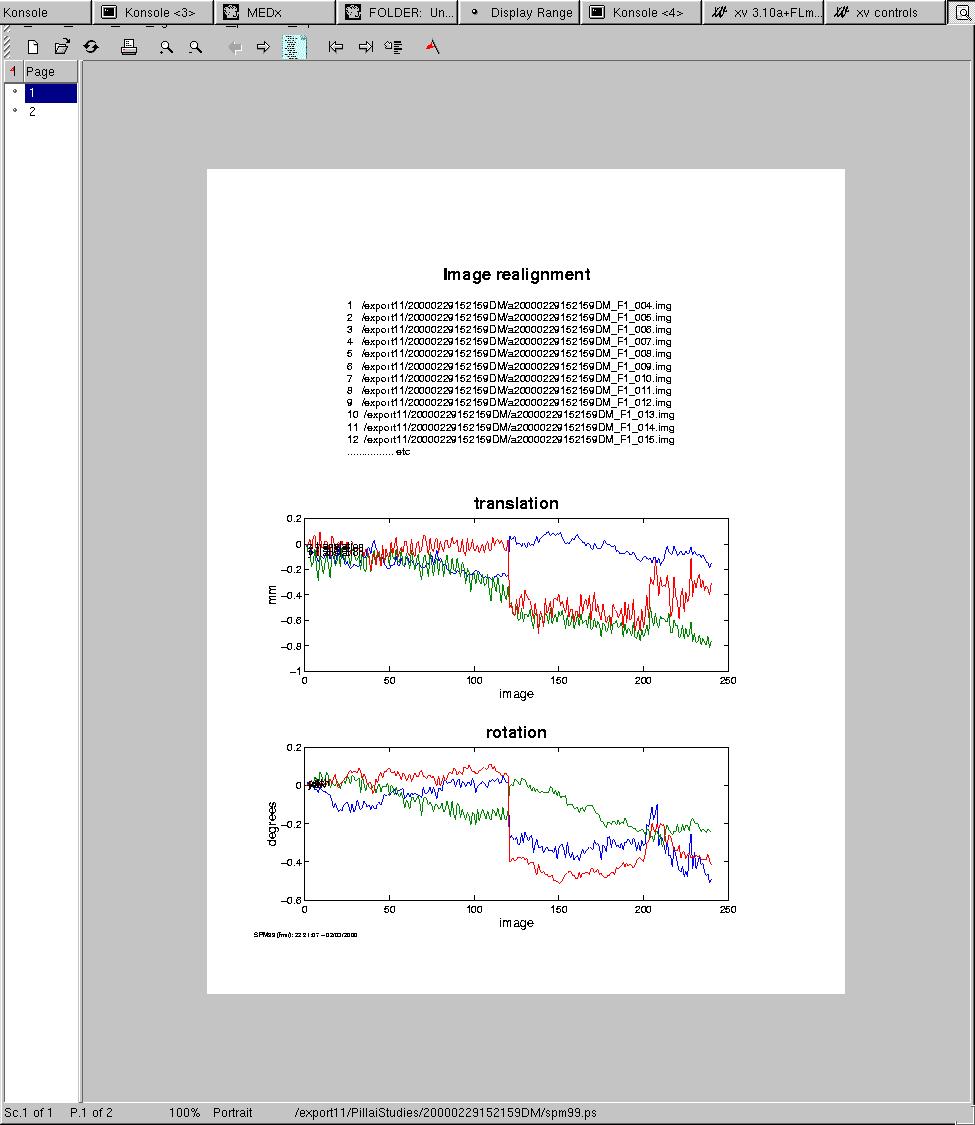
48 ep2d_fid_66b1190_62.ekc 22 slices 2.0 mm skip 1.0 ascending Orientation: Transverse oblique: T --> C, - 15 o
49 ep2d_fid_66b1190_62.ekc Phase encode: A P (for symmetry) FOV: 230x230 mm: Voxels: 1.8 x 1.8 x 3.0 mm
50 ep2d_fid_66b1190_62.ekc
51 ep2d_fid_60b2080_62_64.ekc Enables up to 64 slices in one file We do 32 slices every 3 seconds 64 x 64 matrix All slices are written into x 512 image (mosaic mode) Reduces I/O time necessary to write data to disk

52 ep2d_fid_60b2080_62_64.ekc
53 GRASS fmri Technique TR: 70 msec TE: 40 msec Flip angle: 40 o Matrix: 128 x 256 Slice thickness: 6 mm Acquisition: 9.4 sec per image
54 Computing options MRI scanner software AFNI STIMULATE SPM Other approaches Brain Voyager MEDx
55 Data processing may include the following Correction for differences in time of acquisition across slices Examine the data Motion detection Plot center-of-mass time course for each slice Stimulate View Cine loop of T2* weighted images As native signal intensity As a difference image (a. la. DSA) fmri activation in periphery of brain is an indication of motion
56 Data processing may include the following Motion correction Discard images showing obvious motion Image re-registration to correct for rigid body motion 2D (3 parameters) 3D (6 parameters)
57 Data processing may include the following Baseline flattening 0th order 1st order 2nd order fmri image calculation Clustering of activated voxels
58 Data processing may include the following Image fusion With T2* weighted images With T1 weighted images Multi Planar Reconstruction of fused images Volume rendering of fused images

59 Motion: Peripheral Brain Activation
60 3D Image Registration
61 Image numbe r Re f erence Value Reference Curve Ref erence Curve Series
62 Cross Correlation
63 fmri on T2* weighted EPI images

64 fmri on T1 weighted images

65 fmri on Orthogonal T1 weighted images
66 EPI (single shot) Sequence Problems Nyquist ghosts: N/2 ghosts caused by odd and even echo asymmetries Can apply a correction by measuring the asymmetry with a phase reference FID with the phase encode gradient switched off just prior to each single shot image

67 GHOSTS!
68 Artifacts in fmri images Subject motion Bulk Physiologic Cardiac Respiratory Acquisition time (long is bad: like mammography)
69 Artifacts in fmri images ROI (Are there magnetic susceptibility artifacts in the ROI) Geometric distortion Poor shimming results in additional geometric distortion (particularly in gradient recalled EPI images)

70 Susceptibility Artifact in Orthogonal T2* images

71 Phantom Susceptibility in a T2* Weighted Sequence
72 Physiologic Motion Bulk motion Settling
73 Physiologic Motion Respiration Susceptibility changes caused by movement of chest during respiration (phase changes of 2o 6o at 40 msec) Slow varying Affects inferior images Proportional to B0 Proportional to TE
74 Physiologic Motion Cardiac pulsations Non-rigid body movement of brain parenchyma caused by cardiac pulsations (Brain changes shape) Image dependent brain deformation in EPI Image dependent motion artifact in FLASH Can produce artifacts as large as a few % of BOLD contrast Occurs near CSF (Brainstem, Cerebellum)
75 Motion of as little as 0.1 pixels can seriously degrade fmri images because the MRI signal variation from pixel to pixel is larger than the BOLD effect that is being measured.
76 Motion prevention An Ounce of Prevention is Worth A Pound of Cure Padding Expandable foam (Alpha Cradle) Vacuum bags Hammock Bite bar Contour masks
77 Motion prevention Real time Pressure sensors Infrared systems

78 fmri Head Fixation
White Pixel Artifact. Caused by a noise spike during acquisition Spike in K-space <--> sinusoid in image space
 White Pixel Artifact Caused by a noise spike during acquisition Spike in K-space sinusoid in image space Susceptibility Artifacts Off-resonance artifacts caused by adjacent regions with different
CHAPTER 9: Magnetic Susceptibility Effects in High Field MRI
 Figure 1. In the brain, the gray matter has substantially more blood vessels and capillaries than white matter. The magnified image on the right displays the rich vasculature in gray matter forming porous,
MRI Physics II: Gradients, Imaging
 MRI Physics II: Gradients, Imaging Douglas C., Ph.D. Dept. of Biomedical Engineering University of Michigan, Ann Arbor Magnetic Fields in MRI B 0 The main magnetic field. Always on (0.5-7 T) Magnetizes
Basic fmri Design and Analysis. Preprocessing
 Basic fmri Design and Analysis Preprocessing fmri Preprocessing Slice timing correction Geometric distortion correction Head motion correction Temporal filtering Intensity normalization Spatial filtering
SPM8 for Basic and Clinical Investigators. Preprocessing. fmri Preprocessing
 SPM8 for Basic and Clinical Investigators Preprocessing fmri Preprocessing Slice timing correction Geometric distortion correction Head motion correction Temporal filtering Intensity normalization Spatial
FMRI Pre-Processing and Model- Based Statistics
 FMRI Pre-Processing and Model- Based Statistics Brief intro to FMRI experiments and analysis FMRI pre-stats image processing Simple Single-Subject Statistics Multi-Level FMRI Analysis Advanced FMRI Analysis
HST.583 Functional Magnetic Resonance Imaging: Data Acquisition and Analysis Fall 2008
 MIT OpenCourseWare http://ocw.mit.edu HST.583 Functional Magnetic Resonance Imaging: Data Acquisition and Analysis Fall 2008 For information about citing these materials or our Terms of Use, visit: http://ocw.mit.edu/terms.
Diffusion MRI Acquisition. Karla Miller FMRIB Centre, University of Oxford
 Diffusion MRI Acquisition Karla Miller FMRIB Centre, University of Oxford karla@fmrib.ox.ac.uk Diffusion Imaging How is diffusion weighting achieved? How is the image acquired? What are the limitations,
Functional MRI in Clinical Research and Practice Preprocessing
 Functional MRI in Clinical Research and Practice Preprocessing fmri Preprocessing Slice timing correction Geometric distortion correction Head motion correction Temporal filtering Intensity normalization
EPI Data Are Acquired Serially. EPI Data Are Acquired Serially 10/23/2011. Functional Connectivity Preprocessing. fmri Preprocessing
 Functional Connectivity Preprocessing Geometric distortion Head motion Geometric distortion Head motion EPI Data Are Acquired Serially EPI Data Are Acquired Serially descending 1 EPI Data Are Acquired
SPM8 for Basic and Clinical Investigators. Preprocessing
 SPM8 for Basic and Clinical Investigators Preprocessing fmri Preprocessing Slice timing correction Geometric distortion correction Head motion correction Temporal filtering Intensity normalization Spatial
HST.583 Functional Magnetic Resonance Imaging: Data Acquisition and Analysis Fall 2006
 MIT OpenCourseWare http://ocw.mit.edu HST.583 Functional Magnetic Resonance Imaging: Data Acquisition and Analysis Fall 2006 For information about citing these materials or our Terms of Use, visit: http://ocw.mit.edu/terms.
MRI. When to use What sequences. Outline 2012/09/19. Sequence: Definition. Basic Principles: Step 2. Basic Principles: Step 1. Govind Chavhan, MD
 MRI When to use What sequences Govind Chavhan, MD Assistant Professor and Staff Radiologist The Hospital For Sick Children, Toronto Planning Acquisition Post processing Interpretation Patient history and
MR Advance Techniques. Vascular Imaging. Class III
 MR Advance Techniques Vascular Imaging Class III 1 Vascular Imaging There are several methods that can be used to evaluate the cardiovascular systems with the use of MRI. MRI will aloud to evaluate morphology
Slide 1. Technical Aspects of Quality Control in Magnetic Resonance Imaging. Slide 2. Annual Compliance Testing. of MRI Systems.
 Slide 1 Technical Aspects of Quality Control in Magnetic Resonance Imaging Slide 2 Compliance Testing of MRI Systems, Ph.D. Department of Radiology Henry Ford Hospital, Detroit, MI Slide 3 Compliance Testing
Supplementary Figure 1
 Supplementary Figure 1 BOLD and CBV functional maps showing EPI versus line-scanning FLASH fmri. A. Colored BOLD and CBV functional maps are shown in the highlighted window (green frame) of the raw EPI
Role of Parallel Imaging in High Field Functional MRI
 Role of Parallel Imaging in High Field Functional MRI Douglas C. Noll & Bradley P. Sutton Department of Biomedical Engineering, University of Michigan Supported by NIH Grant DA15410 & The Whitaker Foundation
This Time. fmri Data analysis
 This Time Reslice example Spatial Normalization Noise in fmri Methods for estimating and correcting for physiologic noise SPM Example Spatial Normalization: Remind ourselves what a typical functional image
Lucy Phantom MR Grid Evaluation
 Lucy Phantom MR Grid Evaluation Anil Sethi, PhD Loyola University Medical Center, Maywood, IL 60153 November 2015 I. Introduction: The MR distortion grid, used as an insert with Lucy 3D QA phantom, is
HST.583 Functional Magnetic Resonance Imaging: Data Acquisition and Analysis Fall 2008
 MIT OpenCourseWare http://ocw.mit.edu HST.583 Functional Magnetic Resonance Imaging: Data Acquisition and Analysis Fall 2008 For information about citing these materials or our Terms of Use, visit: http://ocw.mit.edu/terms.
Module 5: Dynamic Imaging and Phase Sharing. (true-fisp, TRICKS, CAPR, DISTAL, DISCO, HYPR) Review. Improving Temporal Resolution.
 Review. Improving Temporal Resolution.") MRES 7005 - Fast Imaging Techniques Module 5: Dynamic Imaging and Phase Sharing (true-fisp, TRICKS, CAPR, DISTAL, DISCO, HYPR) Review Improving Temporal Resolution True-FISP (I) True-FISP (II) Keyhole
MRES 7005 - Fast Imaging Techniques Module 5: Dynamic Imaging and Phase Sharing (true-fisp, TRICKS, CAPR, DISTAL, DISCO, HYPR) Review Improving Temporal Resolution True-FISP (I) True-FISP (II) Keyhole
Fmri Spatial Processing
 Educational Course: Fmri Spatial Processing Ray Razlighi Jun. 8, 2014 Spatial Processing Spatial Re-alignment Geometric distortion correction Spatial Normalization Smoothing Why, When, How, Which Why is
Clinical Importance. Aortic Stenosis. Aortic Regurgitation. Ultrasound vs. MRI. Carotid Artery Stenosis
 Clinical Importance Rapid cardiovascular flow quantitation using sliceselective Fourier velocity encoding with spiral readouts Valve disease affects 10% of patients with heart disease in the U.S. Most
SPATIO-TEMPORAL DATA ANALYSIS WITH NON-LINEAR FILTERS: BRAIN MAPPING WITH fmri DATA
 Image Anal Stereol 2000;19:189-194 Original Research Paper SPATIO-TEMPORAL DATA ANALYSIS WITH NON-LINEAR FILTERS: BRAIN MAPPING WITH fmri DATA KARSTEN RODENACKER 1, KLAUS HAHN 1, GERHARD WINKLER 1 AND
Dynamic Contrast enhanced MRA
 Dynamic Contrast enhanced MRA Speaker: Yung-Chieh Chang Date : 106.07.22 Department of Radiology, Taichung Veterans General Hospital, Taichung, Taiwan 1 Outline Basic and advanced principles of Diffusion
Analysis of Functional MRI Timeseries Data Using Signal Processing Techniques
 Analysis of Functional MRI Timeseries Data Using Signal Processing Techniques Sea Chen Department of Biomedical Engineering Advisors: Dr. Charles A. Bouman and Dr. Mark J. Lowe S. Chen Final Exam October
COBRE Scan Information
 COBRE Scan Information Below is more information on the directory structure for the COBRE imaging data. Also below are the imaging parameters for each series. Directory structure: var/www/html/dropbox/1139_anonymized/human:
ADNI, ADNI_QH, SURVEY. Geometry. connection
 ADNI, ADNI_QH, SURVEY Geometry Coil selection = Head connection = d Multi coil Homogeneity correction ne FOV (mm) = 250.00 RFOV (%) = 100.00 Foldover suppression Matrix scan = 256 reconstruction = 256
Advanced MRI Techniques (and Applications)
") Advanced MRI Techniques (and Applications) Jeffry R. Alger, PhD Department of Neurology Ahmanson-Lovelace Brain Mapping Center Brain Research Institute Jonsson Comprehensive Cancer Center University of
Advanced MRI Techniques (and Applications) Jeffry R. Alger, PhD Department of Neurology Ahmanson-Lovelace Brain Mapping Center Brain Research Institute Jonsson Comprehensive Cancer Center University of
(a Scrhon5 R2iwd b. P)jc%z 5. ivcr3. 1. I. ZOms Xn,s. 1E IDrAS boms. EE225E/BIOE265 Spring 2013 Principles of MRI. Assignment 8 Solutions
jc%z 5. ivcr3. 1. I. ZOms Xn,s. 1E IDrAS boms. EE225E/BIOE265 Spring 2013 Principles of MRI. Assignment 8 Solutions") EE225E/BIOE265 Spring 2013 Principles of MRI Miki Lustig Assignment 8 Solutions 1. Nishimura 7.1 P)jc%z 5 ivcr3. 1. I Due Wednesday April 10th, 2013 (a Scrhon5 R2iwd b 0 ZOms Xn,s r cx > qs 4-4 8ni6 4
EE225E/BIOE265 Spring 2013 Principles of MRI Miki Lustig Assignment 8 Solutions 1. Nishimura 7.1 P)jc%z 5 ivcr3. 1. I Due Wednesday April 10th, 2013 (a Scrhon5 R2iwd b 0 ZOms Xn,s r cx > qs 4-4 8ni6 4
HST.583 Functional Magnetic Resonance Imaging: Data Acquisition and Analysis Fall 2006
 MIT OpenCourseWare http://ocw.mit.edu HST.583 Functional Magnetic Resonance Imaging: Data Acquisition and Analysis Fall 2006 For information about citing these materials or our Terms of Use, visit: http://ocw.mit.edu/terms.
Philips MRI Protocol Dump Created on Comment Software Stream
 Page 1 of 5 Philips MRI Protocol Dump Created on 2/17/2011 4:11:01 PM Comment Created by ExamCard_to_XML with inputs: "J:\ADNI GO - ADNI 2 Phantom5.ExamCard" on system (BU SCHOOL OF MEDICINE :: 192.168.71.10)
Functional MRI data preprocessing. Cyril Pernet, PhD
 Functional MRI data preprocessing Cyril Pernet, PhD Data have been acquired, what s s next? time No matter the design, multiple volumes (made from multiple slices) have been acquired in time. Before getting
Lab Location: MRI, B2, Cardinal Carter Wing, St. Michael s Hospital, 30 Bond Street
 Lab Location: MRI, B2, Cardinal Carter Wing, St. Michael s Hospital, 30 Bond Street MRI is located in the sub basement of CC wing. From Queen or Victoria, follow the baby blue arrows and ride the CC south
Abbie M. Diak, PhD Loyola University Medical Center Dept. of Radiation Oncology
 Abbie M. Diak, PhD Loyola University Medical Center Dept. of Radiation Oncology Outline High Spectral and Spatial Resolution MR Imaging (HiSS) What it is How to do it Ways to use it HiSS for Radiation
Following on from the two previous chapters, which considered the model of the
 Chapter 5 Simulator validation Following on from the two previous chapters, which considered the model of the simulation process and how this model was implemented in software, this chapter is concerned
SIEMENS MAGNETOM Verio syngo MR B17
 \\USER\Dr. Behrmann\routine\Ilan\ep2d_bold_PMU_resting TA: 8:06 PAT: Voxel size: 3.03.03.0 mm Rel. SNR: 1.00 USER: ep2d_bold_pmu Properties Special sat. Prio Recon System Before measurement Body After
Magnetic Resonance Elastography (MRE) of Liver Disease
 of Liver Disease") Magnetic Resonance Elastography (MRE) of Liver Disease Authored by: Jennifer Dolan Fox, PhD VirtualScopics Inc. jennifer_fox@virtualscopics.com 1-585-249-6231 1. Overview of MRE Imaging MRE is a magnetic
Magnetic Resonance Elastography (MRE) of Liver Disease Authored by: Jennifer Dolan Fox, PhD VirtualScopics Inc. jennifer_fox@virtualscopics.com 1-585-249-6231 1. Overview of MRE Imaging MRE is a magnetic
Last Time. This Time. Thru-plane dephasing: worse at long TE. Local susceptibility gradients: thru-plane dephasing
 Motion Correction Last Time Mutual Information Optimiation Decoupling Translation & Rotation Interpolation SPM Example (Least Squares & MI) A Simple Derivation This Time Reslice example SPM Example : Remind
Scan Acceleration with Rapid Gradient-Echo
 Scan Acceleration with Rapid Gradient-Echo Hsiao-Wen Chung ( 鍾孝文 ), Ph.D., Professor Dept. Electrical Engineering, National Taiwan Univ. Dept. Radiology, Tri-Service General Hospital 1 of 214 The Need
fmri Image Preprocessing
 fmri Image Preprocessing Rick Hoge, Ph.D. Laboratoire de neuroimagerie vasculaire (LINeV) Centre de recherche de l institut universitaire de gériatrie de Montréal, Université de Montréal Outline Motion
n o r d i c B r a i n E x Tutorial DSC Module
 m a k i n g f u n c t i o n a l M R I e a s y n o r d i c B r a i n E x Tutorial DSC Module Please note that this tutorial is for the latest released nordicbrainex. If you are using an older version please
Breast MRI Accreditation Program Clinical Image Quality Guide
 Breast MRI Accreditation Program Clinical Image Quality Guide Introduction This document provides guidance on breast MRI clinical image quality and describes the criteria used by the ACR Breast MRI Accreditation
Supplementary methods
 Supplementary methods This section provides additional technical details on the sample, the applied imaging and analysis steps and methods. Structural imaging Trained radiographers placed all participants
The SIMRI project A versatile and interactive MRI simulator *
 COST B21 Meeting, Lodz, 6-9 Oct. 2005 The SIMRI project A versatile and interactive MRI simulator * H. Benoit-Cattin 1, G. Collewet 2, B. Belaroussi 1, H. Saint-Jalmes 3, C. Odet 1 1 CREATIS, UMR CNRS
CORRECTION OF IMAGE DISTORTION IN ECHO PLANAR IMAGE SERIES USING PHASE AND INTENSITY. Ning Xu. Dissertation. Submitted to the Faculty of the
 CORRECTION OF IMAGE DISTORTION IN ECHO PLANAR IMAGE SERIES USING PHASE AND INTENSITY By Ning Xu Dissertation Submitted to the Faculty of the Graduate School of Vanderbilt University in partial fulfillment
Imaging Notes, Part IV
 BME 483 MRI Notes 34 page 1 Imaging Notes, Part IV Slice Selective Excitation The most common approach for dealing with the 3 rd (z) dimension is to use slice selective excitation. This is done by applying
Brilliance CT Big Bore.
 1 2 2 There are two methods of RCCT acquisition in widespread clinical use: cine axial and helical. In RCCT with cine axial acquisition, repeat CT images are taken each couch position while recording respiration.
MEDICAL IMAGE ANALYSIS
 SECOND EDITION MEDICAL IMAGE ANALYSIS ATAM P. DHAWAN g, A B IEEE Engineering in Medicine and Biology Society, Sponsor IEEE Press Series in Biomedical Engineering Metin Akay, Series Editor +IEEE IEEE PRESS
Module 4. K-Space Symmetry. Review. K-Space Review. K-Space Symmetry. Partial or Fractional Echo. Half or Partial Fourier HASTE
 MRES 7005 - Fast Imaging Techniques Module 4 K-Space Symmetry Review K-Space Review K-Space Symmetry Partial or Fractional Echo Half or Partial Fourier HASTE Conditions for successful reconstruction Interpolation
The cost of parallel imaging in functional MRI of the human brain
 Magnetic Resonance Imaging 24 (2006) 1 5 Original contributions The cost of parallel imaging in functional MRI of the human brain Henry Lqtcke4, Klaus-Dietmar Merboldt, Jens Frahm Biomedizinische NMR Forschungs
New Technology Allows Multiple Image Contrasts in a Single Scan
 These images were acquired with an investigational device. PD T2 T2 FLAIR T1 MAP T1 FLAIR PSIR T1 New Technology Allows Multiple Image Contrasts in a Single Scan MR exams can be time consuming. A typical
ACQUIRING AND PROCESSING SUSCEPTIBILITY WEIGHTED IMAGING (SWI) DATA ON GE 3.0T
 DATA ON GE 3.0T") ACQUIRING AND PROCESSING SUSCEPTIBILITY WEIGHTED IMAGING (SWI) DATA ON GE 3.0T Revision date: 12/13/2010 Overview Susceptibility Weighted Imaging (SWI) is a relatively new data acquisition and processing
ACQUIRING AND PROCESSING SUSCEPTIBILITY WEIGHTED IMAGING (SWI) DATA ON GE 3.0T Revision date: 12/13/2010 Overview Susceptibility Weighted Imaging (SWI) is a relatively new data acquisition and processing
The organization of the human cerebral cortex estimated by intrinsic functional connectivity
 1 The organization of the human cerebral cortex estimated by intrinsic functional connectivity Journal: Journal of Neurophysiology Author: B. T. Thomas Yeo, et al Link: https://www.ncbi.nlm.nih.gov/pubmed/21653723
BME I5000: Biomedical Imaging
 1 Lucas Parra, CCNY BME I5000: Biomedical Imaging Lecture 4 Computed Tomography Lucas C. Parra, parra@ccny.cuny.edu some slides inspired by lecture notes of Andreas H. Hilscher at Columbia University.
Motion Artifacts and Suppression in MRI At a Glance
 Motion Artifacts and Suppression in MRI At a Glance Xiaodong Zhong, PhD MR R&D Collaborations Siemens Healthcare MRI Motion Artifacts and Suppression At a Glance Outline Background Physics Common Motion
Acquisition Methods for fmri at 7 Tesla
 Acquisition Methods for fmri at 7 Tesla Peter J. Koopmans, Nuffield department for clinical neurosciences, University of Oxford, UK fmri at 7 Tesla is mostly used in the context of high spatial resolution
Correction for EPI Distortions Using Multi-Echo Gradient-Echo Imaging
 Correction for EPI Distortions Using Multi-Echo Gradient-Echo Imaging Nan-kuei Chen and Alice M. Wyrwicz* Magnetic Resonance in Medicine 41:1206 1213 (1999) A novel and effective technique is described
ThE ultimate, INTuITIVE Mr INTErFAcE
 ThE ultimate, INTuITIVE Mr INTErFAcE Empowering you to do more The revolutionary Toshiba M-power user interface takes Mr performance and flexibility to levels higher than ever before. M-power is able to
A Virtual MR Scanner for Education
 A Virtual MR Scanner for Education Hackländer T, Schalla C, Trümper A, Mertens H, Hiltner J, Cramer BM Hospitals of the University Witten/Herdecke, Department of Radiology Wuppertal, Germany Purpose A
Use of MRI in Radiotherapy: Technical Consideration
 Use of MRI in Radiotherapy: Technical Consideration Yanle Hu, PhD Department of Radiation Oncology, Mayo Clinic Arizona 04/07/2018 2015 MFMER slide-1 Conflict of Interest: None 2015 MFMER slide-2 Objectives
Noise and Artifacts in FMRI
 Noise and Artifacts in FMRI Instructor: Luis Hernandez-Garcia, Ph.D. Associate Research Professor FMRI Laboratory, Biomedical Engineering FMRI analysis - synopsis of what you ll do next week 1. Formulate
M R I Physics Course
 M R I Physics Course Multichannel Technology & Parallel Imaging Nathan Yanasak, Ph.D. Jerry Allison Ph.D. Tom Lavin, B.S. Department of Radiology Medical College of Georgia References: 1) The Physics of
Introduction to Neuroimaging Janaina Mourao-Miranda
 Introduction to Neuroimaging Janaina Mourao-Miranda Neuroimaging techniques have changed the way neuroscientists address questions about functional anatomy, especially in relation to behavior and clinical
HST.583 Functional Magnetic Resonance Imaging: Data Acquisition and Analysis Fall 2008
 MIT OpenCourseWare http://ocw.mit.edu HST.583 Functional Magnetic Resonance Imaging: Data Acquisition and Analysis Fall 2008 For information about citing these materials or our Terms of Use, visit: http://ocw.mit.edu/terms.
Applications Guide for Interleaved
 Applications Guide for Interleaved rephase/dephase MRAV Authors: Yongquan Ye, Ph.D. Dongmei Wu, MS. Tested MAGNETOM Systems : 7TZ, TRIO a Tim System, Verio MR B15A (N4_VB15A_LATEST_20070519) MR B17A (N4_VB17A_LATEST_20090307_P8)
Removal of EPI Nyquist Ghost Artifacts With Two- Dimensional Phase Correction
 Removal of EPI Nyquist Ghost Artifacts With Two- Dimensional Phase Correction Nan-kuei Chen 1,5 and Alice M. Wyrwicz 4 * Magnetic Resonance in Medicine 51:147 153 (004) Odd even echo inconsistencies result
Table of Contents. IntroLab < SPMLabs < Dynevor TWiki
 Table of Contents Lab 1: Introduction to SPM and data checking...1 Goals of this Lab...1 Prerequisites...1 An SPM Installation...1 SPM Defaults...2 L/R Brain Orientation...2 Memory Use for Data Processing...2
Journal of Articles in Support of The Null Hypothesis
 Data Preprocessing Martin M. Monti, PhD UCLA Psychology NITP 2016 Typical (task-based) fmri analysis sequence Image Pre-processing Single Subject Analysis Group Analysis Journal of Articles in Support
This exercise uses one anatomical data set (ANAT1) and two functional data sets (FUNC1 and FUNC2).
 and two functional data sets (FUNC1 and FUNC2).") Exploring Brain Anatomy This week s exercises will let you explore the anatomical organization of the brain to learn some of its basic properties, as well as the location of different structures. The human
Exploring Brain Anatomy This week s exercises will let you explore the anatomical organization of the brain to learn some of its basic properties, as well as the location of different structures. The human
Correction of Partial Volume Effects in Arterial Spin Labeling MRI
 Correction of Partial Volume Effects in Arterial Spin Labeling MRI By: Tracy Ssali Supervisors: Dr. Keith St. Lawrence and Udunna Anazodo Medical Biophysics 3970Z Six Week Project April 13 th 2012 Introduction
Partial k-space Reconstruction
 Chapter 2 Partial k-space Reconstruction 2.1 Motivation for Partial k- Space Reconstruction a) Magnitude b) Phase In theory, most MRI images depict the spin density as a function of position, and hence
Image Acquisition Systems
 Image Acquisition Systems Goals and Terminology Conventional Radiography Axial Tomography Computer Axial Tomography (CAT) Magnetic Resonance Imaging (MRI) PET, SPECT Ultrasound Microscopy Imaging ITCS
Sources of Distortion in Functional MRI Data
 Human Brain Mapping 8:80 85(1999) Sources of Distortion in Functional MRI Data Peter Jezzard* and Stuart Clare FMRIB Centre, Department of Clinical Neurology, University of Oxford, Oxford, UK Abstract:
Partial k-space Recconstruction
 Partial k-space Recconstruction John Pauly September 29, 2005 1 Motivation for Partial k-space Reconstruction a) Magnitude b) Phase In theory, most MRI images depict the spin density as a function of position,
Motion Correction in fmri by Mapping Slice-to-Volume with Concurrent Field-Inhomogeneity Correction
 Motion Correction in fmri by Mapping Slice-to-Volume with Concurrent Field-Inhomogeneity Correction Desmond T.B. Yeo 1,2, Jeffery A. Fessler 2, and Boklye Kim 1 1 Department of Radiology, University of
A more accurate account of the effect of k-space sampling and signal decay on the effective spatial resolution in functional MRI
 A more accurate account of the effect of k-space sampling and signal decay on the effective spatial resolution in functional MRI Denis Chaimow 1 and Amir Shmuel 1,2 1 Centre for Magnetic Resonance Research
Accelerated MRI Techniques: Basics of Parallel Imaging and Compressed Sensing
 Accelerated MRI Techniques: Basics of Parallel Imaging and Compressed Sensing Peng Hu, Ph.D. Associate Professor Department of Radiological Sciences PengHu@mednet.ucla.edu 310-267-6838 MRI... MRI has low
Basic principles of MR image analysis. Basic principles of MR image analysis. Basic principles of MR image analysis
 Basic principles of MR image analysis Basic principles of MR image analysis Julien Milles Leiden University Medical Center Terminology of fmri Brain extraction Registration Linear registration Non-linear
I.e. Sex differences in child appetitive traits and Eating in the Absence of Hunger:
 Supplementary Materials I. Evidence of sex differences on eating behavior in children I.e. Sex differences in child appetitive traits and Eating in the Absence of Hunger: Table 2. Parent Report for Child
CS/NEUR125 Brains, Minds, and Machines. Due: Wednesday, April 5
 CS/NEUR125 Brains, Minds, and Machines Lab 8: Using fmri to Discover Language Areas in the Brain Due: Wednesday, April 5 In this lab, you will analyze fmri data from an experiment that was designed to
MRI Imaging Options. Frank R. Korosec, Ph.D. Departments of Radiology and Medical Physics University of Wisconsin Madison
 MRI Imaging Options Frank R. Korosec, Ph.D. Departments of Radiolog and Medical Phsics Universit of Wisconsin Madison f.korosec@hosp.wisc.edu As MR imaging becomes more developed, more imaging options
Field Maps. 1 Field Map Acquisition. John Pauly. October 5, 2005
 Field Maps John Pauly October 5, 25 The acquisition and reconstruction of frequency, or field, maps is important for both the acquisition of MRI data, and for its reconstruction. Many of the imaging methods
surface Image reconstruction: 2D Fourier Transform
 2/1/217 Chapter 2-3 K-space Intro to k-space sampling (chap 3) Frequenc encoding and Discrete sampling (chap 2) Point Spread Function K-space properties K-space sampling principles (chap 3) Basic Contrast
Bayesian Inference of Hemodynamic Changes in Functional Arterial Spin Labeling Data
 Bayesian Inference of Hemodynamic Changes in Functional Arterial Spin Labeling Data Mark W. Woolrich, 1, * Peter Chiarelli, 1 Daniel Gallichan, 1 Joanna Perthen, 2 and Thomas T. Liu 2 Magnetic Resonance
UNIVERSITY OF SOUTHAMPTON
 UNIVERSITY OF SOUTHAMPTON PHYS2007W1 SEMESTER 2 EXAMINATION 2014-2015 MEDICAL PHYSICS Duration: 120 MINS (2 hours) This paper contains 10 questions. Answer all questions in Section A and only two questions
Magnetic Resonance Angiography
 Magnetic Resonance Angiography Course: Advance MRI (BIOE 594) Instructors: Dr Xiaohong Joe Zhou Dr. Shadi Othman By, Nayan Pasad Phase Contrast Angiography By Moran 1982, Bryan et. Al. 1984 and Moran et.
MB-EPI PCASL. Release Notes for Version February 2015
 MB-EPI PCASL Release Notes for Version 1.0 20 February 2015 1 Background High-resolution arterial spin labeling (ASL) imaging is highly desirable in both neuroscience research and clinical applications
CP Generalize Concepts in Abstract Multi-dimensional Image Model Component Semantics. David Clunie.
 CP-1390 - Generalize Concepts in Abstract Multi-dimensional Image Model Semantics Page 1 STATUS Date of Last Update Person Assigned Submitter Name Submission Date Assigned 2014/06/09 David Clunie mailto:dclunie@dclunie.com
REDUCTION OF NOISE DUE TO TASK CORRELATED MOTION IN EVENT RELATED OVERT WORD GENERATION FUNCTIONAL MAGNETIC RESONANCE IMAGING PARADIGMS
 REDUCTION OF NOISE DUE TO TASK CORRELATED MOTION IN EVENT RELATED OVERT WORD GENERATION FUNCTIONAL MAGNETIC RESONANCE IMAGING PARADIGMS By KAUNDINYA S. GOPINATH A DISSERTATION PRESENTED TO THE GRADUATE
XI Conference "Medical Informatics & Technologies" VALIDITY OF MRI BRAIN PERFUSION IMAGING METHOD
 XI Conference "Medical Informatics & Technologies" - 2006 medical imaging, MRI, brain perfusion Bartosz KARCZEWSKI 1, Jacek RUMIŃSKI 1 VALIDITY OF MRI BRAIN PERFUSION IMAGING METHOD Brain perfusion imaging
Introduction to fmri. Pre-processing
 Introduction to fmri Pre-processing Tibor Auer Department of Psychology Research Fellow in MRI Data Types Anatomical data: T 1 -weighted, 3D, 1/subject or session - (ME)MPRAGE/FLASH sequence, undistorted
SIEMENS MAGNETOM Avanto syngo MR B15
 \\USER\INVESTIGATORS\Ravi\ADNI-phantom\QC Phantom-Localizer TA: 0:10 PAT: Voxel size: 1.9 1.5 8.0 mm Rel. SNR: 1.00 SIEMENS: gre Properties Prio Recon Before measurement After measurement Load to viewer
High performance MRI simulations of motion on multi-gpu systems
 Xanthis et al. Journal of Cardiovascular Magnetic Resonance 2014, 16:48 RESEARCH Open Access High performance MRI simulations of motion on multi-gpu systems Christos G Xanthis 1,2, Ioannis E Venetis 3
Cognitive States Detection in fmri Data Analysis using incremental PCA
 Department of Computer Engineering Cognitive States Detection in fmri Data Analysis using incremental PCA Hoang Trong Minh Tuan, Yonggwan Won*, Hyung-Jeong Yang International Conference on Computational
Computational Medical Imaging Analysis
 Computational Medical Imaging Analysis Chapter 2: Image Acquisition Systems Jun Zhang Laboratory for Computational Medical Imaging & Data Analysis Department of Computer Science University of Kentucky
GE Healthcare CLINICAL GALLERY. Discovery * MR750w 3.0T. This brochure is intended for European healthcare professionals.
 GE Healthcare CLINICAL GALLERY Discovery * MR750w 3.0T This brochure is intended for European healthcare professionals. NEURO PROPELLER delivers high resolution, motion insensitive imaging in all planes.
design as a constrained maximization problem. In principle, CODE seeks to maximize the b-value, defined as, where
 Optimal design of motion-compensated diffusion gradient waveforms Óscar Peña-Nogales 1, Rodrigo de Luis-Garcia 1, Santiago Aja-Fernández 1,Yuxin Zhang 2,3, James H. Holmes 2,Diego Hernando 2,3 1 Laboratorio
Medical Imaging Image Quality, Diffusion MRI, Functional MRI
 G16.4426/EL5823/BE6203 Medical Imaging Image Quality, Diffusion MRI, Functional MRI Riccardo Lattanzi, Ph.D. Assistant Professor Department of Radiology, NYU School of Medicine Department of Electrical
Outline: Contrast-enhanced MRA
 Outline: Contrast-enhanced MRA Background Technique Clinical Indications Future Directions Disclosures: GE Health Care: Research support Consultant: Bracco, Bayer The Basics During rapid IV infusion, Gadolinium