Advanced Visual Medicine: Techniques for Visual Exploration & Analysis
|
|
|
- Archibald Willis
- 5 years ago
- Views:
Transcription


1 Advanced Visual Medicine: Techniques for Visual Exploration & Analysis Interactive Visualization of Multimodal Volume Data for Neurosurgical Planning Felix Ritter, MeVis Research Bremen
2 Multimodal Neurosurgical Planning 2

3 Multimodal Neurosurgical Planning Is a resection possible? What type of resection can be performed? What is the risk of the surgical intervention? What is an optimal access path to the lesion? 2


4 Topics to Work on Combining multimodal volume data Enhancement of risk structures Accentuation of spatial relations Reduction of interaction for exploration Visualization of access path to lesion Supporting brain-surface intervention 3
5 Medical Volume Data Common medical volume data: MRI (magnetic resonance imaging), high soft tissue contrast for visualization of anatomical details fmri (functional magnetic resonance imaging), for detection of the brain s activation areas DTI (diffusion tensor imaging), for reconstruction of the brain s nerve tracts CT (computer tomography), just used in special cases due the high radiation exposure, e.g. skull bone infiltrated by tumor tissue PET (positron emission tomography), nuclear imaging technique to detect functional processes 4
6 Multimodal Neurosurgical Planning A tempting assumption: - Combination of all available imaging modalities can identify all relevant structures! Limitations: - Models behind the involved techniques fmri can give hints about functional areas (Accumulation of oxygen) Fiber-Tracking can give hints about axonal pathways (Diffusion of water) - Usefulness of combinations A better interpretation: - Combination of different images may give additional information but may also introduce limitations and additional complexity 5
7 Multimodality Visualization 6
8 Multimodality Visualization 6

9 Multimodality Visualization? 6


 & fmri (yellow")
10 Multimodal Volume Rendering Visualization of CT & MR data for visualization of implanted electrodes for epilepsy surgery CT, MR & MRA MR (black/white), PET (red) & fmri (yellow and white) Johanna Beyer et al., VIS


11 Multimodal Volume Rendering Combined MRI, fmri visualization Friedemann Rößler et al., SimVis 2006 Werner Jainek et al., EuroVIS

12 Multimodal Volume Rendering MRI, fmri & DTI Alexander Köhn et al., EG 07 9


13 Dealing with Inaccuracies in Multimodal Neurosurgical Planning Accuracy is limited! Sources for limitations are manifold Limited accuracy may become an issue - If the awareness about the limitations gets lost - If the impression of high accuracy is falsely created - If decisions are made, that are not valid based on the given accuracy Limited accuracy turns into inaccuracy Inaccuracy is unavoidable We must be aware of it! 10
14 Sources of Inaccuracies Image registration - Spatial alignment is prerequisite for overlaying different images - Automatic rigid registration 11

15 Sources of Inaccuracies Image registration - Spatial alignment is prerequisite for overlaying different images - Automatic rigid registration 11
16 Sources of Inaccuracies Image registration - Spatial alignment is prerequisite for overlaying different images - Automatic rigid registration Problem: - Spatial deformation 12
17 Sources of Inaccuracies Image registration - Spatial alignment is prerequisite for overlaying different images - Automatic rigid registration Problem: - Spatial deformation 12
18 Sources of Inaccuracies Image registration - Spatial alignment is prerequisite for overlaying different images - Automatic rigid registration Problem: - Spatial deformation Problem: - Different voxel-sizes, slicethickness, interslice-gap 13


19 Sources of Inaccuracies Image registration - Spatial alignment is prerequisite for overlaying different images - Automatic rigid registration Problem: - Spatial deformation Problem: - Different voxel-sizes, slicethickness, interslice-gap 13
20 Extraction of Anatomical Structures T1-weighted ce T1-weighted substraction vessel mask T2-weighted FLAIR extraction brain mask masking masking masking masking 14


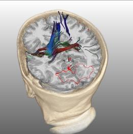
21 Risk Structures MRI fmri DTI anatomical data activation areas nerve tracts vessel and brain mask volume rendering fmri visualization fiber tracking 15
22 Multimodal Volume Visualization 16
23 Multimodal Volume Visualization Combined exploration of inner risk structures and brain surface anatomy difficult 16
24 Multimodal Volume Visualization Combined exploration of inner risk structures and brain surface anatomy difficult Simple solution: Dual views - Internal view: visualization of internal structures (risk structures, occluded by skull and brain) - External view: visualization of opaque anatomy (skull / brain). Requires cutting tools for exploration of inner structures. 16

25 Visualization Complexity Not all areas of the brain are of interest, e.g: - Fading of color saturation of fmri areas far away of the ROI and trajectory - Fibers far away are just visualized as outlined silhouettes - Using distance-based selection of transfer function and shader 17
26 Visualization Complexity Not all areas of the brain are of interest, e.g: - Fading of color saturation of fmri areas far away of the ROI and trajectory - Fibers far away are just visualized as outlined silhouettes - Using distance-based selection of transfer function and shader 17

27 Fiber Clustering Group geometrically similar or related fibers acquired by DTI. Improve perception of fiber bundles and connectivity. Improve interaction with fiber bundles. Avoid user-biased quantification results. Klein et al., SPIE

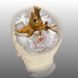
28 Improving Localization Depth perception of individual structures within the brain s hull difficult Visualizing the principal axes of the volume restricted by brain s hull improves localization 19
29 Distance Ring At some orientations one of the axes could be hidden or occluded by other structures Distance Ring indicates location of ROI in view direction - Minimal distance: distance ring is completely open - Maximal distance: distance ring is completely closed 20

30 Distance Ring At some orientations one of the axes could be hidden or occluded by other structures Distance Ring indicates location of ROI in view direction - Minimal distance: distance ring is completely open - Maximal distance: distance ring is completely closed 20
31 Distance Ring Tracing ray through brain mask front [0] position [0..1] back [1] 21


 Brain structures after")
32 Superficial Landmarks Superficial landmarks at surface of skull and brain to support navigation: Relate anatomical structures of head to those landmarks (e.g. nose, ears) Brain structures after opening the skull are known 22
33 Visualization of Contrast Enhanced Structures 23
34 Visualization of Contrast Enhanced Structures 23
35 Visualization of Contrast Enhanced Structures 23
36 Visualization of Contrast Enhanced Structures 23
37 Visualization of Contrast Enhanced Structures 23
38 Visualization of Contrast Enhanced Structures 23
39 Visualization of Contrast Enhanced Structures 23
40 Visualization of Contrast Enhanced Structures 23
41 Automatic Transfer Function Adaptation of Diseased Tissue 24
42 Automatic Transfer Function Adaptation of Diseased Tissue 24
43 Automatic Transfer Function Adaptation of Diseased Tissue 24
44 Automatic Transfer Function Adaptation of Diseased Tissue 24
45 Automatic Transfer Function Adaptation of Diseased Tissue 24
46 Automatic Transfer Function Adaptation of Diseased Tissue 24

47 Automatic Transfer Function Adaptation of Diseased Tissue weighted FLAIR masking 25
48 Automatic Transfer Function Adaptation of Diseased Tissue weighted FLAIR masking 25
49 Automatic Transfer Function Adaptation of Diseased Tissue weighted FLAIR masking 25

50 Automatic Transfer Function Adaptation of Diseased Tissue weighted FLAIR masking 25

51 Two-stage Rendering Pipeline Rieder et al., VCBM
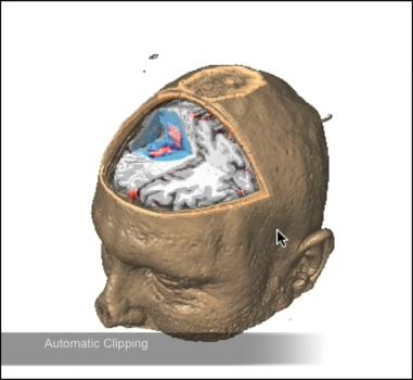
52 Automatic Axis-Aligned Clipping 27
53 Automatic Axis-Aligned Clipping How to reduce interaction required for exploration? 27
54 Automatic Axis-Aligned Clipping How to reduce interaction required for exploration? Dividing rendered volume into octant sectors 27
55 Automatic Axis-Aligned Clipping How to reduce interaction required for exploration? Dividing rendered volume into octant sectors Automatically discard sector located between view point and center of some ROI 27
56 Automatic Axis-Aligned Clipping 28
57 Automatic Axis-Aligned Clipping 28


58 Automatic Axis-Aligned Clipping Partitioning space into 26 different sectors 29

59 Automatic Axis-Aligned Clipping 30
60 Visualization of the Access Path 31
61 Visualization of the Access Path How to support planning of access to a deep-seated lesion? 31


62 Visualization of the Access Path How to support planning of access to a deep-seated lesion? Virtual access path from incision point to target Rieder et al., EuroVIS


63 Visualization of the Access Path How to support planning of access to a deep-seated lesion? Virtual access path from incision point to target Orthogonal cutting plane along the trajectory for detailed exploration Rieder et al., EuroVIS
64 Visualization of the Access Path The virtual access path: simplified cylinder geometry from incision point to ROI for visualization Pincision PROI Fdistance 32

65 Visualization of the Access Path Problem: Incorrect illumination of homogeneous regions (white matter, grey matter) due to ill-defined gradients at cuts Solution: Transfer normals of cutting geometry into cutting surface of volume (consistent shading [Weiskopf et al., TVCG 2003]) 33

66 Visualization of the Access Path 34
67 Brain Peeling 35
68 Brain Peeling How to explore anatomical details in the vicinity of the brain surface with cutting planes? 35
69 Brain Peeling How to explore anatomical details in the vicinity of the brain surface with cutting planes? Peel brain surface step by step: - Compute distance field (DF) from brain mask - Discard voxel if value in DF lower than distance threshold 35

70 Brain Peeling Rieder et al., VCBM
71 Conclusions Transfer function adaptation important for robustness Automated volume clipping facilitates exploration Virtual access path could become integral part of preoperative planning Trajectory aligned cutting plane supports inspection along access path Brain peeling allows the surgeon to inspect anatomical details in the vicinity of the brain surface Awareness about the limitations important! 37
72 Acknowledgments This presentation would have been impossible without the support of my colleagues: Horst Hahn Jan Klein Alexander Köhn Christian Rieder Florian Weiler 38
A Workflow Optimized Software Platform for Multimodal Neurosurgical Planning and Monitoring
 A Workflow Optimized Software Platform for Multimodal Neurosurgical Planning and Monitoring Eine Workflow Optimierte Software Umgebung für Multimodale Neurochirurgische Planung und Verlaufskontrolle A
Multimodal Visualization of DTI and fmri Data using Illustrative Methods
 Multimodal Visualization of DTI and fmri Data using Illustrative Methods Silvia Born 1, Werner Jainek 1, Mario Hlawitschka 2, Gerik Scheuermann 3, Christos Trantakis 4, Jürgen Meixensberger 4, Dirk Bartz
NeuroQLab A Software Assistant for Neurosurgical Planning and Quantitative Image Analysis
 NeuroQLab A Software Assistant for Neurosurgical Planning and Quantitative Image Analysis Florian Weiler 1, Jan Rexilius 2, Jan Klein 1, Horst K. Hahn 1 1 Fraunhofer MEVIS, Universitätsallee 29, 28359
Automatic Quantification of DTI Parameters along Fiber Bundles
 Automatic Quantification of DTI Parameters along Fiber Bundles Jan Klein 1, Simon Hermann 1, Olaf Konrad 1, Horst K. Hahn 1, and Heinz-Otto Peitgen 1 1 MeVis Research, 28359 Bremen Email: klein@mevis.de
Automatic Quantification of DTI Parameters along Fiber Bundles
 Automatic Quantification of DTI Parameters along Fiber Bundles Jan Klein, Simon Hermann, Olaf Konrad, Horst K. Hahn, Heinz-Otto Peitgen MeVis Research, 28359 Bremen Email: klein@mevis.de Abstract. We introduce
Fiber Selection from Diffusion Tensor Data based on Boolean Operators
 Fiber Selection from Diffusion Tensor Data based on Boolean Operators D. Merhof 1, G. Greiner 2, M. Buchfelder 3, C. Nimsky 4 1 Visual Computing, University of Konstanz, Konstanz, Germany 2 Computer Graphics
Computational Medical Imaging Analysis Chapter 4: Image Visualization
 Computational Medical Imaging Analysis Chapter 4: Image Visualization Jun Zhang Laboratory for Computational Medical Imaging & Data Analysis Department of Computer Science University of Kentucky Lexington,
Medical Images Analysis and Processing
 Medical Images Analysis and Processing - 25642 Emad Course Introduction Course Information: Type: Graduated Credits: 3 Prerequisites: Digital Image Processing Course Introduction Reference(s): Insight
Medical Image Analysis
 Computer assisted Image Analysis VT04 29 april 2004 Medical Image Analysis Lecture 10 (part 1) Xavier Tizon Medical Image Processing Medical imaging modalities XRay,, CT Ultrasound MRI PET, SPECT Generic
A Ray-based Approach for Boundary Estimation of Fiber Bundles Derived from Diffusion Tensor Imaging
 A Ray-based Approach for Boundary Estimation of Fiber Bundles Derived from Diffusion Tensor Imaging M. H. A. Bauer 1,3, S. Barbieri 2, J. Klein 2, J. Egger 1,3, D. Kuhnt 1, B. Freisleben 3, H.-K. Hahn
Medical Image Registration
 Medical Image Registration Submitted by NAREN BALRAJ SINGH SB ID# 105299299 Introduction Medical images are increasingly being used within healthcare for diagnosis, planning treatment, guiding treatment
Computer Aided Surgery 8:49 69 (2003)
") Computer Aided Surgery 8:49 69 (2003) Biomedical Paper Multimodal Image Fusion in Ultrasound-Based Neuronavigation: Improving Overview and Interpretation by Integrating Preoperative MRI with Intraoperative
Computer Aided Surgery 8:49 69 (2003) Biomedical Paper Multimodal Image Fusion in Ultrasound-Based Neuronavigation: Improving Overview and Interpretation by Integrating Preoperative MRI with Intraoperative
NEURO M203 & BIOMED M263 WINTER 2014
 NEURO M203 & BIOMED M263 WINTER 2014 MRI Lab 2: Neuroimaging Connectivity Lab In today s lab we will work with sample diffusion imaging data and the group averaged fmri data collected during your scanning
SIGMI Meeting ~Image Fusion~ Computer Graphics and Visualization Lab Image System Lab
 SIGMI Meeting ~Image Fusion~ Computer Graphics and Visualization Lab Image System Lab Introduction Medical Imaging and Application CGV 3D Organ Modeling Model-based Simulation Model-based Quantification
Diffusion Imaging Visualization
 Diffusion Imaging Visualization Thomas Schultz URL: http://cg.cs.uni-bonn.de/schultz/ E-Mail: schultz@cs.uni-bonn.de 1 Outline Introduction to Diffusion Imaging Basic techniques Glyph-based Visualization
Machine Learning for Medical Image Analysis. A. Criminisi
 Machine Learning for Medical Image Analysis A. Criminisi Overview Introduction to machine learning Decision forests Applications in medical image analysis Anatomy localization in CT Scans Spine Detection
Creating a Vision Channel for Observing Deep-Seated Anatomy in Medical Augmented Reality
 Creating a Vision Channel for Observing Deep-Seated Anatomy in Medical Augmented Reality A Cut-Away Technique for In-Situ Visualization Felix Wimmer 1, Christoph Bichlmeier 1, Sandro M. Heining 2, Nassir
ECE1778 Final Report MRI Visualizer
 ECE1778 Final Report MRI Visualizer David Qixiang Chen Alex Rodionov Word Count: 2408 Introduction We aim to develop a mobile phone/tablet based neurosurgical MRI visualization application with the goal
Complex Fiber Visualization
 Annales Mathematicae et Informaticae 34 (2007) pp. 103 109 http://www.ektf.hu/tanszek/matematika/ami Complex Fiber Visualization Henrietta Tomán a, Róbert Tornai b, Marianna Zichar c a Department of Computer
Survey of Computer-Aided Brain Surgery Tools Yasmine El-Glaly
 Survey of Computer-Aided Brain Surgery Tools Yasmine El-Glaly Abstract In this paper we investigate the interfering of computer systems in the process of neurosurgery. We survey the state-of-the-art algorithms
MEDICAL IMAGE ANALYSIS
 SECOND EDITION MEDICAL IMAGE ANALYSIS ATAM P. DHAWAN g, A B IEEE Engineering in Medicine and Biology Society, Sponsor IEEE Press Series in Biomedical Engineering Metin Akay, Series Editor +IEEE IEEE PRESS
Imaging protocols for navigated procedures
 9732379 G02 Rev. 1 2015-11 Imaging protocols for navigated procedures How to use this document This document contains imaging protocols for navigated cranial, DBS and stereotactic, ENT, and spine procedures
n o r d i c B r a i n E x Tutorial DTI Module
 m a k i n g f u n c t i o n a l M R I e a s y n o r d i c B r a i n E x Tutorial DTI Module Please note that this tutorial is for the latest released nordicbrainex. If you are using an older version please
Volume visualization. Volume visualization. Volume visualization methods. Sources of volume visualization. Sources of volume visualization
 Volume visualization Volume visualization Volumes are special cases of scalar data: regular 3D grids of scalars, typically interpreted as density values. Each data value is assumed to describe a cubic
Lecture 6: Medical imaging and image-guided interventions
 ME 328: Medical Robotics Winter 2019 Lecture 6: Medical imaging and image-guided interventions Allison Okamura Stanford University Updates Assignment 3 Due this Thursday, Jan. 31 Note that this assignment
Fiber Selection from Diffusion Tensor Data based on Boolean Operators
 Fiber Selection from Diffusion Tensor Data based on Boolean Operators D. Merhofl, G. Greiner 2, M. Buchfelder 3, C. Nimsky4 1 Visual Computing, University of Konstanz, Konstanz, Germany 2 Computer Graphics
syngo.mr Neuro 3D: Your All-In-One Post Processing, Visualization and Reporting Engine for BOLD Functional and Diffusion Tensor MR Imaging Datasets
 syngo.mr Neuro 3D: Your All-In-One Post Processing, Visualization and Reporting Engine for BOLD Functional and Diffusion Tensor MR Imaging Datasets Julien Gervais; Lisa Chuah Siemens Healthcare, Magnetic
DIFFUSION TENSOR IMAGING ANALYSIS. Using Analyze
 DIFFUSION TENSOR IMAGING ANALYSIS Using Analyze 2 Table of Contents 1. Introduction page 3 2. Loading DTI Data page 4 3. Computing DTI Maps page 5 4. Defining ROIs for Fiber Tracking page 6 5. Visualizing
Automatic Subthalamic Nucleus Targeting for Deep Brain Stimulation. A Validation Study
 Automatic Subthalamic Nucleus Targeting for Deep Brain Stimulation. A Validation Study F. Javier Sánchez Castro a, Claudio Pollo a,b, Jean-Guy Villemure b, Jean-Philippe Thiran a a École Polytechnique
Generation of Hulls Encompassing Neuronal Pathways Based on Tetrahedralization and 3D Alpha Shapes
 Generation of Hulls Encompassing Neuronal Pathways Based on Tetrahedralization and 3D Alpha Shapes Dorit Merhof 1,2, Martin Meister 1, Ezgi Bingöl 1, Peter Hastreiter 1,2, Christopher Nimsky 2,3, Günther
A Generic Lie Group Model for Computer Vision
 A Generic Lie Group Model for Computer Vision Within this research track we follow a generic Lie group approach to computer vision based on recent physiological research on how the primary visual cortex
Diffusion Tensor Imaging and Reading Development
 Diffusion Tensor Imaging and Reading Development Bob Dougherty Stanford Institute for Reading and Learning Reading and Anatomy Every brain is different... Not all brains optimized for highly proficient
METK The Medical Exploration Toolkit
 METK The Medical Exploration Toolkit Christian Tietjen 1, Konrad Mühler 1, Felix Ritter 2, Olaf Konrad 2, Milo Hindennach 2, Bernhard Preim 1 1 Institut für Simulation und Graphik, Otto-von-Guericke-Universität
Medical Visualization - Volume Rendering. J.-Prof. Dr. Kai Lawonn
 Medical Visualization - Volume Rendering J.-Prof. Dr. Kai Lawonn Medical Visualization Pipeline Acquisition Filtering/Enhancement Mapping Rendering Data are given Data are processed e.g., feature extraction
Stroke Quantification Tool (Sonia) Ver User Manual
 Ver User Manual") Stroke Quantification Tool (Sonia) Ver. 1.0 User Manual English. 12/2016 Rev. 1.0 www.wakeup-stroke.eu 1 Table of Contents 1. Introduction...3 2. Installation...4 3. Data Import...5 4. Registration...7
Stroke Quantification Tool (Sonia) Ver. 1.0 User Manual English. 12/2016 Rev. 1.0 www.wakeup-stroke.eu 1 Table of Contents 1. Introduction...3 2. Installation...4 3. Data Import...5 4. Registration...7
Automatic Alignment of Pre- and Post-Interventional Liver CT Images for Assessment of Radiofrequency Ablation
 Automatic Alignment of Pre- and Post-Interventional Liver CT Images for Assessment of Radiofrequency Ablation Christian Rieder a, Stefan Wirtz a, Jan Strehlow a, Stephan Zidowitz a, Philipp Bruners b,
Computational Medical Imaging Analysis
 Computational Medical Imaging Analysis Chapter 1: Introduction to Imaging Science Jun Zhang Laboratory for Computational Medical Imaging & Data Analysis Department of Computer Science University of Kentucky
Video Registration Virtual Reality for Non-linkage Stereotactic Surgery
 Video Registration Virtual Reality for Non-linkage Stereotactic Surgery P.L. Gleason, Ron Kikinis, David Altobelli, William Wells, Eben Alexander III, Peter McL. Black, Ferenc Jolesz Surgical Planning
Introduction to Medical Image Processing
 Introduction to Medical Image Processing Δ Essential environments of a medical imaging system Subject Image Analysis Energy Imaging System Images Image Processing Feature Images Image processing may be
BrainSpace V2.1: Geometric mapping and diffusion-based software for cross-subject multimodality brain imaging informatics
 Brief Introduction to BrainSpace V2.1 BrainSpace V2.1: Geometric mapping and diffusion-based software for cross-subject multimodality brain imaging informatics Graphics and Imaging Laboratory Wayne State
Math in image processing
 Math in image processing Math in image processing Nyquist theorem Math in image processing Discrete Fourier Transformation Math in image processing Image enhancement: scaling Math in image processing Image
ThE ultimate, INTuITIVE Mr INTErFAcE
 ThE ultimate, INTuITIVE Mr INTErFAcE Empowering you to do more The revolutionary Toshiba M-power user interface takes Mr performance and flexibility to levels higher than ever before. M-power is able to
Introduction to Neuroimaging Janaina Mourao-Miranda
 Introduction to Neuroimaging Janaina Mourao-Miranda Neuroimaging techniques have changed the way neuroscientists address questions about functional anatomy, especially in relation to behavior and clinical
Volume rendering for interactive 3-d segmentation
 Volume rendering for interactive 3-d segmentation Klaus D. Toennies a, Claus Derz b a Dept. Neuroradiology, Inst. Diagn. Radiology, Inselspital Bern, CH-3010 Berne, Switzerland b FG Computer Graphics,
Modern Medical Image Analysis 8DC00 Exam
 Parts of answers are inside square brackets [... ]. These parts are optional. Answers can be written in Dutch or in English, as you prefer. You can use drawings and diagrams to support your textual answers.
Interactive segmentation of vascular structures in CT images for liver surgery planning
 Interactive segmentation of vascular structures in CT images for liver surgery planning L. Wang¹, C. Hansen¹, S.Zidowitz¹, H. K. Hahn¹ ¹ Fraunhofer MEVIS, Institute for Medical Image Computing, Bremen,
Methods for data preprocessing
 Methods for data preprocessing John Ashburner Wellcome Trust Centre for Neuroimaging, 12 Queen Square, London, UK. Overview Voxel-Based Morphometry Morphometry in general Volumetrics VBM preprocessing
Multimodal Image Fusion Of The Human Brain
 Multimodal Image Fusion Of The Human Brain Isis Lázaro(1), Jorge Marquez(1), Juan Ortiz(2), Fernando Barrios(2) isislazaro@gmail.com Centro de Ciencias Aplicadas y Desarrollo Tecnológico, UNAM Circuito
FROM IMAGE RECONSTRUCTION TO CONNECTIVITY ANALYSIS: A JOURNEY THROUGH THE BRAIN'S WIRING. Francesca Pizzorni Ferrarese
 FROM IMAGE RECONSTRUCTION TO CONNECTIVITY ANALYSIS: A JOURNEY THROUGH THE BRAIN'S WIRING Francesca Pizzorni Ferrarese Pipeline overview WM and GM Segmentation Registration Data reconstruction Tractography
Advanced MRI Techniques (and Applications)
") Advanced MRI Techniques (and Applications) Jeffry R. Alger, PhD Department of Neurology Ahmanson-Lovelace Brain Mapping Center Brain Research Institute Jonsson Comprehensive Cancer Center University of
Advanced MRI Techniques (and Applications) Jeffry R. Alger, PhD Department of Neurology Ahmanson-Lovelace Brain Mapping Center Brain Research Institute Jonsson Comprehensive Cancer Center University of
Neuroimaging and mathematical modelling Lesson 2: Voxel Based Morphometry
 Neuroimaging and mathematical modelling Lesson 2: Voxel Based Morphometry Nivedita Agarwal, MD Nivedita.agarwal@apss.tn.it Nivedita.agarwal@unitn.it Volume and surface morphometry Brain volume White matter
better images mean better results
 better images mean better results A better way for YOU and YOUR patient brought to you by Advanced Neuro analysis with access to studies wherever you need it Advanced Neuro from Invivo Advancements in
MR IMAGE SEGMENTATION
 MR IMAGE SEGMENTATION Prepared by : Monil Shah What is Segmentation? Partitioning a region or regions of interest in images such that each region corresponds to one or more anatomic structures Classification
SIGMI. ISL & CGV Joint Research Proposal ~Image Fusion~
 SIGMI ISL & CGV Joint Research Proposal ~Image Fusion~ Introduction Research Diagram What CGV Lab is interested in What ISL is interested in Research Plan Research Diagram Medical Imaging and Application
Automatic MS Lesion Segmentation by Outlier Detection and Information Theoretic Region Partitioning Release 0.00
 Automatic MS Lesion Segmentation by Outlier Detection and Information Theoretic Region Partitioning Release 0.00 Marcel Prastawa 1 and Guido Gerig 1 Abstract July 17, 2008 1 Scientific Computing and Imaging
Evaluation of Local Filter Approaches for Diffusion Tensor based Fiber Tracking
 Evaluation of Local Filter Approaches for Diffusion Tensor based Fiber Tracking D. Merhof 1, M. Buchfelder 2, C. Nimsky 3 1 Visual Computing, University of Konstanz, Konstanz 2 Department of Neurosurgery,
Abstract. 1. Introduction
 A New Automated Method for Three- Dimensional Registration of Medical Images* P. Kotsas, M. Strintzis, D.W. Piraino Department of Electrical and Computer Engineering, Aristotelian University, 54006 Thessaloniki,
Automated segmentation methods for liver analysis in oncology applications
 University of Szeged Department of Image Processing and Computer Graphics Automated segmentation methods for liver analysis in oncology applications Ph. D. Thesis László Ruskó Thesis Advisor Dr. Antal
2D-3D Registration using Gradient-based MI for Image Guided Surgery Systems
 2D-3D Registration using Gradient-based MI for Image Guided Surgery Systems Yeny Yim 1*, Xuanyi Chen 1, Mike Wakid 1, Steve Bielamowicz 2, James Hahn 1 1 Department of Computer Science, The George Washington
GE Healthcare CLINICAL GALLERY. Discovery * MR750w 3.0T. This brochure is intended for European healthcare professionals.
 GE Healthcare CLINICAL GALLERY Discovery * MR750w 3.0T This brochure is intended for European healthcare professionals. NEURO PROPELLER delivers high resolution, motion insensitive imaging in all planes.
Normalization for clinical data
 Normalization for clinical data Christopher Rorden, Leonardo Bonilha, Julius Fridriksson, Benjamin Bender, Hans-Otto Karnath (2012) Agespecific CT and MRI templates for spatial normalization. NeuroImage
Improved Navigated Spine Surgery Utilizing Augmented Reality Visualization
 Improved Navigated Spine Surgery Utilizing Augmented Reality Visualization Zein Salah 1,2, Bernhard Preim 1, Erck Elolf 3, Jörg Franke 4, Georg Rose 2 1Department of Simulation and Graphics, University
RADIOMICS: potential role in the clinics and challenges
 27 giugno 2018 Dipartimento di Fisica Università degli Studi di Milano RADIOMICS: potential role in the clinics and challenges Dr. Francesca Botta Medical Physicist Istituto Europeo di Oncologia (Milano)
Introduction. Illustrative rendering is also often called non-photorealistic rendering (NPR)
") Introduction Illustrative rendering is also often called non-photorealistic rendering (NPR) we shall use these terms here interchangeably NPR offers many opportunities for visualization that conventional
Introduction Illustrative rendering is also often called non-photorealistic rendering (NPR) we shall use these terms here interchangeably NPR offers many opportunities for visualization that conventional
fmri/dti analysis using Dynasuite
 fmri/dti analysis using Dynasuite Contents 1 Logging in 2 Finding patient session 3 Viewing and adjusting images 4 Checking brain segmentation 5 Checking image registration 6 Seeing fmri results 7 Saving
11/18/ CPT Preauthorization Groupings Effective January 1, Computerized Tomography (CT) Abdomen 6. CPT Description SEGR CT01
 Abdomen 6. CPT Description SEGR CT01") Computerized Tomography (CT) 6 & 101 5 Upper Extremity 11 Lower Extremity 12 Head 3 Orbit 1 Sinus 2 Neck 4 7 Cervical Spine 8 Thoracic Spine 9 Lumbar Spine 10 Colon 13 CPT Description SEGR 74150 74160
Computerized Tomography (CT) 6 & 101 5 Upper Extremity 11 Lower Extremity 12 Head 3 Orbit 1 Sinus 2 Neck 4 7 Cervical Spine 8 Thoracic Spine 9 Lumbar Spine 10 Colon 13 CPT Description SEGR 74150 74160
UNIVERSITY OF SOUTHAMPTON
 UNIVERSITY OF SOUTHAMPTON PHYS2007W1 SEMESTER 2 EXAMINATION 2014-2015 MEDICAL PHYSICS Duration: 120 MINS (2 hours) This paper contains 10 questions. Answer all questions in Section A and only two questions
Utilizing Salient Region Features for 3D Multi-Modality Medical Image Registration
 Utilizing Salient Region Features for 3D Multi-Modality Medical Image Registration Dieter Hahn 1, Gabriele Wolz 2, Yiyong Sun 3, Frank Sauer 3, Joachim Hornegger 1, Torsten Kuwert 2 and Chenyang Xu 3 1
A method of visualization of a brain neural pathway by using critical points and target regions
 Modelling in Medicine and Biology VII 309 A method of visualization of a brain neural pathway by using critical points and target regions A. Doi 1, H. Fujimura 2, M. Nagano 1, T. Inoue 3 & A. Ogawa 3 1
Sensor-aided Milling with a Surgical Robot System
 1 Sensor-aided Milling with a Surgical Robot System Dirk Engel, Joerg Raczkowsky, Heinz Woern Institute for Process Control and Robotics (IPR), Universität Karlsruhe (TH) Engler-Bunte-Ring 8, 76131 Karlsruhe
3D Surface Reconstruction of the Brain based on Level Set Method
 3D Surface Reconstruction of the Brain based on Level Set Method Shijun Tang, Bill P. Buckles, and Kamesh Namuduri Department of Computer Science & Engineering Department of Electrical Engineering University
Diffusion model fitting and tractography: A primer
 Diffusion model fitting and tractography: A primer Anastasia Yendiki HMS/MGH/MIT Athinoula A. Martinos Center for Biomedical Imaging 03/18/10 Why n how Diffusion model fitting and tractography 0/18 Why
Scalar Data. CMPT 467/767 Visualization Torsten Möller. Weiskopf/Machiraju/Möller
 Scalar Data CMPT 467/767 Visualization Torsten Möller Weiskopf/Machiraju/Möller Overview Basic strategies Function plots and height fields Isolines Color coding Volume visualization (overview) Classification
Norbert Schuff VA Medical Center and UCSF
 Norbert Schuff Medical Center and UCSF Norbert.schuff@ucsf.edu Medical Imaging Informatics N.Schuff Course # 170.03 Slide 1/67 Objective Learn the principle segmentation techniques Understand the role
HST.583 Functional Magnetic Resonance Imaging: Data Acquisition and Analysis Fall 2008
 MIT OpenCourseWare http://ocw.mit.edu HST.583 Functional Magnetic Resonance Imaging: Data Acquisition and Analysis Fall 2008 For information about citing these materials or our Terms of Use, visit: http://ocw.mit.edu/terms.
Network connectivity via inference over curvature-regularizing line graphs
 Network connectivity via inference over curvature-regularizing line graphs Asian Conference on Computer Vision Maxwell D. Collins 1,2, Vikas Singh 2,1, Andrew L. Alexander 3 1 Department of Computer Sciences
Visualisation : Lecture 1. So what is visualisation? Visualisation
 So what is visualisation? UG4 / M.Sc. Course 2006 toby.breckon@ed.ac.uk Computer Vision Lab. Institute for Perception, Action & Behaviour Introducing 1 Application of interactive 3D computer graphics to
Scalar Data. Visualization Torsten Möller. Weiskopf/Machiraju/Möller
 Scalar Data Visualization Torsten Möller Weiskopf/Machiraju/Möller Overview Basic strategies Function plots and height fields Isolines Color coding Volume visualization (overview) Classification Segmentation
The University of Chicago. Center for EPR Imaging in Vivo Physiology. Image Registration. Boris Epel
 The University of Chicago Center for EPR Imaging in Vivo Physiology Image Registration Boris Epel Imaging Methods are Complimentary CT MRI EPRI High resolution anatomic images Quantitative Poor soft tissue
Leksell SurgiPlan. Powerful planning for success
 Leksell SurgiPlan Powerful planning for success Making a difference in surgical planning Leksell SurgiPlan Leksell SurgiPlan is an advanced image-based neurosurgical planning software, specifically designed
REAL-TIME ADAPTIVITY IN HEAD-AND-NECK AND LUNG CANCER RADIOTHERAPY IN A GPU ENVIRONMENT
 REAL-TIME ADAPTIVITY IN HEAD-AND-NECK AND LUNG CANCER RADIOTHERAPY IN A GPU ENVIRONMENT Anand P Santhanam Assistant Professor, Department of Radiation Oncology OUTLINE Adaptive radiotherapy for head and
Leksell SurgiPlan Overview. Powerful planning for surgical success
 Leksell SurgiPlan Overview Powerful planning for surgical success Making a Difference in Surgical Planning Leksell SurgiPlan Leksell SurgiPlan is an advanced image-based neuro surgical planning software,
Deformable Segmentation using Sparse Shape Representation. Shaoting Zhang
 Deformable Segmentation using Sparse Shape Representation Shaoting Zhang Introduction Outline Our methods Segmentation framework Sparse shape representation Applications 2D lung localization in X-ray 3D
Where are we now? Structural MRI processing and analysis
 Where are we now? Structural MRI processing and analysis Pierre-Louis Bazin bazin@cbs.mpg.de Leipzig, Germany Structural MRI processing: why bother? Just use the standards? SPM FreeSurfer FSL However:
BrainSuite Lab Exercises. presented at the UCLA/NITP Advanced Neuroimaging Summer Program 29 July 2014
 BrainSuite Lab Exercises presented at the UCLA/NITP Advanced Neuroimaging Summer Program 29 July 2014 1. Opening and Displaying an MRI Start BrainSuite Drag and drop the T1 image from the native space
Image Registration I
 Image Registration I Comp 254 Spring 2002 Guido Gerig Image Registration: Motivation Motivation for Image Registration Combine images from different modalities (multi-modality registration), e.g. CT&MRI,
Joint CI-JAI advanced accelerator lecture series Imaging and detectors for medical physics Lecture 1: Medical imaging
 Joint CI-JAI advanced accelerator lecture series Imaging and detectors for medical physics Lecture 1: Medical imaging Dr Barbara Camanzi barbara.camanzi@stfc.ac.uk Course layout Day AM 09.30 11.00 PM 15.30
An MRI-based Attenuation Correction Method for Combined PET/MRI Applications
 An MRI-based Attenuation Correction Method for Combined PET/MRI Applications Baowei Fei *, Xiaofeng Yang, Hesheng Wang Departments of Radiology, Emory University, Atlanta, GA Department of Biomedical Engineering
ECSE 626 Project Report Multimodality Image Registration by Maximization of Mutual Information
 ECSE 626 Project Report Multimodality Image Registration by Maximization of Mutual Information Emmanuel Piuze McGill University Montreal, Qc, Canada. epiuze@cim.mcgill.ca Abstract In 1997, Maes et al.
Computational Neuroanatomy
 Computational Neuroanatomy John Ashburner john@fil.ion.ucl.ac.uk Smoothing Motion Correction Between Modality Co-registration Spatial Normalisation Segmentation Morphometry Overview fmri time-series kernel
3D Slicer Overview. Andras Lasso, PhD PerkLab, Queen s University
 3D Slicer Overview Andras Lasso, PhD PerkLab, Queen s University Right tool for the job Technological prototype Research tool Clinical tool Can it be done? Jalopnik.com Innovative, not robust, usually
Virtual Cutting in Medical Data. Institute for Real-time Computer Systems and Robotics. University of Karlsruhe, Department for Computer Science
 Virtual Cutting in Medical Data A. Mazura and S. Seifert Institute for Real-time Computer Systems and Robotics Prof. Dr. U. Rembold, Prof. Dr. R. Dillmann University of Karlsruhe, Department for Computer
Image Registration. Prof. Dr. Lucas Ferrari de Oliveira UFPR Informatics Department
 Image Registration Prof. Dr. Lucas Ferrari de Oliveira UFPR Informatics Department Introduction Visualize objects inside the human body Advances in CS methods to diagnosis, treatment planning and medical
Physical bases of X-ray diagnostics
 Physical bases of X-ray diagnostics Dr. István Voszka Possibilities of X-ray production (X-ray is produced, when charged particles of high velocity are stopped) X-ray tube: Relatively low accelerating
Automatic Cerebral Aneurysm Detection in Multimodal Angiographic Images
 Automatic Cerebral Aneurysm Detection in Multimodal Angiographic Images Clemens M. Hentschke, Oliver Beuing, Rosa Nickl and Klaus D. Tönnies Abstract We propose a system to automatically detect cerebral
3D Voxel-Based Volumetric Image Registration with Volume-View Guidance
 3D Voxel-Based Volumetric Image Registration with Volume-View Guidance Guang Li*, Huchen Xie, Holly Ning, Deborah Citrin, Jacek Copala, Barbara Arora, Norman Coleman, Kevin Camphausen, and Robert Miller
Biomedical Image Processing
 Biomedical Image Processing Jason Thong Gabriel Grant 1 2 Motivation from the Medical Perspective MRI, CT and other biomedical imaging devices were designed to assist doctors in their diagnosis and treatment
Medical Visualization - Illustrative Visualization 2 (Summary) J.-Prof. Dr. Kai Lawonn
 J.-Prof. Dr. Kai Lawonn") Medical Visualization - Illustrative Visualization 2 (Summary) J.-Prof. Dr. Kai Lawonn Hatching 2 Hatching Motivation: Hatching in principle curvature direction Interrante et al. 1995 3 Hatching Hatching
Medical Visualization - Illustrative Visualization 2 (Summary) J.-Prof. Dr. Kai Lawonn Hatching 2 Hatching Motivation: Hatching in principle curvature direction Interrante et al. 1995 3 Hatching Hatching
NIH Public Access Author Manuscript Med Image Comput Comput Assist Interv. Author manuscript; available in PMC 2010 November 10.
 NIH Public Access Author Manuscript Med Image Comput Comput Assist Interv. Author manuscript; available in PMC 2010 November 10. Published in final edited form as: Med Image Comput Comput Assist Interv.
Automatic Vascular Tree Formation Using the Mahalanobis Distance
 Automatic Vascular Tree Formation Using the Mahalanobis Distance Julien Jomier, Vincent LeDigarcher, and Stephen R. Aylward Computer-Aided Diagnosis and Display Lab, Department of Radiology The University