Q.Clear. Steve Ross, Ph.D.
|
|
|
- Harold Dixon
- 6 years ago
- Views:
Transcription
1 Steve Ross, Ph.D. Accurate quantitation (SUV - Standardized Uptake Value) is becoming more important as clinicians seek to utilize PET imaging for more than just diagnosing and staging disease, but also for treatment monitoring. The significant challenge with delivering consistently accurate SUV measurements in PET imaging is that lesion size, volume and contrast recovery are highly impacted by today s reconstruction algorithm. technology is a step forward in providing quantitation accuracy while not sacrificing excellent image quality in PET imaging. This new approach considers all aspects of the imaging chain and the cumulative effect of all improvements, from small to large, to make PET/CT imaging an accurate tool for enabling both confident diagnosis and precise treatment response assessment.

2 INTRODUCTION There is renewed interest in the PET imaging community in obtaining quantitative information about lesion uptake values. Because of this, improvements in image reconstruction algorithms that offer the ability to obtain more accurate estimates of uptake in lesions are of significant interest. Many advancements have been made towards this end, including improvements in system corrections such as randoms and fully 3D scatters [1-2], advances in motion correction [3], modeling of the system point spread function [4], and the inclusion of more accurate projectors and the incorporation of all system corrections into a fully 3D PET image reconstruction model [5]. A core tenet of PET image reconstruction algorithms is the modeling of the underlying PET Poisson noise statistics. Iterative image reconstruction techniques that accurately model the inherent PET physics are generally preferred over standard analytic methods including filtered backprojection (FBP) because of significant increases in signal-to-noise (SNR). The most commonly used clinical PET reconstruction algorithm is the Ordered Subsets Expectation Maximization () algorithm. offers the advantage of accurately modeling the underlying PET physics and also generating the PET images in clinically relevant times via accelerated convergence through the use of subsets. One drawback to the algorithm is that it generally cannot be run to full convergence because the noise in the image grows with each iteration. To compensate for this, the algorithm is generally stopped after a predetermined number of iterations, typically two to four, resulting in an underconverged image. Because PET contrast recovery depends on convergence rates, an under-converged image may produce bias, directly impacting lesion quantitation. To address the effects of convergence and provide more accuracy in PET quantitation, the Regularized Reconstruction iterative algorithm () is being introduced, incorporating prior knowledge about the image quality into the reconstruction. This prior knowledge is incorporated as a term in the algorithm discouraging differences in neighboring image voxel values. By incorporating this factor into the reconstruction algorithm, the algorithm can be run to full convergence and provide more accurate quantitation levels and improved SNR over. acquired data. For Poisson data, the goal of maximizing that probability (expressed here as its logarithm) can be described mathematically by the objective function, (1) where y i represents the measured PET coincidence data, x is the image estimate, and P is the system geometry matrix. The ML-EM algorithm provides an update equation which, given an image that isn t at the maximum of this function, generates a new image with an increased likelihood. Repeating, or iterating, this algorithm to convergence, when the update equation does not substantially change the image, yields the ML image. accelerates the convergence of ML by performing updates based only on a portion of the data at once, which approximates the ML-EM solution in much less reconstruction time. Due to the low volume of information in a conventional clinical PET acquisition, reconstructions using ML-EM or suffer from high noise when run to its full convergence. To avoid this result, the algorithm is typically stopped after 2-4 iterations. This is an effective method to reduce noise in the PET images, but the noise reduction comes at a cost of reduced quantitative accuracy and the potential introduction of distortions in small objects. This effect is demonstrated with a simple simulation as shown in Figure 1. Two small active objects of the same size and activity level are simulated, with one of the small objects placed between the two large elliptical objects of uniform activity distribution. Reconstructed images are shown using with 2 iterations and 25 iterations. While the fully converged 25 iteration image shows much improved contrast and a significant reduction in spatial distortions, excessive noise is introduced. Partial Convergence (2 iterations) Full Convergence (25 iterations) AND CONVERGENCE The most widely used reconstruction algorithm in PET clinical imaging is, an accelerated variant of the ML-EM algorithm [6-8]. The goal of ML-EM is to find the most likely image given the data, defined as the image that maximizes the likelihood (or statistical probability) of producing the Figure 1. Phantom simulation demonstrating the effects of convergence. The image on the left is reconstructed with two iterations of ; on the right, 25 iterations. Note the spatial distortion and loss of counts in the indicated feature at 2 iterations.

3 A clinical example of this effect is shown in Figure 2. Excessive noise is introduced into the high iteration images. To avoid this, an under-converged image, like the two-iterations image shown, is typically used in clinical practice. Convergence Challenges 1 iteration 2 iterations 10 iterations 25 iterations Figure 2. Clinical data reconstructed with a range of iterations. Excessive noise is introduced as the image reaches convergence. Q.CLEAR is a Bayesian penalized likelihood (PL) reconstruction algorithm [9-10] which incorporates an additional term in the objective function of equation 1. This term increases as image noise increases, reducing the objective function, which has the effect of steering the optimization algorithm away from noisier images. This allows the algorithm to reach full convergence without the detrimental effects of excessive noise found with. The PL objective function is written as:, (2) where the first several terms are the same as in Equation 1, R(x) is a penalty to control noise and β controls the relative strength of the regularizing term relative to the data statistics. makes use of the Relative Difference Penalty (RDP) [11] which has the advantage of providing activity dependent noise control. The RDP is given by, (3) where w j and w k are relative weights for different components of the function and γ is a tunable parameter which controls edge preservation. uses the Block Sequential Regularized Expectation Maximization (BSREM) algorithm to solve equation 2 [12-13]. BSREM algorithm allows every single image voxel to achieve 100% convergence despite that did not seek for convergence and may achieve partial convergence, full convergence or over convergence in one single image. The regularization is also modulated to allow an optimal tradeoff between image quality and quantitation. The penalty is designed such that edges are preserved while background image noise is kept low, thus providing superior image quality [14]. Because is always run to convergence, iterations and subsets are no longer inputs provided to the user as is commonly done in. In addition, since the image noise is controlled inside the iterative reconstruction as part of regularization, post filters are not necessary; this is shown in the process flow map of Figure 3.
4 Conventional Iterative Reconstruction Reconstruction STOP before the noise gets too high Deadtime/ Normal Detector Geometry Modeling (LOR) Randoms s Scatter Attenuation Image filtering Partially converged image Scanner correction Object model Deadtime/ Normal Detector Geometry Modeling (LOR) Randoms s P.S.F Scatter ULD-CT Attenuation Reconstruction STOP At full convergence Regularization Fully converged image Scanner correction Object model Figure 3. Process flow maps for conventional iterative reconstruction and. is a fully convergent iterative reconstruction method. Unlike, controls noise as part of the regularization process inside iterative reconstruction. FULL CONVERGENCE WITH NO COMPROMISES PHANTOM ANALYSIS Anthropomorphic Phantoms To demonstrate that full convergence using Q.CLEAR doesn t compromise image quality, SNR has been measured on two anthropomorphic phantoms filled and scanned on the Discovery* PET/CT 710. The first phantom was an Anthropomorphic Torso Phantom TM (Data Spectrum Inc, Durham, NC) as shown in Figure 4. The phantom was modified by the manufacturer so that 10 mm diameter hollow spherical inserts could be placed in varying locations within the phantom. The phantom was filled with 18 F-FDG and the spheres were filled to a target of four times the background. The second phantom scanned was a modified Extended Oval Phantom TM (also from Data Spectrum) as shown in Figure 5. The phantom was modified to include two mounted bottles, and each was filled to a similar density as the lung background of the anthropomorphic phantom. In addition, another bottle mounted in the middle of the phantom was filled to twice the background activity in order to emulate a hot organ. The phantom also contained spherical inserts to mimic lesions. These spheres were filled to 4 times the background activity concentration. In the Extended Oval phantom, a 10 mm diameter sphere was placed into one lung insert. In the background, two spheres (10 mm, 13 mm) were located between the lungs and the hot organ (see Figure 4). A 40 million count PET/CT study was done on the Discovery PET/CT 710 scanner to simulate a single frame of a representative clinical exam. Data acquired from both phantoms was reconstructed with and. The images were filtered with a 4 mm transaxial Gaussian filter and a light axial filter [1:6:1] to control noise. The images were reconstructed with β=350 and no post filtering. All corrections were applied inside the iterative reconstruction loop as shown in Figure 3 for both and. The images reconstructed using, shown in Figure 4 and Figure 5, demonstrate superior contrast to the images, particularly in the lung region. This is attributed to the slow convergence of in the cold background. The SNR results demonstrate a significant improvement with over, with particular improvement shown in the lung region but a clear benefit throughout the entire image.
 modified to include 10 mm and 13 mm spherical inserts, a lung region and an")

 SNR measurements where SNR is defined as signal in a sphere ROI divided by the standard deviation of large uniform")
 Anthropmorphic Torso Phantom TM (Data Spectrum, Durham, NC) modified to include 10 mm spherical inserts.")
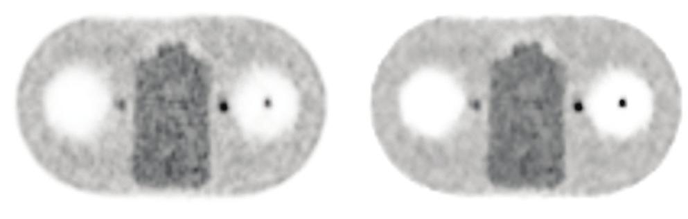
5 (a) (a) (b) (b) (c) SNR (c) SNR mm Lung 10 mm Bkg 13 mm Bkg Figure 5 (a) Extended Oval Phantom TM (Data Spectrum, Durham, NC) modified to include 10 mm and 13 mm spherical inserts, a lung region and an adjacent hot organ. (b) Reconstructed images from a 40M count study acquired on a Discovery PET/CT 710. images reconstructed using 2 iterations/24 subsets and a post-filtering of a 4 mm FWHM Gaussian in-plane and a [1:6:1] weighted axial filter. images reconstructed with β=350 and no post filtering. (c) SNR measurements where SNR is defined as signal in a sphere ROI divided by the standard deviation of large uniform background VOI mm Lung 10 mm Bkg 10 mm Edge Figure 4. (a) Anthropmorphic Torso Phantom TM (Data Spectrum, Durham, NC) modified to include 10 mm spherical inserts. (b) Reconstructed images from a 40M count study acquired on a Discovery PET/CT 710. images reconstructed using 2 iterations/24 subsets and a post-filtering of a 4 mm FWHM Gaussian in-plane and a [1:6:1] weighted axial filter. images reconstructed with β=350 and no post filtering. (c) SNR measurements where SNR is defined as signal in a sphere ROI divided by the standard deviation of large uniform background VOI.

 and PET image quality (SNR) over on both the Discovery PET/CT 610 and Discovery PET/CT 710. (a) SUV mean: 6.")
. images are shown with β=350.")
6 NEMA Image Quality Phantom A NEMA Image Quality IEC Phantom (Data Spectrum, Durham, NC) shown was filled with 18 F-FDG and scanned on the Discovery PET/CT 710. Images were reconstructed with and. As described previously, all corrections were applied inside the iterative loop for both algorithms. images with 2 and 25 iterations are shown along with a image in Figure 6. The 25 iteration image shows improved contrast over the 2 iteration image; however, there is also a significant increase in image noise. shows up to 2 times improvement in PET quantitation accuracy (SUVmean) and PET image quality (SNR) over on both the Discovery PET/CT 610 and Discovery PET/CT 710. (a) SUV mean: 6.4 g/ml (b) 2 iter 25 iter SUV mean: 10.9 g/ml Figure 6. (a) NEMA Image Quality Phantom (Data Spectrum, Durham, NC) images from a study acquired on a Discovery PET/CT 710. images are shown for 2 iterations and 25 iterations and a post-filtering of a 6.4 mm FWHM Gaussian in-plane and a [1:4:1] weighted axial filter Standard axial filter). images are shown with β=350. QUANTITATION AND VISUAL IMPROVEMENTS ON CLINICAL DATA PET/CT scans acquired on Discovery PET/CT 690 and Discovery PET/CT 710 systems were reconstructed with and. All corrections were applied inside the iterative reconstruction loop for both and. and reconstructed images are shown in Figures 7, 8 and 9. The images show a significant increase in SUVmean with. Figure 7 shows whole-body 18 F -FDG clinical images acquired on the Discovery PET/CT 690. The image was reconstructed using 2 iterations/24 subsets and a post-filter of a 4 mm FWHM Gaussian in-plane and a [1:6:1] weighted axial filter. The image was reconstructed with a β=350 and no post filtering. Figure 8 shows 68 Ga-DOTATOC clinical images acquired on a Discovery PET/CT 710, which also demonstrates a significant increase in SUV mean with. The image was reconstructed using 2 iterations/24 subsets and a post-filter of a 6.4 mm FWHM Gaussian in-plane and a [1:4:1] weighted axial filter. The image was reconstructed with a β=400. Figure 7. Clinical 18 F-FDG images acquired on a Discovery PET/CT 690. The image was reconstructed using 2 iterations/24 subsets and a post-filtering of a 4 mm FWHM Gaussian in-plane and a [1:6:1] weighted axial filter ( Light axial filter). The image was reconstructed with β=350. (a) SUV mean: 4.2 g/ml (b) SUV mean: 11.1 g/ml Figure 8. Clinical 68 Ga images acquired on a Discovery PET/CT 710. The image was reconstructed using 2 iterations/24 subsets and a post-filtering of a 6.4 mm FWHM Gaussian in-plane and a [1:4:1] weighted axial filter ( Standard axial filter). The image was reconstructed with β=400.


7 Figure 9 shows 18 F-FDG clinical images acquired on the Discovery PET/CT 600 which also demonstrates a significant increase in SUV mean with. The image was reconstructed using 2 iterations/32 subsets and a post-filter of a 6.4 mm FWHM Gaussian in-plane and a [1:4:1] weighted axial filter. The image was reconstructed with a β=200 and no post filtering. Figure 10 shows 18 F-FDG brain images from two exams acquired on a Discovery PET/CT 690 and reconstructed with and. The images were reconstructed with 3 iterations/32 subsets and a post-filter of 2.5 mm. The images were reconstructed with a β=150 and demonstrate excellent contrast and image quality in both brain exams. (a) Exam1 SUV mean: 1.38 g/ml (b) Exam2 SUV mean: 6.33 g/ml Figure 9. Whole-body 18 F-FDG clinical images acquired on the Discovery PET/CT 600. The image was reconstructed using 2 iterations/32 subsets and a post-filter of a 6.4 mm FWHM Gaussian in-plane and a [1:4:1] weighted axial filter. The image was reconstructed with a β=200 and no post filtering. Figure 10. Two clinical 18 F-FDG brain acquisitions from a Discovery PET/CT 690. The images were reconstructed using 3 iterations/32 subsets and a post-filtering of a 2.5 mm FWHM Gaussian in-plane. The images were reconstructed with β=150. SUMMARY Many recent advancements have been made towards improved quantitative accuracy in PET imaging. A current impediment towards superior quantitative imaging in PET was the necessary under-convergence required in to control image noise. provides a fully convergent PET image reconstruction technique, enabled by controlling image noise through regularized reconstruction. was designed to provide excellent image quality and consistent and accurate quantitation. Like any change in reconstruction processing, the effects of the algorithm should be considered when evaluating longitudinal studies. The results presented here demonstrate that provides an increase in both SNR and SUV recovery over. As such, is the foundation in the next generation of quantitative imaging.
8 REFERENCES [1] C. Stearns, D. McDaniel, S. Kohlmyer, P. Arul, B. Geiser and V. Shanmugam, Random coincidence estimation from single event rates on the Discovery ST PET/CT scanner, IEEE Nuclear Science Symposium Conference Record, [2] M. Iatrou, R. Manjeshwar, S. Ross, K. Thielemans, and C. Stearns, 3D implementation of scatter estimation in 3D PET, IEEE Nuclear Science Symposium Conference Record, 2006, pp [3] S. D. Wollenweber, G. Gopalakrishnan, K. Thielemans, and R. M. Manjeshwar, Evaluation of the Accuracy and Robustness of a Motion Algorithm for PET Using a Novel Phantom Approach, 2012, IEEE Transactions on Nuclear Science, Vol. 59 (1), pp [4] A. Alessio, C. Stearns, S. Tong, S. Ross, S. Kohlmyer, A. Ganin, P. Kinahan, Application and evaluation of a measured spatially variant system model for PET image reconstruction, IEEE Transactions on Medical Imaging, 2010, Vol. 29 (3), pp [5] R. Manjeshwar, S. Ross, M. Iatrou, T. Deller, and C. Stearns, Fully 3D PET iterative reconstruction using distance-driven projectors and native scanner geometry, IEEE Nuclear Science Symposium Conference Record, 2006, pp [6] L.A. Shepp and Y. Vardi, Maximum likelihood estimation for emission tomography, IEEE Transactions on Medical Imaging, 1982, Vol 1, pp , [7] K. Lange and R. Carson, EM reconstruction algorithms for emission and transmission tomography, Journal of Computer Assisted Tomogrphay, 1984, Vol. 8, pp., [8] H.M. Hudson and R.S. Larkin, Accelerated image reconstruction using ordered subsets of projection data, IEEE Transactions on Medical Imaging, 1994, Vol 16, pp [9] C-T.Chen, V. E. Johnson, W. H. Wong, X. Hul, and C. E. Metz, Bayesian Image Reconstruction in Positron Emission Tomography, IEEE Transactions on Nuclear Science, 1990, Vol. 37 (2), pp [10] E.U. Mumcuoglu, R. Leahy, S.R.Cherry, Z. Zhou, Fast gradient-based methods for Bayesian reconstruction of transmission and emission PET images, IEEE Transactions on Medical. Imaging, 1994, Vol. 13 (4), pp [11] J. Nuyts, D. Beque, P. Dupont, L. Mortelmans, A concave prior penalizing relative differences for maximum-a-posteriori reconstruction in emission tomography, IEEE Transactions on Nuclear Science, 2002, Vol. 49 (1), pp [12] A. R. De Pierro and M. E. B. Yamagishi, Fast EM-like methods for maximum a posteriori estimates in emission tomography, IEEE Transactions on Medical Imaging, 2001, Vol. 20, pp [13] S. Ahn and J.A. Fessler, Globally convergent image reconstruction for emission tomography using relaxed ordered subsets algorithms, IEEE Transactions on Medical Imaging, 2003, Vol. 22 (5), pp [14] E. Asma, S. Ahn, S. Ross, A. Chen, and R. Manjeshwar, Accurate and consistent lesion quantitation with clinically acceptable penalized likelihood images, IEEE Nuclear Science Symposium Conference Record, 2012.
9 About GE Healthcare GE Healthcare provides transformational medical technologies and services to meet the demand for increased access, enhanced quality and more affordable healthcare around the world. GE (NYSE: GE) works on things that matter - great people and technologies taking on tough challenges. From medical imaging, software & IT, patient monitoring and diagnostics to drug discovery, biopharmaceutical manufacturing technologies and performance improvement solutions, GE Healthcare helps medical professionals deliver great healthcare to their patients. GE Healthcare 3000 N Grandview Blvd Waukesha, WI USA General Electric Company-All rights reserved. General Electric Company reserves the right to make changes in specifications and features shown herein, or discontinue any products described at any time without notice or obligation. Please contact your GE representative for the most current information. GE, GE Monogram and imagination at work are trademarks of General Electric Company. *Trademark of General Electric Company. All third-party trademarks are the property of their respective owners. GE Healthcare, a division of General Electric Company. DOC , Rev 3
Implementation and evaluation of a fully 3D OS-MLEM reconstruction algorithm accounting for the PSF of the PET imaging system
 Implementation and evaluation of a fully 3D OS-MLEM reconstruction algorithm accounting for the PSF of the PET imaging system 3 rd October 2008 11 th Topical Seminar on Innovative Particle and Radiation
Review of PET Physics. Timothy Turkington, Ph.D. Radiology and Medical Physics Duke University Durham, North Carolina, USA
 Review of PET Physics Timothy Turkington, Ph.D. Radiology and Medical Physics Duke University Durham, North Carolina, USA Chart of Nuclides Z (protons) N (number of neutrons) Nuclear Data Evaluation Lab.
CT NOISE POWER SPECTRUM FOR FILTERED BACKPROJECTION AND ITERATIVE RECONSTRUCTION
 CT NOISE POWER SPECTRUM FOR FILTERED BACKPROJECTION AND ITERATIVE RECONSTRUCTION Frank Dong, PhD, DABR Diagnostic Physicist, Imaging Institute Cleveland Clinic Foundation and Associate Professor of Radiology
Workshop on Quantitative SPECT and PET Brain Studies January, 2013 PUCRS, Porto Alegre, Brasil Corrections in SPECT and PET
 Workshop on Quantitative SPECT and PET Brain Studies 14-16 January, 2013 PUCRS, Porto Alegre, Brasil Corrections in SPECT and PET Físico João Alfredo Borges, Me. Corrections in SPECT and PET SPECT and
Simultaneous measurement of noise and spatial resolution in PET phantom images
 IOP PUBLISHING Phys. Med. Biol. 55 (2010) 1069 1081 PHYSICS IN MEDICINE AND BIOLOGY doi:10.1088/0031-9155/55/4/011 Simultaneous measurement of noise and spatial resolution in PET phantom images Martin
A Weighted Least Squares PET Image Reconstruction Method Using Iterative Coordinate Descent Algorithms
 A Weighted Least Squares PET Image Reconstruction Method Using Iterative Coordinate Descent Algorithms Hongqing Zhu, Huazhong Shu, Jian Zhou and Limin Luo Department of Biological Science and Medical Engineering,
Modeling and Incorporation of System Response Functions in 3D Whole Body PET
 Modeling and Incorporation of System Response Functions in 3D Whole Body PET Adam M. Alessio, Member IEEE, Paul E. Kinahan, Senior Member IEEE, and Thomas K. Lewellen, Senior Member IEEE University of
Introduc)on to PET Image Reconstruc)on. Tomographic Imaging. Projec)on Imaging. Types of imaging systems
on to PET Image Reconstruc)on. Tomographic Imaging. Projec)on Imaging. Types of imaging systems") Introduc)on to PET Image Reconstruc)on Adam Alessio http://faculty.washington.edu/aalessio/ Nuclear Medicine Lectures Imaging Research Laboratory Division of Nuclear Medicine University of Washington Fall
Introduc)on to PET Image Reconstruc)on Adam Alessio http://faculty.washington.edu/aalessio/ Nuclear Medicine Lectures Imaging Research Laboratory Division of Nuclear Medicine University of Washington Fall
Improvement of contrast using reconstruction of 3D Image by PET /CT combination system
 Available online at www.pelagiaresearchlibrary.com Advances in Applied Science Research, 2013, 4(1):285-290 ISSN: 0976-8610 CODEN (USA): AASRFC Improvement of contrast using reconstruction of 3D Image
Assessment of OSEM & FBP Reconstruction Techniques in Single Photon Emission Computed Tomography Using SPECT Phantom as Applied on Bone Scintigraphy
 Assessment of OSEM & FBP Reconstruction Techniques in Single Photon Emission Computed Tomography Using SPECT Phantom as Applied on Bone Scintigraphy Physics Department, Faculty of Applied Science,Umm Al-Qura
Clinically feasible reconstruction of 3D whole-body PET/CT data using blurred anatomical labels
 INSTITUTE OF PHYSICSPUBLISHING Phys. Med. Biol. 47 (00) 0 PHYSICS INMEDICINE AND BIOLOGY PII: S003-955(0)56-9 Clinically feasible reconstruction of 3D whole-body PET/CT data using blurred anatomical labels
The Design and Implementation of COSEM, an Iterative Algorithm for Fully 3-D Listmode Data
 IEEE TRANSACTIONS ON MEDICAL IMAGING, VOL. 20, NO. 7, JULY 2001 633 The Design and Implementation of COSEM, an Iterative Algorithm for Fully 3-D Listmode Data Ron Levkovitz, Dmitry Falikman*, Michael Zibulevsky,
8/2/2017. Disclosure. Philips Healthcare (Cleveland, OH) provided the precommercial
 provided the precommercial") 8//0 AAPM0 Scientific Symposium: Emerging and New Generation PET: Instrumentation, Technology, Characteristics and Clinical Practice Aug Wednesday 0:4am :pm Solid State Digital Photon Counting PET/CT Instrumentation
8//0 AAPM0 Scientific Symposium: Emerging and New Generation PET: Instrumentation, Technology, Characteristics and Clinical Practice Aug Wednesday 0:4am :pm Solid State Digital Photon Counting PET/CT Instrumentation
A Fast GPU-Based Approach to Branchless Distance-Driven Projection and Back-Projection in Cone Beam CT
 A Fast GPU-Based Approach to Branchless Distance-Driven Projection and Back-Projection in Cone Beam CT Daniel Schlifske ab and Henry Medeiros a a Marquette University, 1250 W Wisconsin Ave, Milwaukee,
Image reconstruction for PET/CT scanners: past achievements and future challenges
 Review Image reconstruction for PET/CT scanners: past achievements and future challenges PET is a medical imaging modality with proven clinical value for disease diagnosis and treatment monitoring. The
White Paper. EQ PET: Achieving NEMAreferenced. Technologies. Matthew Kelly, PhD, Siemens Healthcare
 White Paper EQ PET: Achieving NEMAreferenced SUV Across Technologies Matthew Kelly, PhD, Siemens Healthcare Table of Contents Introduction 1 Case Study 1 Cross-Scanner Response Assessment 2 Clinical Example
Semi-Quantitative Metrics in Positron Emission Tomography. Michael Adams. Department of Biomedical Engineering Duke University.
 Semi-Quantitative Metrics in Positron Emission Tomography by Michael Adams Department of Biomedical Engineering Duke University Date: Approved: Timothy G. Turkington, Supervisor Adam P. Wax Terence Z.
The Effects of PET Reconstruction Parameters on Radiotherapy Response Assessment. and an Investigation of SUV peak Sampling Parameters.
 The Effects of PET Reconstruction Parameters on Radiotherapy Response Assessment and an Investigation of SUV peak Sampling Parameters by Leith Rankine Graduate Program in Medical Physics Duke University
Introduction to Positron Emission Tomography
 Planar and SPECT Cameras Summary Introduction to Positron Emission Tomography, Ph.D. Nuclear Medicine Basic Science Lectures srbowen@uw.edu System components: Collimator Detector Electronics Collimator
Projection Space Maximum A Posterior Method for Low Photon Counts PET Image Reconstruction
 Proection Space Maximum A Posterior Method for Low Photon Counts PET Image Reconstruction Liu Zhen Computer Department / Zhe Jiang Wanli University / Ningbo ABSTRACT In this paper, we proposed a new MAP
Unconstrained image reconstruction with resolution modelling does not have a unique solution
 Nuyts EJNMMI Physics 2014, 1:98 COMMENTARY OpenAccess Unconstrained image reconstruction with resolution modelling does not have a unique solution Johan Nuyts Correspondence: johan.nuyts@uz.kuleuven.be
Low-Dose Dual-Energy CT for PET Attenuation Correction with Statistical Sinogram Restoration
 Low-Dose Dual-Energy CT for PET Attenuation Correction with Statistical Sinogram Restoration Joonki Noh, Jeffrey A. Fessler EECS Department, The University of Michigan Paul E. Kinahan Radiology Department,
Technological Advances and Challenges: Experience with Time-Of-Flight PET Combined with 3T MRI. Floris Jansen, GE Healthcare July, 2015
 Technological Advances and Challenges: Experience with Time-Of-Flight PET Combined with 3T MRI Floris Jansen, GE Healthcare July, 2015 PET/MR 101 : challenges Thermal Workflow & Apps RF interactions?!!
PURE. ViSION Edition PET/CT. Patient Comfort Put First.
 PURE ViSION Edition PET/CT Patient Comfort Put First. 2 System features that put patient comfort and safety first. Oncology patients deserve the highest levels of safety and comfort during scans. Our Celesteion
NIH Public Access Author Manuscript J Nucl Med. Author manuscript; available in PMC 2010 February 9.
 NIH Public Access Author Manuscript Published in final edited form as: J Nucl Med. 2010 February ; 51(2): 237. doi:10.2967/jnumed.109.068098. An Assessment of the Impact of Incorporating Time-of-Flight
STATISTICAL image reconstruction methods have shown
 Globally Convergent Ordered Subsets Algorithms: Application to Tomography Sangtae Ahn and Jeffrey A. Fessler Abstract We present new algorithms for penalized-likelihood image reconstruction: modified BSREM
In the short span since the introduction of the first
 Performance Characteristics of a Newly Developed PET/CT Scanner Using NEMA Standards in 2D and 3D Modes Osama Mawlawi, PhD 1 ; Donald A. Podoloff, MD 2 ; Steve Kohlmyer, MS 3 ; John J. Williams 3 ; Charles
UvA-DARE (Digital Academic Repository) Motion compensation for 4D PET/CT Kruis, M.F. Link to publication
 Motion compensation for 4D PET/CT Kruis, M.F. Link to publication") UvA-DARE (Digital Academic Repository) Motion compensation for 4D PET/CT Kruis, M.F. Link to publication Citation for published version (APA): Kruis, M. F. (2014). Motion compensation for 4D PET/CT General
UvA-DARE (Digital Academic Repository) Motion compensation for 4D PET/CT Kruis, M.F. Link to publication Citation for published version (APA): Kruis, M. F. (2014). Motion compensation for 4D PET/CT General
THE methods of maximum likelihood (ML) and maximum
 and maximum") IEEE TRANSACTIONS ON NUCLEAR SCIENCE, VOL. 55, NO. 3, JUNE 2008 953 On Iterative Bayes Algorithms for Emission Tomography Jun Ma Abstract In this paper we formulate a new approach to medical image reconstruction
IEEE TRANSACTIONS ON NUCLEAR SCIENCE, VOL. 55, NO. 3, JUNE 2008 953 On Iterative Bayes Algorithms for Emission Tomography Jun Ma Abstract In this paper we formulate a new approach to medical image reconstruction
Time-of-Flight Technology
 Medical Review Time-of-Flight Technology Bing Bai, PhD Clinical Sciences Manager, PET/CT Canon Medical Systems INTRODUCTION Improving the care for every patient while providing a high standard care to
Monte-Carlo-Based Scatter Correction for Quantitative SPECT Reconstruction
 Monte-Carlo-Based Scatter Correction for Quantitative SPECT Reconstruction Realization and Evaluation Rolf Bippus 1, Andreas Goedicke 1, Henrik Botterweck 2 1 Philips Research Laboratories, Aachen 2 Fachhochschule
Spiral ASSR Std p = 1.0. Spiral EPBP Std. 256 slices (0/300) Kachelrieß et al., Med. Phys. 31(6): , 2004
 Kachelrieß et al., Med. Phys. 31(6): , 2004") Spiral ASSR Std p = 1.0 Spiral EPBP Std p = 1.0 Kachelrieß et al., Med. Phys. 31(6): 1623-1641, 2004 256 slices (0/300) Advantages of Cone-Beam Spiral CT Image quality nearly independent of pitch Increase
Spiral ASSR Std p = 1.0 Spiral EPBP Std p = 1.0 Kachelrieß et al., Med. Phys. 31(6): 1623-1641, 2004 256 slices (0/300) Advantages of Cone-Beam Spiral CT Image quality nearly independent of pitch Increase
Deviceless respiratory motion correction in PET imaging exploring the potential of novel data driven strategies
 g Deviceless respiratory motion correction in PET imaging exploring the potential of novel data driven strategies Presented by Adam Kesner, Ph.D., DABR Assistant Professor, Division of Radiological Sciences,
Ordered subsets algorithms for transmission tomography
 Ordered subsets algorithms for transmission tomography HErdoğan and J A Fessler 4415 EECS Bldg., 1301 Beal Ave., University of Michigan, Ann Arbor, MI 48109-2122USA Abstract. The ordered subsets EM (OSEM)
PET Image Reconstruction using Anatomical Information through Mutual Information Based Priors
 2005 IEEE Nuclear Science Symposium Conference Record M11-354 PET Image Reconstruction using Anatomical Information through Mutual Information Based Priors Sangeetha Somayajula, Evren Asma, and Richard
Continuation Format Page
 C.1 PET with submillimeter spatial resolution Figure 2 shows two views of the high resolution PET experimental setup used to acquire preliminary data [92]. The mechanics of the proposed system are similar
Evaluation of Penalty Design in Penalized Maximum- likelihood Image Reconstruction for Lesion Detection
 Evaluation of Penalty Design in Penalized Maximum- likelihood Image Reconstruction for Lesion Detection Li Yang, Andrea Ferrero, Rosalie J. Hagge, Ramsey D. Badawi, and Jinyi Qi Supported by NIBIB under
Resolution and Noise Properties of MAP Reconstruction for Fully 3-D PET
 IEEE TRANSACTIONS ON MEDICAL IMAGING, VOL. 19, NO. 5, MAY 2000 493 Resolution and Noise Properties of MAP Reconstruction for Fully 3-D PET Jinyi Qi*, Member, IEEE, and Richard M. Leahy, Member, IEEE Abstract
GE Healthcare. Vivid 7 Dimension 08 Cardiovascular ultrasound system
 GE Healthcare Vivid 7 Dimension 08 Cardiovascular ultrasound system ltra Definition. Technology. Performance. Start with a system that s proven its worth in LV quantification and 4D imaging. Then add even
SINGLE-PHOTON emission computed tomography
 1458 IEEE TRANSACTIONS ON NUCLEAR SCIENCE, VOL. 59, NO. 4, AUGUST 2012 SPECT Imaging With Resolution Recovery Andrei V. Bronnikov Abstract Single-photon emission computed tomography (SPECT) is a method
MAXIMUM a posteriori (MAP) or penalized ML image
 or penalized ML image") 42 IEEE TRANSACTIONS ON MEDICAL IMAGING, VOL. 25, NO. 1, JANUARY 2006 Mean and Covariance Properties of Dynamic PET Reconstructions From List-Mode Data Evren Asma and Richard M. Leahy* Abstract We derive
42 IEEE TRANSACTIONS ON MEDICAL IMAGING, VOL. 25, NO. 1, JANUARY 2006 Mean and Covariance Properties of Dynamic PET Reconstructions From List-Mode Data Evren Asma and Richard M. Leahy* Abstract We derive
PET image reconstruction algorithms based on maximum
 IEEE TRANSACTIONS ON MEDICAL IMAGING, VOL. 18, NO. 4, APRIL 1999 293 A Theoretical Study of the Contrast Recovery and Variance of MAP Reconstructions from PET Data Jinyi Qi,* Member, IEEE, and Richard
664 IEEE TRANSACTIONS ON NUCLEAR SCIENCE, VOL. 52, NO. 3, JUNE 2005
 664 IEEE TRANSACTIONS ON NUCLEAR SCIENCE, VOL. 52, NO. 3, JUNE 2005 Attenuation Correction for the NIH ATLAS Small Animal PET Scanner Rutao Yao, Member, IEEE, Jürgen Seidel, Jeih-San Liow, Member, IEEE,
Axial block coordinate descent (ABCD) algorithm for X-ray CT image reconstruction
 algorithm for X-ray CT image reconstruction") Axial block coordinate descent (ABCD) algorithm for X-ray CT image reconstruction Jeffrey A. Fessler and Donghwan Kim EECS Department University of Michigan Fully 3D Image Reconstruction Conference July
Axial block coordinate descent (ABCD) algorithm for X-ray CT image reconstruction Jeffrey A. Fessler and Donghwan Kim EECS Department University of Michigan Fully 3D Image Reconstruction Conference July
Medical Imaging BMEN Spring 2016
 Name Medical Imaging BMEN 420-501 Spring 2016 Homework #4 and Nuclear Medicine Notes All questions are from the introductory Powerpoint (based on Chapter 7) and text Medical Imaging Signals and Systems,
Validation of New Gibbs Priors for Bayesian Tomographic Reconstruction Using Numerical Studies and Physically Acquired Data 1
 Validation of New Gibbs Priors for Bayesian Tomographic Reconstruction Using Numerical Studies and Physically Acquired Data 1 S. J. Lee # Member, IEEE, Y. Choi x Member, IEEE, and G. Gindi { Member, IEEE
Characterization of a Time-of-Flight PET Scanner based on Lanthanum Bromide
 2005 IEEE Nuclear Science Symposium Conference Record M04-8 Characterization of a Time-of-Flight PET Scanner based on Lanthanum Bromide J. S. Karp, Senior Member, IEEE, A. Kuhn, Member, IEEE, A. E. Perkins,
3-D Monte Carlo-based Scatter Compensation in Quantitative I-131 SPECT Reconstruction
 3-D Monte Carlo-based Scatter Compensation in Quantitative I-131 SPECT Reconstruction Yuni K. Dewaraja, Member, IEEE, Michael Ljungberg, Member, IEEE, and Jeffrey A. Fessler, Member, IEEE Abstract- we
GE Healthcare CLINICAL GALLERY. Discovery * MR750w 3.0T. This brochure is intended for European healthcare professionals.
 GE Healthcare CLINICAL GALLERY Discovery * MR750w 3.0T This brochure is intended for European healthcare professionals. NEURO PROPELLER delivers high resolution, motion insensitive imaging in all planes.
High-resolution 3D Bayesian image reconstruction using the micropet small-animal scanner
 Phys. Med. Biol. 43 (1998) 1001 1013. Printed in the UK PII: S0031-9155(98)90627-3 High-resolution 3D Bayesian image reconstruction using the micropet small-animal scanner Jinyi Qi, Richard M Leahy, Simon
QIBA PET Amyloid BC March 11, Agenda
 QIBA PET Amyloid BC March 11, 2016 - Agenda 1. QIBA Round 6 Funding a. Deadlines b. What projects can be funded, what cannot c. Discussion of projects Mechanical phantom and DRO Paul & John? Any Profile
An Efficient Technique For Multi-Phase Model Based Iterative Reconstruction
 1 An Efficient Technique For Multi-Phase Model Based Iterative Reconstruction Shiyu Xu, Debashish Pal and Jean-Baptiste Thibault Abstract Multi-phase scan is a fundamental CT acquisition technology used
Single and Multipinhole Collimator Design Evaluation Method for Small Animal SPECT
 Single and Multipinhole Collimator Design Evaluation Method for Small Animal SPECT Kathleen Vunckx, Dirk Bequé, Michel Defrise and Johan Nuyts Abstract High resolution functional imaging of small animals
An overview of fast convergent ordered-subsets reconstruction methods for emission tomography based on the incremental EM algorithm
 An overview of fast convergent ordered-subsets reconstruction methods for emission tomography based on the incremental EM algorithm Ing-Tsung Hsiao a Parmeshwar Khurd b Anand Rangaraan c and Gene Gindi
EMISSION tomography, which includes positron emission
 1248 IEEE TRANSACTIONS ON NUCLEAR SCIENCE, VOL. 53, NO. 3, JUNE 2006 Aligning Emission Tomography and MRI Images by Optimizing the Emission-Tomography Image Reconstruction Objective Function James E. Bowsher,
Customizable and Advanced Software for Tomographic Reconstruction
 Customizable and Advanced Software for Tomographic Reconstruction 1 What is CASToR? Open source toolkit for 4D emission (PET/SPECT) and transmission (CT) tomographic reconstruction Focus on generic, modular
Partial Volume Effect correction in PET using regularized iterative deconvolution with variance control based on local topology
 This is an author-created, un-copyedited version of an article accepted for publication in Physics in Medicine and Biology. IOP Publishing Ltd is not responsible for any errors or omissions in this version
Noise weighting with an exponent for transmission CT
 doi:10.1088/2057-1976/2/4/045004 RECEIVED 13 January 2016 REVISED 4 June 2016 ACCEPTED FOR PUBLICATION 21 June 2016 PUBLISHED 27 July 2016 PAPER Noise weighting with an exponent for transmission CT Gengsheng
Iterative SPECT reconstruction with 3D detector response
 Iterative SPECT reconstruction with 3D detector response Jeffrey A. Fessler and Anastasia Yendiki COMMUNICATIONS & SIGNAL PROCESSING LABORATORY Department of Electrical Engineering and Computer Science
Penalized-Likelihood Reconstruction for Sparse Data Acquisitions with Unregistered Prior Images and Compressed Sensing Penalties
 Penalized-Likelihood Reconstruction for Sparse Data Acquisitions with Unregistered Prior Images and Compressed Sensing Penalties J. W. Stayman* a, W. Zbijewski a, Y. Otake b, A. Uneri b, S. Schafer a,
Validation of GEANT4 for Accurate Modeling of 111 In SPECT Acquisition
 Validation of GEANT4 for Accurate Modeling of 111 In SPECT Acquisition Bernd Schweizer, Andreas Goedicke Philips Technology Research Laboratories, Aachen, Germany bernd.schweizer@philips.com Abstract.
Quantitative Statistical Methods for Image Quality Assessment
 Quantitative Statistical Methods for Image Quality Assessment The Harvard community has made this article openly available. Please share how this access benefits you. Your story matters Citation Dutta,
SPECT QA and QC. Bruce McBride St. Vincent s Hospital Sydney.
 SPECT QA and QC Bruce McBride St. Vincent s Hospital Sydney. SPECT QA and QC What is needed? Why? How often? Who says? QA and QC in Nuclear Medicine QA - collective term for all the efforts made to produce
Clinically Feasible Reconstruction of 3D Whole-Body PET/CT Data Using Blurred Anatomical Labels
 Clinically Feasible Reconstruction of 3D Whole-Body PET/CT Data Using Blurred Anatomical Labels Claude Comtat 1, Paul E. Kinahan 2, Jeffrey A. Fessler 3, Thomas Beyer 4, David W. Townsend 2, Michel Defrise
PET Quantification using STIR
 PET Quantification using STIR STIR User s Meeting Charalampos Tsoumpas, PhD King s College London Hammersmith Imanet 1 PET Quantification Image elements should correspond to concentration of the injected
Super-resolution Reconstruction of Fetal Brain MRI
 Super-resolution Reconstruction of Fetal Brain MRI Ali Gholipour and Simon K. Warfield Computational Radiology Laboratory Children s Hospital Boston, Harvard Medical School Worshop on Image Analysis for
The Impact of Point Spread Function Modeling on Scan Duration in PET Imaging
 November 21, Volume 2, Issue 3 Original Article The Impact of Point Spread Function Modeling on Scan Duration in PET Imaging Sahar Ahangari 1, 2, Pardis Ghafarian 3, 4, *, Mahnaz Shekari 1, 2, Hossein
Fully 4D List-Mode Reconstruction with Temporal Basis Function Estimation
 5 IEEE Nuclear Science Symposium Conference Record M5-8 Fully D List-Mode Reconstruction with Temporal Basis Function Estimation Andrew J. Reader, Florent C. Sureau, Claude Comtat, Régine Trébossen and
Multi-slice CT Image Reconstruction Jiang Hsieh, Ph.D.
 Multi-slice CT Image Reconstruction Jiang Hsieh, Ph.D. Applied Science Laboratory, GE Healthcare Technologies 1 Image Generation Reconstruction of images from projections. textbook reconstruction advanced
STATISTICAL positron emission tomography (PET) image
 image") IEEE TRANSACTIONS ON MEDICAL IMAGING, VOL. 23, NO. 9, SEPTEMBER 2004 1057 Accurate Estimation of the Fisher Information Matrix for the PET Image Reconstruction Problem Quanzheng Li, Student Member, IEEE,
IEEE TRANSACTIONS ON MEDICAL IMAGING, VOL. 23, NO. 9, SEPTEMBER 2004 1057 Accurate Estimation of the Fisher Information Matrix for the PET Image Reconstruction Problem Quanzheng Li, Student Member, IEEE,
SUV Analysis of F-18 FDG PET Imaging in the Vicinity of the Bladder. Colleen Marie Allen. Graduate Program in Medical Physics Duke University
 SUV Analysis of F-18 FDG PET Imaging in the Vicinity of the Bladder by Colleen Marie Allen Graduate Program in Medical Physics Duke University Date: Approved: Timothy Turkington, Supervisor Terence Wong
If it matters to you, it matters to us
 If it matters to you, it matters to us Philips clinical innovations in nuclear medicine Innovation with insight We understand that clinical innovations are only as valuable as the day-to-day difference
Scatter Correction for Positron Emission Mammography
 Scatter Correction for Positron Emission Mammography Jinyi Qi and Ronald H. Huesman Center for Functional Imaging, Lawrence Berkeley National Laboratory, Berkeley, CA 9472, USA. E-mail: {jqi,rhhuesman}@lbl.gov
Corso di laurea in Fisica A.A Fisica Medica 5 SPECT, PET
 Corso di laurea in Fisica A.A. 2007-2008 Fisica Medica 5 SPECT, PET Step 1: Inject Patient with Radioactive Drug Drug is labeled with positron (β + ) emitting radionuclide. Drug localizes
RADIOMICS: potential role in the clinics and challenges
 27 giugno 2018 Dipartimento di Fisica Università degli Studi di Milano RADIOMICS: potential role in the clinics and challenges Dr. Francesca Botta Medical Physicist Istituto Europeo di Oncologia (Milano)
Reconstruction in CT and relation to other imaging modalities
 Reconstruction in CT and relation to other imaging modalities Jørgen Arendt Jensen November 1, 2017 Center for Fast Ultrasound Imaging, Build 349 Department of Electrical Engineering Center for Fast Ultrasound
Motion Correction in PET Image. Reconstruction
 Motion Correction in PET Image Reconstruction Wenjia Bai Wolfson College Supervisors: Professor Sir Michael Brady FRS FREng Dr David Schottlander D.Phil. Transfer Report Michaelmas 2007 Abstract Positron
THE general task in emission computed tomography (CT)
") 228 IEEE TRANSACTIONS ON MEDICAL IMAGING, VOL. 17, NO. 2, APRIL 1998 List-Mode Likelihood: EM Algorithm and Image Quality Estimation Demonstrated on 2-D PET Lucas Parra* and Harrison H. Barrett, Member,
228 IEEE TRANSACTIONS ON MEDICAL IMAGING, VOL. 17, NO. 2, APRIL 1998 List-Mode Likelihood: EM Algorithm and Image Quality Estimation Demonstrated on 2-D PET Lucas Parra* and Harrison H. Barrett, Member,
MR IMAGE SEGMENTATION
 MR IMAGE SEGMENTATION Prepared by : Monil Shah What is Segmentation? Partitioning a region or regions of interest in images such that each region corresponds to one or more anatomic structures Classification
Respiratory Motion Compensation for Simultaneous PET/MR Based on Strongly Undersampled Radial MR Data
 Respiratory Motion Compensation for Simultaneous PET/MR Based on Strongly Undersampled Radial MR Data Christopher M Rank 1, Thorsten Heußer 1, Andreas Wetscherek 1, and Marc Kachelrieß 1 1 German Cancer
An object-oriented library for 3D PET reconstruction using parallel computing
 An object-oriented library for 3D PET reconstruction using parallel computing Claire Labbé 1, K. Thielemans 2, D. Belluzzo 3, V. Bettinardi 3, M.C. Gilardi 3, D.S. Hague 4, M. Jacobson 5, S. Kaiser 6,
Quantitative imaging for clinical dosimetry
 Quantitative imaging for clinical dosimetry Irène Buvat Laboratoire d Imagerie Fonctionnelle U678 INSERM - UPMC CHU Pitié-Salpêtrière, Paris buvat@imed.jussieu.fr http://www.guillemet.org/irene Methodology
Advanced Image Reconstruction Methods for Photoacoustic Tomography
 Advanced Image Reconstruction Methods for Photoacoustic Tomography Mark A. Anastasio, Kun Wang, and Robert Schoonover Department of Biomedical Engineering Washington University in St. Louis 1 Outline Photoacoustic/thermoacoustic
Performance Evaluation of the Philips Gemini PET/CT System
 Performance Evaluation of the Philips Gemini PET/CT System Rebecca Gregory, Mike Partridge, Maggie A. Flower Joint Department of Physics, Institute of Cancer Research, Royal Marsden HS Foundation Trust,
Collimator-detector response compensation in molecular SPECT reconstruction using STIR framework
 Collimator-detector response compensation in molecular SPECT reconstruction using STIR framework Hojjat Mahani 1,2, Gholamreza Raisali 1, Alireza Kamali-Asl 3 and Mohammad Reza Ay 2,4 Original Article
Globally Convergent Image Reconstruction for Emission Tomography Using Relaxed Ordered Subsets Algorithms
 IEEE TRANSACTIONS ON MEDICAL IMAGING, VOL. 22, NO. 5, MAY 2003 613 Globally Convergent Image Reconstruction Emission Tomography Using Relaxed Ordered Subsets Algorithms Sangtae Ahn*, Student Member, IEEE,
GE Healthcare. AppsLinq* remote courses catalogue
 GE Healthcare AppsLinq* remote courses catalogue AppsLinq * remote training for MR Magnetic Resonance AppsLinq* a GE Training in Partnership (TiP) program, revolutionizes applications training with live,
High Temporal-Resolution Dynamic PET Image Reconstruction Using A New Spatiotemporal Kernel Method
 1 High Temporal-Resolution Dynamic PET Image Reconstruction Using A New Spatiotemporal Kernel Method Guobao Wang Abstract Current clinical dynamic PET has an effective temporal resolution of 5-10 seconds,
Concepts, Applications, and Requirements for Quantitative SPECT/CT. Conflict of Interest Disclosure
 Concepts, Applications, and Requirements for Quantitative SPECT/CT Eric C. Frey, Ph.D. (efrey@jhmi.edu) Division of Medical Imaging Physics Russell H. Morgan Department of Radiology and Radiological Science
Evaluation of Spectrum Mismatching using Spectrum Binning Approach for Statistical Polychromatic Reconstruction in CT
 Evaluation of Spectrum Mismatching using Spectrum Binning Approach for Statistical Polychromatic Reconstruction in CT Qiao Yang 1,4, Meng Wu 2, Andreas Maier 1,3,4, Joachim Hornegger 1,3,4, Rebecca Fahrig
Incorporating HYPR de-noising within iterative PET reconstruction (HYPR-OSEM)
") Home Search Collections Journals About Contact us My IOPscience Incorporating HYPR de-noising within iterative PET reconstruction (HYPR-OSEM) This content has been downloaded from IOPscience. Please scroll
Home Search Collections Journals About Contact us My IOPscience Incorporating HYPR de-noising within iterative PET reconstruction (HYPR-OSEM) This content has been downloaded from IOPscience. Please scroll
1. Deployment of a framework for drawing a correspondence between simple figure of merits (FOM) and quantitative imaging performance in CT.
 and quantitative imaging performance in CT.") Progress report: Development of assessment and predictive metrics for quantitative imaging in chest CT Subaward No: HHSN6801000050C (4a) PI: Ehsan Samei Reporting Period: month 1-18 Deliverables: 1. Deployment
Progress report: Development of assessment and predictive metrics for quantitative imaging in chest CT Subaward No: HHSN6801000050C (4a) PI: Ehsan Samei Reporting Period: month 1-18 Deliverables: 1. Deployment
Cherenkov Radiation. Doctoral Thesis. Rok Dolenec. Supervisor: Prof. Dr. Samo Korpar
 Doctoral Thesis Time-of-Flight Time-of-Flight Positron Positron Emission Emission Tomography Tomography Using Using Cherenkov Cherenkov Radiation Radiation Rok Dolenec Supervisor: Prof. Dr. Samo Korpar
Temperature Distribution Measurement Based on ML-EM Method Using Enclosed Acoustic CT System
 Sensors & Transducers 2013 by IFSA http://www.sensorsportal.com Temperature Distribution Measurement Based on ML-EM Method Using Enclosed Acoustic CT System Shinji Ohyama, Masato Mukouyama Graduate School
Conflicts of Interest Nuclear Medicine and PET physics reviewer for the ACR Accreditation program
 James R Halama, PhD Loyola University Medical Center Conflicts of Interest Nuclear Medicine and PET physics reviewer for the ACR Accreditation program Learning Objectives 1. Be familiar with recommendations
Non-Homogeneous Updates for the Iterative Coordinate Descent Algorithm
 Non-Homogeneous Updates for the Iterative Coordinate Descent Algorithm Zhou Yu a, Jean-Baptiste Thibault b, Charles A. Bouman a, Ken D. Sauer c, and Jiang Hsieh b a School of Electrical Engineering, Purdue
MEDICAL IMAGE ANALYSIS
 SECOND EDITION MEDICAL IMAGE ANALYSIS ATAM P. DHAWAN g, A B IEEE Engineering in Medicine and Biology Society, Sponsor IEEE Press Series in Biomedical Engineering Metin Akay, Series Editor +IEEE IEEE PRESS
Introduction to Emission Tomography
 Introduction to Emission Tomography Gamma Camera Planar Imaging Robert Miyaoka, PhD University of Washington Department of Radiology rmiyaoka@u.washington.edu Gamma Camera: - collimator - detector (crystal
USING cone-beam geometry with pinhole collimation,
 IEEE TRANSACTIONS ON NUCLEAR SCIENCE, VOL. 56, NO. 3, JUNE 2009 687 A Backprojection-Based Parameter Estimation Technique for Skew-Slit Collimation Jacob A. Piatt, Student Member, IEEE, and Gengsheng L.
Slide 1. Technical Aspects of Quality Control in Magnetic Resonance Imaging. Slide 2. Annual Compliance Testing. of MRI Systems.
 Slide 1 Technical Aspects of Quality Control in Magnetic Resonance Imaging Slide 2 Compliance Testing of MRI Systems, Ph.D. Department of Radiology Henry Ford Hospital, Detroit, MI Slide 3 Compliance Testing