TomoTherapy Related Projects. An image guidance alternative on Tomo Low dose MVCT reconstruction Patient Quality Assurance using Sinogram
|
|
|
- Brian Bell
- 5 years ago
- Views:
Transcription
1 TomoTherapy Related Projects An image guidance alternative on Tomo Low dose MVCT reconstruction Patient Quality Assurance using Sinogram


 with a scanning duration of 30 seconds.")

.")
. For MVCT imaging registration, we used bony and soft tissue to align the MVCT to the kvct.")
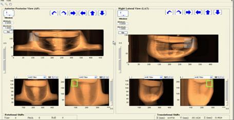
, cranial-caudal (CC), and anterior-posterior (AP) directions.")
2 Development of A Novel Image Guidance Alternative for Patient Localization using Topographic Images for TomoTherapy X. Sharon Qi*, PhD, Benjamin White, PhD, Daniel Low, PhD Department of Radiation Oncology, David of Geffen School of Medicine at UCLA, Los Angeles, CA *xqi@mednet.ucla.edu. Tel: (310) FAX: (310) OBJECTIVES The unique geometric design and integrated on-board imaging system allows for acquisition of topographic images on Tomotherapy Hi ART system. The objective is to develop a faster and lower dose topogram based image registration for TomoTherapy as an alternative image guidance tool to volumetric MVCT. METHODS Topogram procedures were created and performed for three anthropomorphic phantoms, including head (20 cm length), thorax (45 cm length) and spine (17 cm length) phantoms. The topogram procedures consisted of four couch speeds, 1 cm/s, 2 cm/s, 3 cm/s, and 3.99 cm/s (maximum couch speed) with a scanning duration of 30 seconds. All MLC leaves were opened to provide the largest field of view. A data compression factor of 1 was used for the topographic scans. An air scan was acquired to normalize the raw detector output. For patient localization, two topograms were acquired for each phantom and four different couch speeds at gantry angles of 0 and 90. To obtain the correct reference image from the simulation CT, a digitally reconstructed topogram (DRT, Figure 1) was generated in the TomoTherapy beam and detector geometry. To assess daily setup errors, known shifts were applied to the phantom for the topographic images (Figure 2). Image registration was performed by rigidly aligning the visible anatomy of the phantom in the topogram with the anatomy of the created DRT (Figure 3). MVCT scans were performed for the same phantoms using the normal imaging jaw width (4 mm). For MVCT imaging registration, we used bony and soft tissue to align the MVCT to the kvct. The MVCT was set up with known shifts from the kvct position. The shifts derived from topogram were compared to the MVCT image. RESULTS MVCT yielded positional offsets of 2 mm in the medio-lateral (ML), cranial-caudal (CC), and anterior-posterior (AP) directions. For the thorax phantom, the relative positional errors observed in all topograms (with different couch speeds) were 1.5 mm in the ML, 8.1 mm in the CC, and 2.5 mm in the AP direction. Between couch speeds of 1 cm/s, 2 cm/s and 3 cm/s, the relative shifts were within 3 mm in all ML and AP directions, and 5 mm in CC direction; while for fast couch speed such as 4.0 cm/s, larger relative shifts in CC direction between different couch speeds was seen (Table 1). At maximum couch speed, the topogram was markedly blurred in the CC direction. However, the blurred voxels resulted in a high positional error of 8.1 mm in the CC direction. The errors in the ML and AP directions were on the order of the slice thickness of the MVCT, which are at clinical acceptable level. Based on our preliminary measurements, a couch speed of 3 cm/s might be the fastest couch speed achievable with reasonable positional errors due to voxel deformation in the topogram. Compared with the imaging acquisition time of 3-5 min for MVCT scans, the time required to acquire clinically acceptable topograms for the selected phantom were significantly longer using topograms. Based on these results, a couch speed of 3 cm/s is the fasted couch speed achievable with reasonable positional errors due to voxel deformation in the topogram. Figure 2: Reconstructed MV TomoTherapy Topograms at couch speeds of 1 and 4 cm/s generated in the TomoTherapy beam and detector geometry. (a) and (b)couch speed of 1 cm/s; (c) and (d) couch speed of 4.0 cm/s. (a) (b) Figure 3: MV topogram based registration by rigid aligning the reconstructed Topogram to the DRT for (a) a thorax phantom and (b) a head phantom. (c) (d) (d) Figure 1: Digitally Reconstructed Topogram (the reference image) generated from the CT simulation in the TomoTherapy beam and detector geometry for a thorax phantom. (a) anteroposterior MV topogram and (b) left lateral MV topogram. (a) (b) (a) (b) *(b) Courtesy of Dr. Kevin Moore. Table 1: Relative positional errors at different couch speeds using MV Topogram registration versus MVCT registration on TomoTherapy. Direction CONCLUSION Topograms with proper couch speed (<3 cm/s) provide reliable patient localization while significantly reducing pre-treatment imaging time as well as imaging doses. Topogram can be used as an alternative and/or additional patient alignment tool to MVCT on TomoTherapy. References: * Relative positional errors for the topograms at different couch speeds. 1. Morre, KL et al., Fast, low-dose patent localization on TomoTherapy via Tomogram registration, Med. Phys. 37 (8), 4068 (2010). 2. Meeks, SL et al., Performance characterization of megavoltage computed tomography imaging on a helical tomotherapy unit, Med. Phys. 32 (8), 2673 (2005). 3. Keller, H et al., Monte Carlo study of a highly efficient gas ionization detector for megavoltage imaging and image-guided radiotherapy, Med. Phys. 29 (2), 165 (2002). ICCR 2013, 6-9 May 2013 Melbourne, Australia
3
4

5 ASTRO 2013 Oral
, (b), and (c) are from FBP, TV, TF with 100% data; (d), (e), and (f) are from FBP, TV, TF with 25% data.")
6 FIG. 2. Resolution slice reconstruction results from the Siemens image quality phantom. (a), (b), and (c) are from FBP, TV, TF with 100% data; (d), (e), and (f) are from FBP, TV, TF with 25% data. The zoom-in details are shown for the ROI in the selected square.
, (b), and (c) are from FBP, TV, and TF with 100% data; (d), (e), and")
7 FIG. 4. H&N patient results. (a), (b), and (c) are from FBP, TV, and TF with 100% data; (d), (e), and (f) are from FBP, TV, and TF with 25% data.
:081919.")
8 Med Phys Aug;40(8): doi: /
9 Clinical Outcome Assessment TCP/NTCP Clinical dose-response assessment Outcome prediction
10
11 Fig. 2. Dose response curves for malignant gliomas at 0.5, 1.0, and 1.5 years. The patient survival rates given by Walker et al. (9) and Salazar et al. ( 10 and 11) are considered. The lines represent the fitting to the data points for the survival rates given at 0.5, 1.0, and 1.5 years, respectively. The fitting yields: α = 0.06 ± 0.05 Gy 1 and α/β = 10.0 ± 15.1 Gy. The error bars represent statistical uncertainty.
12 Fig. 5. Fitting of different grades of astrocytomas using the data of Salazar et al. ( 10 and 11). For Grade 3 astrocytoma, our fitting yields: α = 0.11 ± 0.10 Gy 1 and α/β = 5.8 ± 11.8 Gy; whereas, for Grade 4 astrocytoma, α = 0.04 ± 0.06 Gy 1 and α/β = 5.6 ± 9.4 Gy. Int. J. Radiation Oncology Biol. Phys., Vol. 64, No. 5, pp , 2006
13 Data Mining Data analysis Time series analysis
14

15 ICCR 2013, 6-9 May 2013 Melbourne, Australia Analysis of Patient Setup Errors and Autocorrelation for Head-and-neck Radiotherapy Using Volumetric Computed Tomography X. Sharon Qi, Angie Hu Department of Radiation Oncology, David of Geffen School of Medicine at UCLA, Los Angeles, CA, USA
REAL-TIME ADAPTIVITY IN HEAD-AND-NECK AND LUNG CANCER RADIOTHERAPY IN A GPU ENVIRONMENT
 REAL-TIME ADAPTIVITY IN HEAD-AND-NECK AND LUNG CANCER RADIOTHERAPY IN A GPU ENVIRONMENT Anand P Santhanam Assistant Professor, Department of Radiation Oncology OUTLINE Adaptive radiotherapy for head and
7/31/2011. Learning Objective. Video Positioning. 3D Surface Imaging by VisionRT
 CLINICAL COMMISSIONING AND ACCEPTANCE TESTING OF A 3D SURFACE MATCHING SYSTEM Hania Al-Hallaq, Ph.D. Assistant Professor Radiation Oncology The University of Chicago Learning Objective Describe acceptance
Tomotherapy Physics. Machine Twinning and Quality Assurance. Emilie Soisson, MS
 Tomotherapy Physics Machine Twinning and Quality Assurance Emilie Soisson, MS Tomotherapy at UW- Madison Treating for nearly 5 years Up to ~45 patients a day on 2 tomo units Units twinned to facilitate
Automated Quality Assurance for Image-Guided Radiation Therapy
 JOURNAL OF APPLIED CLINICAL MEDICAL PHYSICS, VOLUME 10, NUMBER 1, WINTER 2009 Automated Quality Assurance for Image-Guided Radiation Therapy Eduard Schreibmann, a Eric Elder, Tim Fox Department of Radiation
Clinical Prospects and Technological Challenges for Multimodality Imaging Applications in Radiotherapy Treatment Planning
 Clinical Prospects and Technological Challenges for Multimodality Imaging Applications in Radiotherapy Treatment Planning Issam El Naqa, PhD Assistant Professor Department of Radiation Oncology Washington
DUAL-ENERGY CT IN PROTON THERAPY
 10/31/17 DUAL-ENERGY CT IN PROTON THERAPY Isabel Almeida, MAASTRO Clinic 7th NCS Lustrum Symposium 1 10/31/17 http://zonptc.bouwwebcam.nl https://www.youtube.com/watch?v=3vvvf5bqn7g Range uncertainties
Estimating 3D Respiratory Motion from Orbiting Views
 Estimating 3D Respiratory Motion from Orbiting Views Rongping Zeng, Jeffrey A. Fessler, James M. Balter The University of Michigan Oct. 2005 Funding provided by NIH Grant P01 CA59827 Motivation Free-breathing
3/27/2012 WHY SPECT / CT? SPECT / CT Basic Principles. Advantages of SPECT. Advantages of CT. Dr John C. Dickson, Principal Physicist UCLH
 3/27/212 Advantages of SPECT SPECT / CT Basic Principles Dr John C. Dickson, Principal Physicist UCLH Institute of Nuclear Medicine, University College London Hospitals and University College London john.dickson@uclh.nhs.uk
1. Learn to incorporate QA for surface imaging
 Hania Al-Hallaq, Ph.D. Assistant Professor Radiation Oncology The University of Chicago ***No disclosures*** 1. Learn to incorporate QA for surface imaging into current QA procedures for IGRT. 2. Understand
GPU applications in Cancer Radiation Therapy at UCSD. Steve Jiang, UCSD Radiation Oncology Amit Majumdar, SDSC Dongju (DJ) Choi, SDSC
 Choi, SDSC") GPU applications in Cancer Radiation Therapy at UCSD Steve Jiang, UCSD Radiation Oncology Amit Majumdar, SDSC Dongju (DJ) Choi, SDSC Conventional Radiotherapy SIMULATION: Construciton, Dij Days PLANNING:
GPU applications in Cancer Radiation Therapy at UCSD Steve Jiang, UCSD Radiation Oncology Amit Majumdar, SDSC Dongju (DJ) Choi, SDSC Conventional Radiotherapy SIMULATION: Construciton, Dij Days PLANNING:
ph fax Deming Way Middleton WI USA
 www.standardimaging.com 800-261-4446. ph 608-831-0025. fax 608-831-2202 3120 Deming Way Middleton WI 53562-1461 USA 2007 Standard Imaging, Inc. 1239-21 (03.07) beam qa LINAC AND TOMOTHERAPY QA The NEW
TG-148 overview. Introduction. System Overview. System Overview. QA for Helical Tomotherapy: Report of the AAPM Task Group 148. Conflict of Interest:
 QA or Helical Tomotherapy: Report o the AAPM Tas Group 148 Members: Conlict o Interest: r. John Balog owns TomoTherapy stoc. Katja Langen (Co-chair) Nio Papaniolaou (Co-chair) Walter Grant Richard Crilly
Evaluation report. X-ray tomographic image guided radiotherapy systems CEP10071
 Evaluation report X-ray tomographic image guided radiotherapy systems CEP10071 March 2010 Contents 2 Summary... 3 Introduction... 6 Product description... 9 Methods... 14 Technical performance... 23 Purchasing...
Investigation of tilted dose kernels for portal dose prediction in a-si electronic portal imagers
 Investigation of tilted dose kernels for portal dose prediction in a-si electronic portal imagers Krista Chytyk MSc student Supervisor: Dr. Boyd McCurdy Introduction The objective of cancer radiotherapy
Monte Carlo methods in proton beam radiation therapy. Harald Paganetti
 Monte Carlo methods in proton beam radiation therapy Harald Paganetti Introduction: Proton Physics Electromagnetic energy loss of protons Distal distribution Dose [%] 120 100 80 60 40 p e p Ionization
Michael Speiser, Ph.D.
 IMPROVED CT-BASED VOXEL PHANTOM GENERATION FOR MCNP MONTE CARLO Michael Speiser, Ph.D. Department of Radiation Oncology UT Southwestern Medical Center Dallas, TX September 1 st, 2012 CMPWG Workshop Medical
Development of Video Image Guided Setup (VIGS) System for Tomotherapy: Preliminary Study
 System for Tomotherapy: Preliminary Study") Development of Video Image Guided Setup (VIGS) System for Tomotherapy: Preliminary Study Jin Sung Kim*, Sang Gyu Ju*, Chae Seon Hong*, Jaewon Jeong, Kihong Son*, Jung Suk Shin*, Eunheak Shin*, Sung Hwan
Development of Video Image Guided Setup (VIGS) System for Tomotherapy: Preliminary Study Jin Sung Kim*, Sang Gyu Ju*, Chae Seon Hong*, Jaewon Jeong, Kihong Son*, Jung Suk Shin*, Eunheak Shin*, Sung Hwan
8/3/2016. Image Guidance Technologies. Introduction. Outline
 8/3/26 Session: Image Guidance Technologies and Management Strategies Image Guidance Technologies Jenghwa Chang, Ph.D.,2 Department of Radiation Medicine, Northwell Health 2 Hofstra Northwell School of
DAILY LINAC QA BEAM QA
 BEAM QA DAILY LINAC QA The QA BeamChecker Plus allows for fast, reliable, and uncomplicated daily QA of Varian, Elekta, Siemens, and Accuray Treatment Machines. The QA BeamChecker Plus is specifically
A simple method to test geometrical reliability of digital reconstructed radiograph (DRR)
") JOURNAL OF APPLIED CLINICAL MEDICAL PHYSICS, VOLUME 11, NUMBER 1, WINTER 2010 A simple method to test geometrical reliability of digital reconstructed radiograph (DRR) Stefania Pallotta, a Marta Bucciolini
JOURNAL OF APPLIED CLINICAL MEDICAL PHYSICS, VOLUME 11, NUMBER 1, WINTER 2010 A simple method to test geometrical reliability of digital reconstructed radiograph (DRR) Stefania Pallotta, a Marta Bucciolini
Acknowledgments and financial disclosure
 AAPM 2012 Annual Meeting Digital breast tomosynthesis: basic understanding of physics principles James T. Dobbins III, Ph.D., FAAPM Director, Medical Physics Graduate Program Ravin Advanced Imaging Laboratories
Image Guidance and Beam Level Imaging in Digital Linacs
 Image Guidance and Beam Level Imaging in Digital Linacs Ruijiang Li, Ph.D. Department of Radiation Oncology Stanford University School of Medicine 2014 AAPM Therapy Educational Course Disclosure Research
radiotherapy Andrew Godley, Ergun Ahunbay, Cheng Peng, and X. Allen Li NCAAPM Spring Meeting 2010 Madison, WI
 GPU-Accelerated autosegmentation for adaptive radiotherapy Andrew Godley, Ergun Ahunbay, Cheng Peng, and X. Allen Li agodley@mcw.edu NCAAPM Spring Meeting 2010 Madison, WI Overview Motivation Adaptive
Hidenobu Tachibana The Cancer Institute Hospital of JFCR, Radiology Dept. The Cancer Institute of JFCR, Physics Dept.
 2-D D Dose-CT Mapping in Geant4 Hidenobu Tachibana The Cancer Institute Hospital of JFCR, Radiology Dept. The Cancer Institute of JFCR, Physics Dept. Table of Contents Background & Purpose Materials Methods
Radiotherapy Plan Competition TomoTherapy Planning System. Dmytro Synchuk. Ukrainian Center of TomoTherapy, Kirovograd, Ukraine
 Radiotherapy Plan Competition 2016 TomoTherapy Planning System Dmytro Synchuk Ukrainian Center of TomoTherapy, Kirovograd, Ukraine Beam Geometry 6MV fan beam 3 jaw options 1.0, 2.5 and 5 cm 64 leaves binary
Determination of rotations in three dimensions using two-dimensional portal image registration
 Determination of rotations in three dimensions using two-dimensional portal image registration Anthony E. Lujan, a) James M. Balter, and Randall K. Ten Haken Department of Nuclear Engineering and Radiological
Effects of the difference in tube voltage of the CT scanner on. dose calculation
 Effects of the difference in tube voltage of the CT scanner on dose calculation Dong Joo Rhee, Sung-woo Kim, Dong Hyeok Jeong Medical and Radiological Physics Laboratory, Dongnam Institute of Radiological
Volumetric Modulated Arc Therapy - Clinical Implementation. Outline. Acknowledgement. History of VMAT. IMAT Basics of IMAT
 Volumetric Modulated Arc Therapy - Clinical Implementation Daliang Cao, PhD, DABR Swedish Cancer Institute, Seattle, WA Acknowledgement David M. Shepard, Ph.D. Muhammad K. N. Afghan, Ph.D. Fan Chen, Ph.D.
Digital Tomosynthesis for Target Localization
 Digital Tomosynthesis for Target Localization Fang-Fang Yin, Devon Godfrey, Lei Ren Jacqueline Maurer, Jackie Q-L Wu Duke University Medical Center Acknowledgements Duke Radiation Oncology faculty and
Financial disclosure. Onboard imaging modality for IGRT
 Tetrahedron Beam Computed Tomography Based On Multi-Pixel X- Ray Source and Its Application in Image Guided Radiotherapy Tiezhi Zhang, Ph.D. Advanced X-ray imaging Lab Financial disclosure Patent royalty
Initial Clinical Experience with 3D Surface Image Guidance
 Initial Clinical Experience with 3D Surface Image Guidance Amanda Havnen-Smith, Ph.D. Minneapolis Radiation Oncology Ridges Radiation Therapy Center Burnsville, MN April 20 th, 2012 Non-funded research
ADVANCING CANCER TREATMENT
 The RayPlan treatment planning system makes proven, innovative RayStation technology accessible to clinics that need a cost-effective and streamlined solution. Fast, efficient and straightforward to use,
3DVH : SUN NUCLEAR On The Accuracy Of The corporation Planned Dose Perturbation Algorithm Your Most Valuable QA and Dosimetry Tools *Patent Pending
 3DVH : On The Accuracy Of The Planned Dose Perturbation Algorithm SUN NUCLEAR corporation Your Most Valuable QA and Dosimetry Tools *Patent Pending introduction State-of-the-art IMRT QA of static gantry
The team. Disclosures. Ultrasound Guidance During Radiation Delivery: Confronting the Treatment Interference Challenge.
 Ultrasound Guidance During Radiation Delivery: Confronting the Treatment Interference Challenge Dimitre Hristov Radiation Oncology Stanford University The team Renhui Gong 1 Magdalena Bazalova-Carter 1
A novel method to correct for pitch and yaw patient setup errors in helical tomotherapy
 A novel method to correct for pitch and yaw patient setup errors in helical tomotherapy Sarah A. Boswell a Department of Medical Physics, University of Wisconsin, Madison, Wisconsin 53706 Robert Jeraj
Significance of time-dependent geometries for Monte Carlo simulations in radiation therapy. Harald Paganetti
 Significance of time-dependent geometries for Monte Carlo simulations in radiation therapy Harald Paganetti Modeling time dependent geometrical setups Key to 4D Monte Carlo: Geometry changes during the
GPU-based Fast Cone Beam CT Reconstruction from Undersampled and Noisy Projection Data via Total Variation
 GPU-based Fast Cone Beam CT Reconstruction from Undersampled and Noisy Projection Data via Total Variation 5 10 15 20 25 30 35 Xun Jia Department of Radiation Oncology, University of California San Diego,
Optimization of CT Simulation Imaging. Ingrid Reiser Dept. of Radiology The University of Chicago
 Optimization of CT Simulation Imaging Ingrid Reiser Dept. of Radiology The University of Chicago Optimization of CT imaging Goal: Achieve image quality that allows to perform the task at hand (diagnostic
Shadow casting. What is the problem? Cone Beam Computed Tomography THE OBJECTIVES OF DIAGNOSTIC IMAGING IDEAL DIAGNOSTIC IMAGING STUDY LIMITATIONS
 Cone Beam Computed Tomography THE OBJECTIVES OF DIAGNOSTIC IMAGING Reveal pathology Reveal the anatomic truth Steven R. Singer, DDS srs2@columbia.edu IDEAL DIAGNOSTIC IMAGING STUDY Provides desired diagnostic
Image Quality Assessment and Quality Assurance of Advanced Imaging Systems for IGRT. AAPM Penn-Ohio Chapter Sep 25, 2015 Soyoung Lee, PhD
 Image Quality Assessment and Quality Assurance of Advanced Imaging Systems for IGRT AAPM Penn-Ohio Chapter Sep 25, 2015 Soyoung Lee, PhD 1 Outline q Introduction q Imaging performances in 4D-CBCT Image
CT vs. VolumeScope: image quality and dose comparison
 CT vs. VolumeScope: image quality and dose comparison V.N. Vasiliev *a, A.F. Gamaliy **b, M.Yu. Zaytsev b, K.V. Zaytseva ***b a Russian Sci. Center of Roentgenology & Radiology, 86, Profsoyuznaya, Moscow,
Automated Image Analysis Software for Quality Assurance of a Radiotherapy CT Simulator
 Automated Image Analysis Software for Quality Assurance of a Radiotherapy CT Simulator Andrew J Reilly Imaging Physicist Oncology Physics Edinburgh Cancer Centre Western General Hospital EDINBURGH EH4
Optimization of Image Guided Radiation Therapy Using. Limited Angle Projections
 Optimization of Image Guided Radiation Therapy Using Limited Angle Projections by Lei Ren Medical Physics Graduate Program Duke University Date: Approved: Fang-Fang Yin, Supervisor Shiva K. Das Q. Jackie
Using a research real-time control interface to go beyond dynamic MLC tracking
 in partnership with Using a research real-time control interface to go beyond dynamic MLC tracking Dr. Simeon Nill Joint Department of Physics at The Institute of Cancer Research and the Royal Marsden
MR-guided radiotherapy: Vision, status and research at the UMC Utrecht. Dipl. Ing. Dr. Markus Glitzner
 MR-guided radiotherapy: Vision, status and research at the UMC Utrecht Dipl. Ing. Dr. Markus Glitzner About myself Training Medizintechnik TU Graz PhD UMC Utrecht Clinical work Software implementation
Comparison of internal and external dose conversion factors using ICRP adult male and MEET Man voxel model phantoms.
 Comparison of internal and external dose conversion factors using ICRP adult male and MEET Man voxel model phantoms. D.Leone, A.Häußler Intitute for Nuclear Waste Disposal, Karlsruhe Institute for Technology,
Brilliance CT Big Bore.
 1 2 2 There are two methods of RCCT acquisition in widespread clinical use: cine axial and helical. In RCCT with cine axial acquisition, repeat CT images are taken each couch position while recording respiration.
Helical 4D CT pitch management for the Brilliance CT Big Bore in clinical practice
 JOURNAL OF APPLIED CLINICAL MEDICAL PHYSICS, VOLUME 16, NUMBER 3, 2015 Helical 4D CT pitch management for the Brilliance CT Big Bore in clinical practice Guido Hilgers, a Tonnis Nuver, André Minken Department
Is deformable image registration a solved problem?
 Is deformable image registration a solved problem? Marcel van Herk On behalf of the imaging group of the RT department of NKI/AVL Amsterdam, the Netherlands DIR 1 Image registration Find translation.deformation
Dose Distributions. Purpose. Isodose distributions. To familiarize the resident with dose distributions and the factors that affect them
 Dose Distributions George Starkschall, Ph.D. Department of Radiation Physics U.T. M.D. Anderson Cancer Center Purpose To familiarize the resident with dose distributions and the factors that affect them
ADVANCING CANCER TREATMENT
 3 ADVANCING CANCER TREATMENT SUPPORTING CLINICS WORLDWIDE RaySearch is advancing cancer treatment through pioneering software. We believe software has un limited potential, and that it is now the driving
Influence of electron density spatial distribution and X-ray beam quality during CT simulation on dose calculation accuracy
 JOURNAL OF APPLIED CLINICAL MEDICAL PHYSICS, VOLUME 12, NUMBER 3, summer 2011 Influence of electron density spatial distribution and X-ray beam quality during CT simulation on dose calculation accuracy
Preface. Med. Phys. 35(9), , Mechanical QA. Radiation Survey Mechanical tests Light radiation Table, Collimator, Gantry Jaws.
, , Mechanical QA. Radiation Survey Mechanical tests Light radiation Table, Collimator, Gantry Jaws.") AAPM-SAM-2012-Das (1) Beam Data Collection and Commissioning for Linear Accelerators: Technical Considerations and Recommendations Preface Indra J. Das, PhD, FAAPM, FACR, FASTRO Department of Radiation
AAPM-SAM-2012-Das (1) Beam Data Collection and Commissioning for Linear Accelerators: Technical Considerations and Recommendations Preface Indra J. Das, PhD, FAAPM, FACR, FASTRO Department of Radiation
Motion artifact detection in four-dimensional computed tomography images
 Motion artifact detection in four-dimensional computed tomography images G Bouilhol 1,, M Ayadi, R Pinho, S Rit 1, and D Sarrut 1, 1 University of Lyon, CREATIS; CNRS UMR 5; Inserm U144; INSA-Lyon; University
Evaluation of 3D Gamma index calculation implemented in two commercial dosimetry systems
 University of Wollongong Research Online Faculty of Engineering and Information Sciences - Papers: Part A Faculty of Engineering and Information Sciences 2015 Evaluation of 3D Gamma index calculation implemented
CT NOISE POWER SPECTRUM FOR FILTERED BACKPROJECTION AND ITERATIVE RECONSTRUCTION
 CT NOISE POWER SPECTRUM FOR FILTERED BACKPROJECTION AND ITERATIVE RECONSTRUCTION Frank Dong, PhD, DABR Diagnostic Physicist, Imaging Institute Cleveland Clinic Foundation and Associate Professor of Radiology
Respiratory Motion Compensation for C-arm CT Liver Imaging
 Respiratory Motion Compensation for C-arm CT Liver Imaging Aline Sindel 1, Marco Bögel 1,2, Andreas Maier 1,2, Rebecca Fahrig 3, Joachim Hornegger 1,2, Arnd Dörfler 4 1 Pattern Recognition Lab, FAU Erlangen-Nürnberg
Correction of megavoltage cone-beam CT images of the pelvic region based on phantom measurements for dose calculation purposes
 JOURNAL OF APPLIED CLINICAL MEDICAL PHYSICS, VOLUME 10, NUMBER 1, WINTER 2009 Correction of megavoltage cone-beam CT images of the pelvic region based on phantom measurements for dose calculation purposes
Megan A. Wood, M.S. Under the direction of Larry DeWerd, Ph.D. University of Wisconsin Medical Radiation Research Center (UWMRRC)
") Megan A. Wood, M.S. Under the direction of Larry DeWerd, Ph.D. University of Wisconsin Medical Radiation Research Center (UWMRRC) NCCAAPM Spring Meeting May 3, 2013 Introduction FFF background Project
Megan A. Wood, M.S. Under the direction of Larry DeWerd, Ph.D. University of Wisconsin Medical Radiation Research Center (UWMRRC) NCCAAPM Spring Meeting May 3, 2013 Introduction FFF background Project
Spiral CT. Protocol Optimization & Quality Assurance. Ge Wang, Ph.D. Department of Radiology University of Iowa Iowa City, Iowa 52242, USA
 Spiral CT Protocol Optimization & Quality Assurance Ge Wang, Ph.D. Department of Radiology University of Iowa Iowa City, Iowa 52242, USA Spiral CT Protocol Optimization & Quality Assurance Protocol optimization
Four-Dimensional MVCT Imaging and Spatial Translation of Helical Tomotherapy Dose Distributions Using Sinogram Modification
 University of Tennessee, Knoxville Trace: Tennessee Research and Creative Exchange Masters Theses Graduate School 12-2006 Four-Dimensional MVCT Imaging and Spatial Translation of Helical Tomotherapy Dose
Thank-You Members of TG147 TG 147: QA for nonradiographic
 Thank-You Members of TG147 TG 147: QA for nonradiographic localization and positioning systems Twyla Willoughby, M.S. Medical Physicist Clinical AAPM Meeting March 2013 Department of Radiation Oncology
AIDR 3D Iterative Reconstruction:
 Iterative Reconstruction: Integrated, Automated and Adaptive Dose Reduction Erin Angel, PhD Manager, Clinical Sciences, CT Canon Medical Systems USA Iterative Reconstruction 1 Since the introduction of
Protocol. Technical evaluation of X-ray tomographic image-guided radiotherapy devices CEP10070
 Protocol Technical evaluation of X-ray tomographic image-guided radiotherapy devices CEP10070 March 2010 Contents 2 Introduction... 3 Protocol design and validation... 4 General information... 7 Technical
Validation of GEANT4 for Accurate Modeling of 111 In SPECT Acquisition
 Validation of GEANT4 for Accurate Modeling of 111 In SPECT Acquisition Bernd Schweizer, Andreas Goedicke Philips Technology Research Laboratories, Aachen, Germany bernd.schweizer@philips.com Abstract.
An Automated Image-based Method for Multi-Leaf Collimator Positioning Verification in Intensity Modulated Radiation Therapy
 An Automated Image-based Method for Multi-Leaf Collimator Positioning Verification in Intensity Modulated Radiation Therapy Chenyang Xu 1, Siemens Corporate Research, Inc., Princeton, NJ, USA Xiaolei Huang,
MEDICAL IMAGING 2nd Part Computed Tomography
 MEDICAL IMAGING 2nd Part Computed Tomography Introduction 2 In the last 30 years X-ray Computed Tomography development produced a great change in the role of diagnostic imaging in medicine. In convetional
Scaling Calibration in the ATRACT Algorithm
 Scaling Calibration in the ATRACT Algorithm Yan Xia 1, Andreas Maier 1, Frank Dennerlein 2, Hannes G. Hofmann 1, Joachim Hornegger 1,3 1 Pattern Recognition Lab (LME), Friedrich-Alexander-University Erlangen-Nuremberg,
Design and performance characteristics of a Cone Beam CT system for Leksell Gamma Knife Icon
 Design and performance characteristics of a Cone Beam CT system for Leksell Gamma Knife Icon WHITE PAPER Introduction Introducing an image guidance system based on Cone Beam CT (CBCT) and a mask immobilization
Philips SPECT/CT Systems
 Philips SPECT/CT Systems Ling Shao, PhD Director, Imaging Physics & System Analysis Nuclear Medicine, Philips Healthcare June 14, 2008 *Presented SNM08 Categorical Seminar - Quantitative SPECT and PET
IAEA-TECDOC-1583 Commissioning of Radiotherapy Treatment Planning Systems: Testing for Typical External Beam Treatment Techniques
 IAEA-TECDOC-1583 Commissioning of Radiotherapy Treatment Planning Systems: Testing for Typical External Beam Treatment Techniques Report of the Coordinated Research Project (CRP) on Development of Procedures
8/3/2017. Contour Assessment for Quality Assurance and Data Mining. Objective. Outline. Tom Purdie, PhD, MCCPM
 Contour Assessment for Quality Assurance and Data Mining Tom Purdie, PhD, MCCPM Objective Understand the state-of-the-art in contour assessment for quality assurance including data mining-based techniques
A Study of Motion Tracking Accuracy of Robotic Radiosurgery Using a Novel CCD Camera Based End-to-end Test System
 A Study of Motion Tracking Accuracy of Robotic Radiosurgery Using a Novel CCD Camera Based End-to-end Test System Lei Wang 1, Brett Nelson 2,Youming Yang 1 1. Department of Radiation Oncology, Stanford
A learning-based patient repositioning method from limited-angle projections
 A learning-based patient repositioning method from limited-angle projections Chen-Rui Chou 1, C. Brandon Frederick 2, Sha Chang 3, and Stephen M. Pizer 1,2,3 1 Department of Computer Science, UNC-Chapel
Op#miza#on of an On- Board Imaging System for Rapid Radiotherapy. Erica Kemmerling, Meng Wu, He Yang, and Rebecca Fahrig
 Op#miza#on of an On- Board Imaging System for Rapid Radiotherapy Erica Kemmerling, Meng Wu, He Yang, and Rebecca Fahrig Problem: mo#on during treatment Two overlapping thorax views during normal breathing
Quantitative imaging for clinical dosimetry
 Quantitative imaging for clinical dosimetry Irène Buvat Laboratoire d Imagerie Fonctionnelle U678 INSERM - UPMC CHU Pitié-Salpêtrière, Paris buvat@imed.jussieu.fr http://www.guillemet.org/irene Methodology
Digital phantoms for the evaluation of a software used for an automatic analysis of the Winston-Lutz test in image guided radiation therapy
 Author manuscript, published in "Medical Imaging 008: Physics of Medical Imaging, San Diego, CA, USA : United States (008)" DOI : 10.1117/1.768668 Digital phantoms for the evaluation of a software used
The Near Future in Cardiac CT Image Reconstruction
 SCCT 2010 The Near Future in Cardiac CT Image Reconstruction Marc Kachelrieß Institute of Medical Physics (IMP) Friedrich-Alexander Alexander-University Erlangen-Nürnberg rnberg www.imp.uni-erlangen.de
Simulation of Mammograms & Tomosynthesis imaging with Cone Beam Breast CT images
 Simulation of Mammograms & Tomosynthesis imaging with Cone Beam Breast CT images Tao Han, Chris C. Shaw, Lingyun Chen, Chao-jen Lai, Xinming Liu, Tianpeng Wang Digital Imaging Research Laboratory (DIRL),
Tomotherapy archive structure and new software tool for loading and advanced analysis of data contained in it
 reports of practical oncology and radiotherapy 1 6 (2 0 1 1) 58 64 available at www.sciencedirect.com journal homepage: http://www.rpor.eu/ Original article Tomotherapy archive structure and new software
VALIDATION OF DIR. Raj Varadhan, PhD, DABMP Minneapolis Radiation Oncology
 VALIDATION OF DIR Raj Varadhan, PhD, DABMP Minneapolis Radiation Oncology Overview Basics: Registration Framework, Theory Discuss Validation techniques Using Synthetic CT data & Phantoms What metrics to
A Generation Methodology for Numerical Phantoms with Statistically Relevant Variability of Geometric and Physical Properties
 A Generation Methodology for Numerical Phantoms with Statistically Relevant Variability of Geometric and Physical Properties Steven Dolly 1, Eric Ehler 1, Yang Lou 2, Mark Anastasio 2, Hua Li 2 (1) University
Evaluation of AutoQA Lite TM Image Quality Measurement Software
 Evaluation of AutoQA Lite TM Image Quality Measurement Software Andrew J Reilly Imaging Physicist Oncology Physics Edinburgh Cancer Centre Western General Hospital EDINBURGH EH4 2XU Phone: 0131 537 1161
CHAPTER 9 INFLUENCE OF SMOOTHING ALGORITHMS IN MONTE CARLO DOSE CALCULATIONS OF CYBERKNIFE TREATMENT PLANS: A LUNG PHANTOM STUDY
 148 CHAPTER 9 INFLUENCE OF SMOOTHING ALGORITHMS IN MONTE CARLO DOSE CALCULATIONS OF CYBERKNIFE TREATMENT PLANS: A LUNG PHANTOM STUDY 9.1 INTRODUCTION 9.1.1 Dose Calculation Algorithms Dose calculation
Computer-Tomography II: Image reconstruction and applications
 Computer-Tomography II: Image reconstruction and applications Prof. Dr. U. Oelfke DKFZ Heidelberg Department of Medical Physics (E040) Im Neuenheimer Feld 280 69120 Heidelberg, Germany u.oelfke@dkfz.de
An approximate cone beam reconstruction algorithm for gantry-tilted CT
 An approximate cone beam reconstruction algorithm for gantry-tilted CT Ming Yan a, Cishen Zhang ab, Hongzhu Liang a a School of Electrical & Electronic Engineering, Nanyang Technological University, Singapore;
Acknowledgments. Ping Xia, Ph.D., UCSF. Pam Akazawa, CMD, UCSF. Cynthia Chuang, Ph.D., UCSF
 Page 1 Quality Assurance of IMRT Delivery Systems - Siemens Lynn J. Verhey, Ph.D. Professor and Vice-Chair UCSF Dept. of Radiation Oncology AAPM 22 Annual Meeting, Montreal Acknowledgments Ping Xia, Ph.D.,
Introduction. Quality Assurance for Image- Guided Radiation Therapy. Justification for IGRT. Image-Guided Radiation Therapy
 Introduction Quality Assurance for Image- Guided Radiation Therapy Jean-Pierre Bissonnette, Ph.D., MCCPM Princess Margaret Hospital, Toronto, Canada IGRT What is it? Rationale Equipment Quality Assurance
A fluence convolution method to account for respiratory motion in three-dimensional dose calculations of the liver: A Monte Carlo study
 A fluence convolution method to account for respiratory motion in three-dimensional dose calculations of the liver: A Monte Carlo study Indrin J. Chetty, a) Mihaela Rosu, Neelam Tyagi, Lon H. Marsh, Daniel
Background. Outline. Radiographic Tomosynthesis: Image Quality and Artifacts Reduction 1 / GE /
 Radiographic Tomosynthesis: Image Quality and Artifacts Reduction Baojun Li, Ph.D Department of Radiology Boston University Medical Center 2012 AAPM Annual Meeting Background Linear Trajectory Tomosynthesis
Analysis of Radiation Transport through Multileaf Collimators Using BEAMnrc Code
 American Journal of Biomedical Engineering 216, 6(4): 124-131 DOI: 1.5923/j.ajbe.21664.3 Analysis of Radiation Transport through Multileaf Collimators Using BEAMnrc Code Ankit Kajaria 1,*, Neeraj Sharma
2005 IEEE Nuclear Science Symposium Conference Record M10-2
 25 IEEE Nuclear Science Symposium Conference Record M1-2 Estimating 3D Respiratory Motion from Orbiting Views Rongping Zeng 1, Jeffrey A. Fessler 1, and James M. Balter 2 rzeng@eecs.umich.edu, fessler@eecs.umich.edu,
Micro-CT Methodology Hasan Alsaid, PhD
 Micro-CT Methodology Hasan Alsaid, PhD Preclinical & Translational Imaging LAS, PTS, GlaxoSmithKline 20 April 2015 Provide basic understanding of technical aspects of the micro-ct Statement: All procedures
Auto-Segmentation Using Deformable Image Registration. Disclosure. Objectives 8/4/2011
 Auto-Segmentation Using Deformable Image Registration Lei Dong, Ph.D. Dept. of Radiation Physics University of Texas MD Anderson Cancer Center, Houston, Texas AAPM Therapy Educational Course Aug. 4th 2011
Feasibility study of performing IGRT system daily QA using a commercial QA device
 JOURNAL OF APPLIED CLINICAL MEDICAL PHYSICS, VOLUME 12, NUMBER 3, summer 2011 Feasibility study of performing IGRT system daily QA using a commercial QA device Jean L.Peng, 1 Darren Kahler, 2 Jonathan
A closer look at CT scanning
 Vet Times The website for the veterinary profession https://www.vettimes.co.uk A closer look at CT scanning Author : Charissa Lee, Natalie Webster Categories : General, Vets Date : April 3, 2017 A basic
Mathematical methods and simulations tools useful in medical radiation physics
 Mathematical methods and simulations tools useful in medical radiation physics Michael Ljungberg, professor Department of Medical Radiation Physics Lund University SE-221 85 Lund, Sweden Major topic 1:
Motion Compensation from Short-Scan Data in Cardiac CT
 Motion Compensation from Short-Scan Data in Cardiac CT Juliane Hahn 1,2, Thomas Allmendinger 1, Herbert Bruder 1, and Marc Kachelrieß 2 1 Siemens Healthcare GmbH, Forchheim, Germany 2 German Cancer Research
Using a handheld stereo depth camera to overcome limited field-of-view in simulation imaging for radiation therapy treatment planning
 Using a handheld stereo depth camera to overcome limited field-of-view in simulation imaging for radiation therapy treatment planning Cesare Jenkins Departments of Radiation Oncology, Stanford University,
Combination of Markerless Surrogates for Motion Estimation in Radiation Therapy
 Combination of Markerless Surrogates for Motion Estimation in Radiation Therapy CARS 2016 T. Geimer, M. Unberath, O. Taubmann, C. Bert, A. Maier June 24, 2016 Pattern Recognition Lab (CS 5) FAU Erlangen-Nu