Evaluation of AutoQA Lite TM Image Quality Measurement Software
|
|
|
- Cameron Holt
- 5 years ago
- Views:
Transcription
1 Evaluation of AutoQA Lite TM Image Quality Measurement Software Andrew J Reilly Imaging Physicist Oncology Physics Edinburgh Cancer Centre Western General Hospital EDINBURGH EH4 2XU Phone: Fax: andrew.reilly@luht.scot.nhs.uk Web:
2 Overview Overview of AutoQA Lite TM software Our context and requirements Evaluation criteria Evaluation results Example applications Conclusions
3 Overview of AutoQA Lite TM Written by The Institute for Radiological Image Sciences (IRIS) Inc ( UK Distributor: Southern-Scientific Approx cost: VAT (exchange rate dependent) Features Fully automatic analysis of axial images from Catphan 410, 412, 424, 440, 500, InnerVision (Toshiba) and Siemens CT phantoms. Plans for GE and ACR phantoms in future. On-screen / printable report of results Results stored in accessible database Trend analysis facility Version 1.5 evaluated over 18 month period
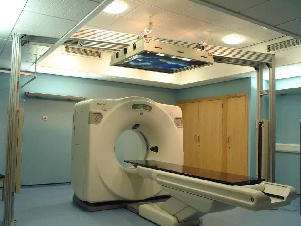
4 Context: Edinburgh Cancer Centre GE HiSpeed FX/i single slice, helical scanner Primarily for radiotherapy treatment planning Frequent QA daily, fortnightly, monthly Strict, tight tolerances ±10 HU on CT number calibration, ± 1 mm on geometry Unusual set-up external lasers, flat couch-top, etc. Catphan 500 Possibility of future use with Sim-CT system
5
6 Our Ideal Analysis Software Quick and easy to use for routine QA Fully automatic in as many different set-up configurations as possible Accurate, reliable, robust Knowledge of implemented algorithms Understand limitations and caveats Ability to access results for further analysis outside software BUT able to deal with unusual situations Manual methods when automatic algorithms fail
7 Evaluation Criteria 1. Software installation 2. Import of CT image files 3. Quality, accuracy and robustness of image analysis and result reporting 4. Retrospective trend analysis 5. Overall impressions (user interface, stability, documentation etc)
8 1. Software Installation Tested on Windows NT and 2000 machines Installation fully automatic and generally smooth Utilises Hardlock USB or Parallel port dongle, requiring installation of additional drivers Merge DICOM server NOT automatically installed Installation not described in manual but is relatively straightforward Automatically starts on login Uninstall program does not remove all icons and files
9 2. Import of CT Image Files Supports DICOM 3.0 files Implicit VR Little Endian format ONLY Two import methods Copy files into appropriate directory for automatic detection by software Send files across network to supplied DICOM server Exhaustively tested both of these
10 Import via Copying Files Images must be placed in c:\images\ct Strict naming convention No additional periods, dashes, etc in file names i.e. standard DICOM UID names not permitted Tested various DICOM formats Import of implicit VR LE images worked correctly All other DICOM formats failed Attempted import directly from CD-ROM written by GE Advantage Workstation FAILED! Images are stored as explicit VR LE Therefore, must be converted to correct format before importing into AutoQA software
11 Import via Network Transfer Fiddly to set-up but once correct worked very smoothly Practical end-user implementation EITHER, requires direct access to network configuration options of CT scanner or workstation OR, copy DICOM files from scanner to CD-ROM / optical disk and send to AutoQA server via local DICOM link in physics Tested both of above and both worked well

12 AutoQA Lite TM Browser
13 AutoQA Lite TM Browser Merges all image series under a particular study into one series Can be difficult to identify image required Image selection is a bit unstable Possible to start analysis without selecting an image! Causes software to crash Images which software cannot interpret appear in the browser as odd entries Delete option does not always work properly
14 3. Evaluation of Analysis Features Scanned all Catphan 500 slices using standard protocol Patient head-first, supine 120 kv, 200 ma, 1.5 s rotation, 5 mm slice width 25 cm diameter scan and reconstruction FOVs Standard reconstruction and processing filters Analysed alignment, MTF and uniformity slices using software Compared results against manual analysis and Dundee / ImPACT measurement software

15 Analysis Summary: Alignment Slice Bars: Centres give z alignment and rotation FWHM above background gives slice width CT Number linearity inserts Holes + phantom edges Pixel size calculation x and y alignment
 (Various methods exist) Retro-recon at 5cm")
16 Analysis Summary: MTF Slice Fourier transform of point spread function gives modulation transfer function (MTF) (Various methods exist) Retro-recon at 5cm FOV
17 Analysis Summary: Uniformity Slice Assess noise by considering mean and standard deviation in central and peripheral ROIs
18 Analysis Summary: Uniformity Slice Consider average horizontal and vertical profiles Fractional uniformity = fraction of pixels in profile lying within tolerance limits about mean (typically ±2σ)
19 Test Scenarios Phantom accurately aligned Experiment with range of fields of view Experiment with range of nominal slice widths Reconstruction so that (0,0,0) is not at centre of image Patient orientations other than HFS First slice at z=i500 mm rather than z=0 Geometrical offsets large and small shifts in x, y, z directions Plus combinations of all three Horizontal and vertical rotations
20 Accurately Aligned Phantom Most measurements agree well with both manual and Dundee/ImPACT software analyses Pixel size exact agreement x, y, z alignment exact agreement CT Num Linearity agree ±0.5% Slice width agree ±2% Phantom rotation x and y agree, z overestimated MTF agree ±8% with Dundee and GE software Noise agree within ±5% Fractional uniformity agree within ±10% Not clear what tolerances are used in calculation

21 AutoQA Lite TM Report
22 Different FOVs Experimented with 25, 35 and 2x50 cm FOV 50 cm FOV can be specified using either Large SFOV or Large-S FOV 25 cm and 35 cm FOV were successful Agreement with manual measurements similar to ideal case Analysis failed for 50 cm FOV Error: Cannot find edges of phantom
23 Different Slice Widths Scanned alignment slice using 1, 2, 3, 5, 7, 10 and 1 mm slice width All calculated widths agree with manual analysis to within ±4% Discrepancy increases with larger slice width Due to location of ROIs for background value? Number of images in report printout is confused
24 Offset Reconstruction Image reconstructed so that centre of slice is 2 cm right and 2 cm anterior of scan centre Centre position correctly calculated Rest of analysis carried out to same accuracy as before NB: AutoQA Lite TM coordinates are in frame of image rather than in anatomical directions
25 Patient Orientation Scanned phantom with patient Head-First Prone Analysis failed Software only works if image slices are in the negative z direction (inferior under this protocol) with respect to the alignment slice Could turn phantom round, but awkward
26 Alignment Slice Offset by z=50 cm CT-simulator specific test Verifies travel of couch from external isocentre to centre of scanner Software automatically identifies slices and analyses images correctly BUT quotes wrong z position for alignment slice Appears to define alignment slice as z=0 and calculates offsets of other slices relative to this
27 Geometrical Offsets Shift phantom in x, y and z directions X -5 mm X +10 mm Y 5 mm Y + 10 mm Z 3 mm Z +8 mm All interpreted correctly, within experimental uncertainty - Shift identified but quoted as 5.2 mm - Slice width correct so phantom bars OK Combination of x, y and z shifts: (5,10,-5) mm Interpreted correctly Note: X and Y shifts are quoted with direction, but z shift is not (i.e. no sign or direction indicator)
28 Phantom Rotation 2 rotation about x axis (superior end to anterior) Calculated correctly by software Approx 4 rotation about y axis (superior end to right) Angle significantly underestimated (2.3 ) Could be due to phantom alignment problem Observation across entire study Phantom repeatedly scanned in same position Calculated angle varies by ±0.2 Rotation of phantom in image plane appears to be slightly overestimated in comparison with manual analysis (+0.2 ) Horizontal and vertical axis rotations are not quoted with sign.
29 4. Retrospective Trend Analysis All results are stored in one of two databases Monitor database routine QA measurements Service database more specialised measurements Databases in DBase IV format Can be loaded into Excel for graphing, etc. Basic reports can be generated showing all results for particular scanner Results outside IEC tolerances are highlighted

30 Example Report
31 5. General Impressions (1) User Interface Generally easy to use Stability When images are in acceptable format, very good If bad images are imported, program can crash or hang Can be difficult to recover from crash without knowing exactly what went wrong
32 5. General Impressions (2) Documentation PDF Format on CD-ROM Describes basic analysis algorithms (similar to Catphan manual) More DICOM information and advice would be helpful More information on structure of phantom definition files would be helpful Customer Support Excellent support available via phone / to IRIS Inc Advice can be given on tuning phantom definition files, etc
33 Comparison of Analysis Methods Full manual analysis Dundee / ImPACT software + manual analysis AutoQA Lite TM 2 hours 30 mins 5 mins AutoQA LiteTM is fully automatic Great for routine QA No manual methods when automatic facility doesn t work (but there is the possibility of modifying configuration files under guidance from IRIS Inc)
34 Application: Reproducibility of Alignment against CT Lasers Catphan aligned on CT lasers and alignment slice scanned Catphan removed, realigned and scanned again. Repeated to give 10 scans in total Repeated for external laser gantry All scans analysed by AutoQA Lite TM Results: Z position out by 0.5 mm (external) and 0.7 mm (internal) Alignment accuracy (1σ) of ±0.1 mm (external) and 0.2 mm (internal)
35 Conclusions When it works it works very well! Vast majority of the measurements performed agree well with external measurement tools Software performs satisfactorily in a wide range of scenarios Once images are imported, analysis is nearly instantaneous Very useful for routine QA programme Limitations MUST be understood before use DICOM file formats, patient orientation, FOV, etc. Glitches in user interface which must be avoided
36 Acknowledgements AutoQA Lite TM Software The IRIS Inc Steve Dyer ( Southern Scientific Keith Thompson ( IDL Image Measurement Software Dundee Medical Physics ( David Sutton Julie Smyth Benedikt Lubbers Alexander Dietzel ImPACT ( Nick Keat
Automated Image Analysis Software for Quality Assurance of a Radiotherapy CT Simulator
 Automated Image Analysis Software for Quality Assurance of a Radiotherapy CT Simulator Andrew J Reilly Imaging Physicist Oncology Physics Edinburgh Cancer Centre Western General Hospital EDINBURGH EH4
ImPACT. Information Leaflet No. 1: CT Scanner Acceptance Testing
 ImPACT Information Leaflet No. 1: CT Scanner Acceptance Testing Version 1.02, 18/05/01 CONTENTS: 1. SCOPE OF LEAFLET 2. GENERAL PRINCIPLES OF ACCEPTANCE AND COMMISSIONING 2.1 PHANTOMS 2.2 EXPOSURE AND
RITtrend allows you to effectively manage all your physics QA data in one powerful and customizable package.
 So much is asked of medical physicists these days We Can Help Automated Radiation Therapy Phantom Analysis in Seconds QC for Therapy OBI s and CT simulators. Radia s Catphan/OBI module performs analysis
C a t p h a n / T h e P h a n t o m L a b o r a t o r y
 C a t p h a n 5 0 0 / 6 0 0 T h e P h a n t o m L a b o r a t o r y C a t p h a n 5 0 0 / 6 0 0 Internationally recognized for measuring the maximum obtainable performance of axial, spiral and multi-slice
S. Guru Prasad, Ph.D., DABR
 PURPOSE S. Guru Prasad, Ph.D., DABR Director of Medical Physics IAEA Consultant NorthShore University Health System and University of Chicago, Pritzker School of Medicine Current TPS utilize more information
Initial Clinical Experience with 3D Surface Image Guidance
 Initial Clinical Experience with 3D Surface Image Guidance Amanda Havnen-Smith, Ph.D. Minneapolis Radiation Oncology Ridges Radiation Therapy Center Burnsville, MN April 20 th, 2012 Non-funded research
Lucy Phantom MR Grid Evaluation
 Lucy Phantom MR Grid Evaluation Anil Sethi, PhD Loyola University Medical Center, Maywood, IL 60153 November 2015 I. Introduction: The MR distortion grid, used as an insert with Lucy 3D QA phantom, is
Slide 1. Technical Aspects of Quality Control in Magnetic Resonance Imaging. Slide 2. Annual Compliance Testing. of MRI Systems.
 Slide 1 Technical Aspects of Quality Control in Magnetic Resonance Imaging Slide 2 Compliance Testing of MRI Systems, Ph.D. Department of Radiology Henry Ford Hospital, Detroit, MI Slide 3 Compliance Testing
CT NOISE POWER SPECTRUM FOR FILTERED BACKPROJECTION AND ITERATIVE RECONSTRUCTION
 CT NOISE POWER SPECTRUM FOR FILTERED BACKPROJECTION AND ITERATIVE RECONSTRUCTION Frank Dong, PhD, DABR Diagnostic Physicist, Imaging Institute Cleveland Clinic Foundation and Associate Professor of Radiology
7/31/2011. Learning Objective. Video Positioning. 3D Surface Imaging by VisionRT
 CLINICAL COMMISSIONING AND ACCEPTANCE TESTING OF A 3D SURFACE MATCHING SYSTEM Hania Al-Hallaq, Ph.D. Assistant Professor Radiation Oncology The University of Chicago Learning Objective Describe acceptance
IAEA-TECDOC-1583 Commissioning of Radiotherapy Treatment Planning Systems: Testing for Typical External Beam Treatment Techniques
 IAEA-TECDOC-1583 Commissioning of Radiotherapy Treatment Planning Systems: Testing for Typical External Beam Treatment Techniques Report of the Coordinated Research Project (CRP) on Development of Procedures
Tomotherapy Physics. Machine Twinning and Quality Assurance. Emilie Soisson, MS
 Tomotherapy Physics Machine Twinning and Quality Assurance Emilie Soisson, MS Tomotherapy at UW- Madison Treating for nearly 5 years Up to ~45 patients a day on 2 tomo units Units twinned to facilitate
Design and performance characteristics of a Cone Beam CT system for Leksell Gamma Knife Icon
 Design and performance characteristics of a Cone Beam CT system for Leksell Gamma Knife Icon WHITE PAPER Introduction Introducing an image guidance system based on Cone Beam CT (CBCT) and a mask immobilization
DAILY LINAC QA BEAM QA
 BEAM QA DAILY LINAC QA The QA BeamChecker Plus allows for fast, reliable, and uncomplicated daily QA of Varian, Elekta, Siemens, and Accuray Treatment Machines. The QA BeamChecker Plus is specifically
Automated Quality Assurance for Image-Guided Radiation Therapy
 JOURNAL OF APPLIED CLINICAL MEDICAL PHYSICS, VOLUME 10, NUMBER 1, WINTER 2009 Automated Quality Assurance for Image-Guided Radiation Therapy Eduard Schreibmann, a Eric Elder, Tim Fox Department of Radiation
Future is Effortless
 AQUATM Radiology is a machine Quality Assurance management solution that standardizes, streamlines and automates QA procedures throughout rediology department and across multiple sites. AQUATM Radiology
Protocol. Technical evaluation of X-ray tomographic image-guided radiotherapy devices CEP10070
 Protocol Technical evaluation of X-ray tomographic image-guided radiotherapy devices CEP10070 March 2010 Contents 2 Introduction... 3 Protocol design and validation... 4 General information... 7 Technical
TomoTherapy Related Projects. An image guidance alternative on Tomo Low dose MVCT reconstruction Patient Quality Assurance using Sinogram
 TomoTherapy Related Projects An image guidance alternative on Tomo Low dose MVCT reconstruction Patient Quality Assurance using Sinogram Development of A Novel Image Guidance Alternative for Patient Localization
Optimisation of Toshiba Aquilion ONE Volume Imaging
 Optimisation of Toshiba Aquilion ONE Volume Imaging Jane Edwards, RPRSG Royal Free London NHS Foundation Trust Dr Mufudzi Maviki, Plymouth Hospitals NHS Trust Background In 2011/12 Radiology at RFH was
Quality control phantoms and protocol for a tomography system
 Quality control phantoms and protocol for a tomography system Lucía Franco 1 1 CT AIMEN, C/Relva 27A O Porriño Pontevedra, Spain, lfranco@aimen.es Abstract Tomography systems for non-destructive testing
TG-148 overview. Introduction. System Overview. System Overview. QA for Helical Tomotherapy: Report of the AAPM Task Group 148. Conflict of Interest:
 QA or Helical Tomotherapy: Report o the AAPM Tas Group 148 Members: Conlict o Interest: r. John Balog owns TomoTherapy stoc. Katja Langen (Co-chair) Nio Papaniolaou (Co-chair) Walter Grant Richard Crilly
3/27/2012 WHY SPECT / CT? SPECT / CT Basic Principles. Advantages of SPECT. Advantages of CT. Dr John C. Dickson, Principal Physicist UCLH
 3/27/212 Advantages of SPECT SPECT / CT Basic Principles Dr John C. Dickson, Principal Physicist UCLH Institute of Nuclear Medicine, University College London Hospitals and University College London john.dickson@uclh.nhs.uk
PET-CT in Radiation Treatment Planning
 PET-CT in Radiation Treatment Planning TA van de Water, Radiotherapeutic Institute Friesland, Leeuwarden JA van Dalen, Isala, Zwolle ACKNOWLEDGEMENTS A. van der Schaaf (University of Groningen, University
Performance Evaluation of the Philips Gemini PET/CT System
 Performance Evaluation of the Philips Gemini PET/CT System Rebecca Gregory, Mike Partridge, Maggie A. Flower Joint Department of Physics, Institute of Cancer Research, Royal Marsden HS Foundation Trust,
Annexure XII SPECIFICATIONS FOR A NEW STATE OF ART 16 SLICE ALL PURPOSE C. T. SCANNER
 Annexure XII SPECIFICATIONS FOR A NEW STATE OF ART 16 SLICE ALL PURPOSE C. T. SCANNER A) Scanner Design X-Ray generator and tube: 1. Scanner: Whole body spiral CT scanner (16 slices) of latest technology.
SPECT QA and QC. Bruce McBride St. Vincent s Hospital Sydney.
 SPECT QA and QC Bruce McBride St. Vincent s Hospital Sydney. SPECT QA and QC What is needed? Why? How often? Who says? QA and QC in Nuclear Medicine QA - collective term for all the efforts made to produce
Unique Features of the GE Senoclaire Tomosynthesis System. Tyler Fisher, M.S., DABR Therapy Physics, Inc.
 Unique Features of the GE Senoclaire Tomosynthesis System Tyler Fisher, M.S., DABR Therapy Physics, Inc. Conflict of Interest Disclosure I have no conflicts to disclose. Learning Objectives Overview of
Quick Reference Datasheet For All RIT113 Packages
 Quick Reference Datasheet For All RIT113 Packages For Rotational Therapies, IMRT & TG142 Highlights and selected product information only. A complete TG142 brochure is available. For more information on
Evaluation report. X-ray tomographic image guided radiotherapy systems CEP10071
 Evaluation report X-ray tomographic image guided radiotherapy systems CEP10071 March 2010 Contents 2 Summary... 3 Introduction... 6 Product description... 9 Methods... 14 Technical performance... 23 Purchasing...
A study on image quality provided by a kilovoltage cone-beam computed tomography
 JOURNAL OF APPLIED CLINICAL MEDICAL PHYSICS, VOLUME 14, NUMBER 1, 2013 A study on image quality provided by a kilovoltage cone-beam computed tomography Julia Garayoa a and Pablo Castro Servicio de Radiofísica,
INTERNATIONAL STANDARD
 INTERNATIONAL STANDARD IEC 60601-2-44 2001 AMENDMENT 1 2002-09 Amendment 1 Medical electrical equipment Part 2-44: Particular requirements for the safety of X-ray equipment for computed tomography Amendement
연구용유방초음파질관리 원광대학병원김혜원
 연구용유방초음파질관리 원광대학병원김혜원 Why US QC? Quality control (QC) testing of ultrasound scanners is important to verify the proper and consistent operation of these devices. main goal ; quality improvement Guidelines
ph fax Deming Way Middleton WI USA
 www.standardimaging.com 800-261-4446. ph 608-831-0025. fax 608-831-2202 3120 Deming Way Middleton WI 53562-1461 USA 2007 Standard Imaging, Inc. 1239-21 (03.07) beam qa LINAC AND TOMOTHERAPY QA The NEW
Implementation and evaluation of a fully 3D OS-MLEM reconstruction algorithm accounting for the PSF of the PET imaging system
 Implementation and evaluation of a fully 3D OS-MLEM reconstruction algorithm accounting for the PSF of the PET imaging system 3 rd October 2008 11 th Topical Seminar on Innovative Particle and Radiation
Comparison of the Imaging Performance of CT Scanners, Issue 11
 Imaging Performance Assessment of CT Scanners Comparison of the Imaging Performance of CT Scanners, Issue 11 Comparison data is presented on the following scanners : Elscint Twin Flash IGE HiSpeed Advantage
Optimization of CT Simulation Imaging. Ingrid Reiser Dept. of Radiology The University of Chicago
 Optimization of CT Simulation Imaging Ingrid Reiser Dept. of Radiology The University of Chicago Optimization of CT imaging Goal: Achieve image quality that allows to perform the task at hand (diagnostic
INSTRUCTIONS RPD INFORMATION RPD PRODUCT INFORMATION. Address 5218 Barthel Industrial Drive Albertville, MN Website
 Expect Service INSTRUCTIONS Radiation Products Design Inc RPD INFORMATION Address 5218 Barthel Industrial Drive Albertville, MN 55301 Website www.rpdinc.com Email sales@rpdinc.com Phone 763-497-2071 or
Spiral CT. Protocol Optimization & Quality Assurance. Ge Wang, Ph.D. Department of Radiology University of Iowa Iowa City, Iowa 52242, USA
 Spiral CT Protocol Optimization & Quality Assurance Ge Wang, Ph.D. Department of Radiology University of Iowa Iowa City, Iowa 52242, USA Spiral CT Protocol Optimization & Quality Assurance Protocol optimization
An approach for measuring the spatial orientations of a computed-tomography simulation system
 JOURNAL OF APPLIED CLINICAL MEDICAL PHYSICS, VOLUME 15, NUMBER 2, 2014 An approach for measuring the spatial orientations of a computed-tomography simulation system Meng Chia Wu, a Ramani Ramaseshan Department
BLUEPRINT TM 3D Planning Software - v1.4 scan protocol
 S H O U L D E R Solutions by Tornier 3 D P L A N N I N G S O F T W A R E B L U E P R I N T T M 3 D P L A N N I N G S O F T W A R E S C A N P R O T O C O L BLUEPRINT TM 3D Planning Software - v1.4 scan
DoseLab for TomoTherapy. AAPM Task Group 148 Background
 DoseLab for TomoTherapy Performing TG-148 QA with DoseLab Christine Gnaster, MS, DABR DoseLab Product Manager AAPM Task Group 148 Background TG-148 could be likened to the TG-142 for TomoTherapy machines
Virtual Phantoms for IGRT QA
 TM Virtual Phantoms for IGRT QA Why ImSimQA? ImSimQA was developed to overcome the limitations of physical phantoms for testing modern medical imaging and radiation therapy software systems, when there
An Acquisition Geometry-Independent Calibration Tool for Industrial Computed Tomography
 4th International Symposium on NDT in Aerospace 2012 - Tu.3.A.3 An Acquisition Geometry-Independent Calibration Tool for Industrial Computed Tomography Jonathan HESS *, Patrick KUEHNLEIN *, Steven OECKL
URGENT IMPORTANT FIELD SAFETY NOTIFICATION
 Subject: Incorrect Movement of the Treatment Table Product: MOSAIQ Scope: Sites affected will be those: 1. Running MOSAIQ and, 2. Treating on linear accelerators with the RATM license Notification Released:
Performance and characteristics of an IR localizing system for radiation therapy
 JOURNAL OF APPLIED CLINICAL MEDICAL PHYSICS, VOLUME 7, NUMBER 2, SPRING 2006 Performance and characteristics of an IR localizing system for radiation therapy Yulia Lyatskaya, 1 Hsiao-Ming Lu, 2 and Lee
XiO DICOM Conformance Statement
 XiO DICOM Conformance Statement For Release 5.10 IMPAC Medical Systems, Inc. Document ID: LEDDCMXIO0001 Language: English Copyright statement 2014 IMPAC Medical Systems, Inc. All rights reserved. Do not
Machine and Physics Data Guide
 WWW..COM Machine and Physics Data Guide STANDARD IMAGING, INC. 3120 Deming Way Middleton, WI 53562-1461 May / 2008 2008 Standard Imaging, Inc. TEL 800.261.4446 TEL 608.831.0025 FAX 608.831.2202 www.standardimaging.com
Computed Tomography & 3D Metrology Application of the VDI/VDE Directive 2630 and Optimization of the CT system
 Computed Tomography & 3D Metrology Application of the VDI/VDE Directive 2630 and Optimization of the CT system ECNDT 2014 Prague October 6-10, 2014 Dr. Eberhard Neuser Dr. Alexander Suppes Imagination
Effects of the difference in tube voltage of the CT scanner on. dose calculation
 Effects of the difference in tube voltage of the CT scanner on dose calculation Dong Joo Rhee, Sung-woo Kim, Dong Hyeok Jeong Medical and Radiological Physics Laboratory, Dongnam Institute of Radiological
Optimized Design of 3D Laser Triangulation Systems
 The Scan Principle of 3D Laser Triangulation Triangulation Geometry Example of Setup Z Y X Target as seen from the Camera Sensor Image of Laser Line The Scan Principle of 3D Laser Triangulation Detektion
Clinical implementation of photon beam flatness measurements to verify beam quality
 JOURNAL OF APPLIED CLINICAL MEDICAL PHYSICS, VOLUME 16, NUMBER 6, 2015 Clinical implementation of photon beam flatness measurements to verify beam quality Simon Goodall, a Nicholas Harding, Jake Simpson,
The team. Disclosures. Ultrasound Guidance During Radiation Delivery: Confronting the Treatment Interference Challenge.
 Ultrasound Guidance During Radiation Delivery: Confronting the Treatment Interference Challenge Dimitre Hristov Radiation Oncology Stanford University The team Renhui Gong 1 Magdalena Bazalova-Carter 1
7/13/2015 EVALUATION OF NONLINEAR RECONSTRUCTION METHODS. Outline. This is a decades-old challenge
 EVALUATION OF NONLINEAR RECONSTRUCTION METHODS Kyle J. Myers, Ph.D. Director, Division of Imaging, Diagnostics, and Software Reliability Office of Science and Engineering Laboratories, CDRH, FDA 2 Outline
Iterative SPECT reconstruction with 3D detector response
 Iterative SPECT reconstruction with 3D detector response Jeffrey A. Fessler and Anastasia Yendiki COMMUNICATIONS & SIGNAL PROCESSING LABORATORY Department of Electrical Engineering and Computer Science
PURE. ViSION Edition PET/CT. Patient Comfort Put First.
 PURE ViSION Edition PET/CT Patient Comfort Put First. 2 System features that put patient comfort and safety first. Oncology patients deserve the highest levels of safety and comfort during scans. Our Celesteion
1. Learn to incorporate QA for surface imaging
 Hania Al-Hallaq, Ph.D. Assistant Professor Radiation Oncology The University of Chicago ***No disclosures*** 1. Learn to incorporate QA for surface imaging into current QA procedures for IGRT. 2. Understand
Version 5.6. Quick Start Guide
 WWW..COM Version 5.6 Quick Start Guide STANDARD IMAGING, INC. 3120 Deming Way Middleton, WI 53562-1461 Jul / 2018 2018 Standard Imaging, Inc. TEL 800.261.4446 TEL 608.831.0025 FAX 608.831.2202 DOC # 80714-05
CT vs. VolumeScope: image quality and dose comparison
 CT vs. VolumeScope: image quality and dose comparison V.N. Vasiliev *a, A.F. Gamaliy **b, M.Yu. Zaytsev b, K.V. Zaytseva ***b a Russian Sci. Center of Roentgenology & Radiology, 86, Profsoyuznaya, Moscow,
MapCHECK 2 & 3DVH. The Gold Standard for 2D Arrays
 MapCHECK 2 & 3DVH The Gold Standard for 2D Arrays Your Most Valuable QA and Dosimetry Tools THE GOLD STANDARD FOR 2D ARRAYS The MapCHECK 2 is the world s most selected independent 2D measurement array.
Calypso the Easy Way to Create Part Programs
 Industrial Measuring Technology from Carl Zeiss Calypso the Easy Way to Create Part Programs We make it visible. Philosophy Visual Metrology TM CAD model feature characteristics Elements are selected to
Medical Image Processing: Image Reconstruction and 3D Renderings
 Medical Image Processing: Image Reconstruction and 3D Renderings 김보형 서울대학교컴퓨터공학부 Computer Graphics and Image Processing Lab. 2011. 3. 23 1 Computer Graphics & Image Processing Computer Graphics : Create,
Reduction of Metal Artifacts in Computed Tomographies for the Planning and Simulation of Radiation Therapy
 Reduction of Metal Artifacts in Computed Tomographies for the Planning and Simulation of Radiation Therapy T. Rohlfing a, D. Zerfowski b, J. Beier a, P. Wust a, N. Hosten a, R. Felix a a Department of
Retrospective Spiral Respiratory Correlated Imaging with Varian RPM
 Retrospective Spiral Respiratory Correlated Imaging with Varian RPM This is a Quick Step Guide for Retrospective Spiral Respiratory Correlated Imaging (4D CT) using the Varian RPM device v1.7 with the
Design and development of a phantom for tomosynthesis with potential for automated analysis via the cloud
 Received: 21 August 2017 Revised: 25 October 2017 Accepted: 7 January 2018 DOI: 10.1002/acm2.12297 MEDICAL IMAGING Design and development of a phantom for tomosynthesis with potential for automated analysis
MapCHECK 2 & 3DVH The Gold Standard for 2D Arrays
 MapCHECK 2 & 3DVH The Gold Standard for 2D Arrays Your Most Valuable QA and Dosimetry Tools THE GOLD STANDARD FOR 2D ARRAYS The MapCHECK 2 is the world s most selected independent 2D measurement array.
Brilliance CT Big Bore.
 1 2 2 There are two methods of RCCT acquisition in widespread clinical use: cine axial and helical. In RCCT with cine axial acquisition, repeat CT images are taken each couch position while recording respiration.
A NEW LASER FOCUS ON PATIENT SAFETY
 A NEW LASER FOCUS ON PATIENT SAFETY PATIENT SAFETY STARTS HERE During CT Simulation, clinics rely on moveable lasers to mark where radiation will enter a patient s body to target the tumor. Fixed lasers
Delta 4. DICOM Conformance Statement D (7)
") Delta 4 DICOM Conformance Statement D001 32 005 01 1 (7) 1 Introduction 1.1 Purpose This conformance statement specifies how the Delta 4 application conforms to the DICOM v3.0 standard. Delta4 uses DICOM
Delta 4 DICOM Conformance Statement D001 32 005 01 1 (7) 1 Introduction 1.1 Purpose This conformance statement specifies how the Delta 4 application conforms to the DICOM v3.0 standard. Delta4 uses DICOM
Developments in Dimensional Metrology in X-ray Computed Tomography at NPL
 Developments in Dimensional Metrology in X-ray Computed Tomography at NPL Wenjuan Sun and Stephen Brown 10 th May 2016 1 Possible factors influencing XCT measurements Components Influencing variables Possible
Lorad FFDM QC Procedures for Medical Physicist. Tao Wu, Ph.D. Hologic, Inc.
 Lorad FFDM QC Procedures for Medical Physicist Tao Wu, Ph.D. Hologic, Inc. Lorad Selenia FFDM Tests Following Lorad QC Manual Collimation Assessment Artifact Evaluation System Resolution Phantom Image
Delta 4 PT. True Volumetric Pre-treatment Verification. The difference is clear
 Delta 4 PT True Volumetric Pre-treatment Verification The difference is clear ScandiDos innovators in advanced volumetric dosimetry solutions ScandiDos ScandiDos is the innovative company that introduced
Quality assurance of a helical tomotherapy machine
 INSTITUTE OF PHYSICS PUBLISHING Phys. Med. Biol. 49 (2004) 2933 2953 PHYSICS IN MEDICINE AND BIOLOGY PII: S0031-9155(04)71892-8 Quality assurance of a helical tomotherapy machine J D Fenwick 1,2,WATomé
A Study of Motion Tracking Accuracy of Robotic Radiosurgery Using a Novel CCD Camera Based End-to-end Test System
 A Study of Motion Tracking Accuracy of Robotic Radiosurgery Using a Novel CCD Camera Based End-to-end Test System Lei Wang 1, Brett Nelson 2,Youming Yang 1 1. Department of Radiation Oncology, Stanford
4 Measurement. and Analysis. 4.1 Overview and Underlying Principles 4-1
 Measurement and Analysis.1 Overview and Underlying Principles.1.1 Introductory Remarks The physics and setup for film dosimetry have been described in the previous chapters. The measurement setup for IMRT
icatvision Quick Reference
 icatvision Quick Reference Navigating the i-cat Interface This guide shows how to: View reconstructed images Use main features and tools to optimize an image. REMINDER Images are displayed as if you are
Applying Hounsfield unit density calibration in SkyScan CT-analyser
 1 Bruker-microCT Method note Applying Hounsfield unit density calibration in SkyScan CT-analyser Hounsfield units (HU) are a standard unit of x-ray CT density, in which air and water are ascribed values
THE WIRELESS PHANTOM PERFORM ACCURATE PATIENT QA IN LESS TIME THAN EVER!
 THE WIRELESS PHANTOM PERFORM ACCURATE PATIENT QA IN LESS TIME THAN EVER! Confidence in complex treatments Modern radiation therapy uses complex plans with techniques such as IMRT, VMAT and Tomotherapy.
Scatter Correction for Dual source Cone beam CT Using the Pre patient Grid. Yingxuan Chen. Graduate Program in Medical Physics Duke University
 Scatter Correction for Dual source Cone beam CT Using the Pre patient Grid by Yingxuan Chen Graduate Program in Medical Physics Duke University Date: Approved: Lei Ren, Supervisor Fang Fang Yin, Chair
1. Deployment of a framework for drawing a correspondence between simple figure of merits (FOM) and quantitative imaging performance in CT.
 and quantitative imaging performance in CT.") Progress report: Development of assessment and predictive metrics for quantitative imaging in chest CT Subaward No: HHSN6801000050C (4a) PI: Ehsan Samei Reporting Period: month 1-18 Deliverables: 1. Deployment
Progress report: Development of assessment and predictive metrics for quantitative imaging in chest CT Subaward No: HHSN6801000050C (4a) PI: Ehsan Samei Reporting Period: month 1-18 Deliverables: 1. Deployment
Spectral analysis of non-stationary CT noise
 Spectral analysis of non-stationary CT noise Kenneth M. Hanson Los Alamos Scientific Laboratory Int. Symposium and Course on Computed Tomography, Las Vegas, April 7-11, 1980 This presentation available
Multi-slice CT Image Reconstruction Jiang Hsieh, Ph.D.
 Multi-slice CT Image Reconstruction Jiang Hsieh, Ph.D. Applied Science Laboratory, GE Healthcare Technologies 1 Image Generation Reconstruction of images from projections. textbook reconstruction advanced
PrecisePLAN 2.00 Import
 Elekta Oncology Systems Ltd. DICOM Conformance Statement For PrecisePLAN 2.00 Import (DICOM Release 2.00) Copyright 2003 Elekta Oncology Systems Ltd. All rights reserved DRC-170-0028-01 DICOM Conformance
Comparison of Scatter Correction Methods for CBCT. Author(s): Suri, Roland E.; Virshup, Gary; Kaissl, Wolfgang; Zurkirchen, Luis
: Suri, Roland E.; Virshup, Gary; Kaissl, Wolfgang; Zurkirchen, Luis") Research Collection Working Paper Comparison of Scatter Correction Methods for CBCT Author(s): Suri, Roland E.; Virshup, Gary; Kaissl, Wolfgang; Zurkirchen, Luis Publication Date: 2010 Permanent Link:
Research Collection Working Paper Comparison of Scatter Correction Methods for CBCT Author(s): Suri, Roland E.; Virshup, Gary; Kaissl, Wolfgang; Zurkirchen, Luis Publication Date: 2010 Permanent Link:
ADVANCING CANCER TREATMENT
 The RayPlan treatment planning system makes proven, innovative RayStation technology accessible to clinics that need a cost-effective and streamlined solution. Fast, efficient and straightforward to use,
CTA HEAD Perfusion AqONE without and with IV Contrast
 CTA HEAD Perfusion AqONE without and with IV Contrast Patient Position Adult Contrast Adult Injection Rate Supine IOML perpendicular to table top. IV: 100 ml with helical head CTA 50 ml without helical
CLASS HOURS: 4 CREDIT HOURS: 4 LABORATORY HOURS: 0
 Revised 10/10 COURSE SYLLABUS TM 220 COMPUTED TOMOGRAPHY PHYSICS CLASS HOURS: 4 CREDIT HOURS: 4 LABORATORY HOURS: 0 CATALOG COURSE DESCRIPTION: This course is one of a three course set in whole body Computed
Overview of Proposed TG-132 Recommendations
 Overview of Proposed TG-132 Recommendations Kristy K Brock, Ph.D., DABR Associate Professor Department of Radiation Oncology, University of Michigan Chair, AAPM TG 132: Image Registration and Fusion Conflict
Thank-You Members of TG147 TG 147: QA for nonradiographic
 Thank-You Members of TG147 TG 147: QA for nonradiographic localization and positioning systems Twyla Willoughby, M.S. Medical Physicist Clinical AAPM Meeting March 2013 Department of Radiation Oncology
CT Protocol Review: Practical Tips for the Imaging Physicist Physicist
 CT Protocol Review: Practical Tips for the Imaging Physicist Physicist Dianna Cody, Ph.D., DABR, FAAPM U.T.M.D. Anderson Cancer Center August 8, 2013 AAPM Annual Meeting Goals Understand purpose and importance
Philips SPECT/CT Systems
 Philips SPECT/CT Systems Ling Shao, PhD Director, Imaging Physics & System Analysis Nuclear Medicine, Philips Healthcare June 14, 2008 *Presented SNM08 Categorical Seminar - Quantitative SPECT and PET
Basics of treatment planning II
 Basics of treatment planning II Sastry Vedam PhD DABR Introduction to Medical Physics III: Therapy Spring 2015 Dose calculation algorithms! Correction based! Model based 1 Dose calculation algorithms!
The DICOM Standard. Miloš Šrámek Austrian Academy of Sciences
 The DICOM Standard Miloš Šrámek Austrian Academy of Sciences Medical Image Formats Typical information present in a file: Image data (unmodified or compressed) Patient identification and demographics Technical
Suitability of a new alignment correction method for industrial CT
 Suitability of a new alignment correction method for industrial CT Matthias Elter 1, Nicole Maass 1, Peter Koch 2 1 Siemens AG, Healthcare Sector, Erlangen, Germany, e-mail: matthias.elter@siemens.com,
8/2/2016. Measures the degradation/distortion of the acquired image (relative to an ideal image) using a quantitative figure-of-merit
 using a quantitative figure-of-merit") Ke Li Assistant Professor Department of Medical Physics and Department of Radiology School of Medicine and Public Health, University of Wisconsin-Madison This work is partially supported by an NIH Grant
Ke Li Assistant Professor Department of Medical Physics and Department of Radiology School of Medicine and Public Health, University of Wisconsin-Madison This work is partially supported by an NIH Grant
Data. ModuLeaf Mini Multileaf Collimator Precision Beam Shaping for Advanced Radiotherapy
 Data ModuLeaf Mini Multileaf Collimator Precision Beam Shaping for Advanced Radiotherapy ModuLeaf Mini Multileaf Collimator Precision Beam Shaping for Advanced Radiotherapy The ModuLeaf Mini Multileaf
LASER SOLUTIONS. Ensure Accurate Setup from Simulation to Treatment. MOVEABLE: CT SIM+ // FIXED: MICRO and MICRO+
 LASER SOLUTIONS Ensure Accurate Setup from Simulation to Treatment MOVEABLE: CT SIM+ // FIXED: MICRO and MICRO+ THE LEADER IN LASER ALIGNMENT Gammex was the first company to replace incandescent lightbulbs
Use of Monte Carlo modelling in radiotherapy linac design. David Roberts, PhD Senior Physicist Elekta
 Use of Monte Carlo modelling in radiotherapy linac design David Roberts, PhD Senior Physicist Elekta Contents Overview of Elekta What we do Where we use Monte Carlo Codes and resources Example : Agility
IMRT and VMAT Patient Specific QA Using 2D and 3D Detector Arrays
 IMRT and VMAT Patient Specific QA Using 2D and 3D Detector Arrays Sotiri Stathakis Outline Why IMRT/VMAT QA AAPM TG218 UPDATE Tolerance Limits and Methodologies for IMRT Verification QA Common sources
Voxar 3D ColonMetrix. Reference Guide
 Voxar 3D ColonMetrix Reference Guide The software described in this document is furnished under a license, and may be used or copied only according to the terms of such license. Toshiba means, Toshiba
Micro-CT Methodology Hasan Alsaid, PhD
 Micro-CT Methodology Hasan Alsaid, PhD Preclinical & Translational Imaging LAS, PTS, GlaxoSmithKline 20 April 2015 Provide basic understanding of technical aspects of the micro-ct Statement: All procedures