MEDICAL IMAGE COMPUTING (CAP 5937) LECTURE 4: Pre-Processing Medical Images (II)
|
|
|
- Mervyn Carter
- 5 years ago
- Views:
Transcription
1 SPRING MEDICAL IMAGE COMPUTING (CAP 5937) LECTURE 4: Pre-Processing Medical Images (II) Dr. Ulas Bagci HEC 221, Center for Research in Computer Vision (CRCV), University of Central Florida (UCF), Orlando, FL bagci@ucf.edu or bagci@crcv.ucf.edu
2 2 Outline Diffusion based Smoothing in Medical Scans Intensity inhomogeneity Correction in MRI
3 3 Recap: How to measure for evaluating noise removal algorithms? Simultaneously suppressing noise and retaining high contrast is difficult trade-off game. Filter Operating Characteristic (FOC) curve captures this trade-off. Residual Noise RN: Relative Standard deviation of intensity within pbject region Contrast RC: - object & background mean; - object & background std. FOC is a curve of 1-RC vs 1-RN.
4 4 Linear and Non-Linear Filtering (Smoothing) Linear approach: Treat every pixel with the exact same convolution. Non-Linear approach: Treat a pixel with varying intensity, depending on its neighborhood qualities.

5 Possible? 5
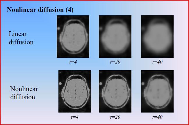
6 6 Perona-Malik (Anisotropic Diffusion) Filtering Perona and Malik propose a nonlinear diffusion method for avoiding the blurring and localization problems of linear diffusion filtering [PAMI 1990]. Smooth the images without removing significant parts of the edges
7 7 Perona-Malik (Anisotropic Diffusion) Filtering Perona and Malik propose a nonlinear diffusion method for avoiding the blurring and localization problems of linear diffusion filtering [PAMI 1990]. Smooth the images without removing significant parts of the edges The smoothing process is considered as diffusion
8 8 Perona-Malik (Anisotropic Diffusion) Filtering Perona and Malik propose a nonlinear diffusion method for avoiding the blurring and localization problems of linear diffusion filtering [PAMI 1990]. Smooth the images without removing significant parts of the edges The smoothing process is considered as diffusion APPROACH: Increase the diffusivity of filter for large (homogeneous) regions, and decrease it nearby edges!
9 9 Perona-Malik (Anisotropic Diffusion) Filtering Perona and Malik propose a nonlinear diffusion method for avoiding the blurring and localization problems of linear diffusion filtering [PAMI 1990]. Smooth the images without removing significant parts of the edges The smoothing process is considered as diffusion APPROACH: Increase the diffusivity of filter for large (homogeneous) regions, and decrease it nearby edges! How can we understand homogenous and edge regions then?
10 10 Perona-Malik (Anisotropic Diffusion) Filtering Perona and Malik propose a nonlinear diffusion method for avoiding the blurring and localization problems of linear diffusion filtering [PAMI 1990]. Smooth the images without removing significant parts of the edges The smoothing process is considered as diffusion APPROACH: Increase the diffusivity of filter for large (homogeneous) regions, and decrease it nearby edges! How can we understand homogenous and edge regions then? Edge likelihood (i.e, gradient for instance) can be measured by
11 11 Perona-Malik (Anisotropic Diffusion) Filtering Perona and Malik propose a nonlinear diffusion method for avoiding the blurring and localization problems of linear diffusion filtering [PAMI 1990]. Smooth the images without removing significant parts of the edges The smoothing process is considered as diffusion APPROACH: Increase the diffusivity of filter for large (homogeneous) regions, and decrease it nearby edges! How can we understand homogenous and edge regions then? Edge likelihood (i.e, gradient for instance) can be measured by Perona-Malik filter is based on
12 12 Perona-Malik (Anisotropic Diffusion) Filtering Perona and Malik propose a nonlinear diffusion method for avoiding the blurring and localization problems of linear diffusion filtering [PAMI 1990]. Smooth the images without removing significant parts of the edges The smoothing process is considered as diffusion APPROACH: Increase the diffusivity of filter for large (homogeneous) regions, and decrease it nearby edges! How can we understand homogenous and edge regions then? Edge likelihood (i.e, gradient for instance) can be measured by Perona-Malik filter is based on where it uses diffusivities such as
13 13 Perona-Malik (Anisotropic Diffusion) Filtering Perona and Malik propose a nonlinear diffusion method for avoiding the blurring and localization problems of linear diffusion filtering [PAMI 1990]. Smooth the images without removing significant parts of the edges The smoothing process is considered as diffusion APPROACH: Increase the diffusivity of filter for large (homogeneous) regions, and decrease it nearby edges! How can we understand homogenous and edge regions then? Edge likelihood (i.e, gradient for instance) can be measured by Perona-Malik filter is based on where it uses diffusivities such as approximation
14 14 General Idea on Anisotropic Diffusivity if (x,y) is a part of an edge è apply little smoothing Else è apply full smoothing
 CONTROLLING THE BLURRING")
15 15 General Idea on Anisotropic Diffusivity if (x,y) is a part of an edge è apply little smoothing Else è apply full smoothing Assume, E is edge likelihood (telling you if you are in homogeneous or edge regions) CONTROLLING THE BLURRING (SMOOTHING) or
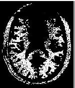
16 16 General Idea on Anisotropic Diffusivity if (x,y) is a part of an edge è apply little smoothing Else è apply full smoothing Define general coefficient (c) for diffusivity:
17 17 General Idea on Anisotropic Diffusivity if (x,y) is a part of an edge è apply little smoothing Else è apply full smoothing Define general coefficient (c) for diffusivity: d=1, direction
18 18 General Idea on Anisotropic Diffusivity if (x,y) is a part of an edge è apply little smoothing Else è apply full smoothing Define general coefficient (c) for diffusivity: Isotropic: Anisotropic:
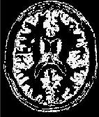
19 Toy Example 19 Original Linear isotropic diffusion (simple Gaussian)

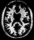
20 Toy Example 20 Original Non-linear isotropic diffusion (edge skipping)
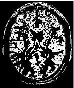
21 Toy Example 21 Original Non-linear anisotropic diffusion
22 22

23 Non-linear Diffusion 23


24 24 Background: BrainWeb GM WM CSF MR Brain simulator
")
25 25 Magnetic Field Inhomogeneity (bias) Credit: K.Friston
 with respect to the external field Different tissues")

26 26 Magnetic Field Inhomogeneity Field inhomogeneity is measured in parts per million (ppm) with respect to the external field Different tissues have different magnetic susceptibilities ð distortions in magnetic field distortions are most noticeable near air-tissue interfaces Credit: K.Friston

27 27 Often hardly noticeable, but registration, segmentation, and thus quantification processes are significantly affected from inhomogeneity field MR Intensity Inhomogeneity (credit: R.Gupta)

28 28 MR Intensity Inhomogeneity Large susceptibility variation in the human brain leads to greater field inhomogeneity and therefore image distortion. (credit: R.Gupta)
29 29 Intensity Inhomogeneity Correction Problem: Imperfections in the RF field cause background variations in MR images. Poses challenges in image segmentation and analysis. Goal: To develop a general method for correcting the variations that fulfills: (R1) no need for user help per scene (R2) no need for accurate prior segmentation (R3) no need for prior knowledge of tissue intensity distribution


30 30 Intensity Inhomogeneity Correction Methods Original Image Inhomogeneity Field Corrected Image
31 31 Bias Correction Approaches Numerous methods have been published in the last two decades I. Prospective Approaches i. Phantom ii. Multicoil iii. Special sequence II. Retrospective Approaches i. Filtering ii. Surface Fitting (intensity or gradient) iii. Segmentation (ML, MAP, FCM, nonparametric, ) iv. Histogram a. High frequency maximization b. Information amximization c. Histogram matching
32 32 Prospective Approaches-Phantom Treat intensity corruption as a systematic error of the MRI acquisition process that can either be minimized by acquiring additional images of a uniform phantom, by acquiring additional images with different coils, or by devising special imaging sequences.
33 33 Prospective Approaches-Phantom Treat intensity corruption as a systematic error of the MRI acquisition process that can either be minimized by acquiring additional images of a uniform phantom, by acquiring additional images with different coils, or by devising special imaging sequences. Oil or water is usually used for phantoms and median filtering is applied for image smoothing.
34 34 Prospective Approaches-Phantom Treat intensity corruption as a systematic error of the MRI acquisition process that can either be minimized by acquiring additional images of a uniform phantom, by acquiring additional images with different coils, or by devising special imaging sequences. Oil or water is usually used for phantoms and median filtering is applied for image smoothing. Warning: The phantom based approach cannot correct for patient-induced inhomogeneity, which is a major drawback of this approach. The remaining intensity inhomogeneity can be as high as 30%
35 35 Prospective Approaches-MultiCoil and Special Sequences Volume and Surface coils Volume coil: induce less inhomogeneity, poor SNR Surface coil: induce severe inhomogeneity, good SNR Method: dividing the filtered surface coil image with the body coil image and smoothing the resulting image Disadvantage: prolonged acquisition time
36 36 Prospective Approaches-MultiCoil and Special Sequences Volume and Surface coils Volume coil: induce less inhomogeneity, poor SNR Surface coil: induce severe inhomogeneity, good SNR Method: dividing the filtered surface coil image with the body coil image and smoothing the resulting image Disadvantage: prolonged acquisition time Sequence design (pulse design) Method: the spatial distribution of the flip angle can be estimated and used to calculate the intensity inhomogeneity. Disadvantage: Hardware design
37 37 Retrospective Approaches Only a few assumptions are needed, therefore these approaches are general.
38 38 Retrospective Approaches Only a few assumptions are needed, therefore these approaches are general. Unlike prospective approaches, these approaches can also correct patient dependent inhomogeneities apart from scanner induced inhomogeneities.
39 39 Retrospective Approaches Only a few assumptions are needed, therefore these approaches are general. Unlike prospective approaches, these approaches can also correct patient dependent inhomogeneities apart from scanner induced inhomogeneities. FILTERING: Filtering methods assume that intensity inhomogeneity is a low-frequency artifact that can be separated from the high-frequency signal of the imaged anatomical structures by low-pass filtering
40 40 Retrospective Approaches Only a few assumptions are needed, therefore these approaches are general. Unlike prospective approaches, these approaches can also correct patient dependent inhomogeneities apart from scanner induced inhomogeneities. FILTERING: Filtering methods assume that intensity inhomogeneity is a low-frequency artifact that can be separated from the high-frequency signal of the imaged anatomical structures by low-pass filtering log v(x) is input image, C N is normalization constant, u(x) corrected image. However, this is true only when imaged anatomical structures are relatively small!!!
41 41 Retrospective Approaches Only a few assumptions are needed, therefore these approaches are general. Unlike prospective approaches, these approaches can also correct patient dependent inhomogeneities apart from scanner induced inhomogeneities. FILTERING: Homomorphic unsharp masking probably the simplest and one of the most commonly used methods b(x) (bias field) is obtained by low-pass filtering of the input image v(x), divided by the constant C N to preserve mean or median intensity

42 42 Prospective Approaches-Filtering/Surface Fitting A representative example of a slice from a rat brain. a: Original image. b: after inhomogeneity correction with the phantom based correction algorithm. Credit: Hui et al, JMRI 2010.
43 43 Retrospective Approaches Only a few assumptions are needed, therefore these approaches are general. Unlike prospective approaches, these approaches can also correct patient dependent inhomogeneities apart from scanner induced inhomogeneities. Segmentation-based approaches: ML (maximum likelihood) or MAP (maximum a posteriori probability) criterion may be used to estimate intensity distribution in MRI FCM (fuzzy c-means) for clustering tissue classes Connectivity criteria is used to enforce smooth labeling
44 44 Retrospective Approaches Only a few assumptions are needed, therefore these approaches are general. Unlike prospective approaches, these approaches can also correct patient dependent inhomogeneities apart from scanner induced inhomogeneities. Histogram-based approaches: directly on image intensity histograms the inhomogeneity field is slowly varying -> it is natural to assume smooth histogram then! N3 method is widely used, the method is iterative and seeks the smooth multiplicative field that maximizes the high frequency content of the distribution of tissue intensity.
45 45 Nonparametric Non-uniform Intensity Normalization (N3-Sled, TMI 1998) Consider the following model of image formation in MR: where at location x, v is measured signal, u is true signal, n is noise. f (bias field) is unknown.
46 46 Nonparametric Non-uniform Intensity Normalization (N3-Sled, TMI 1998) Consider the following model of image formation in MR: where at location x, v is measured signal, u is true signal, n is noise. f (bias field) is unknown. For a noise-free case, to estimate f requires some math. tricks such as
47 47 Nonparametric Non-uniform Intensity Normalization (N3-Sled, TMI 1998) Consider the following model of image formation in MR: where at location x, v is measured signal, u is true signal, n is noise. f (bias field) is unknown. For a noise-free case, to estimate f requires some math. tricks such as
48 48 Nonparametric Non-uniform Intensity Normalization (N3-Sled, TMI 1998) Consider the following model of image formation in MR: where at location x, v is measured signal, u is true signal, n is noise. f (bias field) is unknown. For a noise-free case, to estimate f requires some math. tricks such as Let U,V, and F be the probability densities of
49 49 Nonparametric Non-uniform Intensity Normalization (N3-Sled, TMI 1998) Consider the following model of image formation in MR: where at location x, v is measured signal, u is true signal, n is noise. f (bias field) is unknown. For a noise-free case, to estimate f requires some math. tricks such as Let U,V, and F be the probability densities of (approx.) if u and f are uncorrelated random variables, then
 is unknown. the distribution of their sum is found by For a noise-free case, convolution! to estimate f requires some math.")
50 50 Nonparametric Non-uniform Intensity Normalization (N3-Sled, TMI 1998) Consider the following model of image formation in MR: if u and f are uncorrelated random variables, where at location x, v is measured signal, u is true signal, n is noise. f (bias field) is unknown. the distribution of their sum is found by For a noise-free case, convolution! to estimate f requires some math. tricks such as Let U,V, and F be the probability densities of (approx.) if u and f are uncorrelated random variables, then
51 51 Nonparametric Non-uniform Intensity Normalization (N3-Sled, TMI 1998) 1. Use log domain, and prob. densities of u,v, and f.
52 52 Nonparametric Non-uniform Intensity Normalization (N3-Sled, TMI 1998) 1. Use log domain, and prob. densities of u,v, and f. 2. Guess a kernel f, and estimate u! (since )
53 53 Nonparametric Non-uniform Intensity Normalization (N3-Sled, TMI 1998) 1. Use log domain, and prob. densities of u,v, and f. 2. Guess a kernel f, and estimate u! (since ) 3. Iterate this process until convergence
54 54 Nonparametric Non-uniform Intensity Normalization (N3-Sled, TMI 1998) 1. Use log domain, and prob. densities of u,v, and f. 2. Guess a kernel f, and estimate u! (since ) 3. Iterate this process until convergence The method is based on to maximize the frequency content of the image intensity distribution.
55 55 Nonparametric Non-uniform Intensity Normalization (N3-Sled, TMI 1998) 1. Use log domain, and prob. densities of u,v, and f. 2. Guess a kernel f, and estimate u! (since ) 3. Iterate this process until convergence How do we guess f? (F?)
56 56 Nonparametric Non-uniform Intensity Normalization (N3-Sled, TMI 1998) 1. Use log domain, and prob. densities of u,v, and f. 2. Guess a kernel f, and estimate u! (since ) 3. Iterate this process until convergence How do we guess f? (F?) F is set to be Gaussian field with small variance!
 The approach is simply to propose a distribution for U by sharpening the distribution V, and then to estimate a corresponding smooth field F which produces a distribution close to the one")
57 57 Nonparametric Non-uniform Intensity Normalization (N3-Sled, TMI 1998) 1. Use log domain, and prob. densities of u,v, and f. 2. Guess a kernel f, and estimate u! (since ) The approach is simply to propose a distribution for U by sharpening the distribution V, and then to estimate a corresponding smooth field F which produces a distribution close to the one proposed. 3. Iterate this process until convergence How do we guess f? (F?) F is set to be Gaussian field with small variance!

58 58 ITK Implementation of N3 itkn3mribiasfieldcorrectionimagefiltertest imagedimension inputimage outputimage [shrinkfactor] [maskimage] [numberofiterations] [numberoffittinglevels] [outputbiasfield] Input parameters opt. parameters
![59 ITK Implementation of N3 itkn3mribiasfieldcorrectionimagefiltertest imagedimension inputimage outputimage [shrinkfactor] [maskimage] [numberofiterations]](/docs-images/84/90989851/images/59-4.jpg "[numberoffittinglevels] [outputbiasfield] Input parameters opt. parameters itkn3mribiasfieldcorrectionimagefiltertest 2 t81slice.nii.gz t81corrected.nii.gz 2 t81mask.nii.gz 50 4 t81biasfield.")
59 59 ITK Implementation of N3 itkn3mribiasfieldcorrectionimagefiltertest imagedimension inputimage outputimage [shrinkfactor] [maskimage] [numberofiterations] [numberoffittinglevels] [outputbiasfield] Input parameters opt. parameters itkn3mribiasfieldcorrectionimagefiltertest 2 t81slice.nii.gz t81corrected.nii.gz 2 t81mask.nii.gz 50 4 t81biasfield.nii.gz
")
60 (N3 based method) 60
61 61 Local Histogram Based and Standardization Based Correction Methods Dividing the image into small subvolumes (via fixed thresholding for instance) in which intensity inhomogeneity was supposed to be relatively constant.
62 62 Local Histogram Based and Standardization Based Correction Methods Dividing the image into small subvolumes (via fixed thresholding for instance) in which intensity inhomogeneity was supposed to be relatively constant. Standard intensity scale is assumed (NEXT LECTURE) Similar intensity values are assigned to similar tissues across different images
63 63 Local Histogram Based and Standardization Based Correction Methods Dividing the image into small subvolumes (via fixed thresholding for instance) in which intensity inhomogeneity was supposed to be relatively constant. Standard intensity scale is assumed (NEXT LECTURE) Similar intensity values are assigned to similar tissues across different images Local intensity inhomogeneity was estimated by least square fitting of the intensity histogram model to the actual histogram of a subvolume
64 64 Local Histogram Based and Standardization Based Correction Methods Dividing the image into small subvolumes (via fixed thresholding for instance) in which intensity inhomogeneity was supposed to be relatively constant. Standard intensity scale is assumed (NEXT LECTURE) Similar intensity values are assigned to similar tissues across different images Local intensity inhomogeneity was estimated by least square fitting of the intensity histogram model to the actual histogram of a subvolume The applied histogram model was a finite Gaussian mixture with seven parameters, initialized from the global histogram of the image. (B-splines are used to fit parameters)
65 65 Better (Simpler) Method Standardization Based Correction (SBC) Step 0: Set S c = S, the given scene. Step 1: Standardize S c to the standard intensity gray scale for the particular imaging protocol and body region under consideration and output scene S s ; Step 2: determine m tissue regions S B1, S B2,..., S Bm by using fixed threshold intervals on S s ; Step 3: if S Bi determined in the previous iteration are not much (<0.1%) different from the current S Bi, stop; Step 4: else, estimate background variation in S s as a scene S be, compute corrected scene S c, and go to Step 1;
66 66 SBC-Intuition β ( ) j x β i ( x) O i O j x The existence of discontinuity between inhomogeneity maps (continuous lines) estimated independently from different tissue regions O i and O j. We need a single combined inhomogeneity map for correcting the background intensity variation in the whole image.
67 67 SBC-Intuition 1. Find a weight factor λ to minimize ( c) λβ ( c) 2 β Combine the two inhomogeneity maps β 1 and β 2 to obtain a new discrete inhomogeneity map β d (c): C [0, ) such that, for any c C, δ ( co) ( ) ( ) 3. Determine a 2 nd degree polynomial β that constitutes a LSE fit to β d. The above steps merge O 1 and O 2 and are then repeated until we have only one region and a single unified inhomogeneity map. c C δ ( co) ( ) ( ),, βd ( c) = β ( c) + λβ c δ co, δ co, δ co, δ co, ( )

68 68 Original Corrected
69 69 Standardization Based Correction (SBC) Method GM WM Iteration

70 70 SBC Method Iteration The improvement in correction with increasing number of iterations. Examine the two modes corresponding to WM (large mode) and GM.
 SBC A slice of T1-weighted brain MR image.")
71 71 SBC Method-Comparison Original N3 (Sled et al.) SBC A slice of T1-weighted brain MR image. SBC shows some improvement over N3 particularly as seen in the histogram.
72 72 SBC Method-Comparison Original N3 (Sled et al.) SBC A slice of an abdominal MR image. SBC shows some improvement over N3 particularly as seen in the histogram.


73 73 SBC Method-Comparison Original N3 (Sled et al.) SBC Brainweb T2-weighted MR image. SBC shows some improvement over N3 particularly as seen in the histogram.
74 74 Coefficient of Variation as a quantitative evaluation metric CV (coefficient of variation)?
75 75 Coefficient of Variation as a quantitative evaluation metric CV (coefficient of variation):
76 76 Coefficient of Variation as a quantitative evaluation metric CV (coefficient of variation): For a given tissue class C, CV shows how much intensity inhomogeneity is introduced (existed)
77 77 Coefficient of Variation as a quantitative evaluation metric CV (coefficient of variation): For a given tissue class C, CV shows how much intensity inhomogeneity is introduced (existed) There are some drawbacks in using CV!
78 78 Coefficient of Variation as a quantitative evaluation metric CV (coefficient of variation): For a given tissue class C, CV shows how much intensity inhomogeneity is introduced (existed) There are some drawbacks in using CV! Single tissue class is used C, (alternatively Coef. Of Joint Variation can be used)
79 79 Coefficient of Variation as a quantitative evaluation metric CV (coefficient of variation): For a given tissue class C, CV shows how much intensity inhomogeneity is introduced (existed) There are some drawbacks in using CV! Single tissue class is used C, (alternatively Coef. Of Joint Variation can be used) Sensitive to brightness of the image (change the mean, not the std)
80 80 Coefficient of Variation as a quantitative evaluation metric CV (coefficient of variation): For a given tissue class C, CV shows how much intensity inhomogeneity is introduced (existed) There are some drawbacks in using CV! Single tissue class is used C, (alternatively Coef. Of Joint Variation can be used) Sensitive to brightness of the image (change the mean, not the std)
81 81 SBC Method Quantitative Comparison %cv (GM) %cv (WM) Set Modality Inhomo. Original N3 SBC Original N3 SBC Normal Ms Lesions T1 20% 40% T2 20% 40% PD 20% 40% T1 20% 40% T2 20% 40% PD 20% 40% % cv of tissue intensities in segmented GM and WM regions for twelve simulated MRI scenes from Brainweb before and after correction by N3 SBC
82 82 SBC Method Quantitative Comparison Modality % cv (GM) % cv (WM) Original N3 SBC Original N3 SBC T2 16.7(1.61) 14.9(1.18) 14.7(1.23) 12.9(1.12) 11.5(1.07) 11.2(0.98) PD 7.1( (0.20) 5.6(0.19) 7.8(0.72) 6.6(0.62) 6.2(0.51) The mean and standard deviation of % cv of tissue intensities in segmented GM and WM regions for ten clinical T2- and PD-weighted MRI scenes of MS patients before and after correction by the N3 and SBC methods. SBC > N3; p <
83 83 References and Slide Credits Jayaram K. Udupa, MIPG of University of Pennsylvania, PA. P. Suetens, Fundamentals of Medical Imaging, Cambridge Univ. Press. N. Bryan, Intro. to the science of medical imaging, Cambridge Univ. Press. N. Agam (toy examples) Next Lecture (Preprocessing of Medical Images III) Intensity Standardization in MR Images PET/SPECT Image Denoising (multiplicative noise)
MEDICAL IMAGE COMPUTING (CAP 5937) LECTURE 9: Medical Image Segmentation (III) (Fuzzy Connected Image Segmentation)
 LECTURE 9: Medical Image Segmentation (III) (Fuzzy Connected Image Segmentation)") SPRING 2017 1 MEDICAL IMAGE COMPUTING (CAP 5937) LECTURE 9: Medical Image Segmentation (III) (Fuzzy Connected Image Segmentation) Dr. Ulas Bagci HEC 221, Center for Research in Computer Vision (CRCV),
SPRING 2017 1 MEDICAL IMAGE COMPUTING (CAP 5937) LECTURE 9: Medical Image Segmentation (III) (Fuzzy Connected Image Segmentation) Dr. Ulas Bagci HEC 221, Center for Research in Computer Vision (CRCV),
MR IMAGE SEGMENTATION
 MR IMAGE SEGMENTATION Prepared by : Monil Shah What is Segmentation? Partitioning a region or regions of interest in images such that each region corresponds to one or more anatomic structures Classification
Norbert Schuff VA Medical Center and UCSF
 Norbert Schuff Medical Center and UCSF Norbert.schuff@ucsf.edu Medical Imaging Informatics N.Schuff Course # 170.03 Slide 1/67 Objective Learn the principle segmentation techniques Understand the role
Outlines. Medical Image Processing Using Transforms. 4. Transform in image space
 Medical Image Processing Using Transforms Hongmei Zhu, Ph.D Department of Mathematics & Statistics York University hmzhu@yorku.ca Outlines Image Quality Gray value transforms Histogram processing Transforms
A Model-Independent, Multi-Image Approach to MR Inhomogeneity Correction
 Tina Memo No. 2007-003 Published in Proc. MIUA 2007 A Model-Independent, Multi-Image Approach to MR Inhomogeneity Correction P. A. Bromiley and N.A. Thacker Last updated 13 / 4 / 2007 Imaging Science and
Supplementary methods
 Supplementary methods This section provides additional technical details on the sample, the applied imaging and analysis steps and methods. Structural imaging Trained radiographers placed all participants
Whole Body MRI Intensity Standardization
 Whole Body MRI Intensity Standardization Florian Jäger 1, László Nyúl 1, Bernd Frericks 2, Frank Wacker 2 and Joachim Hornegger 1 1 Institute of Pattern Recognition, University of Erlangen, {jaeger,nyul,hornegger}@informatik.uni-erlangen.de
Classification of Subject Motion for Improved Reconstruction of Dynamic Magnetic Resonance Imaging
 1 CS 9 Final Project Classification of Subject Motion for Improved Reconstruction of Dynamic Magnetic Resonance Imaging Feiyu Chen Department of Electrical Engineering ABSTRACT Subject motion is a significant
Functional MRI in Clinical Research and Practice Preprocessing
 Functional MRI in Clinical Research and Practice Preprocessing fmri Preprocessing Slice timing correction Geometric distortion correction Head motion correction Temporal filtering Intensity normalization
Generalized Scale-Based Image Filtering
 Generalized Scale-Based Image Filtering Andre Souza a, Jayaram K. Udupa a, and Anant Madabhushi a a Medical Image Processing Group, Department of Radiology University of Pennsylvania, Philadelphia, PA
Chapter 3: Intensity Transformations and Spatial Filtering
 Chapter 3: Intensity Transformations and Spatial Filtering 3.1 Background 3.2 Some basic intensity transformation functions 3.3 Histogram processing 3.4 Fundamentals of spatial filtering 3.5 Smoothing
SPM8 for Basic and Clinical Investigators. Preprocessing. fmri Preprocessing
 SPM8 for Basic and Clinical Investigators Preprocessing fmri Preprocessing Slice timing correction Geometric distortion correction Head motion correction Temporal filtering Intensity normalization Spatial
Performance Evaluation of the TINA Medical Image Segmentation Algorithm on Brainweb Simulated Images
 Tina Memo No. 2008-003 Internal Memo Performance Evaluation of the TINA Medical Image Segmentation Algorithm on Brainweb Simulated Images P. A. Bromiley Last updated 20 / 12 / 2007 Imaging Science and
Medical Image Synthesis Methods and Applications
 MR Intensity Scale is Arbitrary This causes problems in most postprocessing methods Inconsistency or algorithm failure 11/5/2015 2 Joint Histogram 1.5 T GE SPGR 3 T Philips MPRAGE 11/5/2015 3 Problem With
An Introduction To Automatic Tissue Classification Of Brain MRI. Colm Elliott Mar 2014
 An Introduction To Automatic Tissue Classification Of Brain MRI Colm Elliott Mar 2014 Tissue Classification Tissue classification is part of many processing pipelines. We often want to classify each voxel
Histograms. h(r k ) = n k. p(r k )= n k /NM. Histogram: number of times intensity level rk appears in the image
 = n k. p(r k )= n k /NM. Histogram: number of times intensity level rk appears in the image") Histograms h(r k ) = n k Histogram: number of times intensity level rk appears in the image p(r k )= n k /NM normalized histogram also a probability of occurence 1 Histogram of Image Intensities Create
Histograms h(r k ) = n k Histogram: number of times intensity level rk appears in the image p(r k )= n k /NM normalized histogram also a probability of occurence 1 Histogram of Image Intensities Create
Lecture 7: Most Common Edge Detectors
 #1 Lecture 7: Most Common Edge Detectors Saad Bedros sbedros@umn.edu Edge Detection Goal: Identify sudden changes (discontinuities) in an image Intuitively, most semantic and shape information from the
Lecture 6: Edge Detection
 #1 Lecture 6: Edge Detection Saad J Bedros sbedros@umn.edu Review From Last Lecture Options for Image Representation Introduced the concept of different representation or transformation Fourier Transform
Fast 3D Mean Shift Filter for CT Images
 Fast 3D Mean Shift Filter for CT Images Gustavo Fernández Domínguez, Horst Bischof, and Reinhard Beichel Institute for Computer Graphics and Vision, Graz University of Technology Inffeldgasse 16/2, A-8010,
MEDICAL IMAGE COMPUTING (CAP 5937) LECTURE 10: Medical Image Segmentation as an Energy Minimization Problem
 LECTURE 10: Medical Image Segmentation as an Energy Minimization Problem") SPRING 06 MEDICAL IMAGE COMPUTING (CAP 97) LECTURE 0: Medical Image Segmentation as an Energy Minimization Problem Dr. Ulas Bagci HEC, Center for Research in Computer Vision (CRCV), University of Central
SPRING 06 MEDICAL IMAGE COMPUTING (CAP 97) LECTURE 0: Medical Image Segmentation as an Energy Minimization Problem Dr. Ulas Bagci HEC, Center for Research in Computer Vision (CRCV), University of Central
Norbert Schuff Professor of Radiology VA Medical Center and UCSF
 Norbert Schuff Professor of Radiology Medical Center and UCSF Norbert.schuff@ucsf.edu 2010, N.Schuff Slide 1/67 Overview Definitions Role of Segmentation Segmentation methods Intensity based Shape based
Digital Image Processing. Image Enhancement - Filtering
 Digital Image Processing Image Enhancement - Filtering Derivative Derivative is defined as a rate of change. Discrete Derivative Finite Distance Example Derivatives in 2-dimension Derivatives of Images
What will we learn? Neighborhood processing. Convolution and correlation. Neighborhood processing. Chapter 10 Neighborhood Processing
 What will we learn? Lecture Slides ME 4060 Machine Vision and Vision-based Control Chapter 10 Neighborhood Processing By Dr. Debao Zhou 1 What is neighborhood processing and how does it differ from point
Image Segmentation. Shengnan Wang
 Image Segmentation Shengnan Wang shengnan@cs.wisc.edu Contents I. Introduction to Segmentation II. Mean Shift Theory 1. What is Mean Shift? 2. Density Estimation Methods 3. Deriving the Mean Shift 4. Mean
Basic fmri Design and Analysis. Preprocessing
 Basic fmri Design and Analysis Preprocessing fmri Preprocessing Slice timing correction Geometric distortion correction Head motion correction Temporal filtering Intensity normalization Spatial filtering
MEDICAL IMAGE COMPUTING (CAP 5937) LECTURE 10: Medical Image Segmentation as an Energy Minimization Problem
 LECTURE 10: Medical Image Segmentation as an Energy Minimization Problem") SPRING 07 MEDICAL IMAGE COMPUTING (CAP 97) LECTURE 0: Medical Image Segmentation as an Energy Minimization Problem Dr. Ulas Bagci HEC, Center for Research in Computer Vision (CRCV), University of Central
SPRING 07 MEDICAL IMAGE COMPUTING (CAP 97) LECTURE 0: Medical Image Segmentation as an Energy Minimization Problem Dr. Ulas Bagci HEC, Center for Research in Computer Vision (CRCV), University of Central
PROCESS > SPATIAL FILTERS
 83 Spatial Filters There are 19 different spatial filters that can be applied to a data set. These are described in the table below. A filter can be applied to the entire volume or to selected objects
SPM8 for Basic and Clinical Investigators. Preprocessing
 SPM8 for Basic and Clinical Investigators Preprocessing fmri Preprocessing Slice timing correction Geometric distortion correction Head motion correction Temporal filtering Intensity normalization Spatial
All images are degraded
 Lecture 7 Image Relaxation: Restoration and Feature Extraction ch. 6 of Machine Vision by Wesley E. Snyder & Hairong Qi Spring 2018 16-725 (CMU RI) : BioE 2630 (Pitt) Dr. John Galeotti The content of these
Learning-based Neuroimage Registration
 Learning-based Neuroimage Registration Leonid Teverovskiy and Yanxi Liu 1 October 2004 CMU-CALD-04-108, CMU-RI-TR-04-59 School of Computer Science Carnegie Mellon University Pittsburgh, PA 15213 Abstract
Biometrics Technology: Image Processing & Pattern Recognition (by Dr. Dickson Tong)
") Biometrics Technology: Image Processing & Pattern Recognition (by Dr. Dickson Tong) References: [1] http://homepages.inf.ed.ac.uk/rbf/hipr2/index.htm [2] http://www.cs.wisc.edu/~dyer/cs540/notes/vision.html
Biometrics Technology: Image Processing & Pattern Recognition (by Dr. Dickson Tong) References: [1] http://homepages.inf.ed.ac.uk/rbf/hipr2/index.htm [2] http://www.cs.wisc.edu/~dyer/cs540/notes/vision.html
Preprocessing II: Between Subjects John Ashburner
 Preprocessing II: Between Subjects John Ashburner Pre-processing Overview Statistics or whatever fmri time-series Anatomical MRI Template Smoothed Estimate Spatial Norm Motion Correct Smooth Coregister
EE795: Computer Vision and Intelligent Systems
 EE795: Computer Vision and Intelligent Systems Spring 2012 TTh 17:30-18:45 FDH 204 Lecture 14 130307 http://www.ee.unlv.edu/~b1morris/ecg795/ 2 Outline Review Stereo Dense Motion Estimation Translational
Segmentation and Grouping
 Segmentation and Grouping How and what do we see? Fundamental Problems ' Focus of attention, or grouping ' What subsets of pixels do we consider as possible objects? ' All connected subsets? ' Representation
Image Processing. Traitement d images. Yuliya Tarabalka Tel.
 Traitement d images Yuliya Tarabalka yuliya.tarabalka@hyperinet.eu yuliya.tarabalka@gipsa-lab.grenoble-inp.fr Tel. 04 76 82 62 68 Noise reduction Image restoration Restoration attempts to reconstruct an
Introduction to Medical Imaging (5XSA0) Module 5
 Module 5") Introduction to Medical Imaging (5XSA0) Module 5 Segmentation Jungong Han, Dirk Farin, Sveta Zinger ( s.zinger@tue.nl ) 1 Outline Introduction Color Segmentation region-growing region-merging watershed
Introduction to Medical Imaging (5XSA0) Module 5 Segmentation Jungong Han, Dirk Farin, Sveta Zinger ( s.zinger@tue.nl ) 1 Outline Introduction Color Segmentation region-growing region-merging watershed
Filters. Advanced and Special Topics: Filters. Filters
 Filters Advanced and Special Topics: Filters Dr. Edmund Lam Department of Electrical and Electronic Engineering The University of Hong Kong ELEC4245: Digital Image Processing (Second Semester, 2016 17)
EPI Data Are Acquired Serially. EPI Data Are Acquired Serially 10/23/2011. Functional Connectivity Preprocessing. fmri Preprocessing
 Functional Connectivity Preprocessing Geometric distortion Head motion Geometric distortion Head motion EPI Data Are Acquired Serially EPI Data Are Acquired Serially descending 1 EPI Data Are Acquired
MEDICAL IMAGE COMPUTING (CAP 5937) LECTURE 19: Machine Learning in Medical Imaging (A Brief Introduction)
 LECTURE 19: Machine Learning in Medical Imaging (A Brief Introduction)") SPRING 2016 1 MEDICAL IMAGE COMPUTING (CAP 5937) LECTURE 19: Machine Learning in Medical Imaging (A Brief Introduction) Dr. Ulas Bagci HEC 221, Center for Research in Computer Vision (CRCV), University
SPRING 2016 1 MEDICAL IMAGE COMPUTING (CAP 5937) LECTURE 19: Machine Learning in Medical Imaging (A Brief Introduction) Dr. Ulas Bagci HEC 221, Center for Research in Computer Vision (CRCV), University
EE795: Computer Vision and Intelligent Systems
 EE795: Computer Vision and Intelligent Systems Spring 2012 TTh 17:30-18:45 WRI C225 Lecture 04 130131 http://www.ee.unlv.edu/~b1morris/ecg795/ 2 Outline Review Histogram Equalization Image Filtering Linear
ADAPTIVE GRAPH CUTS WITH TISSUE PRIORS FOR BRAIN MRI SEGMENTATION
 ADAPTIVE GRAPH CUTS WITH TISSUE PRIORS FOR BRAIN MRI SEGMENTATION Abstract: MIP Project Report Spring 2013 Gaurav Mittal 201232644 This is a detailed report about the course project, which was to implement
Parametric estimate of intensity inhomogeneities. applied to MRI. Martin Styner 1, Christian Brechbühler 2, Gábor Székely 2 and Guido Gerig 1
 Parametric estimate of intensity inhomogeneities 1 applied to MRI Martin Styner 1, Christian Brechbühler 2, Gábor Székely 2 and Guido Gerig 1 1 Dept. of Computer Science, University of North Carolina,
Filtering Images. Contents
 Image Processing and Data Visualization with MATLAB Filtering Images Hansrudi Noser June 8-9, 010 UZH, Multimedia and Robotics Summer School Noise Smoothing Filters Sigmoid Filters Gradient Filters Contents
Comparison between Various Edge Detection Methods on Satellite Image
 Comparison between Various Edge Detection Methods on Satellite Image H.S. Bhadauria 1, Annapurna Singh 2, Anuj Kumar 3 Govind Ballabh Pant Engineering College ( Pauri garhwal),computer Science and Engineering
CS4442/9542b Artificial Intelligence II prof. Olga Veksler
 CS4442/9542b Artificial Intelligence II prof. Olga Veksler Lecture 2 Computer Vision Introduction, Filtering Some slides from: D. Jacobs, D. Lowe, S. Seitz, A.Efros, X. Li, R. Fergus, J. Hayes, S. Lazebnik,
Image Processing
 Image Processing 159.731 Canny Edge Detection Report Syed Irfanullah, Azeezullah 00297844 Danh Anh Huynh 02136047 1 Canny Edge Detection INTRODUCTION Edges Edges characterize boundaries and are therefore
MEDICAL IMAGE NOISE REDUCTION AND REGION CONTRAST ENHANCEMENT USING PARTIAL DIFFERENTIAL EQUATIONS
 MEDICAL IMAGE NOISE REDUCTION AND REGION CONTRAST ENHANCEMENT USING PARTIAL DIFFERENTIAL EQUATIONS Miguel Alemán-Flores, Luis Álvarez-León Departamento de Informática y Sistemas, Universidad de Las Palmas
EECS 556 Image Processing W 09. Image enhancement. Smoothing and noise removal Sharpening filters
 EECS 556 Image Processing W 09 Image enhancement Smoothing and noise removal Sharpening filters What is image processing? Image processing is the application of 2D signal processing methods to images Image
Classification of Abdominal Tissues by k-means Clustering for 3D Acoustic and Shear-Wave Modeling
 1 Classification of Abdominal Tissues by k-means Clustering for 3D Acoustic and Shear-Wave Modeling Kevin T. Looby klooby@stanford.edu I. ABSTRACT Clutter is an effect that degrades the quality of medical
EEM 463 Introduction to Image Processing. Week 3: Intensity Transformations
 EEM 463 Introduction to Image Processing Week 3: Intensity Transformations Fall 2013 Instructor: Hatice Çınar Akakın, Ph.D. haticecinarakakin@anadolu.edu.tr Anadolu University Enhancement Domains Spatial
CS334: Digital Imaging and Multimedia Edges and Contours. Ahmed Elgammal Dept. of Computer Science Rutgers University
 CS334: Digital Imaging and Multimedia Edges and Contours Ahmed Elgammal Dept. of Computer Science Rutgers University Outlines What makes an edge? Gradient-based edge detection Edge Operators From Edges
Automatic Generation of Training Data for Brain Tissue Classification from MRI
 MICCAI-2002 1 Automatic Generation of Training Data for Brain Tissue Classification from MRI Chris A. Cocosco, Alex P. Zijdenbos, and Alan C. Evans McConnell Brain Imaging Centre, Montreal Neurological
SYDE 575: Introduction to Image Processing
 SYDE 575: Introduction to Image Processing Image Enhancement and Restoration in Spatial Domain Chapter 3 Spatial Filtering Recall 2D discrete convolution g[m, n] = f [ m, n] h[ m, n] = f [i, j ] h[ m i,
Digital Image Processing
 Digital Image Processing Jen-Hui Chuang Department of Computer Science National Chiao Tung University 2 3 Image Enhancement in the Spatial Domain 3.1 Background 3.4 Enhancement Using Arithmetic/Logic Operations
Dr. Ulas Bagci
 CAP5415-Computer Vision Lecture 11-Image Segmentation (BASICS): Thresholding, Region Growing, Clustering Dr. Ulas Bagci bagci@ucf.edu 1 Image Segmentation Aim: to partition an image into a collection of
Filtering and Enhancing Images
 KECE471 Computer Vision Filtering and Enhancing Images Chang-Su Kim Chapter 5, Computer Vision by Shapiro and Stockman Note: Some figures and contents in the lecture notes of Dr. Stockman are used partly.
PET Image Reconstruction using Anatomical Information through Mutual Information Based Priors
 2005 IEEE Nuclear Science Symposium Conference Record M11-354 PET Image Reconstruction using Anatomical Information through Mutual Information Based Priors Sangeetha Somayajula, Evren Asma, and Richard
TISSUE classification is a necessary step in many medical
 IEEE TRANSACTIONS ON MEDICAL IMAGING, VOL. 18, NO. 9, SEPTEMBER 1999 737 Adaptive Fuzzy Segmentation of Magnetic Resonance Images Dzung L. Pham, Student Member, IEEE, and Jerry L. Prince,* Member, IEEE
Analysis of Functional MRI Timeseries Data Using Signal Processing Techniques
 Analysis of Functional MRI Timeseries Data Using Signal Processing Techniques Sea Chen Department of Biomedical Engineering Advisors: Dr. Charles A. Bouman and Dr. Mark J. Lowe S. Chen Final Exam October
Automatic Generation of Training Data for Brain Tissue Classification from MRI
 Automatic Generation of Training Data for Brain Tissue Classification from MRI Chris A. COCOSCO, Alex P. ZIJDENBOS, and Alan C. EVANS http://www.bic.mni.mcgill.ca/users/crisco/ McConnell Brain Imaging
Lecture 4: Spatial Domain Transformations
 # Lecture 4: Spatial Domain Transformations Saad J Bedros sbedros@umn.edu Reminder 2 nd Quiz on the manipulator Part is this Fri, April 7 205, :5 AM to :0 PM Open Book, Open Notes, Focus on the material
Phase Difference Reconstruction. Outline
 Advanced MRI Phase Difference Reconstruction Faik Can MERAL Outline Introduction Quantitative Description Arctangent operation ATAN2 Phased-Array Multiple Coil Data Correction of Predictable Phase Errors
Computer Vision I - Basics of Image Processing Part 1
 Computer Vision I - Basics of Image Processing Part 1 Carsten Rother 28/10/2014 Computer Vision I: Basics of Image Processing Link to lectures Computer Vision I: Basics of Image Processing 28/10/2014 2
Image processing. Reading. What is an image? Brian Curless CSE 457 Spring 2017
 Reading Jain, Kasturi, Schunck, Machine Vision. McGraw-Hill, 1995. Sections 4.2-4.4, 4.5(intro), 4.5.5, 4.5.6, 5.1-5.4. [online handout] Image processing Brian Curless CSE 457 Spring 2017 1 2 What is an
Lecture 4 Image Enhancement in Spatial Domain
 Digital Image Processing Lecture 4 Image Enhancement in Spatial Domain Fall 2010 2 domains Spatial Domain : (image plane) Techniques are based on direct manipulation of pixels in an image Frequency Domain
CAD SYSTEM FOR AUTOMATIC DETECTION OF BRAIN TUMOR THROUGH MRI BRAIN TUMOR DETECTION USING HPACO CHAPTER V BRAIN TUMOR DETECTION USING HPACO
 CHAPTER V BRAIN TUMOR DETECTION USING HPACO 145 CHAPTER 5 DETECTION OF BRAIN TUMOR REGION USING HYBRID PARALLEL ANT COLONY OPTIMIZATION (HPACO) WITH FCM (FUZZY C MEANS) 5.1 PREFACE The Segmentation of
Computational Neuroanatomy
 Computational Neuroanatomy John Ashburner john@fil.ion.ucl.ac.uk Smoothing Motion Correction Between Modality Co-registration Spatial Normalisation Segmentation Morphometry Overview fmri time-series kernel
Digital Image Processing. Image Enhancement in the Spatial Domain (Chapter 4)
") Digital Image Processing Image Enhancement in the Spatial Domain (Chapter 4) Objective The principal objective o enhancement is to process an images so that the result is more suitable than the original
Digital Image Processing Image Enhancement in the Spatial Domain (Chapter 4) Objective The principal objective o enhancement is to process an images so that the result is more suitable than the original
Compressed Sensing Reconstructions for Dynamic Contrast Enhanced MRI
 1 Compressed Sensing Reconstructions for Dynamic Contrast Enhanced MRI Kevin T. Looby klooby@stanford.edu ABSTRACT The temporal resolution necessary for dynamic contrast enhanced (DCE) magnetic resonance
CHAPTER 3 TUMOR DETECTION BASED ON NEURO-FUZZY TECHNIQUE
 32 CHAPTER 3 TUMOR DETECTION BASED ON NEURO-FUZZY TECHNIQUE 3.1 INTRODUCTION In this chapter we present the real time implementation of an artificial neural network based on fuzzy segmentation process
Dept of CSE, CIT Gubbi, Tumkur, Mysore, India
 Volume 5, Issue 6, June 2015 ISSN: 2277 128X International Journal of Advanced Research in Computer Science and Software Engineering Research Paper Available online at: www.ijarcsse.com MRI Tissue Segmentation
A Virtual MR Scanner for Education
 A Virtual MR Scanner for Education Hackländer T, Schalla C, Trümper A, Mertens H, Hiltner J, Cramer BM Hospitals of the University Witten/Herdecke, Department of Radiology Wuppertal, Germany Purpose A
Denoising Cryotomograms with IMOD
 Denoising Cryotomograms with IMOD Reasons to Denoise Cryotomograms Easier segmentation of features Presentation Particle picking for subvolume averaging The high-resolution information that you hope to
CS4442/9542b Artificial Intelligence II prof. Olga Veksler
 CS4442/9542b Artificial Intelligence II prof. Olga Veksler Lecture 8 Computer Vision Introduction, Filtering Some slides from: D. Jacobs, D. Lowe, S. Seitz, A.Efros, X. Li, R. Fergus, J. Hayes, S. Lazebnik,
White Pixel Artifact. Caused by a noise spike during acquisition Spike in K-space <--> sinusoid in image space
 White Pixel Artifact Caused by a noise spike during acquisition Spike in K-space sinusoid in image space Susceptibility Artifacts Off-resonance artifacts caused by adjacent regions with different
Computer Vision 2. SS 18 Dr. Benjamin Guthier Professur für Bildverarbeitung. Computer Vision 2 Dr. Benjamin Guthier
 Computer Vision 2 SS 18 Dr. Benjamin Guthier Professur für Bildverarbeitung Computer Vision 2 Dr. Benjamin Guthier 3. HIGH DYNAMIC RANGE Computer Vision 2 Dr. Benjamin Guthier Pixel Value Content of this
Multimedia Computing: Algorithms, Systems, and Applications: Edge Detection
 Multimedia Computing: Algorithms, Systems, and Applications: Edge Detection By Dr. Yu Cao Department of Computer Science The University of Massachusetts Lowell Lowell, MA 01854, USA Part of the slides
Vivekananda. Collegee of Engineering & Technology. Question and Answers on 10CS762 /10IS762 UNIT- 5 : IMAGE ENHANCEMENT.
 Vivekananda Collegee of Engineering & Technology Question and Answers on 10CS762 /10IS762 UNIT- 5 : IMAGE ENHANCEMENT Dept. Prepared by Harivinod N Assistant Professor, of Computer Science and Engineering,
EE795: Computer Vision and Intelligent Systems
 EE795: Computer Vision and Intelligent Systems Spring 2012 TTh 17:30-18:45 FDH 204 Lecture 11 140311 http://www.ee.unlv.edu/~b1morris/ecg795/ 2 Outline Motion Analysis Motivation Differential Motion Optical
Image Processing with Nonparametric Neighborhood Statistics
 Image Processing with Nonparametric Neighborhood Statistics Ross T. Whitaker Scientific Computing and Imaging Institute School of Computing University of Utah PhD Suyash P. Awate University of Pennsylvania,
CHAPTER 3 IMAGE ENHANCEMENT IN THE SPATIAL DOMAIN
 CHAPTER 3 IMAGE ENHANCEMENT IN THE SPATIAL DOMAIN CHAPTER 3: IMAGE ENHANCEMENT IN THE SPATIAL DOMAIN Principal objective: to process an image so that the result is more suitable than the original image
A Novel Iterative Thresholding Algorithm for Compressed Sensing Reconstruction of Quantitative MRI Parameters from Insufficient Data
 A Novel Iterative Thresholding Algorithm for Compressed Sensing Reconstruction of Quantitative MRI Parameters from Insufficient Data Alexey Samsonov, Julia Velikina Departments of Radiology and Medical
Subvoxel Segmentation and Representation of Brain Cortex Using Fuzzy Clustering and Gradient Vector Diffusion
 Subvoxel Segmentation and Representation of Brain Cortex Using Fuzzy Clustering and Gradient Vector Diffusion Ming-Ching Chang Xiaodong Tao GE Global Research Center {changm, taox} @ research.ge.com SPIE
Dense Image-based Motion Estimation Algorithms & Optical Flow
 Dense mage-based Motion Estimation Algorithms & Optical Flow Video A video is a sequence of frames captured at different times The video data is a function of v time (t) v space (x,y) ntroduction to motion
Dynamic Thresholding for Image Analysis
 Dynamic Thresholding for Image Analysis Statistical Consulting Report for Edward Chan Clean Energy Research Center University of British Columbia by Libo Lu Department of Statistics University of British
convolution shift invariant linear system Fourier Transform Aliasing and sampling scale representation edge detection corner detection
 COS 429: COMPUTER VISON Linear Filters and Edge Detection convolution shift invariant linear system Fourier Transform Aliasing and sampling scale representation edge detection corner detection Reading:
Nonrigid Registration using Free-Form Deformations
 Nonrigid Registration using Free-Form Deformations Hongchang Peng April 20th Paper Presented: Rueckert et al., TMI 1999: Nonrigid registration using freeform deformations: Application to breast MR images
Image Registration + Other Stuff
 Image Registration + Other Stuff John Ashburner Pre-processing Overview fmri time-series Motion Correct Anatomical MRI Coregister m11 m 21 m 31 m12 m13 m14 m 22 m 23 m 24 m 32 m 33 m 34 1 Template Estimate
Image Processing Lecture 10
 Image Restoration Image restoration attempts to reconstruct or recover an image that has been degraded by a degradation phenomenon. Thus, restoration techniques are oriented toward modeling the degradation
Digital Image Processing, 2nd ed. Digital Image Processing, 2nd ed. The principal objective of enhancement
 Chapter 3 Image Enhancement in the Spatial Domain The principal objective of enhancement to process an image so that the result is more suitable than the original image for a specific application. Enhancement
Edge-Preserving Denoising for Segmentation in CT-Images
 Edge-Preserving Denoising for Segmentation in CT-Images Eva Eibenberger, Anja Borsdorf, Andreas Wimmer, Joachim Hornegger Lehrstuhl für Mustererkennung, Friedrich-Alexander-Universität Erlangen-Nürnberg
Edge Detection (with a sidelight introduction to linear, associative operators). Images
. Images") Images (we will, eventually, come back to imaging geometry. But, now that we know how images come from the world, we will examine operations on images). Edge Detection (with a sidelight introduction to
Images (we will, eventually, come back to imaging geometry. But, now that we know how images come from the world, we will examine operations on images). Edge Detection (with a sidelight introduction to
Image Processing. Filtering. Slide 1
 Image Processing Filtering Slide 1 Preliminary Image generation Original Noise Image restoration Result Slide 2 Preliminary Classic application: denoising However: Denoising is much more than a simple
A MORPHOLOGY-BASED FILTER STRUCTURE FOR EDGE-ENHANCING SMOOTHING
 Proceedings of the 1994 IEEE International Conference on Image Processing (ICIP-94), pp. 530-534. (Austin, Texas, 13-16 November 1994.) A MORPHOLOGY-BASED FILTER STRUCTURE FOR EDGE-ENHANCING SMOOTHING
Image Enhancement in Spatial Domain (Chapter 3)
") Image Enhancement in Spatial Domain (Chapter 3) Yun Q. Shi shi@njit.edu Fall 11 Mask/Neighborhood Processing ECE643 2 1 Point Processing ECE643 3 Image Negatives S = (L 1) - r (3.2-1) Point processing
Image Enhancement in Spatial Domain (Chapter 3) Yun Q. Shi shi@njit.edu Fall 11 Mask/Neighborhood Processing ECE643 2 1 Point Processing ECE643 3 Image Negatives S = (L 1) - r (3.2-1) Point processing
MEDICAL IMAGE ANALYSIS
 SECOND EDITION MEDICAL IMAGE ANALYSIS ATAM P. DHAWAN g, A B IEEE Engineering in Medicine and Biology Society, Sponsor IEEE Press Series in Biomedical Engineering Metin Akay, Series Editor +IEEE IEEE PRESS
Other Linear Filters CS 211A
 Other Linear Filters CS 211A Slides from Cornelia Fermüller and Marc Pollefeys Edge detection Convert a 2D image into a set of curves Extracts salient features of the scene More compact than pixels Origin
6 credits. BMSC-GA Practical Magnetic Resonance Imaging II
 BMSC-GA 4428 - Practical Magnetic Resonance Imaging II 6 credits Course director: Ricardo Otazo, PhD Course description: This course is a practical introduction to image reconstruction, image analysis
Chapter 3 Set Redundancy in Magnetic Resonance Brain Images
 16 Chapter 3 Set Redundancy in Magnetic Resonance Brain Images 3.1 MRI (magnetic resonance imaging) MRI is a technique of measuring physical structure within the human anatomy. Our proposed research focuses
Introduction to Medical Image Processing
 Introduction to Medical Image Processing Δ Essential environments of a medical imaging system Subject Image Analysis Energy Imaging System Images Image Processing Feature Images Image processing may be