Preprocessing II: Between Subjects John Ashburner
|
|
|
- Conrad Charles
- 5 years ago
- Views:
Transcription
1 Preprocessing II: Between Subjects John Ashburner


2 Pre-processing Overview Statistics or whatever fmri time-series Anatomical MRI Template Smoothed Estimate Spatial Norm Motion Correct Smooth Coregister m m m m m m m m m m m m Spatially normalised Deformation
3 Spatial Normalisation * Brains of different subjects vary in shape and size. * Need to bring them all into a common anatomical space. * Examine homologous regions across subjects * Improve anatomical specificity * Improve sensitivity * Report findings in a common anatomical space (eg MNI space) * In SPM, alignment is achieved by matching grey matter with grey matter and white matter with white matter. * Need to segment.
4 Contents * Segment Use segmentation routine for spatial normalisation * Gaussian mixture model * Intensity non-uniformity correction * Deformed tissue probability maps * Dartel * Smoothing
 * Bias Correction Component * Warping (Non-linear")
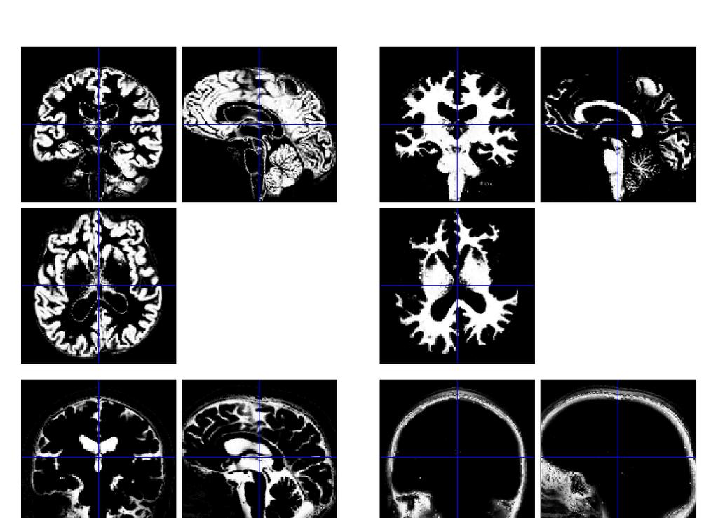
5 Segmentation * Segmentation in SPM8 also estimates a spatial transformation that can be used for spatially normalising images. * It uses a generative model, which involves: * Mixture of Gaussians (MOG) * Bias Correction Component * Warping (Non-linear Registration) Component
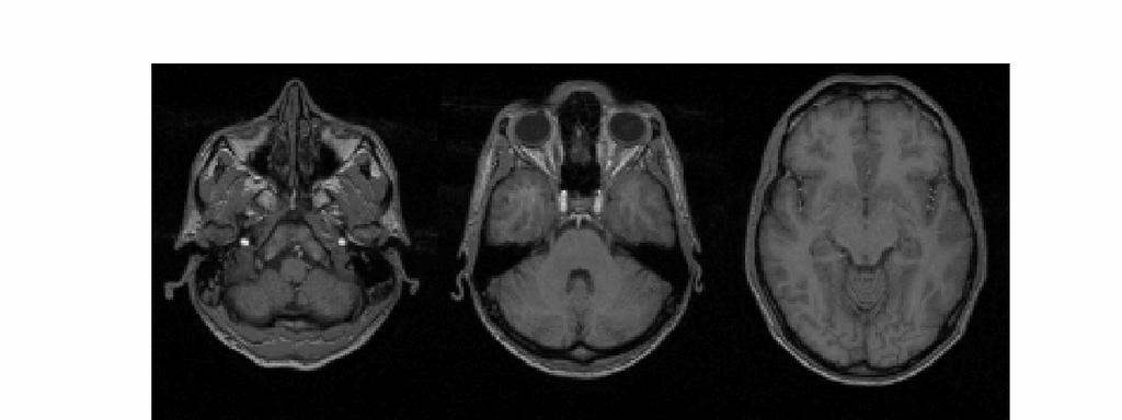
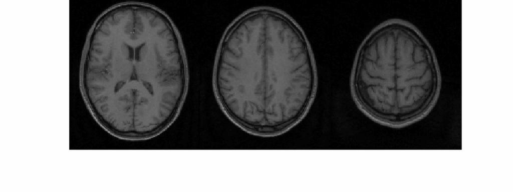
6 Image Intensity Distributions (T1-weighted MRI)
7 Mixture of Gaussians (MOG) * Classification is based on a Mixture of Gaussians model (MOG), which represents the intensity probability density by a number of Gaussian distributions. Frequency Image Intensity
8 Belonging Probabilities Belonging probabilities are assigned by normalising to one.
9 Non-Gaussian Intensity Distributions * Multiple Gaussians per tissue class allow non-gaussian intensity distributions to be modelled. * E.g. accounting for partial volume effects

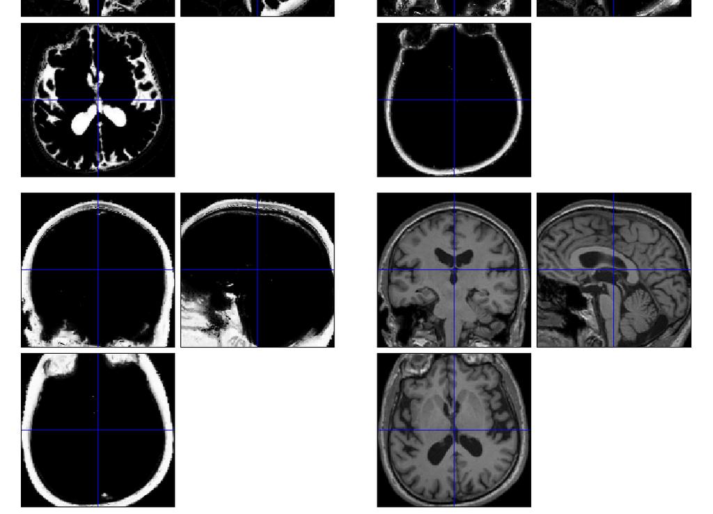
10 Modelling a Bias Field * A bias field is modelled as a linear combination of basis functions. Corrupted image Bias Field Corrected image
11 Tissue Probability Maps * Tissue probability maps (TPMs) are used instead of the proportion of voxels in each Gaussian as the prior. ICBM Tissue Probabilistic Atlases. These tissue probability maps are kindly provided by the International Consortium for Brain Mapping, John C. Mazziotta and Arthur W. Toga.

12 Tissue Probability Maps for New Segment Includes additional non-brain tissue classes (bone, and soft tissue)
13


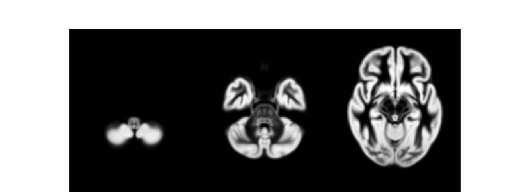
14 Deforming the Tissue Probability Maps * Tissue probability images are deformed so that they can be overlaid on top of the image to segment.
15 Optimisation * The best parameters are those that minimise this objective function. * Optimisation involves finding them. * Begin with starting estimates, and repeatedly change them so that the objective function decreases each time. E = I log ρ i 1 ( β) K i = k = 1 γ k K j= 1 b γ ik j ( α) b ( α) ij 1 2πσ 2 k exp ( ρ ( β) y µ ) i i 2σ 2 k k 2
16 Steepest Descent Start Optimum Alternate between optimising different groups of parameters

17 Spatially normalised BrainWeb phantoms (T1, T2 and PD) Tissue probability maps of GM and WM Cocosco, Kollokian, Kwan & Evans. BrainWeb: Online Interface to a 3D MRI Simulated Brain Database. NeuroImage 5(4):S425 (1997)
18 Contents * Segment * DARTEL * Flow field parameterisation * Objective function * Template creation * Examples * Smooth
.")
19 DARTEL Image Registration * Uses fast approximations * Deformation integrated using scaling and squaring * Uses Levenberg-Marquardt optimiser * Multi-grid matrix solver * Matches GM with GM, WM with WM etc * Diffeomorphic registration takes about 30 mins per image pair ( images). Grey matter template warped to individual Individual scan

20 Evaluations of nonlinear registration algorithms
21 DARTEL * Parameterising the deformation * φ (0) = Identity * φ (1) = u(φ (t) )dt 1 t=0 * u is a flow field to be estimated * Scaling and squaring is used to generate deformations.
22
23 Forward and backward transforms
24 Registration objective function * Simultaneously minimize the sum of: * Matching Term * Drives the matching of the images. * Multinomial assumption * Regularisation term * A measure of deformation roughness * Regularises the registration. * A balance between the two terms.

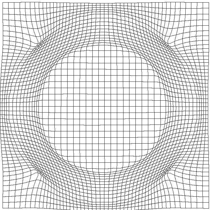
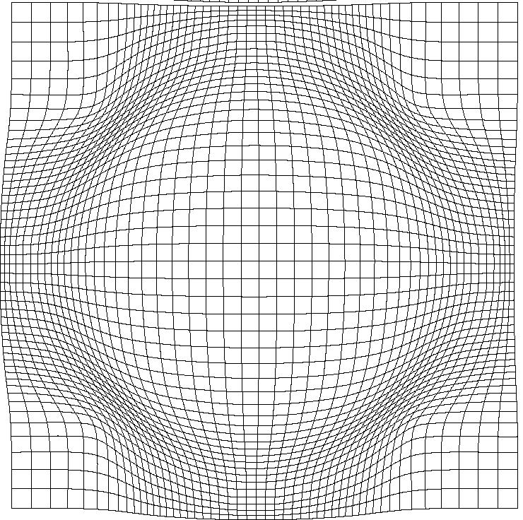
25 Effect of Different Regularisation Terms
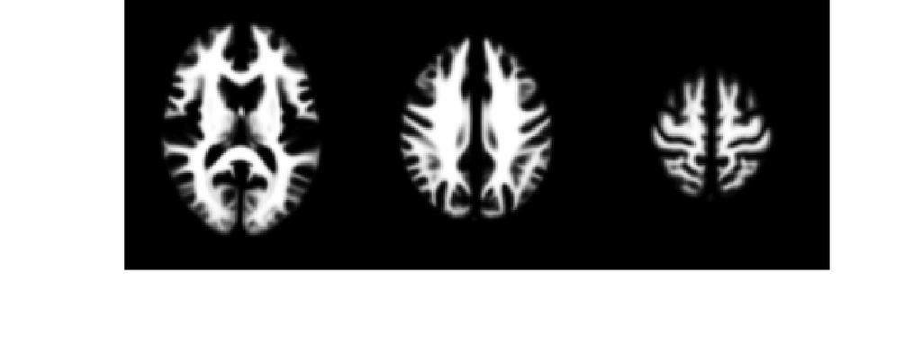
26 Simultaneous registration of GM to GM and WM to WM Subject 1 Grey matter White matter Grey matter White matter Subject 3 Grey matter White matter Subject 2 Grey matter White matter Template Grey matter White matter Subject 4


27 Template Initial Average Iteratively generated from 471 subjects Began with rigidly aligned tissue probability maps Used an inverse consistent formulation After a few iterations Final template

28 Grey matter average of 452 subjects affine

29 Grey matter average of 471 subjects
30 Initial GM images

31 Warped GM images
32
33
34
35
36
37
38
39
40
41 Contents * Segment * Dartel * Smoothing * Compensating for inaccuracies in inter-subject alignment


.")
42 Smooth Blurring is done by convolution. Each voxel after smoothing effectively becomes the result of applying a weighted region of interest (ROI). Before convolution Convolved with a circle Convolved with a Gaussian


43 VBM Pre-processing * Use Segment button for characterising intensity distributions of tissue classes. * DARTEL import, to generate rigidly aligned grey and white matter maps. * DARTEL warping to generate modulated warped grey matter. * Smooth. * Statistics.
44 References * Ashburner & Friston. Unified Segmentation. NeuroImage 26: (2005). * Ashburner. A Fast Diffeomorphic Image Registration Algorithm. NeuroImage 38: (2007). * Ashburner & Friston. Computing average shaped tissue probability templates. NeuroImage 45(2): (2009). * Klein et al. Evaluation of 14 nonlinear deformation algorithms applied to human brain MRI registration. NeuroImage 46(3): (2009).
Image Registration + Other Stuff
 Image Registration + Other Stuff John Ashburner Pre-processing Overview fmri time-series Motion Correct Anatomical MRI Coregister m11 m 21 m 31 m12 m13 m14 m 22 m 23 m 24 m 32 m 33 m 34 1 Template Estimate
Methods for data preprocessing
 Methods for data preprocessing John Ashburner Wellcome Trust Centre for Neuroimaging, 12 Queen Square, London, UK. Overview Voxel-Based Morphometry Morphometry in general Volumetrics VBM preprocessing
Neuroimaging and mathematical modelling Lesson 2: Voxel Based Morphometry
 Neuroimaging and mathematical modelling Lesson 2: Voxel Based Morphometry Nivedita Agarwal, MD Nivedita.agarwal@apss.tn.it Nivedita.agarwal@unitn.it Volume and surface morphometry Brain volume White matter
Computational Neuroanatomy
 Computational Neuroanatomy John Ashburner john@fil.ion.ucl.ac.uk Smoothing Motion Correction Between Modality Co-registration Spatial Normalisation Segmentation Morphometry Overview fmri time-series kernel
An Introduction To Automatic Tissue Classification Of Brain MRI. Colm Elliott Mar 2014
 An Introduction To Automatic Tissue Classification Of Brain MRI Colm Elliott Mar 2014 Tissue Classification Tissue classification is part of many processing pipelines. We often want to classify each voxel
Functional MRI data preprocessing. Cyril Pernet, PhD
 Functional MRI data preprocessing Cyril Pernet, PhD Data have been acquired, what s s next? time No matter the design, multiple volumes (made from multiple slices) have been acquired in time. Before getting
Neuroimage Processing
 Neuroimage Processing Instructor: Moo K. Chung mkchung@wisc.edu Lecture 2-3. General Linear Models (GLM) Voxel-based Morphometry (VBM) September 11, 2009 What is GLM The general linear model (GLM) is a
Voxel-Based Morphometry & DARTEL. Ged Ridgway, London With thanks to John Ashburner and the FIL Methods Group
 Zurich SPM Course 2012 Voxel-Based Morphometry & DARTEL Ged Ridgway, London With thanks to John Ashburner and the FIL Methods Group Aims of computational neuroanatomy * Many interesting and clinically
腦部結構影像 標準化 組織分割 體素型態 本週課程內容. Analysis Softwares. A Course of MRI
 本週課程內容 腦部結構影像 A Course of MRI 盧家鋒助理教授國立陽明大學物理治療暨輔助科技學系 alvin4016@ym.edu.tw 腦部結構影像 空間標準化 (Spatial normalization) 均勻度校正 (Bias correction) 組織分割 (Segmentation) 體素形態學分析 (Voxel-based morphometry, VBM) 影像平滑化
Image Processing for fmri John Ashburner. Wellcome Trust Centre for Neuroimaging, 12 Queen Square, London, UK.
 Iage Processing for fmri John Ashburner Wellcoe Trust Centre for Neuroiaging, 12 Queen Square, London, UK. Contents * Preliinaries * Rigid-Body and Affine Transforations * Optiisation and Objective Functions
Zurich SPM Course Voxel-Based Morphometry. Ged Ridgway (Oxford & UCL) With thanks to John Ashburner and the FIL Methods Group
 With thanks to John Ashburner and the FIL Methods Group") Zurich SPM Course 2015 Voxel-Based Morphometry Ged Ridgway (Oxford & UCL) With thanks to John Ashburner and the FIL Methods Group Examples applications of VBM Many scientifically or clinically interesting
Zurich SPM Course 2015 Voxel-Based Morphometry Ged Ridgway (Oxford & UCL) With thanks to John Ashburner and the FIL Methods Group Examples applications of VBM Many scientifically or clinically interesting
fmri pre-processing Juergen Dukart
 fmri pre-processing Juergen Dukart Outline Why do we need pre-processing? fmri pre-processing Slice time correction Realignment Unwarping Coregistration Spatial normalisation Smoothing Overview fmri time-series
Structural Segmentation
 Structural Segmentation FAST tissue-type segmentation FIRST sub-cortical structure segmentation FSL-VBM voxelwise grey-matter density analysis SIENA atrophy analysis FAST FMRIB s Automated Segmentation
Data pre-processing framework in SPM. Bogdan Draganski
 Data pre-processing fraework in SPM Bogdan Draganski Outline Why do we need pre-processing? Overview Structural MRI pre-processing fmri pre-processing Why do we need pre-processing? What do we want? Reason
Supplementary methods
 Supplementary methods This section provides additional technical details on the sample, the applied imaging and analysis steps and methods. Structural imaging Trained radiographers placed all participants
A Model-Independent, Multi-Image Approach to MR Inhomogeneity Correction
 Tina Memo No. 2007-003 Published in Proc. MIUA 2007 A Model-Independent, Multi-Image Approach to MR Inhomogeneity Correction P. A. Bromiley and N.A. Thacker Last updated 13 / 4 / 2007 Imaging Science and
Structural Segmentation
 Structural Segmentation FAST tissue-type segmentation FIRST sub-cortical structure segmentation FSL-VBM voxelwise grey-matter density analysis SIENA atrophy analysis FAST FMRIB s Automated Segmentation
UBC SPM Course Voxel-Based Morphometry & DARTEL. Ged Ridgway
 UBC SPM Course 2010 Voxel-Based Morphometry & DARTEL Ged Ridgway Aims of computational neuroanatomy * Many interesting and clinically important questions might relate to the shape or local size of regions
1 Introduction Motivation and Aims Functional Imaging Computational Neuroanatomy... 12
 Contents 1 Introduction 10 1.1 Motivation and Aims....... 10 1.1.1 Functional Imaging.... 10 1.1.2 Computational Neuroanatomy... 12 1.2 Overview of Chapters... 14 2 Rigid Body Registration 18 2.1 Introduction.....
FIL SPM Course Spatial Preprocessing
 Registration, noralisation, segentation, VBM Matthan Caan, BIC/CSCA suer school 2012 Neuroiage, 2012 slides copied/adjusted fro: FIL SPM Course Spatial Preprocessing 1. smri 2. DTI 3. fmri By John Ashburner
Introduction to fmri. Pre-processing
 Introduction to fmri Pre-processing Tibor Auer Department of Psychology Research Fellow in MRI Data Types Anatomical data: T 1 -weighted, 3D, 1/subject or session - (ME)MPRAGE/FLASH sequence, undistorted
Spatial Preprocessing
 Spatial Preprocessing Overview of SPM Analysis fmri time-series Design matrix Statistical Parametric Map John Ashburner john@fil.ion.ucl.ac.uk Motion Correction Smoothing General Linear Model Smoothing
MR IMAGE SEGMENTATION
 MR IMAGE SEGMENTATION Prepared by : Monil Shah What is Segmentation? Partitioning a region or regions of interest in images such that each region corresponds to one or more anatomic structures Classification
Detecting Changes In Non-Isotropic Images
 Detecting Changes In Non-Isotropic Images K.J. Worsley 1, M. Andermann 1, T. Koulis 1, D. MacDonald, 2 and A.C. Evans 2 August 4, 1999 1 Department of Mathematics and Statistics, 2 Montreal Neurological
Nonrigid Registration using Free-Form Deformations
 Nonrigid Registration using Free-Form Deformations Hongchang Peng April 20th Paper Presented: Rueckert et al., TMI 1999: Nonrigid registration using freeform deformations: Application to breast MR images
GLM for fmri data analysis Lab Exercise 1
 GLM for fmri data analysis Lab Exercise 1 March 15, 2013 Medical Image Processing Lab Medical Image Processing Lab GLM for fmri data analysis Outline 1 Getting Started 2 AUDIO 1 st level Preprocessing
Automatic Registration-Based Segmentation for Neonatal Brains Using ANTs and Atropos
 Automatic Registration-Based Segmentation for Neonatal Brains Using ANTs and Atropos Jue Wu and Brian Avants Penn Image Computing and Science Lab, University of Pennsylvania, Philadelphia, USA Abstract.
Automatic Generation of Training Data for Brain Tissue Classification from MRI
 Automatic Generation of Training Data for Brain Tissue Classification from MRI Chris A. COCOSCO, Alex P. ZIJDENBOS, and Alan C. EVANS http://www.bic.mni.mcgill.ca/users/crisco/ McConnell Brain Imaging
Signal, Noise and Preprocessing*
 Translational Neuroodeling Unit SNR & Preproc Teporal Spatial General Realign Coreg Noralise Sooth Signal, Noise and Preprocessing* Methods and Models for fmri Analysis Septeber 25 th, 2015 Lars Kasper,
Evaluation of multiple voxel-based morphometry approaches and applications in the analysis of white matter changes in temporal lobe epilepsy
 Evaluation of multiple voxel-based morphometry approaches and applications in the analysis of white matter changes in temporal lobe epilepsy Wenjing Li a, Huiguang He a, Jingjing Lu b, Bin Lv a, Meng Li
CHAPTER 2. Morphometry on rodent brains. A.E.H. Scheenstra J. Dijkstra L. van der Weerd
 CHAPTER 2 Morphometry on rodent brains A.E.H. Scheenstra J. Dijkstra L. van der Weerd This chapter was adapted from: Volumetry and other quantitative measurements to assess the rodent brain, In vivo NMR
Histograms. h(r k ) = n k. p(r k )= n k /NM. Histogram: number of times intensity level rk appears in the image
 = n k. p(r k )= n k /NM. Histogram: number of times intensity level rk appears in the image") Histograms h(r k ) = n k Histogram: number of times intensity level rk appears in the image p(r k )= n k /NM normalized histogram also a probability of occurence 1 Histogram of Image Intensities Create
Histograms h(r k ) = n k Histogram: number of times intensity level rk appears in the image p(r k )= n k /NM normalized histogram also a probability of occurence 1 Histogram of Image Intensities Create
Segmentation of Cerebral MRI Scans Using a Partial Volume Model, Shading Correction, and an Anatomical Prior
 Segmentation of Cerebral MRI Scans Using a Partial Volume Model, Shading Correction, and an Anatomical Prior Aljaž Noe a, Stanislav Kovačič a, James C. Gee b a Faculty of Electrical Engineering, University
SPM Introduction. SPM : Overview. SPM: Preprocessing SPM! SPM: Preprocessing. Scott Peltier. FMRI Laboratory University of Michigan
 SPM Introduction Scott Peltier FMRI Laboratory University of Michigan! Slides adapted from T. Nichols SPM! SPM : Overview Library of MATLAB and C functions Graphical user interface Four main components:
Structural MRI analysis
 Structural MRI analysis volumetry and voxel-based morphometry cortical thickness measurements structural covariance network mapping Boris Bernhardt, PhD Department of Social Neuroscience, MPI-CBS bernhardt@cbs.mpg.de
Where are we now? Structural MRI processing and analysis
 Where are we now? Structural MRI processing and analysis Pierre-Louis Bazin bazin@cbs.mpg.de Leipzig, Germany Structural MRI processing: why bother? Just use the standards? SPM FreeSurfer FSL However:
SPM Introduction SPM! Scott Peltier. FMRI Laboratory University of Michigan. Software to perform computation, manipulation and display of imaging data
 SPM Introduction Scott Peltier FMRI Laboratory University of Michigan Slides adapted from T. Nichols SPM! Software to perform computation, manipulation and display of imaging data 1 1 SPM : Overview Library
for Images A Bayesian Deformation Model
 Statistics in Imaging Workshop July 8, 2004 A Bayesian Deformation Model for Images Sining Chen Postdoctoral Fellow Biostatistics Division, Dept. of Oncology, School of Medicine Outline 1. Introducing
Norbert Schuff VA Medical Center and UCSF
 Norbert Schuff Medical Center and UCSF Norbert.schuff@ucsf.edu Medical Imaging Informatics N.Schuff Course # 170.03 Slide 1/67 Objective Learn the principle segmentation techniques Understand the role
Performance Evaluation of the TINA Medical Image Segmentation Algorithm on Brainweb Simulated Images
 Tina Memo No. 2008-003 Internal Memo Performance Evaluation of the TINA Medical Image Segmentation Algorithm on Brainweb Simulated Images P. A. Bromiley Last updated 20 / 12 / 2007 Imaging Science and
Automatic Generation of Training Data for Brain Tissue Classification from MRI
 MICCAI-2002 1 Automatic Generation of Training Data for Brain Tissue Classification from MRI Chris A. Cocosco, Alex P. Zijdenbos, and Alan C. Evans McConnell Brain Imaging Centre, Montreal Neurological
mritc: A Package for MRI Tissue Classification
 mritc: A Package for MRI Tissue Classification Dai Feng 1 Luke Tierney 2 1 Merck Research Labratories 2 University of Iowa July 2010 Feng & Tierney (Merck & U of Iowa) MRI Tissue Classification July 2010
Norbert Schuff Professor of Radiology VA Medical Center and UCSF
 Norbert Schuff Professor of Radiology Medical Center and UCSF Norbert.schuff@ucsf.edu 2010, N.Schuff Slide 1/67 Overview Definitions Role of Segmentation Segmentation methods Intensity based Shape based
VBM Tutorial. 1 Getting Started. John Ashburner. March 12, 2015
 VBM Tutorial John Ashburner March 12, 2015 1 Getting Started The data provided are a selection of T1-weighted scans from the freely available IXI dataset 1. The overall plan will be to Start up SPM. Check
Normalization for clinical data
 Normalization for clinical data Christopher Rorden, Leonardo Bonilha, Julius Fridriksson, Benjamin Bender, Hans-Otto Karnath (2012) Agespecific CT and MRI templates for spatial normalization. NeuroImage
Fmri Spatial Processing
 Educational Course: Fmri Spatial Processing Ray Razlighi Jun. 8, 2014 Spatial Processing Spatial Re-alignment Geometric distortion correction Spatial Normalization Smoothing Why, When, How, Which Why is
Spatial normalization of injured brains for neuroimaging research: An illustrative introduction of available options
 Spatial normalization of injured brains for neuroimaging research: An illustrative introduction of available options Junghoon Kim, PhD, Brian Avants, PhD, Sunil Patel, MS, and John Whyte, MD, PhD 1 Recent
Basic fmri Design and Analysis. Preprocessing
 Basic fmri Design and Analysis Preprocessing fmri Preprocessing Slice timing correction Geometric distortion correction Head motion correction Temporal filtering Intensity normalization Spatial filtering
Functional MRI in Clinical Research and Practice Preprocessing
 Functional MRI in Clinical Research and Practice Preprocessing fmri Preprocessing Slice timing correction Geometric distortion correction Head motion correction Temporal filtering Intensity normalization
Shape-based Diffeomorphic Registration on Hippocampal Surfaces Using Beltrami Holomorphic Flow
 Shape-based Diffeomorphic Registration on Hippocampal Surfaces Using Beltrami Holomorphic Flow Abstract. Finding meaningful 1-1 correspondences between hippocampal (HP) surfaces is an important but difficult
Spatial Special Preprocessing
 Spatial Special Preprocessing Methods & Models for fmri Data Analysis Septeber 26, 2014 Lars Kasper, PhD TNU & MR-Technology Group Institute for Bioedical Engineering, UZH & ETHZ Generous slide support:
SPM8 for Basic and Clinical Investigators. Preprocessing. fmri Preprocessing
 SPM8 for Basic and Clinical Investigators Preprocessing fmri Preprocessing Slice timing correction Geometric distortion correction Head motion correction Temporal filtering Intensity normalization Spatial
Atlas of Classifiers for Brain MRI Segmentation
 Atlas of Classifiers for Brain MRI Segmentation B. Kodner 1,2, S. H. Gordon 1,2, J. Goldberger 3 and T. Riklin Raviv 1,2 1 Department of Electrical and Computer Engineering, 2 The Zlotowski Center for
Introduction to Neuroimaging Janaina Mourao-Miranda
 Introduction to Neuroimaging Janaina Mourao-Miranda Neuroimaging techniques have changed the way neuroscientists address questions about functional anatomy, especially in relation to behavior and clinical
Voxel-MARS and CMARS: Methods for Early Detection of Alzheimer s Disease by Classification of Structural Brain MRI
 Voxel-MARS and CMARS: Methods for Early Detection of Alzheimer s Disease by Classification of Structural Brain MRI Gerhard-Wilhelm Weber, Poznan University of Technology Alper Çevik, Middle East Technical
Quantitative MRI of the Brain: Investigation of Cerebral Gray and White Matter Diseases
 Quantities Measured by MR - Quantitative MRI of the Brain: Investigation of Cerebral Gray and White Matter Diseases Static parameters (influenced by molecular environment): T, T* (transverse relaxation)
Online Statistical Inference for Quantifying Mandible Growth in CT Images
 Online Statistical Inference for Quantifying Mandible Growth in CT Images Moo K. Chung 1,2, Ying Ji Chuang 2, Houri K. Vorperian 2 1 Department of Biostatistics and Medical Informatics 2 Vocal Tract Development
Computational anatomy with the SPM software
 Available online at www.sciencedirect.com Magnetic Resonance Imaging 27 (2009) 1163 1174 Computational anatomy with the SPM software John Ashburner Wellcome Trust Centre for Neuroimaging, WC1N 3BG London,
Norbert Schuff Professor of Radiology VA Medical Center and UCSF
 Norbert Schuff Professor of Radiology Medical Center and UCSF Norbert.schuff@ucsf.edu Slide 1/67 Overview Definitions Role of Segmentation Segmentation methods Intensity based Shape based Texture based
Ensemble registration: Combining groupwise registration and segmentation
 PURWANI, COOTES, TWINING: ENSEMBLE REGISTRATION 1 Ensemble registration: Combining groupwise registration and segmentation Sri Purwani 1,2 sri.purwani@postgrad.manchester.ac.uk Tim Cootes 1 t.cootes@manchester.ac.uk
A Design Toolbox to Generate Complex Phantoms for the Evaluation of Medical Image Processing Algorithms
 A Design Toolbox to Generate Complex Phantoms for the Evaluation of Medical Image Processing Algorithms Omar Hamo, Georg Nelles, Gudrun Wagenknecht Central Institute for Electronics, Research Center Juelich,
Preprocessing of fmri data
 Preprocessing of fmri data Pierre Bellec CRIUGM, DIRO, UdM Flowchart of the NIAK fmri preprocessing pipeline fmri run 1 fmri run N individual datasets CIVET NUC, segmentation, spatial normalization slice
Brain Extraction, Registration & EPI Distortion Correction
 Brain Extraction, Registration & EPI Distortion Correction What use is Registration? Some common uses of registration: Combining across individuals in group studies: including fmri & diffusion Quantifying
EMSegmenter Tutorial (Advanced Mode)
") EMSegmenter Tutorial (Advanced Mode) Dominique Belhachemi Section of Biomedical Image Analysis Department of Radiology University of Pennsylvania 1/65 Overview The goal of this tutorial is to apply the
EMSegmenter Tutorial (Advanced Mode) Dominique Belhachemi Section of Biomedical Image Analysis Department of Radiology University of Pennsylvania 1/65 Overview The goal of this tutorial is to apply the
Basic principles of MR image analysis. Basic principles of MR image analysis. Basic principles of MR image analysis
 Basic principles of MR image analysis Basic principles of MR image analysis Julien Milles Leiden University Medical Center Terminology of fmri Brain extraction Registration Linear registration Non-linear
Automated MR Image Analysis Pipelines
 Automated MR Image Analysis Pipelines Andy Simmons Centre for Neuroimaging Sciences, Kings College London Institute of Psychiatry. NIHR Biomedical Research Centre for Mental Health at IoP & SLAM. Neuroimaging
Multiple Model Estimation : The EM Algorithm & Applications
 Multiple Model Estimation : The EM Algorithm & Applications Princeton University COS 429 Lecture Dec. 4, 2008 Harpreet S. Sawhney hsawhney@sarnoff.com Plan IBR / Rendering applications of motion / pose
Spatial Normalization using Basis Functions
 Chapter 3 Spatial Normalization using Basis Functions John Ashburner & Karl J. Friston The Wellcome Dept. of Imaging Neuroscience, 12 Queen Square, London WC1N 3BG, UK. Contents 3.1 Introduction.................................
Last Time. This Time. Thru-plane dephasing: worse at long TE. Local susceptibility gradients: thru-plane dephasing
 Motion Correction Last Time Mutual Information Optimiation Decoupling Translation & Rotation Interpolation SPM Example (Least Squares & MI) A Simple Derivation This Time Reslice example SPM Example : Remind
EPI Data Are Acquired Serially. EPI Data Are Acquired Serially 10/23/2011. Functional Connectivity Preprocessing. fmri Preprocessing
 Functional Connectivity Preprocessing Geometric distortion Head motion Geometric distortion Head motion EPI Data Are Acquired Serially EPI Data Are Acquired Serially descending 1 EPI Data Are Acquired
Methodological progress in image registration for ventilation estimation, segmentation propagation and multi-modal fusion
 Methodological progress in image registration for ventilation estimation, segmentation propagation and multi-modal fusion Mattias P. Heinrich Julia A. Schnabel, Mark Jenkinson, Sir Michael Brady 2 Clinical
ADAPTIVE GRAPH CUTS WITH TISSUE PRIORS FOR BRAIN MRI SEGMENTATION
 ADAPTIVE GRAPH CUTS WITH TISSUE PRIORS FOR BRAIN MRI SEGMENTATION Abstract: MIP Project Report Spring 2013 Gaurav Mittal 201232644 This is a detailed report about the course project, which was to implement
SPM8 for Basic and Clinical Investigators. Preprocessing
 SPM8 for Basic and Clinical Investigators Preprocessing fmri Preprocessing Slice timing correction Geometric distortion correction Head motion correction Temporal filtering Intensity normalization Spatial
Introductory Concepts for Voxel-Based Statistical Analysis
 Introductory Concepts for Voxel-Based Statistical Analysis John Kornak University of California, San Francisco Department of Radiology and Biomedical Imaging Department of Epidemiology and Biostatistics
Edge-Preserving MRI Super Resolution Using a High Frequency Regularization Technique
 Edge-Preserving MRI Super Resolution Using a High Frequency Regularization Technique Kaveh Ahmadi Department of EECS University of Toledo, Toledo, Ohio, USA 43606 Email: Kaveh.ahmadi@utoledo.edu Ezzatollah
Measuring longitudinal brain changes in humans and small animal models. Christos Davatzikos
 Measuring longitudinal brain changes in humans and small animal models Christos Davatzikos Section of Biomedical Image Analysis University of Pennsylvania (Radiology) http://www.rad.upenn.edu/sbia Computational
Preprocessing of fmri data (basic)
") Preprocessing of fmri data (basic) Practical session SPM Course 2016, Zurich Andreea Diaconescu, Maya Schneebeli, Jakob Heinzle, Lars Kasper, and Jakob Sieerkus Translational Neuroodeling Unit (TNU) Institute
Preprocessing of fmri data (basic) Practical session SPM Course 2016, Zurich Andreea Diaconescu, Maya Schneebeli, Jakob Heinzle, Lars Kasper, and Jakob Sieerkus Translational Neuroodeling Unit (TNU) Institute
Registration: Rigid vs. Deformable
 Lecture 20 Deformable / Non-Rigid Registration ch. 11 of Insight into Images edited by Terry Yoo, et al. Spring 2017 16-725 (CMU RI) : BioE 2630 (Pitt) Dr. John Galeotti The content of these slides by
Appendix E1. Supplementary Methods. MR Image Acquisition. MR Image Analysis
 RSNA, 2015 10.1148/radiol.2015150532 Appendix E1 Supplementary Methods MR Image Acquisition By using a 1.5-T system (Avanto, Siemens Medical, Erlangen, Germany) under a program of regular maintenance (no
Spline-Based Probabilistic Model for Anatomical Landmark Detection
 Spline-Based Probabilistic Model for Anatomical Landmark Detection Camille Izard 1,2, Bruno Jedynak 1,2, and Craig E.L. Stark 3 1 Laboratoire Paul Painlevé, Université des Sciences et Technologies de Lille,
The Effects of Inter-slice Gap Thickness on Parametric Methods for Diffusion Tensor Imaging Abstract
 The Effects of Inter-slice Gap Thickness on Parametric Methods for Diffusion Tensor Imaging Beth Hutchinson; Neuroscience Training program, Statistics 592 Abstract Although high-resolution, whole-brain
Multi-Atlas Segmentation using Clustering, Local Non-Linear Manifold Embeddings and Target-Specific Templates
 Multi-Atlas Segmentation using Clustering, Local Non-Linear Manifold Embeddings and Target-Specific Templates CHRISTOPH ARTHOFER, BSc., MSc. Thesis submitted to the University of Nottingham for the degree
HHS Public Access Author manuscript Med Image Comput Comput Assist Interv. Author manuscript; available in PMC 2018 March 20.
 Online Statistical Inference for Large-Scale Binary Images Moo K. Chung 1,2, Ying Ji Chuang 2, and Houri K. Vorperian 2 1 Department of Biostatistics and Medical Informatics, University of Wisconsin, Madison,
LOCUS: LOcal Cooperative Unified Segmentation of MRI Brain Scans
 Author manuscript, published in "MICCAI07-10th International Conference on Medical Image Computing and Computer Assisted Intervention, Brisbane : Australia (2007)" DOI : 10.1007/978-3-540-75757-3_27 LOCUS:
Multi-scale Voxel-based Morphometry via Weighted Spherical Harmonic Representation
 Multi-scale Voxel-based Morphometry via Weighted Spherical Harmonic Representation Moo K. Chung 1,2, Li Shen 4, Kim M. Dalton 2, and Richard J. Davidson 2,3 1 Department of Statistics, Biostatistics and
A Nonparametric Bayesian Approach to Detecting Spatial Activation Patterns in fmri Data
 A Nonparametric Bayesian Approach to Detecting Spatial Activation Patterns in fmri Data Seyoung Kim, Padhraic Smyth, and Hal Stern Bren School of Information and Computer Sciences University of California,
Cocozza S., et al. : ALTERATIONS OF FUNCTIONAL CONNECTIVITY OF THE MOTOR CORTEX IN FABRY'S DISEASE: AN RS-FMRI STUDY
 ALTERATIONS OF FUNCTIONAL CONNECTIVITY OF THE MOTOR CORTEX IN FABRY'S DISEASE: AN RS-FMRI STUDY SUPPLEMENTARY MATERIALS Sirio Cocozza, MD 1*, Antonio Pisani, MD, PhD 2, Gaia Olivo, MD 1, Francesco Saccà,
FROM IMAGE RECONSTRUCTION TO CONNECTIVITY ANALYSIS: A JOURNEY THROUGH THE BRAIN'S WIRING. Francesca Pizzorni Ferrarese
 FROM IMAGE RECONSTRUCTION TO CONNECTIVITY ANALYSIS: A JOURNEY THROUGH THE BRAIN'S WIRING Francesca Pizzorni Ferrarese Pipeline overview WM and GM Segmentation Registration Data reconstruction Tractography
IS MRI Based Structure a Mediator for Lead s Effect on Cognitive Function?
 IS MRI Based Structure a Mediator for Lead s Effect on Cognitive Function? Brian Caffo, Sining Chen, Brian Schwartz Department of Biostatistics and Environmental Health Sciences Johns Hopkins University
Subvoxel Segmentation and Representation of Brain Cortex Using Fuzzy Clustering and Gradient Vector Diffusion
 Subvoxel Segmentation and Representation of Brain Cortex Using Fuzzy Clustering and Gradient Vector Diffusion Ming-Ching Chang Xiaodong Tao GE Global Research Center {changm, taox} @ research.ge.com SPIE
Analysis of fmri data within Brainvisa Example with the Saccades database
 Analysis of fmri data within Brainvisa Example with the Saccades database 18/11/2009 Note : All the sentences in italic correspond to informations relative to the specific dataset under study TP participants
Whole Body MRI Intensity Standardization
 Whole Body MRI Intensity Standardization Florian Jäger 1, László Nyúl 1, Bernd Frericks 2, Frank Wacker 2 and Joachim Hornegger 1 1 Institute of Pattern Recognition, University of Erlangen, {jaeger,nyul,hornegger}@informatik.uni-erlangen.de
Surface-based Analysis: Inter-subject Registration and Smoothing
 Surface-based Analysis: Inter-subject Registration and Smoothing Outline Exploratory Spatial Analysis Coordinate Systems 3D (Volumetric) 2D (Surface-based) Inter-subject registration Volume-based Surface-based
Multiple Model Estimation : The EM Algorithm & Applications
 Multiple Model Estimation : The EM Algorithm & Applications Princeton University COS 429 Lecture Nov. 13, 2007 Harpreet S. Sawhney hsawhney@sarnoff.com Recapitulation Problem of motion estimation Parametric
Probabilistic Registration of 3-D Medical Images
 Probabilistic Registration of 3-D Medical Images Mei Chen, Takeo Kanade, Dean Pomerleau, Jeff Schneider CMU-RI-TR-99-16 The Robotics Institute Carnegie Mellon University Pittsburgh, Pennsylvania 1513 July,
Spatial Normalization of Brain Images with Focal Lesions Using Cost Function Masking
 NeuroImage 14, 486 500 (2001) doi:10.1006/nimg.2001.0845, available online at http://www.idealibrary.com on Spatial Normalization of Brain Images with Focal Lesions Using Cost Function Masking Matthew
The organization of the human cerebral cortex estimated by intrinsic functional connectivity
 1 The organization of the human cerebral cortex estimated by intrinsic functional connectivity Journal: Journal of Neurophysiology Author: B. T. Thomas Yeo, et al Link: https://www.ncbi.nlm.nih.gov/pubmed/21653723
Modified Expectation Maximization Method for Automatic Segmentation of MR Brain Images
 Modified Expectation Maximization Method for Automatic Segmentation of MR Brain Images R.Meena Prakash, R.Shantha Selva Kumari 1 P.S.R.Engineering College, Sivakasi, Tamil Nadu, India 2 Mepco Schlenk Engineering
Bayesian Spatiotemporal Modeling with Hierarchical Spatial Priors for fmri
 Bayesian Spatiotemporal Modeling with Hierarchical Spatial Priors for fmri Galin L. Jones 1 School of Statistics University of Minnesota March 2015 1 Joint with Martin Bezener and John Hughes Experiment
FSL Pre-Processing Pipeline
 The Art and Pitfalls of fmri Preprocessing FSL Pre-Processing Pipeline Mark Jenkinson FMRIB Centre, University of Oxford FSL Pre-Processing Pipeline Standard pre-processing: Task fmri Resting-state fmri