A Virtual MR Scanner for Education
|
|
|
- Roxanne Horn
- 5 years ago
- Views:
Transcription
1 A Virtual MR Scanner for Education Hackländer T, Schalla C, Trümper A, Mertens H, Hiltner J, Cramer BM Hospitals of the University Witten/Herdecke, Department of Radiology Wuppertal, Germany
2 Purpose A realistic simulation of a MR scanner is to be developed For the user it should be possible to change all relevant settings of the virtual scanner and to adapt them to the expected pathology Students in education and doctors in training are the target group.
3 Purpose: Details Examination Selection of an adequate pulse sequence Optimization of tissue contrast by adjusting the parameters of the pulse sequence Optimization of the signal to noise ratio (SNR) and the examination time Identification of image artifacts Post processing Windowing Differentiation between image space and k-space
4 Structure of the Presentation 1. A quick tour across the simulation 2. The technical structure of the software 3. A detailed look at the simulation 4. Parameter images 5. Conclusion
5 A Quick Tour: Main Program Window Select the reference phantom for examination

6 A Quick Tour: Reference Phantom Content PD % T1 ms T2 ms 1 Fat Methemoglobin White matter Gray matter Edema Cystic Fluid 89 1, CSF 89 2, Water 100 4, Relaxation times dependent on magnetic field strength. Given values for 1.5T.

7 A Quick Tour: Reference Phantom Close the info window
8 A Quick Tour: Select Inversion-Recovery Select pulse sequence: Inversion-Recovery

9 A Quick Tour: Suppress Cystic Fluid 1. Adjust exam parameter 2. Change TI to 749 to suppress cystic fluid 3. Start examination

10 A Quick Tour: Suppress Fat 1. Select: Four image view Cystic fluid is suppressed 2. Suppress fat: Change TI to Start examination

11 A Quick Tour: Comparison of Results Cystic fluid is suppressed Fat is suppressed
12 Structure of the Presentation 1. A quick tour across the simulation 2. The technical structure of the software 3. A detailed look at the simulation 4. Parameter images 5. Conclusion
13 Objectives User interface is to correspond to a real world scanner Simulation is to be independent of a particular hardware and software platform Pulse sequences are to be easily extensible with a plug-in mechanism
14 Method Programming language: Pure Java 1.2 Internationalized versions: English, German, Estonian, Hardware requirements: Equivalent to a Pentium II 400MHz, 128 MB, 618k (!!) free hard disk space Software requirements: Operating system with a Java JRE 1.2 or higher License: GNU General Public License
15 Real MR Scanner User input: TR, TE,. Real K-space Imag Inverse FFT (r 2 +i 2 ) 1/2 Magnitude image Convert to 12 Bit DICOM Export User input: Window/Center Windowing Image visualization
16 Basics of the Virtual MR Scanner 1. Base of the simulation are parameter images of T1, T2 and PD derived once from a real-world examination 2. Calculation of an intensity image in exchange for the real-world object 3. Superposition of artifacts (e.g. noise) in the image space 4. Transform of the intensity image into the k-space 5. Processing of k-space analog to a real MR scanner
17 Virtual MR: Part 1 Parameter image PD Parameter image T1 Parameter image T2 Parameter image SZ Parameter image Flow DICOM import User input: TR, TE,. Pulse sequence Image Real Imag Image space manipulations: e.g. Motion artifacts, no k-space Manipulation? yes FFT 1 2
18 Virtual MR: Part K-space Real Imag Inverse FFT k-space manipulations: e.g. oversampling, (r 2 +i 2 ) 1/2 Magnitude image Convert to 12 Bit DICOM export User input: Window/Center Windowing Image visualization
19 Structure of the Presentation 1. A quick tour across the simulation 2. The technical structure of the software 3. A detailed look at the simulation 4. Parameter images 5. Conclusion
20 User Interface Image area Control area

21 Histogram View


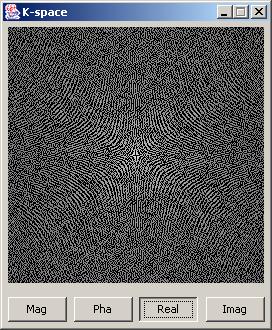
22 K-Space View
23 K-Space Manipulator
24 Available K-Space Manipulations 1 Original image 2 Every 2nd column cleared pixels wide margin cleared 4 16 pixels wide center cleared
25 Windowing 1 Optimum C= 735 W= Maximum C= 2048 W= Brightness C= 0 W= Contrast C= 581 W= 303

26 Adjusting Parameters of a Pulse Sequence
27 General Parameters Parameter Comment Coil Organ coil or body coil Matrix size Fixed value of 256*256 pixels Field of View (FoV) [mm] Rectangle FoV Vertical to horizontal ratio of FoV [1/8] Slice thickness [mm] Number of acquisitions Number of acquisitions/excitations (NEX) Phase oversampling Off / On Frequency oversampling Off / On
28 Pulse Sequence dependent Parameters Pulse-Sequence TR TE TI Flip. TEeff ETL Spin Echo + + Turbo Spin Echo Gradient Echo Spoiled Gradient Echo Refocussed Gradient Echo Saturation Recovery + Inversion Recovery + + +
29 Weighting (Spin Echo) 1 T1 Weighting TR=400, TE=10 2 PD Weighting TR=3000, TE=10 3 T2 Weighting TR=3000, TE=120

30 Signal to Noise Ratio (SNR) 1 SNR = 1,000 Organ coil, 6mm, 1 NEX 2 SNR = Body coil, 6mm, 1 NEX 3 SNR = Body coil, 2mm, 1 NEX 4 SNR = 0,943 Body coil, 2mm, 8 NEX
31 Oversampling PO=Phase Oversampling FO=Frequency Oversampling 1 FoV = 256 FO = no, PO = no SNR = Exam time 4:16 2 FoV = 192 FO = yes, PO = yes SNR = Exam time 8:32 3 FoV = 192 FO = yes, PO = no SNR = Exam time 4:16 4 FoV = 192 FO = no, PO = yes SNR = Exam time 8:32
32 Rectangle FoV 1 FoV = 256 Rec = 8 / 8 PO = no SNR = Exam time 12:48 2 FoV = 256 Rec = 6 / 8 PO = no SNR = Exam time 9:36 3 FoV = 256 Rec = 6 / 8 PO = yes SNR = Exam time 19:12
33 Motion Artifacts 1 No motion 2 Translational motion horizontal = 2 Pix/min 3 Translational motion vertical = 2 Pix/min 4 Periodic motion horizontal = 2 Pix/min frequency = 5 /min
34 Structure of the Presentation 1. A quick tour across the simulation 2. The technical structure of the software 3. A detailed look at the simulation 4. Parameter images 5. Conclusion
35 Calculation of the Parameter Images Patient examination under clinical conditions Spin echo pulse sequences T1: TR= step 30, TE=5 (9 measurements) T2, PD: TR=2440, TE= step 50 (16 measurements) Calculation of T1,T2 and PD using a weighted least square fit Error of the calculated T2 values: ± 5% T1 is uncertain! Possible solution: non-linear curve fit (Marquardt-Levenberg algorithm) or simplex method

36 Editor for Parameter Images Alpha version
37 Structure of the Presentation 1. A quick tour across the simulation 2. The technical structure of the software 3. A detailed look at the simulation 4. Parameter images 5. Conclusion
38 Results On a 500 MHz PC the software calculates an image within 5 to 20 seconds Calculation time depends on Pulse sequence Superposition of artifacts
39 Conclusion An interactive simulation of a real world examination is possible on a standard PC The users can study the operation of a costly and not everywhere available equipment on their desktop Contact: Web: hacklaender@iftm.de
MRI. When to use What sequences. Outline 2012/09/19. Sequence: Definition. Basic Principles: Step 2. Basic Principles: Step 1. Govind Chavhan, MD
 MRI When to use What sequences Govind Chavhan, MD Assistant Professor and Staff Radiologist The Hospital For Sick Children, Toronto Planning Acquisition Post processing Interpretation Patient history and
Breast MRI Accreditation Program Clinical Image Quality Guide
 Breast MRI Accreditation Program Clinical Image Quality Guide Introduction This document provides guidance on breast MRI clinical image quality and describes the criteria used by the ACR Breast MRI Accreditation
New Technology Allows Multiple Image Contrasts in a Single Scan
 These images were acquired with an investigational device. PD T2 T2 FLAIR T1 MAP T1 FLAIR PSIR T1 New Technology Allows Multiple Image Contrasts in a Single Scan MR exams can be time consuming. A typical
Lab Location: MRI, B2, Cardinal Carter Wing, St. Michael s Hospital, 30 Bond Street
 Lab Location: MRI, B2, Cardinal Carter Wing, St. Michael s Hospital, 30 Bond Street MRI is located in the sub basement of CC wing. From Queen or Victoria, follow the baby blue arrows and ride the CC south
HST.583 Functional Magnetic Resonance Imaging: Data Acquisition and Analysis Fall 2008
 MIT OpenCourseWare http://ocw.mit.edu HST.583 Functional Magnetic Resonance Imaging: Data Acquisition and Analysis Fall 2008 For information about citing these materials or our Terms of Use, visit: http://ocw.mit.edu/terms.
HST.583 Functional Magnetic Resonance Imaging: Data Acquisition and Analysis Fall 2006
 MIT OpenCourseWare http://ocw.mit.edu HST.583 Functional Magnetic Resonance Imaging: Data Acquisition and Analysis Fall 2006 For information about citing these materials or our Terms of Use, visit: http://ocw.mit.edu/terms.
MRI image formation 8/3/2016. Disclosure. Outlines. Chen Lin, PhD DABR 3. Indiana University School of Medicine and Indiana University Health
 MRI image formation Indiana University School of Medicine and Indiana University Health Disclosure No conflict of interest for this presentation 2 Outlines Data acquisition Spatial (Slice/Slab) selection
HST.583 Functional Magnetic Resonance Imaging: Data Acquisition and Analysis Fall 2008
 MIT OpenCourseWare http://ocw.mit.edu HST.583 Functional Magnetic Resonance Imaging: Data Acquisition and Analysis Fall 2008 For information about citing these materials or our Terms of Use, visit: http://ocw.mit.edu/terms.
surface Image reconstruction: 2D Fourier Transform
 2/1/217 Chapter 2-3 K-space Intro to k-space sampling (chap 3) Frequenc encoding and Discrete sampling (chap 2) Point Spread Function K-space properties K-space sampling principles (chap 3) Basic Contrast
COBRE Scan Information
 COBRE Scan Information Below is more information on the directory structure for the COBRE imaging data. Also below are the imaging parameters for each series. Directory structure: var/www/html/dropbox/1139_anonymized/human:
Orthopedic MRI Protocols. Philips Panorama HFO
 Orthopedic MRI Protocols Philips Panorama HFO 1 2 Prepared in collaboration with Dr. John F. Feller, Medical Director of Desert Medical Imaging, Palm Springs, CA. Desert Medical Imaging will provide the
SIEMENS MAGNETOM Avanto syngo MR B15
 \\USER\INVESTIGATORS\Ravi\ADNI-phantom\QC Phantom-Localizer TA: 0:10 PAT: Voxel size: 1.9 1.5 8.0 mm Rel. SNR: 1.00 SIEMENS: gre Properties Prio Recon Before measurement After measurement Load to viewer
SIEMENS MAGNETOM Avanto syngo MR B15
 \\USER\INVESTIGATORS\Ravi\ADNI-Subject\Localizer TA: 0:10 PAT: Voxel size: 1.9 1.5 8.0 mm Rel. SNR: 1.00 SIEMENS: gre Properties Prio Recon Before measurement After measurement Load to viewer Inline movie
UNIVERSITY OF SOUTHAMPTON
 UNIVERSITY OF SOUTHAMPTON PHYS2007W1 SEMESTER 2 EXAMINATION 2014-2015 MEDICAL PHYSICS Duration: 120 MINS (2 hours) This paper contains 10 questions. Answer all questions in Section A and only two questions
Module 4. K-Space Symmetry. Review. K-Space Review. K-Space Symmetry. Partial or Fractional Echo. Half or Partial Fourier HASTE
 MRES 7005 - Fast Imaging Techniques Module 4 K-Space Symmetry Review K-Space Review K-Space Symmetry Partial or Fractional Echo Half or Partial Fourier HASTE Conditions for successful reconstruction Interpolation
ADNI, ADNI_QH, SURVEY. Geometry. connection
 ADNI, ADNI_QH, SURVEY Geometry Coil selection = Head connection = d Multi coil Homogeneity correction ne FOV (mm) = 250.00 RFOV (%) = 100.00 Foldover suppression Matrix scan = 256 reconstruction = 256
Slide 1. Technical Aspects of Quality Control in Magnetic Resonance Imaging. Slide 2. Annual Compliance Testing. of MRI Systems.
 Slide 1 Technical Aspects of Quality Control in Magnetic Resonance Imaging Slide 2 Compliance Testing of MRI Systems, Ph.D. Department of Radiology Henry Ford Hospital, Detroit, MI Slide 3 Compliance Testing
Scan Acceleration with Rapid Gradient-Echo
 Scan Acceleration with Rapid Gradient-Echo Hsiao-Wen Chung ( 鍾孝文 ), Ph.D., Professor Dept. Electrical Engineering, National Taiwan Univ. Dept. Radiology, Tri-Service General Hospital 1 of 214 The Need
Exam 8N080 - Introduction MRI
 Exam 8N080 - Introduction MRI Friday January 23 rd 2015, 13.30-16.30h For this exam you may use an ordinary calculator (not a graphical one). In total there are 6 assignments and a total of 65 points can
Gradient-Echo. Spin-Echo. Echo planar. Assessment of Regional Function Assessment of Global. Parallel Imaging. Function. Steady State Imaging
 Gradient-Echo Spin-Echo James W. Goldfarb Ph.D. Department of Research and Education St. Francis Hospital Program in Biomedical Engineering SUNY Stony Brook Echo planar Assessment of Regional Function
Lucy Phantom MR Grid Evaluation
 Lucy Phantom MR Grid Evaluation Anil Sethi, PhD Loyola University Medical Center, Maywood, IL 60153 November 2015 I. Introduction: The MR distortion grid, used as an insert with Lucy 3D QA phantom, is
Philips MRI Protocol Dump Created on Comment Software Stream
 Page 1 of 5 Philips MRI Protocol Dump Created on 2/17/2011 4:11:01 PM Comment Created by ExamCard_to_XML with inputs: "J:\ADNI GO - ADNI 2 Phantom5.ExamCard" on system (BU SCHOOL OF MEDICINE :: 192.168.71.10)
A Novel Iterative Thresholding Algorithm for Compressed Sensing Reconstruction of Quantitative MRI Parameters from Insufficient Data
 A Novel Iterative Thresholding Algorithm for Compressed Sensing Reconstruction of Quantitative MRI Parameters from Insufficient Data Alexey Samsonov, Julia Velikina Departments of Radiology and Medical
T 1 MAPPING FOR DCE-MRI
 T 1 MAPPING FOR DCE-MRI A dissertation submitted to the Faculty of Medicine, University of Malaya in partial fulfillment of the requirements for the degree of Master of Medical Physics By NURUN NAJWA BINTI
MR Advance Techniques. Vascular Imaging. Class III
 MR Advance Techniques Vascular Imaging Class III 1 Vascular Imaging There are several methods that can be used to evaluate the cardiovascular systems with the use of MRI. MRI will aloud to evaluate morphology
MB-EPI PCASL. Release Notes for Version February 2015
 MB-EPI PCASL Release Notes for Version 1.0 20 February 2015 1 Background High-resolution arterial spin labeling (ASL) imaging is highly desirable in both neuroscience research and clinical applications
SIEMENS MAGNETOM Verio syngo MR B17
 \\USER\Dr. Behrmann\routine\Ilan\ep2d_bold_PMU_resting TA: 8:06 PAT: Voxel size: 3.03.03.0 mm Rel. SNR: 1.00 USER: ep2d_bold_pmu Properties Special sat. Prio Recon System Before measurement Body After
Siemens AG, Healthcare Sector. syngo MR D13 0. Supplement - Parameters and image text 0.
 Siemens AG, Healthcare Sector 0 0 n.a. English Cs2 syngo Neuro Operator MR-05014 630 05/2010 01 02 Informatik, Manual D11 Cape syngo MR D13 0.0 Supplement - Parameters and image text 0. syngo MR D13 0.
Fast Imaging UCLA. Class Business. Class Business. Daniel B. Ennis, Ph.D. Magnetic Resonance Research Labs. Tuesday (3/7) from 6-9pm HW #1 HW #2
 from 6-9pm HW #1 HW #2") Fast Imaging Daniel B. Ennis, Ph.D. Magnetic Resonance Research Labs Class Business Tuesday (3/7) from 6-9pm 6:00-7:30pm Groups Avanto Sara Said, Yara Azar, April Pan Skyra Timothy Marcum, Diana Lopez,
Fast Imaging Daniel B. Ennis, Ph.D. Magnetic Resonance Research Labs Class Business Tuesday (3/7) from 6-9pm 6:00-7:30pm Groups Avanto Sara Said, Yara Azar, April Pan Skyra Timothy Marcum, Diana Lopez,
Qualitative Comparison of Conventional and Oblique MRI for Detection of Herniated Spinal Discs
 Qualitative Comparison of Conventional and Oblique MRI for Detection of Herniated Spinal Discs Doug Dean Final Project Presentation ENGN 2500: Medical Image Analysis May 16, 2011 Outline Review of the
Supplementary Information
 Supplementary Information Magnetic resonance imaging reveals functional anatomy and biomechanics of a living dragon tree Linnea Hesse 1,2,*, Tom Masselter 1,2,3, Jochen Leupold 4, Nils Spengler 5, Thomas
MEDICAL IMAGE COMPUTING (CAP 5937) LECTURE 4: Pre-Processing Medical Images (II)
 LECTURE 4: Pre-Processing Medical Images (II)") SPRING 2016 1 MEDICAL IMAGE COMPUTING (CAP 5937) LECTURE 4: Pre-Processing Medical Images (II) Dr. Ulas Bagci HEC 221, Center for Research in Computer Vision (CRCV), University of Central Florida (UCF),
SPRING 2016 1 MEDICAL IMAGE COMPUTING (CAP 5937) LECTURE 4: Pre-Processing Medical Images (II) Dr. Ulas Bagci HEC 221, Center for Research in Computer Vision (CRCV), University of Central Florida (UCF),
Measuring baseline whole-brain perfusion on GE 3.0T using arterial spin labeling (ASL) MRI
 MRI") Measuring baseline whole-brain perfusion on GE 3.0T using arterial spin labeling (ASL) MRI Revision date: 09/15/2008 Overview This document describes the procedure for measuring baseline whole-brain perfusion
Measuring baseline whole-brain perfusion on GE 3.0T using arterial spin labeling (ASL) MRI Revision date: 09/15/2008 Overview This document describes the procedure for measuring baseline whole-brain perfusion
A Transmission Line Matrix Model for Shielding Effects in Stents
 A Transmission Line Matrix Model for hielding Effects in tents Razvan Ciocan (1), Nathan Ida (2) (1) Clemson University Physics and Astronomy Department Clemson University, C 29634-0978 ciocan@clemon.edu
GE Healthcare CLINICAL GALLERY. Discovery * MR750w 3.0T. This brochure is intended for European healthcare professionals.
 GE Healthcare CLINICAL GALLERY Discovery * MR750w 3.0T This brochure is intended for European healthcare professionals. NEURO PROPELLER delivers high resolution, motion insensitive imaging in all planes.
SIEMENS MAGNETOM Skyra syngo MR D13
 Page 1 of 8 SIEMENS MAGNETOM Skyra syngo MR D13 \\USER\CIND\StudyProtocols\PTSA\*dm_ep2d_mono70_b0_p2_iso2.0 TA:1:05 PAT:2 Voxel size:2.0 2.0 2.0 mm Rel. SNR:1.00 :epse Properties Routine Prio Recon Load
Magnetic Resonance Elastography (MRE) of Liver Disease
 of Liver Disease") Magnetic Resonance Elastography (MRE) of Liver Disease Authored by: Jennifer Dolan Fox, PhD VirtualScopics Inc. jennifer_fox@virtualscopics.com 1-585-249-6231 1. Overview of MRE Imaging MRE is a magnetic
Magnetic Resonance Elastography (MRE) of Liver Disease Authored by: Jennifer Dolan Fox, PhD VirtualScopics Inc. jennifer_fox@virtualscopics.com 1-585-249-6231 1. Overview of MRE Imaging MRE is a magnetic
XI Signal-to-Noise (SNR)
") XI Signal-to-Noise (SNR) Lecture notes by Assaf Tal n(t) t. Noise. Characterizing Noise Noise is a random signal that gets added to all of our measurements. In D it looks like this: while in D
XI Signal-to-Noise (SNR) Lecture notes by Assaf Tal n(t) t. Noise. Characterizing Noise Noise is a random signal that gets added to all of our measurements. In D it looks like this: while in D
CHAPTER 9: Magnetic Susceptibility Effects in High Field MRI
 Figure 1. In the brain, the gray matter has substantially more blood vessels and capillaries than white matter. The magnified image on the right displays the rich vasculature in gray matter forming porous,
MRI Physics II: Gradients, Imaging
 MRI Physics II: Gradients, Imaging Douglas C., Ph.D. Dept. of Biomedical Engineering University of Michigan, Ann Arbor Magnetic Fields in MRI B 0 The main magnetic field. Always on (0.5-7 T) Magnetizes
Page 1 of 9. Protocol: adult_other_adni3basichumanprotocol25x_ _ _1. 3 Plane Localizer. 3 Plane Localizer PATIENT POSITION
 3 Localizer FOV 26.0 Slice Thickness 5.0 Slice Spacing 0.0 Freq 256 Phase 128 3-PLANE 3 Localizer Unswap Phase Correction Gradient Echo Imaging Options Seq, Fast Recon All Images 3 Localizer Pause / SCIC
M R I Physics Course
 M R I Physics Course Multichannel Technology & Parallel Imaging Nathan Yanasak, Ph.D. Jerry Allison Ph.D. Tom Lavin, B.S. Department of Radiology Medical College of Georgia References: 1) The Physics of
syngo MR E11 Operator Manual Ortho Answers for life.
 www.siemens.com/healthcare syngo MR E11 Operator Manual Ortho Answers for life. syngo MR E11 Operator Manual Ortho Legend Indicates a hint Is used to provide information on how to avoid operating errors
Midterm Review
 Midterm Review - 2017 EE369B Concepts Noise Simulations with Bloch Matrices, EPG Gradient Echo Imaging 1 About the Midterm Monday Oct 30, 2017. CCSR 4107 Up to end of C2 1. Write your name legibly on this
White Pixel Artifact. Caused by a noise spike during acquisition Spike in K-space <--> sinusoid in image space
 White Pixel Artifact Caused by a noise spike during acquisition Spike in K-space sinusoid in image space Susceptibility Artifacts Off-resonance artifacts caused by adjacent regions with different
SIEMENS MAGNETOM TrioTim syngo MR B17
 \\USER\KNARRGROUP\MultiBand\LavretskyMultiBand\trufi localizer 3-plane TA: 5.1 s PAT: Voxel size: 1.2 1.2 5. Rel. SNR: 1.00 SIEMENS: trufi Load to stamp Slice group 1 Slices 1 Dist. factor 20 % Phase enc.
SIEMENS MAGNETOM Verio syngo MR B15V
 \\USER\ZAHID_RESEARCH\MS\No Name\3D SWI TA: 6:39 PAT: 2 Voxel size: 1.0 0.5 2.0 mm Rel. SNR: 1.00 SIEMENS: gre Properties Prio Recon Before measurement After measurement Load to viewer Inline movie Auto
Role of Parallel Imaging in High Field Functional MRI
 Role of Parallel Imaging in High Field Functional MRI Douglas C. Noll & Bradley P. Sutton Department of Biomedical Engineering, University of Michigan Supported by NIH Grant DA15410 & The Whitaker Foundation
2.1 Signal Production. RF_Coil. Scanner. Phantom. Image. Image Production
 An Extensible MRI Simulator for Post-Processing Evaluation Remi K.-S. Kwan?, Alan C. Evans, and G. Bruce Pike McConnell Brain Imaging Centre, Montreal Neurological Institute, McGill University, Montreal,
Magnetic Resonance Angiography
 Magnetic Resonance Angiography Course: Advance MRI (BIOE 594) Instructors: Dr Xiaohong Joe Zhou Dr. Shadi Othman By, Nayan Pasad Phase Contrast Angiography By Moran 1982, Bryan et. Al. 1984 and Moran et.
MRI Imaging Options. Frank R. Korosec, Ph.D. Departments of Radiology and Medical Physics University of Wisconsin Madison
 MRI Imaging Options Frank R. Korosec, Ph.D. Departments of Radiolog and Medical Phsics Universit of Wisconsin Madison f.korosec@hosp.wisc.edu As MR imaging becomes more developed, more imaging options
Field Maps. 1 Field Map Acquisition. John Pauly. October 5, 2005
 Field Maps John Pauly October 5, 25 The acquisition and reconstruction of frequency, or field, maps is important for both the acquisition of MRI data, and for its reconstruction. Many of the imaging methods
Parallel Magnetic Resonance Imaging (pmri): How Does it Work, and What is it Good For?
: How Does it Work, and What is it Good For?") Parallel Magnetic Resonance Imaging (pmri): How Does it Work, and What is it Good For? Nathan Yanasak, Ph.D. Chair, AAPM TG118 Department of Radiology Georgia Regents University Overview Phased-array coils
Parallel Magnetic Resonance Imaging (pmri): How Does it Work, and What is it Good For? Nathan Yanasak, Ph.D. Chair, AAPM TG118 Department of Radiology Georgia Regents University Overview Phased-array coils
Measuring baseline whole-brain perfusion on GE 3.0T using arterial spin labeling (ASL) MRI
 MRI") Measuring baseline whole-brain perfusion on GE 3.0T using arterial spin labeling (ASL) MRI Revision date: 11/20/2006 Overview This document describes the procedure for measuring baseline whole-brain perfusion
Measuring baseline whole-brain perfusion on GE 3.0T using arterial spin labeling (ASL) MRI Revision date: 11/20/2006 Overview This document describes the procedure for measuring baseline whole-brain perfusion
MSK EXTREME. V-SPEC Technology HIGH FIELD 1.0 TESLA TRULY OPEN, DEDICATED MRI. The Power of High Field. Rapid ROI. Optimized Performance
 MSK EXTREME HIGH FIELD 1.0 TESLA TRULY OPEN, DEDICATED MRI The Power of High Field Rapid ROI Optimized Performance High Throughput Ease of Use Increased Patient Comfort V-SPEC Technology THE POWER OF HIGH
Institutionen för medicin och hälsa
 Institutionen för medicin och hälsa Department of Medical and Health Sciences Master Thesis Synthetic MRI for visualization of quantitative MRI Examensarbete utfört i medicinsk teknik vid Tekniska högskolan
Supplementary methods
 Supplementary methods This section provides additional technical details on the sample, the applied imaging and analysis steps and methods. Structural imaging Trained radiographers placed all participants
A novel noise removal using homomorphic normalization for multi-echo knee MRI
 A novel noise removal using homomorphic normalization for multi-echo knee MRI Xuenan Cui 1a),HakilKim 1b), Seongwook Hong 1c), and Kyu-Sung Kwack 2d) 1 School of Information and Communication Engineering,
(a Scrhon5 R2iwd b. P)jc%z 5. ivcr3. 1. I. ZOms Xn,s. 1E IDrAS boms. EE225E/BIOE265 Spring 2013 Principles of MRI. Assignment 8 Solutions
jc%z 5. ivcr3. 1. I. ZOms Xn,s. 1E IDrAS boms. EE225E/BIOE265 Spring 2013 Principles of MRI. Assignment 8 Solutions") EE225E/BIOE265 Spring 2013 Principles of MRI Miki Lustig Assignment 8 Solutions 1. Nishimura 7.1 P)jc%z 5 ivcr3. 1. I Due Wednesday April 10th, 2013 (a Scrhon5 R2iwd b 0 ZOms Xn,s r cx > qs 4-4 8ni6 4
EE225E/BIOE265 Spring 2013 Principles of MRI Miki Lustig Assignment 8 Solutions 1. Nishimura 7.1 P)jc%z 5 ivcr3. 1. I Due Wednesday April 10th, 2013 (a Scrhon5 R2iwd b 0 ZOms Xn,s r cx > qs 4-4 8ni6 4
Chapter 3 Set Redundancy in Magnetic Resonance Brain Images
 16 Chapter 3 Set Redundancy in Magnetic Resonance Brain Images 3.1 MRI (magnetic resonance imaging) MRI is a technique of measuring physical structure within the human anatomy. Our proposed research focuses
Development and Evaluation of a New Method for Measuring of Signal-to-Noise Ratio in Magnetic Resonance Images
 Development and Evaluation of a New Method for Measuring of Signal-to-Noise Ratio in Magnetic Resonance Images Poster No.: C-0707 Congress: ECR 2014 Type: Scientific Exhibit Authors: A. Fukuyama, K. Imai,
Single Breath-hold Abdominal T 1 Mapping using 3-D Cartesian Sampling and Spatiotemporally Constrained Reconstruction
 Single Breath-hold Abdominal T 1 Mapping using 3-D Cartesian Sampling and Spatiotemporally Constrained Reconstruction Felix Lugauer 1,3, Jens Wetzl 1, Christoph Forman 2, Manuel Schneider 1, Berthold Kiefer
Diffusion MRI Acquisition. Karla Miller FMRIB Centre, University of Oxford
 Diffusion MRI Acquisition Karla Miller FMRIB Centre, University of Oxford karla@fmrib.ox.ac.uk Diffusion Imaging How is diffusion weighting achieved? How is the image acquired? What are the limitations,
Functional analysis with DTI and diffusion-neurography of cranial nerves
 Functional analysis with DTI and diffusion-neurography of cranial nerves Poster No.: C-1942 Congress: ECR 2013 Type: Educational Exhibit Authors: J. P. Martínez Barbero, T. Martín Noguerol, A. Luna Alcalá;
Functional MRI. Jerry Allison, Ph. D. Medical College of Georgia
 Functional MRI Jerry Allison, Ph. D. Medical College of Georgia BOLD Imaging Technique Blood Oxygen Level Dependent contrast can be used to map brain function Right Hand Motor Task Outline fmri BOLD Contrast
Following on from the two previous chapters, which considered the model of the
 Chapter 5 Simulator validation Following on from the two previous chapters, which considered the model of the simulation process and how this model was implemented in software, this chapter is concerned
연구용유방초음파질관리 원광대학병원김혜원
 연구용유방초음파질관리 원광대학병원김혜원 Why US QC? Quality control (QC) testing of ultrasound scanners is important to verify the proper and consistent operation of these devices. main goal ; quality improvement Guidelines
BME I5000: Biomedical Imaging
 1 Lucas Parra, CCNY BME I5000: Biomedical Imaging Lecture 4 Computed Tomography Lucas C. Parra, parra@ccny.cuny.edu some slides inspired by lecture notes of Andreas H. Hilscher at Columbia University.
Diffusion Mapping with FireVoxel Quick Start Guide
 Diffusion Mapping with FireVoxel Quick Start Guide Medical image analysis tool developed by Artem Mikheev and Henry Rusinek Radiology Department, NYU School of Medicine Original version prepared by Jinyu
Clinical Importance. Aortic Stenosis. Aortic Regurgitation. Ultrasound vs. MRI. Carotid Artery Stenosis
 Clinical Importance Rapid cardiovascular flow quantitation using sliceselective Fourier velocity encoding with spiral readouts Valve disease affects 10% of patients with heart disease in the U.S. Most
The SIMRI project A versatile and interactive MRI simulator *
 COST B21 Meeting, Lodz, 6-9 Oct. 2005 The SIMRI project A versatile and interactive MRI simulator * H. Benoit-Cattin 1, G. Collewet 2, B. Belaroussi 1, H. Saint-Jalmes 3, C. Odet 1 1 CREATIS, UMR CNRS
Super-resolution Reconstruction of Fetal Brain MRI
 Super-resolution Reconstruction of Fetal Brain MRI Ali Gholipour and Simon K. Warfield Computational Radiology Laboratory Children s Hospital Boston, Harvard Medical School Worshop on Image Analysis for
ADNI GO - ADNI 2 Human7 (9) 38:30.1
 38:30.1") Philips RI Protocol Dump Create on 11/25/2013 11:40:24 A Comment Create by ExamCar_to_XL with inputs: "K:\ADNI GO - ADNI 2 Human7.ExamCar" on system (BU SCHOOL OF EDICINE :: 192.168.71.10) Software Stream
Philips RI Protocol Dump Create on 11/25/2013 11:40:24 A Comment Create by ExamCar_to_XL with inputs: "K:\ADNI GO - ADNI 2 Human7.ExamCar" on system (BU SCHOOL OF EDICINE :: 192.168.71.10) Software Stream
MIDAS Signal Calibration
 Contents MIDAS Signal Calibration A. Maudsley, 2004-2007 Introduction... 1 The Calibration Phantom... 2 Calibration Method... 4 Additional Considerations... 11 Acknowledgements... 12 Introduction The MR
EE795: Computer Vision and Intelligent Systems
 EE795: Computer Vision and Intelligent Systems Spring 2012 TTh 17:30-18:45 WRI C225 Lecture 02 130124 http://www.ee.unlv.edu/~b1morris/ecg795/ 2 Outline Basics Image Formation Image Processing 3 Intelligent
Correction of Partial Volume Effects in Arterial Spin Labeling MRI
 Correction of Partial Volume Effects in Arterial Spin Labeling MRI By: Tracy Ssali Supervisors: Dr. Keith St. Lawrence and Udunna Anazodo Medical Biophysics 3970Z Six Week Project April 13 th 2012 Introduction
Applications Guide for Interleaved
 Applications Guide for Interleaved rephase/dephase MRAV Authors: Yongquan Ye, Ph.D. Dongmei Wu, MS. Tested MAGNETOM Systems : 7TZ, TRIO a Tim System, Verio MR B15A (N4_VB15A_LATEST_20070519) MR B17A (N4_VB17A_LATEST_20090307_P8)
Abbie M. Diak, PhD Loyola University Medical Center Dept. of Radiation Oncology
 Abbie M. Diak, PhD Loyola University Medical Center Dept. of Radiation Oncology Outline High Spectral and Spatial Resolution MR Imaging (HiSS) What it is How to do it Ways to use it HiSS for Radiation
Imaging protocols for navigated procedures
 9732379 G02 Rev. 1 2015-11 Imaging protocols for navigated procedures How to use this document This document contains imaging protocols for navigated cranial, DBS and stereotactic, ENT, and spine procedures
A Study of Medical Image Analysis System
 Indian Journal of Science and Technology, Vol 8(25), DOI: 10.17485/ijst/2015/v8i25/80492, October 2015 ISSN (Print) : 0974-6846 ISSN (Online) : 0974-5645 A Study of Medical Image Analysis System Kim Tae-Eun
Optimizing Flip Angle Selection in Breast MRI for Accurate Extraction and Visualization of T1 Tissue Relaxation Time
 Optimizing Flip Angle Selection in Breast MRI for Accurate Extraction and Visualization of T1 Tissue Relaxation Time GEORGIOS KETSETZIS AND MICHAEL BRADY Medical Vision Laboratory Department of Engineering
Collaborative Sparsity and Compressive MRI
 Modeling and Computation Seminar February 14, 2013 Table of Contents 1 T2 Estimation 2 Undersampling in MRI 3 Compressed Sensing 4 Model-Based Approach 5 From L1 to L0 6 Spatially Adaptive Sparsity MRI
SIEMENS MAGNETOM Symphony syngo MR A30
 \\USER\ADNI STUDY\MAIN PROTOCOL\HUMAN PROTOCOL\localizer Scan Time: 9.2 [s] Voxel size: 2.2 1.1 10.0 [mm] Rel. SNR: 1.00 SIEMENS: gre Slice group 1 Slice group 2 Slice group 3 280 [mm] 10 [mm] 20 [ms]
Regional Phase Correction of Inversion-Recovery MR Images
 MAGNETIC RESONANCE IN MEDICINE 14,56-67 ( 1990) Regional Phase Correction of Inversion-Recovery MR Images JOSEPH A. BORRELLO, THOMAS L. CHENEVERT, AND ALEX M. AISEN Department of Radiology, University
TEMPLATE-BASED AUTOMATIC SEGMENTATION OF MASSETER USING PRIOR KNOWLEDGE
 TEMPLATE-BASED AUTOMATIC SEGMENTATION OF MASSETER USING PRIOR KNOWLEDGE H.P. Ng 1,, S.H. Ong 3, P.S. Goh 4, K.W.C. Foong 1, 5, W.L. Nowinski 1 NUS Graduate School for Integrative Sciences and Engineering,
MR IMAGE SEGMENTATION
 MR IMAGE SEGMENTATION Prepared by : Monil Shah What is Segmentation? Partitioning a region or regions of interest in images such that each region corresponds to one or more anatomic structures Classification
Classification of Subject Motion for Improved Reconstruction of Dynamic Magnetic Resonance Imaging
 1 CS 9 Final Project Classification of Subject Motion for Improved Reconstruction of Dynamic Magnetic Resonance Imaging Feiyu Chen Department of Electrical Engineering ABSTRACT Subject motion is a significant
FOCUS. Image Based Automatic Shimming Using B 0 Gradients. Installation and Users Guide Version 0.9
 FOCUS Image Based Automatic Shimming Using B 0 Gradients Installation and Users Guide Version 0.9 FOCUS - Field Optimization by Computed Update of Shims Joost A. B. Lohman, Bruker Spectrospin ltd, U.K.
RETROSPECTIVE TECHNIQUES FOR SEGMENTATION OF STRUCTURAL AND FUNCTIONAL MR BRAIN IMAGES
 RETROSPECTIVE TECHNIQUES FOR SEGMENTATION OF STRUCTURAL AND FUNCTIONAL MR BRAIN IMAGES SURESH ANAND SADANANTHAN School of Computer Engineering A thesis submitted to the Nanyang Technological University
CLASS HOURS: 4 CREDIT HOURS: 4 LABORATORY HOURS: 0
 Revised 10/10 COURSE SYLLABUS TM 220 COMPUTED TOMOGRAPHY PHYSICS CLASS HOURS: 4 CREDIT HOURS: 4 LABORATORY HOURS: 0 CATALOG COURSE DESCRIPTION: This course is one of a three course set in whole body Computed
Outline: Contrast-enhanced MRA
 Outline: Contrast-enhanced MRA Background Technique Clinical Indications Future Directions Disclosures: GE Health Care: Research support Consultant: Bracco, Bayer The Basics During rapid IV infusion, Gadolinium
T 2 -Relaxometry for Myelin Water Fraction Extraction Using Wald Distribution and Extended Phase Graph
 T -Relaxometry for Myelin Water Fraction Extraction Using Wald Distribution and Extended Phase Graph Alireza Akhondi-Asl, Onur Afacan, Robert V. Mulkern, Simon K. Warfield Computational Radiology Laboratory,
Compressed Sensing for Rapid MR Imaging
 Compressed Sensing for Rapid Imaging Michael Lustig1, Juan Santos1, David Donoho2 and John Pauly1 1 Electrical Engineering Department, Stanford University 2 Statistics Department, Stanford University rapid
Use of MRI in Radiotherapy: Technical Consideration
 Use of MRI in Radiotherapy: Technical Consideration Yanle Hu, PhD Department of Radiation Oncology, Mayo Clinic Arizona 04/07/2018 2015 MFMER slide-1 Conflict of Interest: None 2015 MFMER slide-2 Objectives
TOPICS 2/5/2006 8:17 PM. 2D Acquisition 3D Acquisition
 TOPICS 2/5/2006 8:17 PM 2D Acquisition 3D Acquisition 2D Acquisition Involves two main steps : Slice Selection Slice selection is accomplished by spatially saturating (single or multi slice imaging) or
A Model-Independent, Multi-Image Approach to MR Inhomogeneity Correction
 Tina Memo No. 2007-003 Published in Proc. MIUA 2007 A Model-Independent, Multi-Image Approach to MR Inhomogeneity Correction P. A. Bromiley and N.A. Thacker Last updated 13 / 4 / 2007 Imaging Science and
This Time. fmri Data analysis
 This Time Reslice example Spatial Normalization Noise in fmri Methods for estimating and correcting for physiologic noise SPM Example Spatial Normalization: Remind ourselves what a typical functional image
Accelerated MRI Techniques: Basics of Parallel Imaging and Compressed Sensing
 Accelerated MRI Techniques: Basics of Parallel Imaging and Compressed Sensing Peng Hu, Ph.D. Associate Professor Department of Radiological Sciences PengHu@mednet.ucla.edu 310-267-6838 MRI... MRI has low
Automated Brain Lesion Detection and Segmentation Using Magnetic Resonance Images
 University of Miami Scholarly Repository Open Access Dissertations Electronic Theses and Dissertations 2015-05-04 Automated Brain Lesion Detection and Segmentation Using Magnetic Resonance Images Nooshin
Fmri Spatial Processing
 Educational Course: Fmri Spatial Processing Ray Razlighi Jun. 8, 2014 Spatial Processing Spatial Re-alignment Geometric distortion correction Spatial Normalization Smoothing Why, When, How, Which Why is