Real Time Process Control with Optical Coherence Tomography
|
|
|
- Clemence McKinney
- 5 years ago
- Views:
Transcription

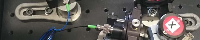
1 Real Time Process Control with Optical Coherence Tomography 16th of June 2016, Ch. Meier Bern HUCE, University OPTOLAB of Applied Science HuCE-optoLabt
2 Overview Short introduction to OCT Systems Resolution and NA, SD OCT and SS OCT, Scanning and Full Field Systems Examples of Real Time Process Control using OCT Braucht mindestens 30 min. Lasik Film ist aus der Präsentation gelöscht
3 Introduction and Theory


4 OCT: Basic principle Comparable with ultrasonic tomography measuring the time delay of back-scattered or back-reflected light Too short time delays for direct measurements interferometric measurements B-scan A-scan, spatial domain


5 3D Imaging by lateral scanning Cross sectional images obtained by scanning in x and y direction A-scan, B-scan, C-scan


6 Diagnostic window Det. Silicon InGaAs PbS or PbSe Apll. Ophthalmic Retina, anterior segment Cardiovascular, Dermatology, other tissue NDT, QA Laser >1.6


7 Time Domain OCT Michelson Interferometer setup with moving reference mirror The signal envelope represent the scattering or reflectivity depth profile
 Spectrometer z 0 z")
8 FD OCT, Spectrometer based Source S(λ) Spectrometer z 0 z 1 z
")


9 FD OCT, Spectrometer based Source S(λ) Spectrometer z 0 z 1 z Interferences due to optical path difference Frequency in k space is proportional to OPD sample Scattering or reflectivity depth profile is obtained by a Fourier transformation
10 Swept Source OCT Detector Intensity a.u
11 Axial Resolution General signal in Frequency Domain General signal in Frequency Domain Gaussian source spectrum -> Gaussian PSF Intensity in a.u. Intensity in a.u Wavelengh in um Distance in um PSF = axial resolution = Coherence Length


12 Resolution Lateral resolution Axial resolution Confocal Microscope OCT Coherence gate
13 Scanning OCT Full Field OCT Cam SLED TD OCT Source S(λ) Detector z 0 z SLED Cam SS 0 z FD OCT Swept Source Detector 0 13 z
14 Scanning versus Full Field OCT SM fiber cam Scattering media Coherence gate Coherence gate Confocal gate

15 Acquisition Time A-scan rate Camera Line rate /khz Swept Source Rate /khz AVIIVA 100 Santec Basler 140 Axsun, Insight, Thorlabs Fraunhofer, AIT 600 OCTLight 850 OptoRes lines/frame, 512 frames A_Scan rate/khz B-scan rate/ Hz C-scan rate/ Hz
16 Examples of Real Time Process Control


")

17 Full Field OCT Phase Sensitive Parallel Optical Coherence Tomography Number of pixels: 300 x 300 Smart pixels (demodulation) Frame rate up to 10^6/s C-scan rate 3-6 Hz (1mm depth)


18 Topographic measuremenents Solder Bumps

19 Layer thickness measurement 19


20 Layer thickness measurement 20


21 Schichtdickenmessung in Schläuchen 21


22 Optimized Laser Head for Contact-Free Osteotomy with real time Depth Control Advanced Osteotomy Tools Robot for bone cutting Clean cuts, better healing


23 Measurement of cutting depth After each laser-shot one B-scan for depth measurement Advanced Osteotomy Tools


24 Ultra-high Resolution OCT Monitoring for Dosimetry Control during Selective Retina Laser Treatment Source: Topcon Coagulation of RPE, photoreceptor cells, choroid Introduced tissue damage is irreversible Excessive tissue damage for RPE-linked pathologies [1] [1] Brinkmann R, and Birngruber R, Medizinische Physik 2007; 17:6 22
")


25 Selective Retina Therapy (SRT) Sub-threshold laser treatment Tissue damage remains limited to the retinal pigment epithelium (RPE) Introduced retinal lesions remain ophthalmoscopically barely visible or invisible Dynamic changes in tissue detected by time-resolved OCT provide real-time feedback for laser dosimetry Retina: en face view. Treatment sites (white) at different power levels and treatment plan

26 Time-resolved OCT data Effects originate in RPE / Bruch s membrane complex and expand to inner retina Signals linked to thermal expansion, thermal vibration and changes in tissue scattering Axial tissue movement in the range of few μm/s up to few m/s detectable ~ 100 mm/s P. Steiner. PhD Thesis

27 Clinical SRT Studies: OCT Visibility P. Steiner. PhD Thesis


28 Damage mechanisms Thermal vibration shockwaves introduced by abrupt heating Thermal expansion long term changes after the pulse, typical relaxation times of tens of ms Rapid dynamic changes Rearrangement of scatterers due to microbubble creation P. Steiner. PhD Thesis

29 Real Time Optical Coherence Tomography Laser Dosimetry control during Selective Retina Therapy


30 Investigation with technical samples P. Morgenthaler Master Thesis 2016


31 Seeing Surgical Laser Surgical laser equipped with measurement and visualization system Enables planning and controlling the surgery Product launch 2014 (CTI 12984)


32 Challenge: Data Processing Algorithms to extend the imaging range Surgery planning by touch screen

33 HuCE-optoLab Thank you for your attention
University of Lübeck, Medical Laser Center Lübeck GmbH Optical Coherence Tomography
 University of Lübeck, Medical Laser Center Lübeck GmbH Optical Coherence Tomography. Theory Dr. Gereon Hüttmann / 009 What is OCT? ( for the MD ) Lichtquelle Probe Detektor Display OCT is Ultrasound with
OPTICAL COHERENCE TOMOGRAPHY:SIGNAL PROCESSING AND ALGORITHM
 OPTICAL COHERENCE TOMOGRAPHY:SIGNAL PROCESSING AND ALGORITHM OCT Medical imaging modality with 1-10 µ m resolutions and 1-2 mm penetration depths High-resolution, sub-surface non-invasive or minimally
Pathology Hinting as the Combination of Automatic Segmentation with a Statistical Shape Model
 Pathology Hinting as the Combination of Automatic Segmentation with a Statistical Shape Model Pascal A. Dufour 12,HannanAbdillahi 3, Lala Ceklic 3,Ute Wolf-Schnurrbusch 23,JensKowal 12 1 ARTORG Center
Pathology Hinting as the Combination of Automatic Segmentation with a Statistical Shape Model
 Pathology Hinting as the Combination of Automatic Segmentation with a Statistical Shape Model Pascal A. Dufour 1,2, Hannan Abdillahi 3, Lala Ceklic 3, Ute Wolf-Schnurrbusch 2,3, and Jens Kowal 1,2 1 ARTORG
Miniature Mirau interferometry for swept-source OCT imaging with applications in dermatology
 Miniature Mirau interferometry for swept-source OCT imaging with applications in dermatology C. Gorecki 1, S. Bargiel 1, J. Lullin 1,, J. Albero 1, N. Passilly 1, S. Perrin 1, L. Froehly 1, W.- S. Wang
The Future of OCT? Swept Source. Todd J. Purkiss, MD, PhD River City Retina Club July 16, 2015
 The Future of OCT? Swept Source Todd J. Purkiss, MD, PhD River City Retina Club July 16, 2015 Background on OCT Optical Coherence Tomography Optical When a light wave meets the interface of two differing
The role of light source in coherence scanning interferometry and optical coherence tomography
 The role of light source in coherence scanning interferometry and optical coherence tomography Dr Rong Su Research Fellow Advanced Manufacturing Research Group Manufacturing Metrology Team Precision manufacturing
Double-Shot 3-D Displacement Field Measurement Using Hyperspectral Interferometry
 Proceedings Double-Shot 3-D Displacement Field Measurement Using Hyperspectral Interferometry Tobias V. Reichold *, Pablo D. Ruiz and Jonathan M. Huntley Wolfson School of Mechanical, Electrical and Manufacturing
Fuzzy C-means Clustering For Retinal Layer Segmentation On High Resolution OCT Images
 Fuzzy C-means Clustering For Retinal Layer Segmentation On High Resolution OCT Images Markus A. Mayer1,2, Ralf P. Tornow3, Joachim Hornegger1, Friedrich E. Kruse3 1 Chair of Pattern Recognition, 2 Graduate
Towards building an anatomically correct solid eye model with volumetric representation of retinal morphology
 Towards building an anatomically correct solid eye model with volumetric representation of retinal morphology Robert J. Zawadzki a *, T. Scott Rowe b, Alfred R. Fuller c, Bernd Hamann c and John S. Werner
Improved image speckle noise reduction and novel dispersion cancellation in Optical Coherence Tomography
 Improved image speckle noise reduction and novel dispersion cancellation in Optical Coherence Tomography by Prabakar Puvanathasan A thesis presented to the University of Waterloo in fulfillment of the
Light and tissue: 2. Two-photon, Optical Coherence Tomography, Photoacoustics
 Light and tissue: 2 Two-photon, Optical Coherence Tomography, Photoacoustics Last lecture: Absorption and Scattering Last lecture: Mean free path Mean free path Transport mean free path Ntziachristos (2010)
Optical Sectioning. Bo Huang. Pharmaceutical Chemistry
 Optical Sectioning Bo Huang Pharmaceutical Chemistry Approaches to 3D imaging Physical cutting Technical difficulty Highest resolution Highest sensitivity Optical sectioning Simple sample prep. No physical
From Eye to Insight Hand-held, contact-free Optical Coherence Tomography for pediatrics ENVISU C2300 OCT Take OCT to your patient FDA 510(k) Cleared
 Cleared") From Eye to Insight Hand-held, contact-free Optical Coherence Tomography for pediatrics ENVISU C2300 OCT Take OCT to your patient FDA 510(k) Cleared ENVISU C-CLASS EASY ON YOUR PATIENT The origins of pathology
From Eye to Insight Hand-held, contact-free Optical Coherence Tomography for pediatrics ENVISU C2300 OCT Take OCT to your patient FDA 510(k) Cleared ENVISU C-CLASS EASY ON YOUR PATIENT The origins of pathology
Signal post processing in frequency domain OCT and OCM using a filter bank approach
 Signal post processing in frequency domain OCT and OCM using a filter bank approach Bernd Hofer 1,2, Boris Považay 1, Boris Hermann 1, Angelika Unterhuber 1, Gerald Matz 2, Franz Hlawatsch 2, Wolfgang
Other Laser Surgery Laser Tonsillectomy Use CO 2 with mirror bouncing system Operation takes 15 minutes, no pain Cauterizes blood vessels & Lymphatic
 Other Laser Surgery Laser Tonsillectomy Use CO 2 with mirror bouncing system Operation takes 15 minutes, no pain Cauterizes blood vessels & Lymphatic vessels no blood in throat Patient eat & drink just
EnFocus Your Upgrade Path to High Performance Intrasurgical OCT
 Your Upgrade Path to High Performance Intrasurgical OCT is FDA 510(k) Cleared > Ultra HD OCT EXTEND YOUR MICROSCOPE S POTENTIAL WITH INTRASURGICAL OCT Brilliant Images, Sub-Surface Knowledge is an intrasurgical
Sensor based adaptive laser micromachining using ultrashort pulse lasers for zero-failure manufacturing
 Sensor based adaptive laser micromachining using ultrashort pulse lasers for zero-failure manufacturing Fraunhofer Institute for Production Technology, Aachen M. Sc. Guilherme Mallmann Prof. Dr.-Ing. Robert
PH880 Topics in Physics
 PH880 Topics in Physics Modern Optical Imaging (Fall 2010) KAIST PH880 11/10/2010 Overview of week 11 Monday: Digital Holographic Tomography Optical Coherence Tomography Wednesday: PhotoacousticTomography
Design and Implementation of Full Field-Optical Coherence Tomography on an Olympus IX73 Microscope. by Rahul Thakur
 Design and Implementation of Full Field-Optical Coherence Tomography on an Olympus IX73 Microscope by Rahul Thakur A Thesis submitted to Faculty of Graduate Studies at The University of Manitoba in partial
Fourier Transform Imaging Spectrometer at Visible Wavelengths
 Fourier Transform Imaging Spectrometer at Visible Wavelengths Noah R. Block Advisor: Dr. Roger Easton Chester F. Carlson Center for Imaging Science Rochester Institute of Technology May 20, 2002 1 Abstract
Product Specifications
 s Optical Coherence Tomography System Inner Vision 2000-WR Signature Approved Checked Author Document number Version C.Chong A.Morosawa D.Ogawa Page 1 / 10 -Ordering code- - WR - - - - - - - - - Customize
Unsurpassed Wide Field Image Quality
 Unsurpassed Wide Field Image Quality EIDON TrueColor Confocal Scanner The latest technology to disclose new imaging quality to eye care professionals in all clinical practices. Many diagnosis of ocular
MEMS SENSOR FOR MEMS METROLOGY
 MEMS SENSOR FOR MEMS METROLOGY IAB Presentation Byungki Kim, H Ali Razavi, F. Levent Degertekin, Thomas R. Kurfess 9/24/24 OUTLINE INTRODUCTION Motivation Contact/Noncontact measurement Optical interferometer
LOW-COHERENCE INTERFEROMETRY, AN ADVANCED TECHNIQUE FOR OPTICAL METROLOGY IN INDUSTRY
 LOW-COHERENCE INTERFEROMETRY, AN ADVANCED TECHNIQUE FOR OPTICAL METROLOGY IN INDUSTRY M. L. Dufour, G. Lamouche, V. Detalle, B. Gauthier, and P. Sammut Industrial Materials Institute, National Research
Optical Active 3D Scanning. Gianpaolo Palma
 Optical Active 3D Scanning Gianpaolo Palma 3D Scanning Taxonomy SHAPE ACQUISTION CONTACT NO-CONTACT NO DESTRUCTIVE DESTRUCTIVE X-RAY MAGNETIC OPTICAL ACOUSTIC CMM ROBOTIC GANTRY SLICING ACTIVE PASSIVE
Multi-GPU accelerated real-time retinal image segmentation
 Multi-GPU accelerated real-time retinal image segmentation by Maxwell Miao B.A.Sc, Simon Fraser University, 2014 Thesis Submitted in Partial Fulfillment of the Requirements for the Degree of Master of
45 µm polystyrene bead embedded in scattering tissue phantom. (a,b) raw images under oblique
 raw images under oblique") Phase gradient microscopy in thick tissue with oblique back-illumination Tim N Ford, Kengyeh K Chu & Jerome Mertz Supplementary Figure 1: Comparison of added versus subtracted raw OBM images 45 µm polystyrene
Phase gradient microscopy in thick tissue with oblique back-illumination Tim N Ford, Kengyeh K Chu & Jerome Mertz Supplementary Figure 1: Comparison of added versus subtracted raw OBM images 45 µm polystyrene
Coherent Microscopy and Optical Coherence Tomography for Biomedical Applications
 Coherent Microscopy and Optical Coherence Tomography for Biomedical Applications Jeremy M. Coupland Tel.: ++44 (0)1509 227506; Fax: ++44 (0)1509 227502; Email: j.m.coupland@lboro.ac.uk Wolfson School of
Confocal Raman Imaging with WITec Sensitivity - Resolution - Speed. Always - Provable - Routinely
 Confocal Raman Imaging with WITec Sensitivity - Resolution - Speed Always - Provable - Routinely WITec GmbH, Ulm, Germany, info@witec.de, www.witec.de A modular microscope series An Example: FLIM optical
GLOBAL MARKETS AND TECHNOLOGIES FOR OPTICAL COHERENCE TOMOGRAPHY (OCT) FOCUS ON TIME DOMAIN AND POLARIZATION SENSITIVE.
 FOCUS ON TIME DOMAIN AND POLARIZATION SENSITIVE.") GLOBAL MARKETS AND TECHNOLOGIES FOR OPTICAL COHERENCE TOMOGRAPHY (OCT) FOCUS ON TIME DOMAIN AND POLARIZATION SENSITIVE HLC150A May 2013 Shalini Shahani Dewan Project Analyst ISBN: 1-56965-345-3 BCC Research
GLOBAL MARKETS AND TECHNOLOGIES FOR OPTICAL COHERENCE TOMOGRAPHY (OCT) FOCUS ON TIME DOMAIN AND POLARIZATION SENSITIVE HLC150A May 2013 Shalini Shahani Dewan Project Analyst ISBN: 1-56965-345-3 BCC Research
Chemical Characterization of Diverse Pharmaceutical Samples by Confocal Raman Microscopy
 Whitepaper Chemical Characterization of Diverse Pharmaceutical Samples by Confocal Raman Microscopy WITec GmbH, Lise-Meitner-Str. 6, 89081 Ulm, Germany, www.witec.de Introduction The development and production
Parallelization. Marcel Jacomet Josef Goette Bern University of Applied Sciences Bfh-Ti HuCE-microLab, Biel/Bienne.
 Josef Goette Bern University of Applied Sciences Bfh-Ti HuCE-microLab, Biel/Bienne Marcel.Jacomet@bfh.ch OCT at ata-path FiFo FT huce.ti.bfh.ch/microlab October 11, 2017 to OCT OCT at ata-path FiFo FT
Rapid Imaging of Microstructure using Spatially Resolved Acoustic Spectroscopy
 1st International Symposium on Laser Ultrasonics: Science, Technology and Applications July 16-18 2008, Montreal, Canada Rapid Imaging of Microstructure using Spatially Resolved Acoustic Spectroscopy Steve
NPL REPORT ENG 8. Overview of tomography techniques to measure wafer thickness in MEMS structures. Andy Robinson & Richard Leach NOT RESTRICTED
 NPL REPORT ENG 8 Overview of tomography techniques to measure wafer thickness in MEMS structures Andy Robinson & Richard Leach June 2008 NOT RESTRICTED Overview of tomography techniques to measure wafer
3D OPTICAL PROFILER MODEL 7503
 3D Optical Profiler MODEL 7503 Features: 3D OPTICAL PROFILER MODEL 7503 Chroma 7503 is a sub-nano 3D Optical Profiler developed using the technology of white light interference to measure and analyze the
7.3 Refractive Index Profiling of Fibers and Fusion Splices
 7.3 Refractive Index Profiling of Fibers and Fusion Splices 199 necessary for measuring the reflectance of optical fiber fusion splices. Fig. 7.10 schematically depicts an OFDR containing a Michelson interferometer
Hyperspectral interferometry for single-shot absolute measurement of 3-D shape and displacement fields
 EPJ Web of Conferences 6, 6 10007 (2010) DOI:10.1051/epjconf/20100610007 Owned by the authors, published by EDP Sciences, 2010 Hyperspectral interferometry for single-shot absolute measurement of 3-D shape
Coherent Gradient Sensing Microscopy: Microinterferometric Technique. for Quantitative Cell Detection
 Coherent Gradient Sensing Microscopy: Microinterferometric Technique for Quantitative Cell Detection Proceedings of the SEM Annual Conference June 7-10, 010 Indianapolis, Indiana USA 010 Society for Experimental
Quantitative Analysis of Cardiomyocyte Dynamics with Optical Coherence Phase Microscopy
 Quantitative Analysis of Cardiomyocyte Dynamics with Optical Coherence Phase Microscopy Rehman Ansari* a,c, Redouane Aherrahrou b, Zouhair Aherrahrou b, Jeanette Erdmann b, Gereon Hüttmann a, and Achim
The new IOLMaster 700 Next generation biometry from ZEISS. With SWEPT Source Biometry
 The new IOLMaster 700 Next generation biometry from ZEISS With SWEPT Source Biometry The moment you get the full picture to make the best decisions for your patients. This is the moment we work for. //
Zeiss VisuMax/MEL for LASIK and Keratoplasty. William W. Culbertson, MD; Bascom Palmer Eye Institute, Miami, FL
 Zeiss VisuMax/MEL for LASIK and Keratoplasty William W. Culbertson, MD; Bascom Palmer Eye Institute, Miami, FL 1 VisuMax Femtosecond Laser Precision Low IOP Low energy Gentle on patient Minimal applanation
Development of automated ultraviolet laser beam profiling system using fluorometric technique
 Development of automated ultraviolet laser beam profiling system using fluorometric technique BB Shrivastava*, NS Benerji, P Bhatnagar, HS Vora a and U Nundy Chemical and Excimer Laser Section a Laser
Optical Topography Measurement of Patterned Wafers
 Optical Topography Measurement of Patterned Wafers Xavier Colonna de Lega and Peter de Groot Zygo Corporation, Laurel Brook Road, Middlefield CT 6455, USA xcolonna@zygo.com Abstract. We model the measurement
What is Frequency Domain Analysis?
 R&D Technical Bulletin P. de Groot 9/3/93 What is Frequency Domain Analysis? Abstract: The Zygo NewView is a scanning white-light interferometer that uses frequency domain analysis (FDA) to generate quantitative
Efficient Signal Processing Algorithms for Optical Doppler Tomography Literature Survey
 Efficient Signal Processing Algorithms for Optical Doppler Tomography Literature Survey University of Texas at Austin Department of Electrical and Computer Engineering Milos Milosevic Wade Schwartzkopf
PalmScan PRO 5 in 1 Biometer Your Complete Biometry System - Fully Configurable
 B-Scan A-Scan Pachymeter Keratometer UBM PalmScan PRO 5 in 1 Biometer Your Complete Biometry System - Fully Configurable A-Scan + IOL Calculator Keratometer Pachymeter B-Scan UBM www.micromedinc.com 818.222.3310
picoemerald Tunable Two-Color ps Light Source Microscopy & Spectroscopy CARS SRS
 picoemerald Tunable Two-Color ps Light Source Microscopy & Spectroscopy CARS SRS 1 picoemerald Two Colors in One Box Microscopy and Spectroscopy with a Tunable Two-Color Source CARS and SRS microscopy
Characterization of stratified media using high-resolution thin film measurement techniques
 Characterization of stratified media using high-resolution thin film measurement techniques Alberto Aguerri Sensofar-Tech, S.L. Crt. N150 Km14.5 IPCT Mòdul TR-20, 08227 Terrassa (Barcelona), Spain E-mail:
Company Presentation Optical Components
 Company Presentation Optical Components A selection of our partners 2 A selection of our partners 3 Optical Technologies Fiber Optics components & fiber optic assemblies Precision optics Acousto-optics
Introduction to Biomedical Imaging
 Alejandro Frangi, PhD Computational Imaging Lab Department of Information & Communication Technology Pompeu Fabra University www.cilab.upf.edu X-ray Projection Imaging Computed Tomography Digital X-ray
New Olympus FV3000RS Laser Scanning Confocal System. Quotation Number V2
 New Olympus FV3000RS Laser Scanning Confocal System Quotation Number 00056013 V2 Quotation Date: 08/08/2018 Revised Date: 07/09/2018 Quotation number: 00056013 Quotation Expiry: 07/11/2018 Macquarie University
SIMULATION AND VISUALIZATION IN THE EDUCATION OF COHERENT OPTICS
 SIMULATION AND VISUALIZATION IN THE EDUCATION OF COHERENT OPTICS J. KORNIS, P. PACHER Department of Physics Technical University of Budapest H-1111 Budafoki út 8., Hungary e-mail: kornis@phy.bme.hu, pacher@phy.bme.hu
3D SWEPT SOURCE OCT. Very high scanning speed: 30,000 A-Scans/sec. 130,800 A-Scans. Cut plane 16 x 16 x 6 mm. Topo/Pachy Map in 0.3 sec.
 3D SWEPT SOURCE OCT FOURIER DOMAIN OCT CASIA SS-1000 Very high scanning speed: 30,000 A-Scans/sec. 130,800 A-Scans Cut plane 16 x 16 x 6 mm Topo/Pachy Map in 0.3 sec. Free adjustable display in 2D and
QUANTITATIVE PHASE IMAGING OF BIOLOGICAL CELLS USING OFF-AXIS METHOD OF WIDE FIELD DIGITAL INTERFERENCE MICROSCOPY (WFDIM)
") http:// QUANTITATIVE PHASE IMAGING OF BIOLOGICAL CELLS USING OFF-AXIS METHOD OF WIDE FIELD DIGITAL INTERFERENCE MICROSCOPY (WFDIM) Pradeep Kumar Behera 1, Dalip Singh Mehta 2 1,2 Physics,Indian Institute
http:// QUANTITATIVE PHASE IMAGING OF BIOLOGICAL CELLS USING OFF-AXIS METHOD OF WIDE FIELD DIGITAL INTERFERENCE MICROSCOPY (WFDIM) Pradeep Kumar Behera 1, Dalip Singh Mehta 2 1,2 Physics,Indian Institute
Optical Ptychography Imaging
 Optical Ptychography Imaging Summer Project Annafee Azad Supervisors: Dr Fucai Zhang Prof Ian Robinson Summer 2014 23 October 2014 Optical Ptychography Imaging P a g e 2 Abstract This report details a
CHAPTER 2: THREE DIMENSIONAL TOPOGRAPHICAL MAPPING SYSTEM. Target Object
 CHAPTER 2: THREE DIMENSIONAL TOPOGRAPHICAL MAPPING SYSTEM 2.1 Theory and Construction Target Object Laser Projector CCD Camera Host Computer / Image Processor Figure 2.1 Block Diagram of 3D Areal Mapper
SPECTRUM. The world s first fully automated Raman AFM. AFM - confocal Raman - SNOM - TERS AFM KPFM. Raman. AFM-Raman characterization of PS-PVAC
 Raman KPFM AFM AFM-Raman characterization of PS-PVAC polymer blend film SPECTRUM The world s first fully automated Raman AFM AFM - confocal Raman - SNOM - TERS The first fully integrated & automated AFM
Quantitative Three-Dimensional Imaging of the Posterior Segment with the Heidelberg Retina Tomograph
 Quantitative Three-Dimensional Imaging of the Posterior Segment with the Heidelberg Retina Tomograph Heidelberg Engineering GmbH, Heidelberg, Germany Contents 1 Introduction... 1 2 Confocal laser scanning
RoHS COMPLIANT 2002/95/EC
 Superlum Broadband Light Sources cblmd-series (2nd Generation) Compact Broadband Light Technical Product Specification Document Number SL.3328.00.000D3 June 2017 Revision 001 ATTENTION ELECTROSTATIC SENSITIVE
Available online at ScienceDirect. Procedia CIRP 10 (2013 ) th CIRP Conference on Computer Aided Tolerancing
 th CIRP Conference on Computer Aided Tolerancing") Available online at www.sciencedirect.com ScienceDirect Procedia CIRP 1 (213 ) 7 76 12th CIRP Conference on Computer Aided Tolerancing Accelerated surface measurement using wavelength scanning interferometer
Available online at www.sciencedirect.com ScienceDirect Procedia CIRP 1 (213 ) 7 76 12th CIRP Conference on Computer Aided Tolerancing Accelerated surface measurement using wavelength scanning interferometer
Leica TCS STED. The Fast Track to Superresolution Technical Documentation
 Leica TCS STED The Fast Track to Superresolution Technical Documentation 8 9 6 7 Inverted research microscope Leica DMI6000 CS Scan head Laser and power supply Computer table Air damped optical table 6
NDD FLIM Systems for Leica SP2 MP and SP5 MP Multiphoton Microscopes
 NDD FLIM Systems for Leica SP2 MP and SP5 MP Multiphoton Microscopes bh FLIM systems for the confocal and the multiphoton versions of the Leica SP2 and SP5 microscopes are available since 2002 [4]. These
Optical Diffraction and Interference using Single Photon Counting
 Optical Diffraction and Interference using Single Photon Counting Optical Diffraction and Interference using Single Photon Counting In this experiment the wave and quantum properties of light can be studied
RoHS COMPLIANT 2002/95/EC
 Document ID: SL.QC... May Revision: ATTENTION ELECTROSTATIC SENSITIVE DEVICES RoHS COMPLIANT /95/EC Page of 9 Contents Product Description / Applications / Features... Mechanical specification... Electrical
Building Your Own 2-Photon Microscope: Challenges, Advantages and Limitations
 Building Your Own 2-Photon : Challenges, Advantages and Limitations Roberto Weigert, Ph.D. Intracellular Membrane Trafficking Unit Oral and Pharyngeal Cancer Branch NIDCR-NIH Building Your Own 2-Photon
Correction of refraction. volume deformation measurements
 Loughborough University Institutional Repository Correction of refraction induced distortion in optical coherence tomography corneal reconstructions for volume deformation measurements This item was submitted
ksa ICE - Integrated Control for Epitaxy
 Introduction The k-space Integrated Control for Epitaxy system (ksa ICE) is a modular in-situ metrology tool designed for today s MOCVD reactors. It combines proven ksa MOS, ksa BandiT, and ksa RateRat
THE DICOM 2013 INTERNATIONAL CONFERENCE & SEMINAR. DICOM Fields of Use. Klaus Neuner. Brainlab AG. Software Project Manager Feldkirchen, Germany
 THE DICOM 2013 INTERNATIONAL CONFERENCE & SEMINAR March 14-16 Bangalore, India DICOM Fields of Use Klaus Neuner Brainlab AG Software Project Manager Feldkirchen, Germany Introduction This presentation
Applications in Ophthalmology
 Applications in Ophthalmology July 2017 Dr. David Leuenberger, Business Development Manager Bernstrasse 388 CH-8953 Dietikon Switzerland Phone +41 58 856 3011 www.optotune.com info@optotune.com Agenda
Roughness Measurement Methodology according to DIN 4768 Using Optical Coherence Tomography (OCT)
") Roughness Measurement Methodology according to DIN 4768 Using Optical Coherence Tomography (OCT) Marcello M. Amaral a *, Marcus P. Raele a, José P. Caly b, Ricardo E. Samad a, Nilson D. Vieira Jr. a, Anderson
Roughness Measurement Methodology according to DIN 4768 Using Optical Coherence Tomography (OCT) Marcello M. Amaral a *, Marcus P. Raele a, José P. Caly b, Ricardo E. Samad a, Nilson D. Vieira Jr. a, Anderson
SPECTRAL EFFECTS OF PHOSPHOR- BASED WHITE LIGHT-EMITTING- DIODE (LED) IN COHERENCE SCANNING INTERFEROMETRY
 IN COHERENCE SCANNING INTERFEROMETRY") SPECTRAL EFFECTS OF PHOSPHOR- BASED WHITE LIGHT-EMITTING- DIODE (LED) IN COHERENCE SCANNING INTERFEROMETRY CHONG WEE KEAT School of Electrical & Electronics Engineering A thesis submitted to in partial
SPECTRAL EFFECTS OF PHOSPHOR- BASED WHITE LIGHT-EMITTING- DIODE (LED) IN COHERENCE SCANNING INTERFEROMETRY CHONG WEE KEAT School of Electrical & Electronics Engineering A thesis submitted to in partial
Progress Towards Low-Cost Compact Metric Adaptive Optics Systems
 Progress Towards Low-Cost Compact Metric Adaptive Optics Systems Justin D. Mansell, Brian Henderson, Brennen Wiesner, Robert Praus, and Steve Coy Active Optical Systems, LLC www.aos-llc.com 1 Outline Introduction
Basic optics. Geometrical optics and images Interference Diffraction Diffraction integral. we use simple models that say a lot! more rigorous approach
 Basic optics Geometrical optics and images Interference Diffraction Diffraction integral we use simple models that say a lot! more rigorous approach Basic optics Geometrical optics and images Interference
College Physics 150. Chapter 25 Interference and Diffraction
 College Physics 50 Chapter 5 Interference and Diffraction Constructive and Destructive Interference The Michelson Interferometer Thin Films Young s Double Slit Experiment Gratings Diffraction Resolution
FDA 510(k)-cleared OCT system for ophthalmic surgery SEE WHAT YOU VE BEEN MISSING. EnFocus intrasurgical OCT
-cleared OCT system for ophthalmic surgery SEE WHAT YOU VE BEEN MISSING. EnFocus intrasurgical OCT") FDA 510(k)-cleared OCT system for ophthalmic surgery SEE WHAT YOU VE BEEN MISSING EnFocus intrasurgical OCT SEE WHAT YOU VE DURING YOUR SU Make the best-informed surgical decisions for your patient with
FDA 510(k)-cleared OCT system for ophthalmic surgery SEE WHAT YOU VE BEEN MISSING EnFocus intrasurgical OCT SEE WHAT YOU VE DURING YOUR SU Make the best-informed surgical decisions for your patient with
SPIcam: an overview. Alan Diercks Institute for Systems Biology 23rd July 2002
 SPIcam: an overview Alan Diercks Institute for Systems Biology diercks@systemsbiology.org 23rd July 2002 1 Outline Overview of instrument CCDs mechanics instrument control performance construction anecdotes
DUB. SkinScanner Skin Measurement with Ultrasound. Dermatology Ÿ Aesthetics Ÿ Research Ÿ Cosmetics. w w w. d u b s k i n s c a n n e r.c o m.
 DUB SkinScanner Skin Measurement with Ultrasound w w w. d u b s k i n s c a n n e r.c o m 22 MHz Dermatology Aesthetics Research Cosmetics Depth = 2.4 mm high frequency ultrasound and more since 1978 tpm
Speckle Variance Optical Coherence Tomography (svoct)
") Speckle Variance Optical Coherence Tomography (svoct) Mohammad Sultan Mahmud, PhD Researcher and Engineer 26 th June, 2013. Motivation Clinical issue: Tumor blood supply differs from that of normal tissue.
Speckle Variance Optical Coherence Tomography (svoct) Mohammad Sultan Mahmud, PhD Researcher and Engineer 26 th June, 2013. Motivation Clinical issue: Tumor blood supply differs from that of normal tissue.
WITec Raman Spectroscopy Solutions
 Confocal Raman Imaging WITec Raman Spectroscopy Solutions -40-20 0 20 CCD cts 500 1000 1500 2000 2500 3000 3000 relative wavenumbers (cm -1 ) www.witec.de WITec UHTS Ultra-High Throughput Spectrometers
10/5/09 1. d = 2. Range Sensors (time of flight) (2) Ultrasonic Sensor (time of flight, sound) (1) Ultrasonic Sensor (time of flight, sound) (2) 4.1.
 (2) Ultrasonic Sensor (time of flight, sound) (1) Ultrasonic Sensor (time of flight, sound) (2) 4.1.") Range Sensors (time of flight) (1) Range Sensors (time of flight) (2) arge range distance measurement -> called range sensors Range information: key element for localization and environment modeling Ultrasonic
Range Sensors (time of flight) (1) Range Sensors (time of flight) (2) arge range distance measurement -> called range sensors Range information: key element for localization and environment modeling Ultrasonic
LASER SLIT LAMP. Optimized for Retina
 VERSATILE GREEN LASER Full-featured, portable green laser for the clinic and operating room. LASER SLIT LAMP Magnification 5x, 8x, 12.5x, 20x, 31x with 12.5x eyepieces (±5 D compensation range) Field of
FOX DIODE LASER. Power Calibration at Fiber Distal Tip Green Aiming Beam Video Touch Screen Intuitive Handling Rapid Amortization
 The world s first diode laser with laser output calibration 810 nm 940 nm 980 nm 1064 nm DIODE LASER Power Calibration at Fiber Distal Tip Green Aiming Beam Video Touch Screen Intuitive Handling Rapid
Vision Based Metal Spectral Analysis using
 1/27 Vision Based Metal Spectral Analysis using Eranga Ukwatta Department of Electrical and Computer Engineering The University of Western Ontario May 25, 2009 2/27 Outline 1 Overview of Element Spectroscopy
Imaging the eye fundus with real-time en-face spectral domain optical coherence tomography
 Imaging the eye fundus with real-time en-face spectral domain optical coherence tomography Adrian Bradu 1,* and Adrian Gh. Podoleanu 1 1 Applied Optics Group, School of Physical Sciences, University of
Development of Optical Coherence Tomography for the Measurement of the Surface Wrinkle of Composite Parts
 Development of Optical Coherence Tomography for the Measurement of the Surface Wrinkle of Composite Parts Yoshiharu KUZE Composite Laboratory Manufacturing Technology Research Department May 9, 2017 1
3D TeraHertz Tomography
 3D TeraHertz Tomography B. Recur, 3 A. Younus, 1 S. Salort, 2 P. Mounaix, 1 B. Chassagne, 2 P. Desbarats, 3 1 LOMA, Université de Bordeaux / CNRS 2 ALPhANOV, Centre Technologique Optique et Lasers, Université
SEE BEYOND. Ophthalmic Surgical Microscope Proveo 8
 SEE BEYOND Ophthalmic Surgical Microscope Proveo 8 One of the benefits with Proveo 8 is the way the illumination is achieved by four coaxial LED lights. The optics of the microscope and the innovative
Image Acquisition Systems
 Image Acquisition Systems Goals and Terminology Conventional Radiography Axial Tomography Computer Axial Tomography (CAT) Magnetic Resonance Imaging (MRI) PET, SPECT Ultrasound Microscopy Imaging ITCS
System for Gauge Blocks Diagnostics
 System for Gauge Blocks Diagnostics Z. Buchta, B. Mikel, M. Čížek, J. Lazar, O. Číp, Institute of Scientific Instruments AS CR, v.v.i., Brno, Czech Republic Abstract: In this paper, a novel principle of
Durham Magneto Optics Ltd. NanoMOKE3
 Durham Magneto Optics Ltd NanoMOKE3 Specifications Marketed by Quantum Design 1 1. Introduction NanoMOKE3 is a new generation of ultra-high sensitivity magnetooptical magnetometer and Kerr microscope.
GPU-Accelerated Optical Coherence Tomography (OCT) Imaging
 Imaging") GPU-Accelerated Optical Coherence Tomography OCT Imaging Kang Zhang Speaer G Global Research General lectric Company Jin U. Kang Department of lectrical and Computer ngineering The Johns Hopins University
GPU-Accelerated Optical Coherence Tomography OCT Imaging Kang Zhang Speaer G Global Research General lectric Company Jin U. Kang Department of lectrical and Computer ngineering The Johns Hopins University
e2v OCTOPLUS USB3 Modular camera platform from e2v
 e2v OCTOPLUS USB3 Modular camera platform from e2v Datasheet Features 2048 pixels CMOS Monochrome LineScan Sensor Pixel size available in 2 versions: 10x20µm and 10x200µm Pixel full well capacity available
Roughness parameters and surface deformation measured by "Coherence Radar" P. Ettl, B. Schmidt, M. Schenk, I. Laszlo, G. Häusler
 Roughness parameters and surface deformation measured by "Coherence Radar" P. Ettl, B. Schmidt, M. Schenk, I. Laszlo, G. Häusler University of Erlangen, Chair for Optics Staudtstr. 7/B2, 91058 Erlangen,
Versatile laser Raman Spectrometer. RMP-500 series
 Versatile laser Raman Spectrometer RMP-500 series About RMP-500 RMP-500 series is a compact and versatile laser Raman spectrometer consisting of a micro Raman probe connected through fiber optics to the
External Triggering Options
 380 Main Street Dunedin, FL 34698 (727) 733-2447 (727) 733-3962 fax External Triggering Options Our S2000 and S1024DW Spectrometers provide four methods of acquiring data. In the Normal Mode, Ocean Optics
Measure at the speed of light...
 Measure at the speed of light... LENSTAR LS 900 Biometry Explore new dimensions... Complete optical biometer including CCT and lens thickness Align once, get all results fast biometrical assessment Non
810 nm. 980 nm FOX nm 12 W VERSATILE FOX 810/980/ 1064/1470 nm IDEAL FOR: DENTAL ENT DERMATOLOGY GYNECOLOGY SURGERY / VET.
 810 1064 980 12 W 980 VERSATILE 810/980/ 1064/1470 IDEAL FOR: DENTAL ENT DERMATOLOGY GYNECOLOGY SURGERY / VET www.arclaser.com PRECISION & PERFECTION CALIBRATION at the distal tip of the fiber = safe and
ZEISS Smartproof 5 Your Integrated Widefield Confocal Microscope for Surface Analysis in Quality Assurance and Quality Control
 Product Information Version 1.0 ZEISS Smartproof 5 Your Integrated Widefield Confocal Microscope for Surface Analysis in Quality Assurance and Quality Control Dedicated Design. Guided Workflow. Trusted
Automatic Segmentation of Layers in outer Retina by Model Selection
 Automatic Segmentation of Layers in outer Retina by Model Selection P. Bose Babu 1 M.Tech.,G. Naga Vasantha 2 1 Assistant Professor, Department of Electronics and Communication Engineering, Andhra Loyola