Optical Sectioning. Bo Huang. Pharmaceutical Chemistry
|
|
|
- Silvia Sullivan
- 6 years ago
- Views:
Transcription

1 Optical Sectioning Bo Huang Pharmaceutical Chemistry
2 Approaches to 3D imaging Physical cutting Technical difficulty Highest resolution Highest sensitivity Optical sectioning Simple sample prep. No physical damage Tomography Micheva & Smith, Neuron (2007)
3 Optical sectioning Confocal microscopy principle Point scanning confocal Multi-point scanning confocal Spinning disk Two-photon microscopy Total internal reflection fluorescence (TIRF) Other methods
 equivalent: the 3D focus of a")
4 The Point Spread Function (PSF) PFS: the three dimensional image of a point object (Almost) equivalent: the 3D focus of a laser

5 The lateral size of the PSF y x Lateral resolution Example: 1.4 NA objective at 550 nm Δ xy 240 nm FWHM = 220 nm nm
6 The axial size of the PSF FWHM = 568 nm z x Axial resolution (works only for low NA system) Example: 1.4 NA objective at 550 nm Δ z 850 nm
7 z resolution z sectioning z Conservation of energy tells that the detector will collect the signal equally well at any z position
8 How to achieve optical Mathematics Deconvolution microscopy Limiting the detection depth Confocal microscopy Limiting the excitation depth z Two-photon microscopy Total internal reflection fluorescence (TIRF) microscopy Single plane illumination microscopy (SPIM)
9 Principle of confocal Pinhole

10 Confocal vs. widefield fluorescence microscopy

11 The pinhole size in confocal microscopy Detection efficiency (PSF det ) = PSF Pinhole Pinhole size should be related to PSF width: - 100x / 1.4 NA 220 nm x 100 = 22 μm - 40x / 1.3 NA 235 nm x 40 = 9.4 μm - 20x / 0.75 NA 407 nm x 20 = 8.1 μm - 10x / 0.45 NA 678 nm x 10 = 6.8 μm
12 The PSF of confocal microscopy Detector Detector = Excitation Detection PSF Confocal = PSF ex PSF det In the case of infinitely small pinhole: PSF Confocal PSF ex 2
13 The confocal volume Integrating PSF confocal over the entire space now gets a finite value Diffraction limited volume: Δ xy 250 nm, Δ z 600 nm
14 The sectioning ability of confocal microscopy Guess: When imaging a thick homogenous sample with the best confocal microscope, what is the fraction of signal coming from outside of the focal
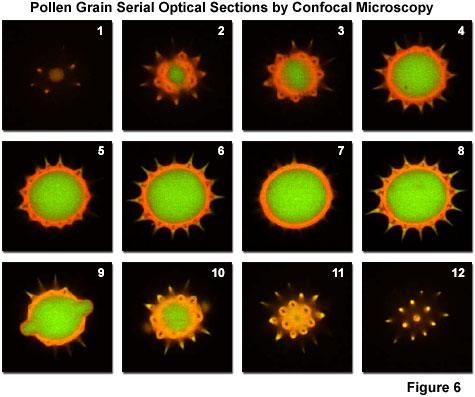

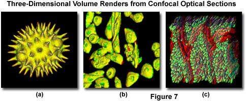
15 Confocal sectioning and 3D reconstruction
 Microscope Controller and image acquisition computer Scan head")
16 Components of a confocal Light source (usually laser based) Detector (PMT or CCD) Microscope Controller and image acquisition computer Scan head Pinhole
17 Point scanning confocal Stage scan Laser scan Hybrid Simple but slow


18 Galvanometer scanning / Two mirrors for x-y raster scan Excitation Detection


19 Averaging multiple scans to enhance S/N 1 frame 8-frame average

20 How to scan faster? Normal galvo mirror: several μs per pixel sec per image Resonance scanner: up to 30 fps The answer is:


21 Spinning disk confocal Scanning with a rotating pinhole array CCD camera detection

22 Spinning disk confocal


23 Spinning disk confocal Scanning direction Up to 2000 frames / sec The disk rotation must match the camera frame rate 12 scanning frames per 360 rotation
24 Multi-photon microscopy Drobizhev et al., J. Phys. Chem. B One-photon (2009) absorption wavelength (nm) S 1 hν 2 hν Fluorescence Emission hν 1 S 0 Two-photon One-photon absorption absorption Two-photon 1400 absorption wavelength (nm) Two-photon excitation: Fluorescence I 2
 2 Recall: PSF Confocal = PSF ex PSF det (PSF ex ) 2 Two-photon excitation is")
25 Optical sectioning without a Fluorescence I 2 PSF 2P.ex = (PSF 1P.ex ) 2 Recall: PSF Confocal = PSF ex PSF det (PSF ex ) 2 Two-photon excitation is localized to the laser focal point
26 Deep tissue imaging Tissue scattering and absorption decrease confocal performance Confocal 100 μm Two-photon

27 Ultrafast laser source for 2PE Continuous wave (CW) laser t To see reasonable 2PE with CW laser: ~ 1W power Pulsed laser Peak power Ti:Sapphire laser, 100 fs, 80 MHz 10 mw avg 1.2 kw peak Pulse duration Repetition rate Average power t

28 Ultrafast laser source for 2PE

29 Two-photon microscopy of in vivo brain function Kherlopian, et al., BMC Systems Biology (2
30 Total internal reflection fluorescence microscopy n 1 n 1 sinθ 1 = n 2 sinθ 2 θ 1 Total internal reflection: n 2 θ 2 θ 1 = 90 sinθ 2 sinθ c = n 1 /n 2 n 1 = 1.33 (water), n 2 = 1.52 (glass), θ c = 61

31 The evanescent wave in TIR The energy of the evanescent wave is localized near the interface. The strength of the evanescent wave field decreases exponentially. The penetration depth is a function of the wavelength and the incident angle.


32 TIRM improves S/N for surface Epifluorescence TIRF
33 What are the applications of Organization of plasma membrane proteins Dynamics of plasma membrane lipids Endocytosis and exocytosis Focal adhesion Growth cone migration Imaging in supported lipid bilayers In vitro reconstituted cytoskeleton filaments Single molecule imaging
34 Through-the-objective TIRF

35 Requirement for TIRF NA = n glass sinθ max n water = n glass sinθ c θ max θ c NA n water 1.33


36 TIRF compatible objectives 1.40 NA Barely enough for laser TIRF 1.45 / 1.49 NA TIRF objective More homogenous illumination field Higher efficiency for lamp TIRF Compromised image quality 1.65 NA Sapphire coverglass Toxic oil
37 TIRF illuminator Focusing the light to the back-focal-plane of the objective Translate the light to the edge of the objective back-aperture Sample plane Objective Back focal plane


38 A home-built TIRF illuminator Focusing lens Field diaphragm Beam expander Translation stage
39 A commercial TIRF


40 COS cells expressing GFP Dan Axelrod, Univ of Michigan

41 Alternating Epi/TIRF Merrifield et al., Cell 2005

42 Aplysia growth cone Andy Schaefer, Paul Forscher, Yale Univ.
43 Oblique angle illumination: the gray zone θ c 2-5 μm An incident angle close but smaller than the critical angle can improve the signal from near-surface objects.
44 Single-plane illumination microscopy (SPIM) Sample rotation or beam scanning Objective Camera

45 Zebra fish embryo development imaged by SPIM Keller et al., Science (200

46 Super-resolution microscopy Projection 50 nm section y x z x 100 nm 3D STORM image of a clathrin-coated pit Wu et al., Nat Cell Biol (201
47 Now the tools are in your hands!
NDD FLIM Systems for Leica SP2 MP and SP5 MP Multiphoton Microscopes
 NDD FLIM Systems for Leica SP2 MP and SP5 MP Multiphoton Microscopes bh FLIM systems for the confocal and the multiphoton versions of the Leica SP2 and SP5 microscopes are available since 2002 [4]. These
Nonlinear optics and two photon microscopy. Table of contents. Sam Whiteley and Seth Parker PHYS 173/BGGN 266 July 13, 2014
 Nonlinear optics and two photon microscopy Sam Whiteley and Seth Parker PHYS 173/BGGN 266 July 13, 2014 Table of contents 1. Introduction 2. Optical setup 3. Initial images and troubleshooting 4. Determining
PH880 Topics in Physics
 PH880 Topics in Physics Modern Optical Imaging (Fall 2010) Overview of week 8 Monday Nonlinear Microscopy Wednesday No class (Mid term week) Quantum Optics Electro- magnetic Optics Wave Optics Ray Optics
EPFL SV PTBIOP BIOP COURSE 2015 OPTICAL SLICING METHODS
 BIOP COURSE 2015 OPTICAL SLICING METHODS OPTICAL SLICING METHODS Scanning Methods Wide field Methods Point Scanning Deconvolution Line Scanning Multiple Beam Scanning Single Photon Multiple Photon Total
Last class. This class. Single molecule imaging Deconvolution. FLIM Confocal
 FLIM, Confocal Last class Single molecule imaging Deconvolution This class FLIM Confocal FLIM Fluorescence Lifetime IMaging FLIM Fluorescence lifetime imaging Easier and more accurate quantitation Much
Confocal vs. Deconvolution
 Confocal vs. Deconvolution Cesare Covino ALEMBIC Advanced Light and Electron Bio-Imaging Center Istituto Scientifico San Raffaele (Milano) www.hsr.it/research/alembic Fluorescence high contrast high sensibility
Confocal Raman Microscope RAMOS
 Confocal Raman Microscope RAMOS 1 future`s Confocal Raman Microscope RAMOS 2 Confocal Raman Microscope RAMOS Ostec Corporate Group produces and offers hi-tech innovative scientific and analytical equipment.
Confocal Raman Imaging with WITec Sensitivity - Resolution - Speed. Always - Provable - Routinely
 Confocal Raman Imaging with WITec Sensitivity - Resolution - Speed Always - Provable - Routinely WITec GmbH, Ulm, Germany, info@witec.de, www.witec.de A modular microscope series An Example: FLIM optical
New Olympus FV3000RS Laser Scanning Confocal System. Quotation Number V2
 New Olympus FV3000RS Laser Scanning Confocal System Quotation Number 00056013 V2 Quotation Date: 08/08/2018 Revised Date: 07/09/2018 Quotation number: 00056013 Quotation Expiry: 07/11/2018 Macquarie University
Building Your Own 2-Photon Microscope: Challenges, Advantages and Limitations
 Building Your Own 2-Photon : Challenges, Advantages and Limitations Roberto Weigert, Ph.D. Intracellular Membrane Trafficking Unit Oral and Pharyngeal Cancer Branch NIDCR-NIH Building Your Own 2-Photon
Lightsheet Z.1. Light Sheet Fluorescence Microscopy by Carl Zeiss. Fabrice Schmitt, Sales Manager Carl ZEISS France
 Lightsheet Z.1 Light Sheet Fluorescence Microscopy by Carl Zeiss Fabrice Schmitt, Sales Manager Carl ZEISS France 12.12.2012 Light Sheet Fluorescence Microscopy (LSFM) Principle The Principle of Light
A SUPER-RESOLUTION MICROSCOPY WITH STANDING EVANESCENT LIGHT AND IMAGE RECONSTRUCTION METHOD
 A SUPER-RESOLUTION MICROSCOPY WITH STANDING EVANESCENT LIGHT AND IMAGE RECONSTRUCTION METHOD Hiroaki Nishioka, Satoru Takahashi Kiyoshi Takamasu Department of Precision Engineering, The University of Tokyo,
Real-world applications of intense light matter interaction beyond the scope of classical micromachining.
 Dr. Lukas Krainer lk@onefive.com CEO Real-world applications of intense light matter interaction beyond the scope of classical micromachining. 1 Management & Company Company Based in Zürich, Switzerland
LIFA KEY FEATURES APPLICATIONS. Fluorescence Lifetime Attachment LIFA 15001A02 16/03/2015
 LIFA Fluorescence Lifetime Attachment LIFA 151A2 16/3/215 The LIFA is a dedicated system for Fluorescence Lifetime Imaging Microscopy (FLIM). It allows the generation of lifetime images on any widefield
Leica TCS STED CW. The Fast Track to Superresolution. Leica TCS STED CW
 The Fast Track to Superresolution content Motivation Concept Realisation Applications - why do researchers need the TCS STED CW? - what is the TCS STED CW based on? - how does the TCS STED CW work? - what
Supplementary Figure 1 Guide stars of progressively longer wavelengths can be used for direct wavefront sensing at increasingly large depth in the
 Supplementary Figure 1 Guide stars of progressively longer wavelengths can be used for direct wavefront sensing at increasingly large depth in the cortex of the living mouse. Typical SH images of guide
Introductory Guide to Light Microscopy - Biomedical Confocal Microscopy
 Introductory Guide to Light Microscopy - Biomedical Confocal Microscopy 7 May 2007 Michael Hooker Microscopy Facility Michael Chua microscopy@unc.edu 843-3268 6007 Thurston Bowles Wendy Salmon wendy_salmon@med.unc.edu
Non-Descanned FLIM Systems for Olympus FV-1000 and FV-300 Multiphoton Microscopes
 Non-Descanned FLIM Systems for Olympus FV-1000 and FV-300 Multiphoton Microscopes Abstract. Recently multiphoton versions of the Olympus FV 1000 and FV 300 laser scanning microscopes have become available.
Leica TCS STED. The Fast Track to Superresolution Technical Documentation
 Leica TCS STED The Fast Track to Superresolution Technical Documentation 8 9 6 7 Inverted research microscope Leica DMI6000 CS Scan head Laser and power supply Computer table Air damped optical table 6
DSU Start-Up instructions
 DSU Start-Up instructions Always: - start with the 10x objective - properly center the stage around the current objective before changing to another objective - when done, leave the 10x objective in standby
Transmitted light Illuminator VIS-LED, color temperature. Epifluorescence lamp module X-Cite 120PC Q (Excelitas
 ELYRA PS.1 Multi-functional fluorescence inverted widefield microscope enabling live-cell imaging, TIRF or HILO illuminion and two super-resolution techniques: structured illuminion microscopy (SIM) and
DeltaVision OMX SR super-resolution microscope. Super-resolution doesn t need to be complicated
 DeltaVision OMX SR super-resolution microscope Super-resolution doesn t need to be complicated Live-cell super-resolution microscopy DeltaVision OMX SR is a compact super-resolution microscope system optimized
Diskovery. Multi-modal Imaging System
 Diskovery Multi-modal Imaging System Diskovery DISKOVERY SYSTEM CONFIGURATOR Diskover More Different tools are required to answer different questions. Now you can answer more questions about a sample during
Indiana Center for Biological Microscopy. BioRad MRC 1024 MP Confocal & Multi-Photon Microscope
 Indiana Center for Biological Microscopy BioRad MRC 1024 MP Confocal & Multi-Photon Microscope Microscope and the Attached Accessories A: B: C: D: E: F: G: H: Mercury Lamp Transmission Light Kr/Ar Laser
E x Direction of Propagation. y B y
 x E x Direction of Propagation k z z y B y An electromagnetic wave is a travelling wave which has time varying electric and magnetic fields which are perpendicular to each other and the direction of propagation,
Vutara 350. Innovation with Integrity. The Fastest, Super-Resolution Microscope Deep 3D Imaging on Live Cells, Quickly and Easily
 Vutara 350 The Fastest, Super-Resolution Microscope Deep 3D Imaging on Live Cells, Quickly and Easily Innovation with Integrity Fluorescence Microscopy Vutara 350 Don t Get Left Behind Bruker s Vutara
Live-cell 3D super-resolution imaging in thick biological samples
 Nature Methods Live-cell 3D super-resolution imaging in thick biological samples Francesca Cella Zanacchi, Zeno Lavagnino, Michela Perrone Donnorso, Alessio Del Bue, Lauria Furia, Mario Faretta & Alberto
3-D. Here red spheres show the location of gold nanoparticles inside/around a cell nucleus.
 3-D The CytoViva 3-D System allows the user can locate objects of interest in a 3-D space. It does this by acquiring multiple Z planes and performing our custom software routines to locate and observe
Tutorial: Instantaneous Measurement of M 2 Beam Propagation Ratio in Real-Time
 Tutorial: Instantaneous Measurement of M 2 Beam Propagation Ratio in Real-Time By Allen M. Cary, Jeffrey L. Guttman, Razvan Chirita, Derrick W. Peterman, Photon Inc A new instrument design allows the M
University of Lübeck, Medical Laser Center Lübeck GmbH Optical Coherence Tomography
 University of Lübeck, Medical Laser Center Lübeck GmbH Optical Coherence Tomography. Theory Dr. Gereon Hüttmann / 009 What is OCT? ( for the MD ) Lichtquelle Probe Detektor Display OCT is Ultrasound with
INFINITY-CORRECTED TUBE LENSES
 INFINITY-CORRECTED TUBE LENSES For use with Infinity-Corrected Objectives Available in Focal Lengths Used by Thorlabs, Nikon, Leica, Olympus, and Zeiss Designs for Widefield and Laser Scanning Applications
 Introduction Introduction Introduction Introduction Introduction use damage for processing! Outline Outline Processing with fs pulses Role of focusing Low-energy processing Processing with fs pulses 10
Raster Image Correlation Spectroscopy and Number and Brightness Analysis
 Raster Image Correlation Spectroscopy and Number and Brightness Analysis 15 Principles of Fluorescence Course Paolo Annibale, PhD On behalf of Dr. Enrico Gratton lfd Raster Image Correlation Spectroscopy
Light and tissue: 2. Two-photon, Optical Coherence Tomography, Photoacoustics
 Light and tissue: 2 Two-photon, Optical Coherence Tomography, Photoacoustics Last lecture: Absorption and Scattering Last lecture: Mean free path Mean free path Transport mean free path Ntziachristos (2010)
LIFA. SPECIFICATIONs. Fluorescence Lifetime Attachment LIFA14001A02 25/02/2014
 LIFA Fluorescence Lifetime Attachment The LIFA is a dedicated system for Fluorescence Lifetime Imaging Microscopy (FLIM). It allows the generation of lifetime images on any widefield fluorescence microscope
Score 1: No backward movement, animal shows shrinker behavior.
 Supplementary Information Axon regeneration in C. elegans after femtosecond laser axotomy M. F. Yanik, H. Cinar, H. N. Cinar, A. D. Chisholm, Y. Jin, and A. Ben-Yakar Methods: The worms were maintained
TN425: A study of fluorescence standards confirms that OptiGrid confocal images are suitable for quantitative microscopy
 TN425: A study of fluorescence standards confirms that OptiGrid confocal images are suitable for quantitative microscopy Introduction The OptiGrid converts the illumination system of a conventional wide
3-D Reconstruction and Measurement of Microtubules from Multiple Angle-Total Internal Reflection Fluorescence Microscopy
 3-D Reconstruction and Measurement of Microtubules from Multiple Angle-Total Internal Reflection Fluorescence Microscopy Qian Yang Alexander Karpikov Derek Toomre James Duncan Yale University New Haven,
Independent Resolution Test of
 Independent Resolution Test of as conducted and published by Dr. Adam Puche, PhD University of Maryland June 2005 as presented by (formerly Thales Optem Inc.) www.qioptiqimaging.com Independent Resolution
Cell-based FLIM Energy-Transfer Measurements Using Alba
 Cell-based FLIM Energy-Transfer Measurements Using Alba ISS, Inc. Introduction This application note describes the use of Alba - Confocal Spectroscopy and Imaging Workstation for the acquisition and analysis
Supplementary Figure 1. Receptive fields evoked in both monkeys End points of saccades evoked using micro-stimulation in the injection sites in both
 Supplementary Figure 1. Receptive fields evoked in both monkeys End points of saccades evoked using micro-stimulation in the injection sites in both monkeys with 0.25mm depth spacing (see Online Methods).
Coherent Diffraction Imaging with Nano- and Microbeams
 Diffraction Imaging with Nano- and Microbeams Why does lensless need? Mark A Pfeifer Cornell High Energy Synchrotron Source Cornell University Ithaca, NY 14850 map322@cornell.edu XLD 2011 June 28, 2011
Lens Implementation on GATE for Optical Imaging Simulation
 2017 IEEE NSS/MIC USA, Atlanta Lens Implementation on GATE for Optical Imaging Simulation Han Gyu Kang 1, Seong Hyun Song 1, Young Been Hang 1, Kyeong Min Kim 2, and Seong Jong Hong 1,3* 1.Dept. of Senior
Confocal Microscope Imaging of Single-Emitter Fluorescence and Hanbury Brown & Twiss Setup for Photon Antibunching. Edward Pei
 Confocal Microscope Imaging of Single-Emitter Fluorescence and Hanbury Brown & Twiss Setup for Photon Antibunching Edward Pei Abstract The purpose of these labs was to study single photon sources and measure
Confocal Microscopy Imaging of Single Emitter Fluorescence and Hanbury Brown, and Twiss Setup for Photon Antibunching. Abstract
 James Maslek 10/26/12 Confocal Microscopy Imaging of Single Emitter Fluorescence and Hanbury Brown, and Twiss Setup for Photon Antibunching Abstract The purpose of this experiment was to observe fluorescence
IMAGING PLATFORMS IN THE FACULTY OF MEDICINE. Guo Jing Lab Manager Faculty Core Facilty June
 IMAGING PLATFORMS IN THE FACULTY OF MEDICINE Guo Jing Lab Manager Faculty Core Facilty June 27 2011 http://www.med.hku.hk/corefac/ Mission Training and education Basic operation Advanced application Imaging
To determine the wavelength of laser light using single slit diffraction
 9 To determine the wavelength of laser light using single slit diffraction pattern 91 Apparatus: Helium-Neon laser or diode laser, a single slit with adjustable aperture width, optical detector and power
LIGHT SHEET MICROSCOPE. Alphα NEW GENERATION. Ground Breaking Technology For Selective Plane Illumination Microscopy
 LIGHT SHEET MICROSCOPE NEW GENERATION 3 Alphα Ground Breaking Technology For Selective Plane Illumination Microscopy Alphα 3 GET THE MOST OUT OF LIGHT SHEET IMAGING Optimal SPIM Architecture SHARP OPTICAL
Introduction to. 3D Scanning Confocal Microscope with Spectrometer
 Introduction to Nanofinder-S 3D Scanning Confocal Microscope with Spectrometer Alexei Kuzmin E-mail: a.kuzmin@cfi.lu.lv Principle of Confocal Microscopy Laser X-Y Excitation Pinhole Excitation Filter Objective
Renishaw invia Raman Microscope (April 2006)
") Renishaw invia Raman Microscope (April 2006) I. Starting the System 1. The main system unit is ON all the time. 2. Switch on the Leica microscope and light source for reflective bright field (BF) imaging.
Renishaw invia Raman Microscope (April 2006) I. Starting the System 1. The main system unit is ON all the time. 2. Switch on the Leica microscope and light source for reflective bright field (BF) imaging.
HOLOGRAPHIC FEMTOSECOND LASER PROCESSING AND THREE-DIMENSIONAL RECORDING IN BIOLOGICAL TISSUES
 Progress In Electromagnetics Research Letters, Vol. 2, 115 123, 2008 HOLOGRAPHIC FEMTOSECOND LASER PROCESSING AND THREE-DIMENSIONAL RECORDING IN BIOLOGICAL TISSUES Y. Hayasaki Department of Optical Science
Bioimage Informatics
 Bioimage Informatics Lecture 10, Spring 2012 Bioimage Data Analysis (II): Applications of Point Feature Detection Techniques: Super Resolution Fluorescence Microscopy Bioimage Data Analysis (III): Edge
MAT 17C - DISCUSSION #4, Counting Proteins
 MAT 17C - DISCUSSION #4, Counting Proteins Visualization of molecules inside a living cell is one of the most important tools of molecular biology in the 21 st century. In fact, it was the subject of the
Chapter 4 Microscopy
 Chapter 4 Microscopy Gabriel Popescu University of Illinois at Urbana Champaign Beckman Institute Quantitative Light Imaging Laboratory http://light.ece.uiuc.edu Principles of Optical Imaging Electrical
45 µm polystyrene bead embedded in scattering tissue phantom. (a,b) raw images under oblique
 raw images under oblique") Phase gradient microscopy in thick tissue with oblique back-illumination Tim N Ford, Kengyeh K Chu & Jerome Mertz Supplementary Figure 1: Comparison of added versus subtracted raw OBM images 45 µm polystyrene
Phase gradient microscopy in thick tissue with oblique back-illumination Tim N Ford, Kengyeh K Chu & Jerome Mertz Supplementary Figure 1: Comparison of added versus subtracted raw OBM images 45 µm polystyrene
Direct measurement of the evanescent field profile produced by objective-based total internal reflection fluorescence
 Journal of Biomedical Optics 111, 014006 January/February 2006 Direct measurement of the evanescent field profile produced by objective-based total internal reflection fluorescence Alexa L. Mattheyses
LSM510 Confocal Microscope Standard Operation Protocol Basic Operation
 LSM510 Confocal Microscope Standard Operation Protocol Basic Operation Please make sure that the COMPRESSED AIR has been TURNED ON prior to the use of the equipment. Kindly inform the administrator if
SPECTRUM. The world s first fully automated Raman AFM. AFM - confocal Raman - SNOM - TERS AFM KPFM. Raman. AFM-Raman characterization of PS-PVAC
 Raman KPFM AFM AFM-Raman characterization of PS-PVAC polymer blend film SPECTRUM The world s first fully automated Raman AFM AFM - confocal Raman - SNOM - TERS The first fully integrated & automated AFM
CFIM MICROSCOPY COURSE TIMETABLE PRINCIPLES OF MICROSCOPY MONDAY 6 TH OF JANUARY 2014 FRIDAY 10 TH OF JANUARY 2014
 MICROSCOPY COURSE TIMETABLE PRINCIPLES OF MICROSCOPY MONDAY 6 TH OF JANUARY 2014 FRIDAY 10 TH OF JANUARY 2014 CONFOCAL AND FLUORESCENCE MICROSCOPY MONDAY 20 TH OF JANUARY 2014 FRIDAY 24 TH OF JANUARY 2014
FLUOVIEW FV1000/FV1200
 FLUOVIEW FV1000/FV1200 UPGRADE TO 3D NANOIMAGING AND SINGLE MOLECULE TRACKING FOR OLYMPUS FLUOVIEW FV1000/FV1200 Within the past few years, several methods have been devised in order to obtain images with
Near Field Observation of a Refractive Index Grating and a Topographical Grating by an Optically Trapped Gold Particle
 Near Field Observation of a Refractive Index Grating and a Topographical Grating by an Optically Trapped Gold Particle Hiroo UKITA and Hirotaka UEMI Ritsumeikan University, Kusatsu-shi, Shiga, 2 Japan
FLEX 2 NEW. Key points. 3D Confocal Raman, 2 lasers, fiber based, AFM combined
 FLEX 2 3D Confocal Raman, 2 lasers, fiber based, AFM combined NEW Key points Compact size 2 lasers, easily switchable 2 confocal operation modes, easily switchable : High Spatial Resolution 35 nm High
2011 Optical Science & Engineering PhD Qualifying Examination Optical Sciences Track: Advanced Optics Time allowed: 90 minutes
 2011 Optical Science & Engineering PhD Qualifying Examination Optical Sciences Track: Advanced Optics Time allowed: 90 minutes Answer all four questions. All questions count equally. 3(a) A linearly polarized
Chapter 24. Wave Optics. Wave Optics. The wave nature of light is needed to explain various phenomena
 Chapter 24 Wave Optics Wave Optics The wave nature of light is needed to explain various phenomena Interference Diffraction Polarization The particle nature of light was the basis for ray (geometric) optics
Chemical Characterization of Pharmaceutical Samples by Confocal Raman Microscopy and Correlative Techniques
 APPLICATION NOTE Chemical Characterization of Pharmaceutical Samples by Confocal Raman Microscopy and Correlative Techniques WITec GmbH, Lise-Meitner-Str. 6, 89081 Ulm, Germany fon +49 (0) 731 140 700,
Leica TCS SPE. Spectacular Imaging! Technical Documentation
 Leica TCS SPE Spectacular Imaging! Technical Documentation Leica TCS SPE Spectacular Imaging Easy to Achieve A Reliable System Affordable Excellence The high resolution spectral confocal Leica TCS SPE
TissueFAXS SL Confocal high throughput configuration (actual appearance of the product may differ)
") TISSUEFAXS CONFOCAL TissueFAXS SL Confocal high throughput configuration (actual appearance of the product may differ) TissueFAXS Confocal provides a unique combination of digital slide scanning and laser
TISSUEFAXS CONFOCAL TissueFAXS SL Confocal high throughput configuration (actual appearance of the product may differ) TissueFAXS Confocal provides a unique combination of digital slide scanning and laser
1. Sample preparation 2. 3D single-molecule tracking: setup and accuracy of 3D localization 3. Experimental setup for TIR fluorescence microscopy
 Electronic Supplementary Material (ESI) for ChemComm. This journal is The Royal Society of Chemistry 2015 Supplementary Information for Restricted Diffusion of Guest Molecules in Polymer Thin Films on
MonoVista CRS+ Raman Microscopes
 MonoVista CRS+ Benefits Deep UV to NIR wavelength range Up to 4 integrated multi-line lasers plus port for large external lasers Dual beam path for UV and VIS/NIR Motorized Laser selection Auto Alignment
Supporting Information for Azimuthal Polarization Filtering for Accurate, Precise, and Robust Single-Molecule Localization Microscopy
 Nano Letters Supporting Information for Azimuthal Polarization Filtering for Accurate, Precise, and Robust Single-Molecule Localization Microscopy Matthew D. Lew, and W. E. Moerner *, Departments of Chemistry
Overview. Etalon-Based FSRS Setup APPLICATION NOTE
 2016 Princeton Instruments, Inc. All rights reserved. Advanced CCD Cameras and Imaging Spectrographs Facilitate Acquisition of Novel Femtosecond Stimulated Raman Spectroscopy Data To Improve SERS Biosensors
Simplified model of ray propagation and outcoupling in TFs.
 Supplementary Figure 1 Simplified model of ray propagation and outcoupling in TFs. After total reflection at the core boundary with an angle α, a ray entering the taper (blue line) hits the taper sidewalls
BME I5000: Biomedical Imaging
 1 Lucas Parra, CCNY BME I5000: Biomedical Imaging Lecture 4 Computed Tomography Lucas C. Parra, parra@ccny.cuny.edu some slides inspired by lecture notes of Andreas H. Hilscher at Columbia University.
Chapter 36. Image Formation
 Chapter 36 Image Formation Apr 22, 2012 Light from distant things We learn about a distant thing from the light it generates or redirects. The lenses in our eyes create images of objects our brains can
Effect of OTF attenuation on single-slice SR-SIM reconstruction
 Effect of OTF attenuation on single-slice SR-SIM reconstruction Supplementary Figure 1: Actin filaments in U2OS cells, labelled with Phalloidin-Atto488, measured on a DeltaVision OMX, excited at 488 nm
Development of automated ultraviolet laser beam profiling system using fluorometric technique
 Development of automated ultraviolet laser beam profiling system using fluorometric technique BB Shrivastava*, NS Benerji, P Bhatnagar, HS Vora a and U Nundy Chemical and Excimer Laser Section a Laser
Mie scattering model for dual-axes confocal architecture
 Mie scattering model for dual-axes confocal architecture Larry K. Wong, a Michael J. Mandella, b Paul Holcomb, c Gordon S. Kino, b Thomas D. Wang a a Division of Gastroenterology, Stanford University School
Samples Carolina sample slides (pollen, algae, ). Clean off oil with lens paper then OpticPad around lens (metal not glass) when done.
. Clean off oil with lens paper then OpticPad around lens (metal not glass) when done.") Bi/BE 227 Winter 2018 Assignment #1 Widefield and confocal laser scanning microscopy Schedule: Jan 3: Lecture Jan 3-12: Students get trained on how to use scopes, start on assignment Jan 3-17: Carrying
Bi/BE 227 Winter 2018 Assignment #1 Widefield and confocal laser scanning microscopy Schedule: Jan 3: Lecture Jan 3-12: Students get trained on how to use scopes, start on assignment Jan 3-17: Carrying
CinCam CCD - Technical Data -
 - Technical Data - SENSOR DATA CCD-1201 CCD-1301 CCD-2301 CCD-2302 Format: 1/2 1/3 2/3 2/3 Active area: 6.5mm x 4.8mm 4.8mm x 3.6mm 9.0mm x 6.7mm 8.5mm x 7.1mm Number of pixel: 1388 x 1038 (1.4MPixel)
ONBI Practical 7: Comparison of techniques
 ONBI Practical 7: Comparison of techniques RM Parton 2014 Aims of practical 7: One of the most common issues confronting people new to microscopy is the confusing array of different techniques available.
DiSPIM A Flexible Dual-View Light Sheet Microscope Platform
 DiSPIM A Flexible Dual-View Light Sheet Microscope Platform ASI s DiSPIM Team: John Zemek Gary Rondeau Jon Daniels President Technical Director DiSPIM Lead Engineer NIH Collaborators and Inventors: Hari
Fluorescence Microscope with Spinning Disk Confocal and Total Internal Reflection Fluorescence Modules
 Open Tender Notification for the procurement of Fluorescence Microscope with Spinning Disk Confocal and Total Internal Reflection Fluorescence Modules at the Indian Institute of Science, Bangalore (Last
Multi-Photon Training
 Multi-Photon Training Overview This training will take approximately 4 hours. The first 2 hours will be spent learning one-on-one with your trainer, while the following 2 hours will be your opportunity
pco.edge electrons 2048 x 1536 pixel 50 fps :1 > 60 % pco. low noise high resolution high speed high dynamic range
 edge 3.1 scientific CMOS camera high resolution 2048 x 1536 pixel low noise 1.1 electrons global shutter USB 3.0 small form factor high dynamic range 27 000:1 high speed 50 fps high quantum efficiency
WAVELENGTH MANAGEMENT
 BEAM DIAGNOS TICS SPECIAL PRODUCTS OEM DETECTORS THZ DETECTORS PHOTO DETECTORS HIGH POWER SOLUTIONS POWER DETECTORS ENERGY DETECTORS MONITORS Camera Accessories WAVELENGTH MANAGEMENT UV CONVERTERS UV Converters
12/7/2012. Biomolecular structure. Diffraction, X-ray crystallography, light- and electron microscopy. CD spectroscopy, mass spectrometry
 phase difference at a given distance constructive/destructive interference Biomolecular structure. Diffraction, X-ray crystallography, light- and electron microscopy. CD spectroscopy, mass spectrometry
ALBERT-LUDWIG UNIVERSITY FREIBURG Institute of Computer Science Chair for Pattern Recognition and Image Processing
 ALBERT-LUDWIG UNIVERSITY FREIBURG Institute of Computer Science Chair for Pattern Recognition and Image Processing Multiview Deblurring for 3-D Images from Light Sheet based Fluorescence Microscopy - Supporting
specular diffuse reflection.
 Lesson 8 Light and Optics The Nature of Light Properties of Light: Reflection Refraction Interference Diffraction Polarization Dispersion and Prisms Total Internal Reflection Huygens s Principle The Nature
Supporting information for: A highly directional room-temperature single. photon device
 Supporting information for: A highly directional room-temperature single photon device Nitzan Livneh,, Moshe G. Harats,, Daniel Istrati, Hagai S. Eisenberg, and Ronen Rapaport,, Applied Physics Department,
Computer Vision. The image formation process
 Computer Vision The image formation process Filippo Bergamasco (filippo.bergamasco@unive.it) http://www.dais.unive.it/~bergamasco DAIS, Ca Foscari University of Venice Academic year 2016/2017 The image
Experiment 8 Wave Optics
 Physics 263 Experiment 8 Wave Optics In this laboratory, we will perform two experiments on wave optics. 1 Double Slit Interference In two-slit interference, light falls on an opaque screen with two closely
Optics for nonlinear microscopy
 Optics for nonlinear microscopy Nonlinear microscopy Dispersion management Compact housing In-line input/output apertures High throughput Robust mechanical design Latest generations of Dispersive Mirrors
Chemical Characterization of Diverse Pharmaceutical Samples by Confocal Raman Microscopy
 Whitepaper Chemical Characterization of Diverse Pharmaceutical Samples by Confocal Raman Microscopy WITec GmbH, Lise-Meitner-Str. 6, 89081 Ulm, Germany, www.witec.de Introduction The development and production
Ray Optics. Lecture 23. Chapter 23. Physics II. Course website:
 Lecture 23 Chapter 23 Physics II Ray Optics Course website: http://faculty.uml.edu/andriy_danylov/teaching/physicsii Let s finish talking about a diffraction grating Diffraction Grating Let s improve (more
Cherenkov Radiation. Doctoral Thesis. Rok Dolenec. Supervisor: Prof. Dr. Samo Korpar
 Doctoral Thesis Time-of-Flight Time-of-Flight Positron Positron Emission Emission Tomography Tomography Using Using Cherenkov Cherenkov Radiation Radiation Rok Dolenec Supervisor: Prof. Dr. Samo Korpar
COHERENCE AND INTERFERENCE
 COHERENCE AND INTERFERENCE - An interference experiment makes use of coherent waves. The phase shift (Δφ tot ) between the two coherent waves that interfere at any point of screen (where one observes the
H.-J. Jordan (NanoFocus Messtechnik GmbH), R. Brodmann (Brodmann Marketing & Vertrieb)
, R. Brodmann (Brodmann Marketing & Vertrieb)") Highly accurate surface measurement by means of white light confocal microscopy Hochgenaue Oberflächenmessung mit Hilfe von konfokalen Weißlichttechniken H.-J. Jordan (NanoFocus Messtechnik GmbH), R. Brodmann
Highly accurate surface measurement by means of white light confocal microscopy Hochgenaue Oberflächenmessung mit Hilfe von konfokalen Weißlichttechniken H.-J. Jordan (NanoFocus Messtechnik GmbH), R. Brodmann
Principles of Light Microscopy
 Monday 8 August 2011 Principles of Light Microscopy 09:00 09:30 Introduction 09:30 10:15 The story of the microscope / 10:15 Coffee 10:30 12:45 Limitations of the eye. Resolution, contrast, magnification.
BSI scmos. High Quantum Efficiency. Cooled Scientific CMOS Camera
 95 BSI scmos High Quantum Efficiency Cooled Scientific CMOS Camera Backside-illuminated scmos technology Opening a new era of high sensitivity imaging applications! The Dhyana 95 is a highly sensitive
SOLAR CELL SURFACE INSPECTION USING 3D PROFILOMETRY
 SOLAR CELL SURFACE INSPECTION USING 3D PROFILOMETRY Prepared by Benjamin Mell 6 Morgan, Ste16, Irvine CA 92618 P: 949.461.9292 F: 949.461.9232 nanovea.com Today's standard for tomorrow's materials. 21