SPECTRUM. The world s first fully automated Raman AFM. AFM - confocal Raman - SNOM - TERS AFM KPFM. Raman. AFM-Raman characterization of PS-PVAC
|
|
|
- Erin Shepherd
- 5 years ago
- Views:
Transcription


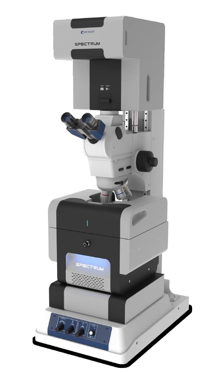

1 Raman KPFM AFM AFM-Raman characterization of PS-PVAC polymer blend film SPECTRUM The world s first fully automated Raman AFM AFM - confocal Raman - SNOM - TERS
.")

. HotSpot - Automatic location of active TERS* region on the probe.")


2 The first fully integrated & automated AFM Raman SNOM TERS system Simultaneous AFM and confocal Raman imaging using different objectives (up to 100x). Free rotation of microscope turret (up to 4 objectives) with AFM probe on the sample. Fully automated and easy to use operation. Automated removal of AFM probe (for low working distance objectives or when AFM probe is not required). HotSpot - Automatic location of active TERS* region on the probe. *Tip Enhanced Raman Scattering. XY motor and XYZ piezo-scanning stage Two simple steps in AFM & Raman / Fluorescence imaging Step 1 Sample survey (with cantilever removed). High resolution and wide field of view. Step 2 Simultaneous AFM and confocal Raman/ Fluorescence imaging of selected area. cantilever automatically moves to the working position and engages 2 Up to 4 objectives can be installed into the turret of the commercial optical microscope. All standard imaging modes are supported. Some AFM modes may require different cantilevers. Easy probe exchange and automated approach allows any advanced AFM mode (>30 modes) to be readily used together with Raman.
.")
 mode for advanced nano-mechanical and electrical properties imaging.")


3 Features Automation Laser/cantilever/photodiode system alignment. Automatic removal of AFM probe (when low working distance objectives are used or when AFM probe is not required). Sample movement. Probe approach & retraction. Scanning parameters adjustment. Unique SPM capabilities Low noise. Sample scanning with resolution down to atomic. Large sample size (up to 50mm 50mm). Special sample holder for slides (75mm 25mm). AFM, STM and tuning fork operation; measurements in liquid. More than 30 advanced SPM modes supported. HybriD (HD) mode for advanced nano-mechanical and electrical properties imaging. Tuning fork tip holder AFM tip holder STM tip holder Holder for operation in liquid Unique integration of SPM with optics for AFM - Raman - SNOM - TERS Upright or Full Transmission configuration. Professional upright microscope with 4 position revolving turret. Various objectives and imaging modes supported. Free rotation of objective turret with AFM probe on the sample. Scanning by laser spot. This option is provided by a very stable scanning mirror with closed loop capacitance sensors. It allows to position the laser spot with a high precision on the tip apex. Both fiber based and free space optical coupling to Raman system is available. Fiber based coupling provides compact and flexible system arrangement. Free space coupling guarantees highest sensitivity. Complete integration is available with NT-MDT, Renishaw and Thermo Scientific Raman microscopes. Scanning Near-field Optical Microscopy. Aperture and apertureless SNOM modes are supported using cantilever or fiber probes. Accessories Temperature control. Heating up to 150 C. Different, easy exchangeable sample holders. Probe exchange accessory. Heating stage Different sample holders
. Laser based cantilever deflection system (for cantilever AFM).")
. SNOM unit (optional).")
4 System Design 1. Specially designed confocal unit equipped with scanning mirror and bright field imaging system Mitutoyo Upright microscope. 4 position turret with different objectives. SPM head with different probe holders (cantilever AFM, STM, tuning fork,liquid). Laser based cantilever deflection system (for cantilever AFM). SPM base: Piezo Scan Stage (100m 100m 10m). Motorized Stage (35mm 35mm). Manual positioner of SPM head (3mm 3mm). Motorized and Piezo drives for objective focusing (optional). Heating stage (optional). SNOM unit (optional) Different optical configurations Upright, with scanning mirror (Standard configuration) Dual Scanning system (3 independent closed-loop scanning axes by sample + 2 by laser spot). Designed for nontransparent samples. Optical resolution down to 400nm simultaneously with AFM. Excitation laser focusing and signal collection are performed by high numerical aperture objective simultaneously with AFM. Laser scanning for automatic location of active TERS region on the probe. SNOM unit for bottom illumination/ collection (Optional) Special unit for fiber input/output, and motorized drive for bottom objective. Objectives with different magnifications can be used. 2 modes: Transmission - detection with PMT of the signal collected with the bottom objective. Collection - excitation with laser from the bottom, collection of the signal with cantilever aperture and registration with the spectrometer detectors.


5 MultiScan 4x 40 µm Multiscan AFM Topography 10x Sample survey with high resolution. Automated high resolution AFM - Raman imaging without limitations of the piezo-scanner range. 40 µm Multiscan Raman map, Si peak intensity 1. Choosing area on the sample (any size, no limitations of the scanner). 2. Automated AFM probe approach. 3. Simultaneous measurement of AFM and confocal Raman/fluorescence maps from multiple areas (automated). 4. Image stitching (automated). 40 µm Multiscan Raman map, Si peak mass center 40 µm Multiscan High resolution bright field image
.")


6 Automation Automated sample positioning (35mm 35mm)* High precision positioning motors equipped with optical sensors allow automated AFM-Raman imaging of any sample areas (within 35mm 35mm travel range). Cantilever deflection system auto alignment It only takes a few seconds to get the laser based cantilever deflection system aligned automatically. Special algorithms provide high speed laser alignment and optimized photodiode positioning. Fully motorized approach by tip Preliminary approach is done by high precision motors. Soft approach by piezo. NT-MDT developed phase sensitive algorithm that guaranties gentle probe approach. Automated probe removal AFM probe is automatically removed - when low working distance objective has to be used or when AFM is not required. The probe exchange procedure became simpler. Special AFM tip holder design and motorized probe holder positioner makes it easy to exchange any probe (AFM, STM, SNOM). AFM holder removed AFM holder in a working position * The movement range is automatically exchanged to 5mm 5mm when the bottom objective is used.

, where")


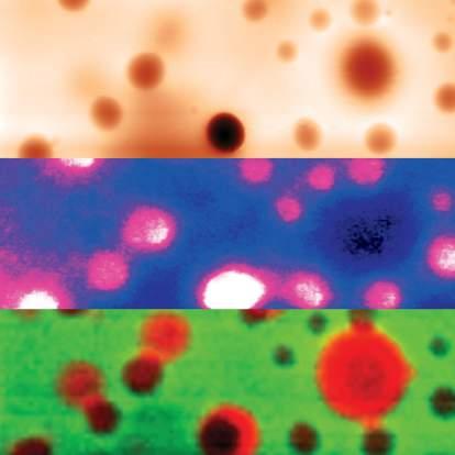
7 Applications AFM for nanomechanical and Raman for Chemical analysis HybriD Mode TM (HD-AFM TM ) is a non-resonant oscillatory mode of atomic force microscopy (AFM), where the tip-sample distance is modulated by use of a quasi-harmonic signal. The tip periodically gets in and out of contact with the sample thousand times per second. Real-time analysis of measured force curves provides the opportunity to extract a wide spectrum of morphological, mechanical and electromagnetic properties of the sample in each scanning point with high spatial resolution. Polymer blend. Scan size: 30x30µm Topography Adhesion F D 3 3 Force Set-point deflection E Modulus Distance E Modulus Residual deformation 1 5 Baseline 2 Adhesion 4 t Adhesion Baseline D Stiffness Overlay of fluorescence (green) and Raman (red) maps HybriD mode principle Nanowires CdS nanowires with polymers Mo oxide nanowires 4µm 4µm 4µm 5µm 5µm AFM Fluorescence map Raman map overlay, CdS (red) and PANI (green) AFM Raman map, Mo oxide peak Graphene Artificial diamond characterization 5µm 5µm 5µm 2µm 2µm AFM Raman map, 2D-band Raman map, G-band AFM Raman map cm -1 band
8 Technical Specification Measuring heads AFM,STM and Tuning Fork Laser based cantilever deflection system, with automated adjustment and targeting Sample Dimensions: up to 50/10mm in diameter/height Sample weight: up to 2kg Heating: from RT to 150 Scanning system Scanning type: by sample Range: 100m 100m 10m (CL) Resolution Noise XY: no more 0.3nm (with closed loop sensors) Noise Z (RMS, Hz bandwith): 0.06nm (typical) Sample positioning system Movement: automated, binded with the videomicroscope Range XY: 35mm 35mm; 5mm 5mm while using with the bottom objective Min. step: 0.35m Approach Automated approach by tip Range: 10mm Optical parts Spectral range*: nm Spectral resolution: down to 0.1 cm -1 (depends on spectrometer and grating used) Objectives: 100x 0.7NA 10x 0.28NA Any other objective (optional) Detectors**: TE cooled (down to -100 C) CCD / EMCCD camera. Photon multiplier (PMT) or avalanche photodiode (APD) in photon counting mode or other type of CCD camera (UV, IR) Photon multiplier for fast confocal laser (Rayleigh) imaging Confocal maps resolution: with the probe, for blue laser with 100x 0.7NA XY: <400nm Z: <800nm without the probe, for blue laser with 100x 1.4NA XY: <200nm Z: <500nm Bottom objective (optional) Movement: motor and piezo (optional) Range: 25mm by motor, 100m by piezo Min. step: 0.1m by motor, 3nm by piezo * Extended spectral ranges (UV, IR) are available as option. ** Depends on the system configuration. SLSP BR
FLEX 2 NEW. Key points. 3D Confocal Raman, 2 lasers, fiber based, AFM combined
 FLEX 2 3D Confocal Raman, 2 lasers, fiber based, AFM combined NEW Key points Compact size 2 lasers, easily switchable 2 confocal operation modes, easily switchable : High Spatial Resolution 35 nm High
MonoVista CRS+ Raman Microscopes
 MonoVista CRS+ Benefits Deep UV to NIR wavelength range Up to 4 integrated multi-line lasers plus port for large external lasers Dual beam path for UV and VIS/NIR Motorized Laser selection Auto Alignment
Certus Standard. NanoScanTechnology. Basic Datasheet. reasoned innovations. Basic Configuration of Scanning Probe Microscope
 NanoScanTechnology reasoned innovations Nano Scan Technology Ltd. Russia, 141700, Dolgoprudny, Zavodskaya St, 7 Phone: +7 (495) 642-40-68 +7 (495) 642-40-67 Skype: NanoScanTech E-mail: info@nanoscantech.ru
Certus Light. NanoScanTechnology. Basic Datasheet. reasoned innovations. Entry Level Scanning Probe Microscope. Scanning Probe Microscope
 NanoScanTechnology reasoned innovations Nano Scan Technology Ltd. Russia, 141700, Dolgoprudny, Zavodskaya St, 7 Phone: +7 (495) 642-40-68 +7 (495) 642-40-67 Skype: NanoScanTech E-mail: info@nanoscantech.ru
MultiView 2000 TM. The First Tip and Sample Scanning Probe Microscope. The Next Evolution in SPM. The Next Evolution in SPM
 MultiView 2000 TM The First Tip and Sample Scanning Probe Microscope MultiView 2000 TM Using Two Award Winning Nanonics 3D FlatScan Stages MultiView 2000TM Top-View (Top) and open position (Bottom). The
Confocal Raman Imaging with WITec Sensitivity - Resolution - Speed. Always - Provable - Routinely
 Confocal Raman Imaging with WITec Sensitivity - Resolution - Speed Always - Provable - Routinely WITec GmbH, Ulm, Germany, info@witec.de, www.witec.de A modular microscope series An Example: FLIM optical
Certus Optic. NanoScanTechnology. Basic Datasheet. reasoned innovations. Integrated Optical and Scanning Probe Microscope
 NanoScanTechnology reasoned innovations Nano Scan Technology Ltd. Russia, 141700, Dolgoprudny, Zavodskaya St, 7 Phone: +7 (495) 642-40-68 +7 (495) 642-40-67 Skype: NanoScanTech E-mail: info@nanoscantech.ru
NTEGRA SPECTRA. AFM - Raman - SNOM - TERS
 AFM - Raman - SNOM - TERS AFM Topography Surface Potential Raman map, G-band Graphene flakes on gold, 30 x 30 μm NTEGRA SPECTRA Atomic Force Microscopy Confocal Raman / Fluorescence Microscopy Scanning
Confocal Raman Microscope RAMOS
 Confocal Raman Microscope RAMOS 1 future`s Confocal Raman Microscope RAMOS 2 Confocal Raman Microscope RAMOS Ostec Corporate Group produces and offers hi-tech innovative scientific and analytical equipment.
Sample Sizes: up to 1 X1 X 1/4. Scanners: 50 X 50 X 17 microns and 15 X 15 X 7 microns
 R-AFM100 For Nanotechnology Researchers Wanting to do routine scanning of nano-structures Instrument Innovators Using AFM as a platform to create a new instrument Educators Teaching students about AFM
Introduction to. 3D Scanning Confocal Microscope with Spectrometer
 Introduction to Nanofinder-S 3D Scanning Confocal Microscope with Spectrometer Alexei Kuzmin E-mail: a.kuzmin@cfi.lu.lv Principle of Confocal Microscopy Laser X-Y Excitation Pinhole Excitation Filter Objective
Versatile laser Raman Spectrometer. RMP-500 series
 Versatile laser Raman Spectrometer RMP-500 series About RMP-500 RMP-500 series is a compact and versatile laser Raman spectrometer consisting of a micro Raman probe connected through fiber optics to the
MultiView Tomorrow s Systems. for Today s Challenges. Benefits of the Multiprobe System
 MultiView 4000 Tomorrow s Systems for Today s Challenges The integration of multiple probes in scanning probe microscopy (SPM) has been a dream since its earliest days of development. Nano-structure research
The Anfatec Level AFM a short description. Atomic Force Microscopy - approved devices for affordable prices
 The Anfatec Level AFM a short description Atomic Force Microscopy - approved devices for affordable prices Our system is complete for almost all typical applications. It provides all basic modes as: contact
Confocal Raman Systems SPECTROSCOPY GROUP
 Confocal Raman Systems SPECTROSCOPY GROUP MonoVista CRS Configuration Option PIXIS TE-Cooled CCD Camera Acton Spectrograph Micro-Raman Interface Witness Camera Optional Laser Micro/Macro Transfer Optics
Depth analysis for laminated film by confocal Raman mapping
 Newest modular 3D Imaging Raman Microspectroscopy System Simple operation and low cost with all the basic features of our top of the line system. Structural images of transparent samples (plastic, film,
Thermo Scientific DXR2 Raman Family
 MOLECULAR SPECTROSCOPY Thermo Scientific DXR2 Raman Family Focus on answers, not the technique Product Specifications Easily adapt to any sample challenge using the Thermo Scientific DXR 2 family of Raman
Renishaw invia Raman Microscope (April 2006)
") Renishaw invia Raman Microscope (April 2006) I. Starting the System 1. The main system unit is ON all the time. 2. Switch on the Leica microscope and light source for reflective bright field (BF) imaging.
Renishaw invia Raman Microscope (April 2006) I. Starting the System 1. The main system unit is ON all the time. 2. Switch on the Leica microscope and light source for reflective bright field (BF) imaging.
Modular Approach and Customized solutions (near field, UV-VIS, etc.)
") Compact & Flexible System Configuration High Resolution: < 0.3cm Measurement down to 10cm Confocal Optics for Microscope and Remote Probe Fully Automated 2D, 3D & 4D Raman Imaging Attachable to AFM, XRD,
Compact & Flexible System Configuration High Resolution: < 0.3cm Measurement down to 10cm Confocal Optics for Microscope and Remote Probe Fully Automated 2D, 3D & 4D Raman Imaging Attachable to AFM, XRD,
WITec Raman Spectroscopy Solutions
 Confocal Raman Imaging WITec Raman Spectroscopy Solutions -40-20 0 20 CCD cts 500 1000 1500 2000 2500 3000 3000 relative wavenumbers (cm -1 ) www.witec.de WITec UHTS Ultra-High Throughput Spectrometers
Leica TCS STED. The Fast Track to Superresolution Technical Documentation
 Leica TCS STED The Fast Track to Superresolution Technical Documentation 8 9 6 7 Inverted research microscope Leica DMI6000 CS Scan head Laser and power supply Computer table Air damped optical table 6
Chemical Characterization of Diverse Pharmaceutical Samples by Confocal Raman Microscopy
 Whitepaper Chemical Characterization of Diverse Pharmaceutical Samples by Confocal Raman Microscopy WITec GmbH, Lise-Meitner-Str. 6, 89081 Ulm, Germany, www.witec.de Introduction The development and production
PInano 1x3 XYZ & XY Piezo Stage Systems
 New: Large Aperture for Slides, Petri Dishes, Heaters & Specimen Holders PInano 1x3 XYZ & XY Piezo Stage Systems Low-Profile, Low-Cost, Nanopositioning Systems for Super-Resolution Microscopy PInano series
Operating Procedure for Horiba Raman Microscope
 Operating Procedure for Horiba Raman Microscope SAFETY Be aware of Laser radiation at all times! Do not remove the covers of the instrument. Components are supplied with 110V electric source. Do not touch
Modular Raman Spectrometers
 Modular Raman Flexible Raman from the Raman Experts horiba.com/scientific Flexible and Affordable Raman The new range of modular Raman spectrometers from HORIBA Scientific allows the user to have a flexible
Attachable to other Advanced analytical tools (e.g AFM, XRD, SEM etc.) Detect and measure deposits in liquid as it is (Particle ID Detection)
 Detect and measure deposits in liquid as it is (Particle ID Detection)") Compact & Flexible System Configuration High Resolution: < 0.3cm Measurement down to 10cm Confocal Optics for Microscope and Remote Probe Fully Automated 2D, 3D & 4D Raman Imaging Attachable to other Advanced
Compact & Flexible System Configuration High Resolution: < 0.3cm Measurement down to 10cm Confocal Optics for Microscope and Remote Probe Fully Automated 2D, 3D & 4D Raman Imaging Attachable to other Advanced
ihr Series horiba.com/osd Research Grade Spectrometers Simply the best imaging spectrometers with no compromise
 ihr Series Research Grade Spectrometers Simply the best imaging spectrometers with no compromise horiba.com/osd Unmatched Flexibility in Applications HORIBA Scientific s Optical Spectroscopy Division
AUTOFOCUS SENSORS & MICROSCOPY AUTOMATION IR LASER SCANNING CONFOCAL MICROSCOPE IRLC DEEP SEE. Now See Deeper than ever before
 AUTOFOCUS SENSORS & MICROSCOPY AUTOMATION IR LASER SCANNING CONFOCAL MICROSCOPE IRLC DEEP SEE Now See Deeper than ever before Review and inspection of non visible subsurface defects Non visible and subsurface
Lecture: P1_Wk3_L5 Contact Mode Scans. Ron Reifenberger Birck Nanotechnology Center Purdue University 2012
 Lecture: Contact Mode Scans Ron Reifenberger Birck Nanotechnology Center Purdue University 2012 1 The Purpose of a Microscope is to Obtain an Image Reflected laser spot Laser Diode Four-Quadrant Photodetector
Characterization of MEMS Devices
 MEMS: Characterization Characterization of MEMS Devices Prasanna S. Gandhi Assistant Professor, Department of Mechanical Engineering, Indian Institute of Technology, Bombay, Recap Fabrication of MEMS Conventional
TissueFAXS SL Confocal high throughput configuration (actual appearance of the product may differ)
") TISSUEFAXS CONFOCAL TissueFAXS SL Confocal high throughput configuration (actual appearance of the product may differ) TissueFAXS Confocal provides a unique combination of digital slide scanning and laser
TISSUEFAXS CONFOCAL TissueFAXS SL Confocal high throughput configuration (actual appearance of the product may differ) TissueFAXS Confocal provides a unique combination of digital slide scanning and laser
Solver PRO-M Scanning Probe Microscope. SPM Controller
 Solver PRO-M Scanning Probe Microscope SPM Controller (models BL022MTM, BL022MRM) Reference Manual June, 2006 Copyright NT-MDT, 2006 Web Page: http://www.ntmdt.com General Information: spm@ntmdt.ru Technical
Confocal Microscopy Imaging of Single Emitter Fluorescence and Hanbury Brown, and Twiss Setup for Photon Antibunching. Abstract
 James Maslek 10/26/12 Confocal Microscopy Imaging of Single Emitter Fluorescence and Hanbury Brown, and Twiss Setup for Photon Antibunching Abstract The purpose of this experiment was to observe fluorescence
M-545 Manual XY Microscope Stage For Olympus, Nikon, Leica & Zeiss Microscopes / PI Piezo Stages
 M-545 Manual XY Microscope Stage For Olympus, Nikon, Leica & Zeiss Microscopes / PI Piezo Stages M-545 manual microscopy stages are designed to accommodate the P-545 PInano series of XY / XYZ Piezo stag
Microscopy. Marc McGuigan North Quincy High School Thursday, May 11, 2006
 Microscopy Marc McGuigan North Quincy High School Thursday, May 11, 006 Outline Activity Introduction Electromagnetic Spectrum Visible Light Light Microscope AFM Scanning Electron Microscopy Near-Field
TABLE OF CONTENTS PRODUCT DESCRIPTION LASERDEC CL200 / CL200HP TECHNICAL DATA DIMENSIONS LASERDEC CL500 / CL500HP TECHNICAL DATA DIMENSIONS
 TABLE OF CONTENTS PRODUCT DESCRIPTION LASERDEC CL200 / CL200HP TECHNICAL DATA DIMENSIONS LASERDEC CL500 / CL500HP TECHNICAL DATA DIMENSIONS ACCESSORIES ATTENUATION UNIT 0 ATTENUATION UNIT 90 BEAM REDUCER
Imaging Spectrometers
 JOBIN YVON Imaging Spectrometers ihr Series Uniquely shaped for uniquely superior performance. ihr Series Imaging Spectrometers A Unique Shape for a Unique Spectrometer The difference between ihr spectrometers
SYNCERITY TM 1024 x 256
 ELEMENTAL ANALYSIS FLUORESCENCE GRATINGS & OEM SPECTROMETERS OPTICAL COMPONENTS PARTICLE CHARACTERIZATION RAMAN SPECTROSCOPIC ELLIPSOMETRY SPR IMAGING SYNCERITY TM 1024 x 256 Open-Electrode TE-Cooled CCD
TRiCAM APPLICATIONS KEY FEATURES. Time Resolved intensified CAMera. TRiCAM 13001A01 31/10/2013
 TRiCAM Time Resolved intensified CAMera The TRiCAM is a compact Intensified CCD camera for scientific and industrial applications that require 1) lowlight level imaging, 2) ultra-short exposures through
LabSpec 6. Spectroscopy Suite. Simply Powerful Software
 LabSpec 6 Spectroscopy Suite Simply Powerful Software The LabSpec 6 spectroscopy suite offers an intuitive platform for Raman, photoluminescence (PL) and cathodoluminescence (CL) spectroscopy. The sixth
1. Motivation 2. Nanopositioning and Nanomeasuring Machine 3. Multi-Sensor Approach 4. Conclusion and Outlook
 Prospects of multi-sensor technology for large-area applications in micro- and nanometrology 08/21/2011-08/25/2011, National Harbor E. Manske 1, G. Jäger 1, T. Hausotte 2 1 Ilmenau University of Technology,
Optical properties and characterization
 Optical properties and characterization Name Picture Description Site Responsible 1 Laser Nd:YAG MAPLE (Matrix Assisted Pulsed Laser Evaporation) system for biomaterials and polymeric thin film deposition
Series Spectrometers PARTICLE CHARACTERIZATION ELEMENTAL ANALYSIS FLUORESCENCE GRATINGS & OEM SPECTROMETERS OPTICAL COMPONENTS RAMAN
 Series Spectrometers ELEMENTAL ANALYSIS FLUORESCENCE GRATINGS & OEM SPECTROMETERS OPTICAL COMPONENTS PARTICLE CHARACTERIZATION RAMAN SPECTROSCOPIC ELLIPSOMETRY SPR IMAGING ihr Series Imaging Spectrometers
M-545 XY Microscope Stage with Ultrasonic Linear Drives High Stability, Low Profile, High Speed, Direct Position Measurement
 M-545 XY Microscope Stage with Ultrasonic Linear Drives High Stability, Low Profile, High Speed, Direct Position Measurement The M-545.2U microscope stage with closed-loop ultrasonic piezo motors provides
Leica TCS SPE. Spectacular Imaging! Technical Documentation
 Leica TCS SPE Spectacular Imaging! Technical Documentation Leica TCS SPE Spectacular Imaging Easy to Achieve A Reliable System Affordable Excellence The high resolution spectral confocal Leica TCS SPE
Confocal Microscope Imaging of Single-Emitter Fluorescence and Hanbury Brown & Twiss Setup for Photon Antibunching. Edward Pei
 Confocal Microscope Imaging of Single-Emitter Fluorescence and Hanbury Brown & Twiss Setup for Photon Antibunching Edward Pei Abstract The purpose of these labs was to study single photon sources and measure
WITec Suite FIVE. Project FIVE Control FIVE Project FIVE+
 Project FIVE Control FIVE Project FIVE+ For further information about WITec Suite please contact us: by phone: +49 (0) 731 140700 by email: info@witec.de WITec Suite FIVE Data Acquisition, Evaluation and
Chemical Characterization of Pharmaceutical Samples by Confocal Raman Microscopy and Correlative Techniques
 APPLICATION NOTE Chemical Characterization of Pharmaceutical Samples by Confocal Raman Microscopy and Correlative Techniques WITec GmbH, Lise-Meitner-Str. 6, 89081 Ulm, Germany fon +49 (0) 731 140 700,
SCALA. Scanning Laser Analyzer
 SCALA Scanning Laser Analyzer SCALA TECHNOLOGY Mecwins offers an optical platform for MEMS characterization using its proprietary technology. SCALA is a complete tool to characterize your devices (cantilevers,
TABLE OF CONTENTS PRODUCT DESCRIPTION CINCAM CCD TECHNICAL DATA SENSOR RESPONSE DIMENSIONS CINCAM CCD LARGE FORMAT TECHNICAL DATA SENSOR RESPONSE
 TABLE OF CONTENTS PRODUCT DESCRIPTION CINCAM CCD TECHNICAL DATA SENSOR RESPONSE DIMENSIONS CINCAM CCD LARGE FORMAT TECHNICAL DATA SENSOR RESPONSE DIMENSIONS CINCAM CMOS TECHNICAL DATA SENSOR RESPONSE DIMENSIONS
Indian Institute of Technology Kanpur Centre for Nanosciences
 Tender Enquiry No.: CNS/2014 15/JUN/01 Date: June 04, 2015 Last Date for submission: June 19, 2015 ATOMIC FORCE MICROSCOPE Sealed Quotations are invited in two bid system (technical and financial bid separately
LIFA KEY FEATURES APPLICATIONS. Fluorescence Lifetime Attachment LIFA 15001A02 16/03/2015
 LIFA Fluorescence Lifetime Attachment LIFA 151A2 16/3/215 The LIFA is a dedicated system for Fluorescence Lifetime Imaging Microscopy (FLIM). It allows the generation of lifetime images on any widefield
3D ATOMIC FORCE MICROSCOPY OF HIGH ASPECT RATIO STRUCTURES. R.W. Herfst
 3D ATOMIC FORCE MICROSCOPY OF HIGH ASPECT RATIO STRUCTURES R.W. Herfst CONTENTS Introduction: why 3D AFM measurements Bottlenecks in AFM based 3D metrology for the semiconductor industry TNO approach to
Spectrograph overview:
 High performance measurement systems Monochromator Family Gilden Photonics offers a range of integrated optical wavelength solutions in customized designs, OEM design, manufacturing and value added resell
Positioning system of a metrological AFM: design considerations
 Positioning system of a metrological AFM: design considerations AFM workshop LNE, Trappes Jan Piot K.U.Leuven Division PMA Overview Introduction General layout metrological AFM Layout of the positioning
Optical Trapping in the Teaching Lab
 BE.309: Biological Instrumentation and Measurement Laboratory GEM4 Summer School Optical Trapping in the Teaching Lab 1 Lab Objective 1. Become familiar with the fundamentals of optical trapping. 2. Learn
WAVELENGTH MANAGEMENT
 Camera Accessories WAVELENGTH MANAGEMENT UV CONVERTERS UV Converters take advantage of a phenomenon called fluorescence to extend the performance range of the Beamage beam profiling camera to ultraviolet
Applications of Piezo Actuators for Space Instrument Optical Alignment
 Year 4 University of Birmingham Presentation Applications of Piezo Actuators for Space Instrument Optical Alignment Michelle Louise Antonik 520689 Supervisor: Prof. B. Swinyard Outline of Presentation
Silicon Avalanche Photodiodes in Dynamic Light Scattering
 Silicon Avalanche Photodiodes in Dynamic Light Scattering August 2016 Introduction This application note describes the use of the ID100 single photon counting detector for the measurement of light scattered
Mu lt i s p e c t r a l
 Viewing Angle Analyser Revolutionary system for full spectral and polarization measurement in the entire viewing angle EZContrastMS80 & EZContrastMS88 ADVANCED LIGHT ANALYSIS by Field iris Fourier plane
NanoLens AFM and Bruker 3D Microscopes Integrated 1000X Inspection Combines for Maximum Metrology Value
 NanoLens AFM and Bruker 3D Microscopes Integrated 1000X Inspection Combines for Maximum Metrology Value Outline Introduction/Administrative Overview of Bruker 3D Optical Microscopes Software, Automation
Lightsheet Z.1. Light Sheet Fluorescence Microscopy by Carl Zeiss. Fabrice Schmitt, Sales Manager Carl ZEISS France
 Lightsheet Z.1 Light Sheet Fluorescence Microscopy by Carl Zeiss Fabrice Schmitt, Sales Manager Carl ZEISS France 12.12.2012 Light Sheet Fluorescence Microscopy (LSFM) Principle The Principle of Light
Building Your Own 2-Photon Microscope: Challenges, Advantages and Limitations
 Building Your Own 2-Photon : Challenges, Advantages and Limitations Roberto Weigert, Ph.D. Intracellular Membrane Trafficking Unit Oral and Pharyngeal Cancer Branch NIDCR-NIH Building Your Own 2-Photon
RAMM Microscope Configuration Guide
 RAMM Microscope Configuration Guide ASI s RAMM frame microscopes are modular open frame alternative to conventional commercial microscopes. Using a RAMM frame may be especially appropriate for instruments
CSM Technical Features //// ULTRA NANOINDENTATION TESTER (UNHT)
") CSM Technical Features //// ULTRA NANOINDENTATION TESTER (UNHT) //// Table of contents //// Table of contents... 2 //// Introduction... 4 //// Key Features... 5 > ULTRA HIGH RESOLUTION AND VERY LOW NOISE
CSM Technical Features //// ULTRA NANOINDENTATION TESTER (UNHT) //// Table of contents //// Table of contents... 2 //// Introduction... 4 //// Key Features... 5 > ULTRA HIGH RESOLUTION AND VERY LOW NOISE
Sensor based adaptive laser micromachining using ultrashort pulse lasers for zero-failure manufacturing
 Sensor based adaptive laser micromachining using ultrashort pulse lasers for zero-failure manufacturing Fraunhofer Institute for Production Technology, Aachen M. Sc. Guilherme Mallmann Prof. Dr.-Ing. Robert
M-545 XY Microscope Stage with Ultrasonic Linear Drives High Stability, Low Profile, High Speed, Direct Position Measurement
 M-545 XY Microscope Stage with Ultrasonic Linear Drives High Stability, Low Profile, High Speed, Direct Position Measurement The M-545.2P microscope stage with closed-loop ultrasonic piezo motors provides
Multi-Photon Training
 Multi-Photon Training Overview This training will take approximately 4 hours. The first 2 hours will be spent learning one-on-one with your trainer, while the following 2 hours will be your opportunity
Optical Sectioning. Bo Huang. Pharmaceutical Chemistry
 Optical Sectioning Bo Huang Pharmaceutical Chemistry Approaches to 3D imaging Physical cutting Technical difficulty Highest resolution Highest sensitivity Optical sectioning Simple sample prep. No physical
Indiana Center for Biological Microscopy. BioRad MRC 1024 MP Confocal & Multi-Photon Microscope
 Indiana Center for Biological Microscopy BioRad MRC 1024 MP Confocal & Multi-Photon Microscope Microscope and the Attached Accessories A: B: C: D: E: F: G: H: Mercury Lamp Transmission Light Kr/Ar Laser
Voyage Confocal Raman Microscope. Raman Solution. True Confocality and True Flexibility. Features:
 Raman Solution MADAtec Srl ITALY WWW.MADATEC.COM Tel. +39 02-36542401 e-mail: sales@madatec.com Voyage Confocal Raman Microscope True Confocality and True Flexibility Features: Optimized True Confocal
Back Illuminated Scientific CMOS
 Prime 95B Scientific CMOS Camera Datasheet CMOS, EMCCD AND CCD CAMERAS FOR LIFE SCIENCES Back Illuminated Scientific CMOS Discovery depends on every photon Primary applications: Super-Resolution Microscopy
Piezo Mechanisms in Optics and Laser Technology
 Piezo Mechanisms in Optics and Laser Technology By Stefan Vorndran, Scott Jordan, Steffen Arnold, PI (Physik Instrumente) Laser technology is crucial for the advancement of many high tech fields from aerospace
WinCamD-LCM 1" CMOS Beam Profiling Camera, SuperSpeed USB 3.0, * nm * model-dependent
 Datasheet WinCamD-LCM 1" CMOS Beam Profiling Camera, SuperSpeed USB 3.0, 190 1610* nm * model-dependent With an 11.3 x 11.3 mm active area, 4.2 Mpixels, 5.5 x 5.5 μm pixels, optical and electronic triggering
LASERS FOR ANALYTICAL INSTRUMENTATION
 LASERS FOR ANALYTICAL INSTRUMENTATION CW Lasers (Broad and Narrow Spectrum) Nanosecond SLM Lasers Accessories ADVANTAGES One size One control interface One voltage (+5 VDC) One software Many wavelengths
LASERS FOR ANALYTICAL INSTRUMENTATION
 LASERS FOR ANALYTICAL INSTRUMENTATION CW Lasers (Broad and Narrow Spectrum) Nanosecond SLM Lasers Accessories ADVANTAGES One size One control interface One voltage (+5 VDC) One software Many wavelengths
Near Field Observation of a Refractive Index Grating and a Topographical Grating by an Optically Trapped Gold Particle
 Near Field Observation of a Refractive Index Grating and a Topographical Grating by an Optically Trapped Gold Particle Hiroo UKITA and Hirotaka UEMI Ritsumeikan University, Kusatsu-shi, Shiga, 2 Japan
TRIBOMETERS MICRO-MACRO TRIBOLOGY TESTING
 TRIBOMETERS MICRO-MACRO TRIBOLOGY TESTING The Tribometer provides highly accurate and repeatable wear friction testing in rotative and linear modes compliant to ISO and ASTM standards. Designed, at the
STANDARD SERIES MONOCHROMATOS FEATURES. Highly Customizable Modular Design. Two Configurable Input and Output Ports
 STANDARD SERIES MONOCHROMATOS FEATURES Highly Customizable Modular Design Two Configurable Input and Output Ports Configurable turret and Grating Options USB2.0 Communication A Full Line of Input and Output
Single Photon Counting Module
 Description Laser Components COUNT series of s has been developed to offer a unique combination of high quantum efficiency, wide dynamic range and ease of use for photon counting applications. Combining
FST s status on EUV Pellicle & Inspection System Development
 FST s status on EUV Pellicle & Inspection System Development OCT.04, 2015 EUV Pellicle TWG @ Imec, nl. Donwon Park FST (Korea) http://www.fstc.co.kr FST Business Segments Division Pellicle TCU (Temperature
4D IMAGING AT YOUR FINGERTIPS Real-time, Portable, High-Resolution Solutions for your Quality Control Needs
 STDO Dynamic 3D 4D Imaging Microscopy Instrument Systems 4D IMAGING AT YOUR FINGERTIPS Real-time, Portable, High-Resolution Solutions for your Quality Control Needs STDO-HOLO Overview: STDO-HOLO enables
Development of AFM Based on Nano Positioning Stage Niandong Jiao 1,2,*, Yuechao Wang 1, Ning Xi 1,3, and Zaili Dong 1
 Development of AFM Based on Nano Positioning Stage Niandong Jiao 1,2,*, Yuechao Wang 1, Ning Xi 1,3, and Zaili Dong 1 1 Shenyang Institute of Automation, Chinese Academy of Sciences, China 2 Graduate School
LaserDec CL - Product Description -
 CO 2 LASER BEAM PROFILER LaserDec CL - Product Description - The high performance LaserDec system is based on industry s unique imaging technique. It is designed for monitoring high-power CO 2 -lasers
Technology and equipment at the Bioimaging Center
 Technology and equipment at the Bioimaging Center Light microscopy / Widefields Name Type Applications and equipment LEICA AF6000LX Nikon BioStation Timelaps imaging, CO2 and temperature controlled (14
Work Instructions for Confocal Laser Scanning Microscopy
 Contents 0.1 To be Verified Before Switch ON......................... 2 0.2 Switch ON Protocol-Single Photon Mode Confocal Laser Scanning Microscope 2 0.3 Switch ON Protocol - Multi Photon Mode Confocal
Single Photon Counting Module COUNT blue -Series
 Single Photon Counting Module COUNT blue -Series Description Laser Components COUNT blue series of Single Photon Counting Modules has been developed to offer a unique combination of high quantum efficiency,
Raman Sample Holders
 Raman Sample Holders Ocean Optics offers several sample holders for Raman analysis of liquids and solids, including a multipurpose holder that can be configured for fluorescence and other measurements.
University of Minnesota Nano Fabrication Center Standard Operating Procedure
 Equipment Name: University of Minnesota Nano Fabrication Center Coral Name: hs-scope Revision Number: 1.5 Model: HS200A Revisionist: M. Fisher Location: Bay 1 Date: 9/12/2013 1 Description The Hyphenated
Compact non-contact 3D surface profiler
 Compact non-contact 3D surface profiler Compact. Flexible. Powerful. S lynx is a new non-contact 3D surface profiler designed for use in industry and research. It has been designed as a compact and versatile
Enabling Optics on the Nanoscale
 Enabling Optics on the Nanoscale Comprehensive solutions for AFM and Raman spectroscopy, Tip-Enhanced Raman Spectroscopy (TERS), Aperture SNOM and Scattering-type SNOM (ssnom), Confocal microscopy, NanoManipulation
Introduction of the Industry's Broadest Tunable Quantum Cascade Lasers. February Block Engineering 1
 Introduction of the Industry's Broadest Tunable Quantum Cascade Lasers February 2014 2014 Block Engineering 1 Widely-Tunable QCL Products LaserTune Mini-QCL Smallest widely-tunable QCL source Embedded
ZEISS Smartproof 5 Your Integrated Widefield Confocal Microscope for Surface Analysis in Quality Assurance and Quality Control
 Product Information Version 1.0 ZEISS Smartproof 5 Your Integrated Widefield Confocal Microscope for Surface Analysis in Quality Assurance and Quality Control Dedicated Design. Guided Workflow. Trusted
Colocalization Module for MountainsMap
 Colocalization Module for MountainsMap Combine surface data from different instrument types - carry out correlative studies For 3D optical profilers & AFM, STM, SEM, fluorescence, Raman, IR & other microscopes
Prizmatix. Optical Specifications. Mic-LED-635 spectrum. BLCC-02 Benchtop LED Current Controller Specifications
 Mic-LED-635 High Power Collimated Red LED Light Source for Fluorescence Microscopy Featuring Prizmatix Modular Design for Multi-Wavelength and Fiberoptic Setup Ver. 01 Introduction The compact Mic-LED-635,
High-Accuracy LIBS with Nanosecond and Picosecond Time Resolution Enabled by Ultrasensitive emiccd Technology
 2015 Princeton Instruments, Inc. All rights reserved. High-Accuracy LIBS with Nanosecond and Picosecond Time Resolution Enabled by Ultrasensitive emiccd Technology The PI-MAX4:1024EMB emiccd camera seamlessly
R0 in µm THP in µm 18 C - 22 C
 ZEISS O-INSPECT Specifications Status: November 2017 System description Type according to ISO 10360-1:2000 O-INSPECT 3/2/2: Column CMM, O-INSPECT 5/4/3 and 8/6/3: Fixed bridge CMM Operating mode motorized
Vibrometer Introduction
 Introduction Hysen www.vibrometry.co.kr 5 OFV Series OFV-5000 Modular Controller VD/DD Decoder Guide-Line Velocity Decoder Decoder Description No.of Ranges Best Resolution Max.Velocity Upper Freq. Limit
ASI Photoport TIRF Injector Instruction Manual
 ASI Photoport TIRF Injector Instruction Manual Applied Scientific Instrumentation, Inc. 29391 W. Enid Rd. Eugene, OR 97402-9533 USA Phone: (800) 706-2284 (541) 461-8181 Fax: (541) 461-4018 Web: www.asiimaging.com
Optimization of integrated optic components by refractive index profile measurements
 EFOC 92 Optimization of integrated optic components by refractive index profile measurements by R. Göring, T. Possner, Fraunhofer Einrichtung für Angewandte Optik und Feinmechanik (Jena, D) Summary Refractive