TG 132: Use of Image Registration and Fusion in RT
|
|
|
- Tracey Blankenship
- 6 years ago
- Views:
Transcription

1 TG 132: Use of Image Registration and Fusion in RT Kristy K Brock, PhD, DABR, FAAPM Associate Professor Department of Radiation Oncology, University of Michigan Chair, AAPM TG 132: Image Registration and Fusion
2 Acknowledgements & Conflict of Interest AAPM Task Group 132 Members I have a licensing agreement for deformable image registration technology with RaySearch Laboratories.
3 Objectives 1. Describe how (deformable) image registration algorithms work? 2. Describe techniques to commission and validate image registration in the clinic (TG 132 recommendations) 3. Illustrate the concepts with clinical examples.
4 Techniques
5 How do they work? Match something Intensity, gradients, boundaries, features Constrain by a function Geometric, physical, biomechanical
6 How do they work? Match something Intensity, gradients, boundaries, features What happens when the intensity correspondence varies? What happens when the gradient isn t there? What happens when the boundaries aren t well defined? What happens with the features aren t visible? Constrain by a function Geometric, physical, biomechanical Can you rely on this model when the match above is missing?
 Contour matching Affine (Translation + Rotation + scaling + shearing) Mean Square Difference Spline (B-spline,")
 Mutual Information Quick, Easy, local Surface-based Manual or autosegmentation Great for 4D CT Modality Biomechanical Gradient")
7 Why? Many Image Registration Techniques Metric Transformation Optimization Your Eye Translation Brain-power Least Squares (Points) Translation + Rotation Simplex Chamfer Matching (surface matching) Contour matching Affine (Translation + Rotation + scaling + shearing) Mean Square Difference Spline (B-spline, Thin Good for same plate modality spline) (x-ray), different Correlation Coefficient contrast/noise Physical (CECT, (optical/fluid CT, CBCT) Works flow, for elastic Multi-body) Mutual Information Quick, Easy, local Surface-based Manual or autosegmentation Great for 4D CT Modality Biomechanical Gradient descent etc
8 Top 3 Similarity Metrics Sum of the Square Differences Mutual Information Contour Propagation

 2 CT 2 CT 1 CT 2")

9 Kessler / UM Sum of Squared Differences subtract one image from the other CT 2 - CT 1 = Difference Image I I (I -I ) 2 CT 2 CT 1 CT 2 CT 1 Individual Intensity Distributions Sum of the Squares of the Differences


10 Kessler / UM Sum of Squared Differences subtract one image from the other CT - MR = Difference Image I CT I MR Individual Intensity Distributions Not Zero This doesn t usually make much sense
11 Mutual Information Maximise the mutual information Marginal Entropies Joint Entropy Mutual Information, I(A,B) H(A) H(B) H(A,B) Sensitivity of results: Vary the vector field and evaluate the change in similarity metric Hub, et. al., IEEE TMI 2009
12 How Reliable is the Max MI? Actually, min -MI -MI -MI dx Min MI Best Solution dx Min MI Best Solution


13 Intensity Variation: Impact on CC/MSD Clear intensity variation No relevant intensity variation, noise/artifact

14 Contour Matching
15 Top 3 Regularizers Thin Plate Splines & B-Splines Weighted basis splines Flow/Optical Gradient driven with regularization Elastic/Biomechanical Material properties: compressibility and stiffness


 affine")
16 Kessler / UM Thin-Plate Splines Liver Bladder n å i=1 T(P) = a 0 + a x x + a y y + a z z + w i U(P - P i ) affine warping
17 Kessler / UM B-Splines Transformation is built up using a set of weighted basis splines DX weighted sum w 1 w 2 w 3 w 4 knots k 1 k 2 k 3 k 4 X X = X + DX = X + w i b(x-k i )

18 Kessler / UM B-Splines Transformation is built up using a set of weighted basis splines DX w 1 w 3 w 4 weighted sum w 2 knots k 1 k 2 k 3 k 4 X X = X + DX = X + w i b(x-k i )

19 Kessler / UM B-Splines Basis function has finite range X = X + DX = X + w i b(x-k i )
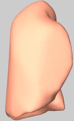
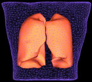
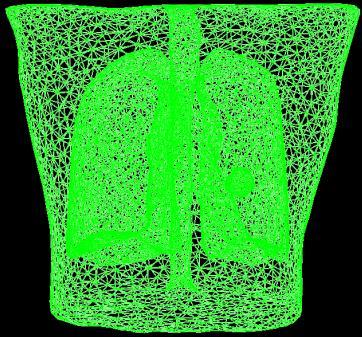
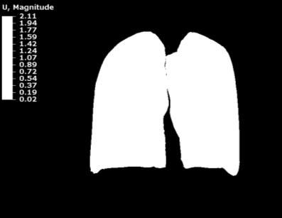
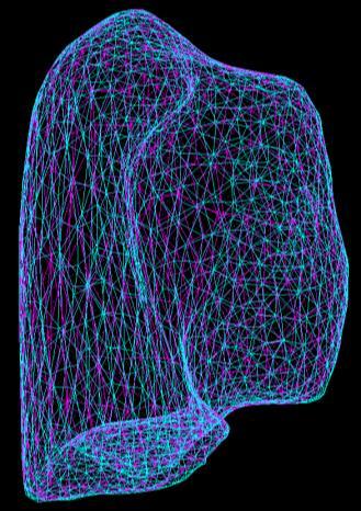
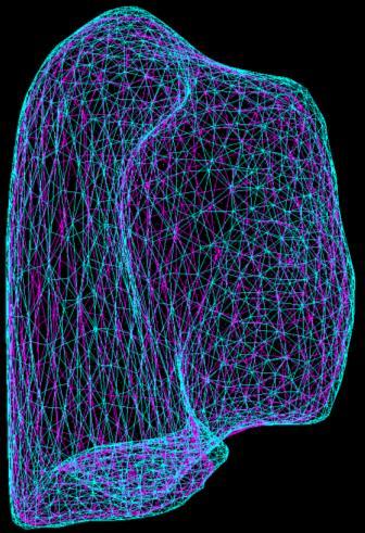
20 Biomechanical Model Inhale Image Surface Mesh Surface Exhale Image Surface Projection Contact Surface Parenchyma (Tetra elements) Bronchial Tree (Shell Elements) Boundary Conditions Finite Element Analysis
21 Commissioning and QA Preliminary recommendations from TG 132* *pending approval from Science Council
22 Clinical Recommendations (1/2) 1. Understand the basic image registration techniques and methods of visualizing image fusion 2. Understand the basic components of the registration algorithm used clinically to ensure its proper use 3. Perform end-to-end tests of imaging, registration, and planning/treatment systems if image registration is performed on a standalone system
23 Clinical Recommendations (2/2) 4. Perform comprehensive commissioning of image registration using the provided digital phantom data (or similar data) as well as clinical data from the user s institution 5. Develop a request and report system to ensure communication and documentation between all users of image registration 6. Establish a patient specific QA practice for efficient evaluation of image registration results
24 Commissioning and QA Understand the whole picture Understand fundamental components of algorithm
25 Understand the basic image registration techniques and methods of visualizing How? image fusion TG report has basic information and references AAPM Virtual Library Several books and review papers
26 Understand the basic components of the registration algorithm used clinically to ensure its proper use How? At minimum, the vendor should disclose: Similarity metric used Why do we need to know the Regularization used Transformation used Optimization method What knobs you can turn and what they do Read white papers implementation?


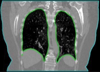
 Deformed")
 Slides")
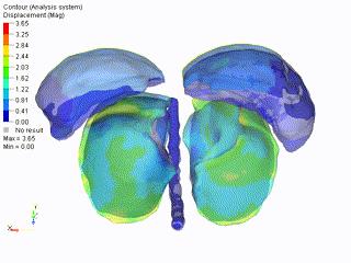
27 New method to validate Deformable Image Registration Deformable 3D Presage dosimeters Control (No Deformation) Deformed (27% Lateral Compression) Slides Courtesy of Mark Oldham and Shiva Das

28 Dosimeter & Deformable Registration-based Dose Accumulation: Dose Distributions Deformed Dosimeter Field Shape Differences DVF-based Accumulation Caution must be used when Field Displacements accumulating dose, especially in regions of the image with homogeneous intensity. Horizontal (Compression Axis) 40% narrower to 175% wider Vertical 33% shorter to 50% taller Slides Courtesy of Mark Oldham and Shiva Das


:")
29 Different DIR Algorithms have Different Strengths and Weaknesses Distribution Coronal Axial Sagittal 3D γ 3%/3mm Measured, Optical CT 96% 1 (control) DIR-predicted, Intensity-based DIR 60% 1 DIR-predicted, Biomechanical Surface projection 91% 2 1. Juang. IJROBP 2013;87(2): M Velec, et al, PRO, 2015
30 Commissioning and QA Understand the whole picture Phantom approach to understand characteristics of Understand algorithm fundamental implementation components of algorithm


31 Perform end-to-end tests of imaging, registration, and planning/treatment systems if image registration is performed on a stand-alone system How? Any simple phantom or solid water Why? It s already mandated
32 Validation Tests and Frequencies Frequency Quality Metric Tolerance Acceptance and Commissioning Annual or Upon Upgrade System end-to-end tests Data Transfer using physics phantom Rigid Registration Accuracy (Digital Phantoms, subset) Deformable Registration Accuracy (Digital Phantoms, subset) Example clinical patient case verification Accurate Baseline Baseline Baseline
33 Why Virtual Phantoms Known attributes (volumes, offsets, deformations, etc.) Testing standardization we all are using the same data Geometric phantoms quantitative validation Anthropomorphic realistic and quantitative


34 Rigid Geometric Data Helps us to learn the impact of the knobs of the registration Validation of most straightforward case Similar to 20x20 field profile * Phantom Data Courtesy of ImSim QA

35 Example Commissioning Tests [mm]
 layer of")
36 Rigid Anatomical Phantom Multi-Modality Translation Offset 1 additional (simple) layer of complexity
37 Deformable Phantom Commissioning Procedure: Run Deformable Image Registration Export DICOM Deformation Vector Field (DVF) Pseudo code provided to compare known DVF with exported DVF Target: 95% of voxels within 2 mm, max error less than 5 mm


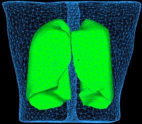
38 Deformable Lung Clinical Lung Data Simulated Deformed Lung *Courtesy DIR-lab, Dr. Castillo
39 Target Tolerances Stationary Image Moving Image Test Tolerance All Datasets Voxel Intensity Exact Basic Phantom Dataset - 2 Each modality image in Basic Phantom Dataset 1 Orientation Rigid Registration Translation Only Exact Maximum cardinal direction error less than 0.5*voxel dimension Basic Phantom Dataset 3 Each modality image in Basic Phantom Dataset 1 Rigid Registration Translation and Rotation Maximum cardinal direction error less than 0.5*voxel dimension Basic Anatomical Dataset - 1 Basic Anatomical Dataset - 2 Registration translation only Basic Anatomical Dataset - 1 Basic Anatomical Dataset - 3 Registration translation only Basic Anatomical Dataset - 1 Basic Anatomical Dataset - 4 Registration translation only Basic Anatomical Dataset - 1 Basic Anatomical Dataset - 5 Registration translation only Maximum cardinal direction error less than 0.5*voxel dimension size Maximum cardinal direction error less than 0.5*voxel dimension size Maximum cardinal direction error less than 0.5*voxel dimension size Maximum cardinal direction error less than 0.5*voxel dimension size Basic Anatomical Dataset - 1 Basic Deformation Dataset - 1 Deformable Registration 95% of voxels within 2 mm Sliding Deformation Dataset - 1 Sliding Deformation Dataset - 2 Deformable Registration max error less than 5 mm 95% of voxels within 2 mm Clinical 4DCT dataset (Deformation can be processed in either direction) Deformable Registration Max error less than 5 mm Mean vector error of all landmark points less than 2 mm Max error less than 5 mm
40 Commissioning and QA Understand the whole picture Phantom approach to understand characteristics of Understand algorithm fundamental Quantitative implementation components of Validation of algorithm Clinical Images

41 What Tools Do we Have? Visual Verification: Excellent tool for established techniques. Not enough for commissioning!
42 Quantitative Validation Techniques Landmark Based Does the registration map a landmark on Image A to the correct position on Image B? Target Registration Error (TRE) Contour Based Does the registration map the contours onto the new image correctly? Dice Similarity Coefficient (DSC) Mean Distance to Agreement (MDA) Digital/Physical Phantoms Compare known motion with registration results


43 Landmark Based (TRE) A B A TRE CT: 512x512x152; 0.09 cm in plane, 0.25 cm slice; GE scanner; 4D CT with Varian RPM Reproducibility of point identification is sub-voxel Gross errors Quantification of local accuracy within the target Increasing the number increases the overall volume quantification Manual technique Can identify max errors
44 That sounds great! Is that enough?
45 Accuracy of Points 1 cm X X X RMS = 0.3 mm

46 Points Don t Tell the Whole Story 1 cm X X X


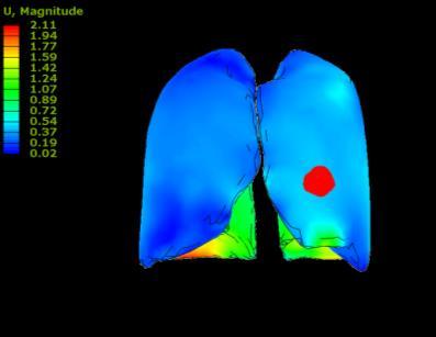
47 Inhale Accuracy of Contours Modeled Exhale Algorithm 1 Error 102 Bronchial Bifs TRE: 8.0 mm DSC > 0.9 Algorithm 2 TRE: 3.7 mm Actual Exhale DSC > 0.9 Modeled Exhale


48 Commissioning and QA Understand the whole picture Phantom approach to understand characteristics of Understand algorithm fundamental Quantitative implementation components of Validation of Documentation algorithm Clinical Images and Evaluation in Clinical Environment
 to be regi")
49 Request Clear identification of the image set(s) to be registered Identification of the primary (e.g. reference) image geometry An understanding of the local region(s) of importance The intended use of the result Target delineation Techniques to use (deformable or rigid) The accuracy required for the final use
50 Report Identify actual images used Indicate the accuracy of registration for local regions of importance and anatomical landmarks Identify any critical inaccuracies to alert the user Verify acceptable tolerances for use Techniques used to perform registration Fused images in report with annotations Documentation from system used for fusion


51 Clinical Example CT no contrast DIR for Multi-Modality Planning Accuracy required: voxel level Uncertainties create a systematic error that propagates throughout the treatment? MRI with contrast


52 Accounting for Uncertainties in Registration Prior to Deformable Registration CT MR coronal sagittal Before After Deformable Registration


53 Accounting for Uncertainties in Prior to Deformable Registration CT Registration MR X Assess uncertainty around GTV GTV (defined on MR, mapped to CT for Tx) Add margin around GTV definition to account for uncertainty when required Region of CT-defined GTV that is missed

 from the static plan Swaminath, IJROBP, in")

54 Clinical Impact of Dose Accumulation Dose Accumulation INH EXH Tx Velec, IJROBP patients 70% of patients have acc dose deviations ( 5%) from the static plan Swaminath, IJROBP, in press 81 patients, 142 liver mets accgtv dose is a better predictor of TTLP compared to minptv dose for liver metastases SBRT
55 Establish a patient specific QA practice for efficient evaluation of image registration results Why? At this point we are still understanding how the the registration is performing on different types of patients How? Visual Verification Spot checks of landmarks Boundary comparison
56 Vendor Recommendations 1. Disclose basic components of their registration algorithm to ensure its proper use 2. Provide the ability to export the registration matrix or deformation vector field for validation 3. Provide tools to qualitatively evaluate the image registration 4. Provide the ability to identify landmarks on 2 images and calculate the TRE from the registration 5. Provide the ability to calculate the DSC and MDA between the contours defined on an image and the contours mapped to the image via image registration 6. Support the integration of a request and report system for image registration
57 TG-132 Product Guidelines for understating of clinical tools Digital (virtual) phantoms Recommendations for commissioning and clinical implementation Recommendations for periodic and patient specific QA/QC Recommendations for clinical processes
58 Summary Deformable registration is a powerful tool that can help us to integrate multi-modality images, understand motion and anatomical changes, and compute an improved estimate of the delivered dose With power comes responsibility we must commission the system prior to use, understand the limitations, and communicate its proper use to clinicians, dosimetrists, therapists, and others TG 132 can help to provide tools, but the individuality of clinical workflows requires individual tests
Overview of Proposed TG-132 Recommendations
 Overview of Proposed TG-132 Recommendations Kristy K Brock, Ph.D., DABR Associate Professor Department of Radiation Oncology, University of Michigan Chair, AAPM TG 132: Image Registration and Fusion Conflict
Image Co-Registration II: TG132 Quality Assurance for Image Registration. Image Co-Registration II: TG132 Quality Assurance for Image Registration
 Image Co-Registration II: TG132 Quality Assurance for Image Registration Preliminary Recommendations from TG 132* Kristy Brock, Sasa Mutic, Todd McNutt, Hua Li, and Marc Kessler *Recommendations are NOT
Good Morning! Thank you for joining us
 Good Morning! Thank you for joining us Deformable Registration, Contour Propagation and Dose Mapping: 101 and 201 Marc Kessler, PhD, FAAPM The University of Michigan Conflict of Interest I receive direct
VALIDATION OF DIR. Raj Varadhan, PhD, DABMP Minneapolis Radiation Oncology
 VALIDATION OF DIR Raj Varadhan, PhD, DABMP Minneapolis Radiation Oncology Overview Basics: Registration Framework, Theory Discuss Validation techniques Using Synthetic CT data & Phantoms What metrics to
Is deformable image registration a solved problem?
 Is deformable image registration a solved problem? Marcel van Herk On behalf of the imaging group of the RT department of NKI/AVL Amsterdam, the Netherlands DIR 1 Image registration Find translation.deformation
Deformable Image Registration, Contour Propagation and Dose Mapping: 101 and 201. Please do not (re)redistribute
redistribute") Deformable Registration, Contour Deformable Registration, Contour Propagation and Dose Mapping: 101 and 201 Marc Kessler, PhD The University of Michigan Jean Pouliot, PhD University of California Learning
Deformable Registration, Contour Deformable Registration, Contour Propagation and Dose Mapping: 101 and 201 Marc Kessler, PhD The University of Michigan Jean Pouliot, PhD University of California Learning
REAL-TIME ADAPTIVITY IN HEAD-AND-NECK AND LUNG CANCER RADIOTHERAPY IN A GPU ENVIRONMENT
 REAL-TIME ADAPTIVITY IN HEAD-AND-NECK AND LUNG CANCER RADIOTHERAPY IN A GPU ENVIRONMENT Anand P Santhanam Assistant Professor, Department of Radiation Oncology OUTLINE Adaptive radiotherapy for head and
Methodological progress in image registration for ventilation estimation, segmentation propagation and multi-modal fusion
 Methodological progress in image registration for ventilation estimation, segmentation propagation and multi-modal fusion Mattias P. Heinrich Julia A. Schnabel, Mark Jenkinson, Sir Michael Brady 2 Clinical
Implementation of Advanced Image Guided Radiation Therapy
 Image Acquisition Course Outline Principles, characteristics& applications of the available modalities Image Processing in the T x room Image guided treatment delivery What can / can t we do in the room
Virtual Phantoms for IGRT QA
 TM Virtual Phantoms for IGRT QA Why ImSimQA? ImSimQA was developed to overcome the limitations of physical phantoms for testing modern medical imaging and radiation therapy software systems, when there
Use of image registration and fusion algorithms and techniques in radiotherapy: Report of the AAPM Radiation Therapy Committee Task Group No.
 Use of image registration and fusion algorithms and techniques in radiotherapy: Report of the AAPM Radiation Therapy Committee Task Group No. 132 Kristy K. Brock a) Department of Imaging Physics, The University
Mutual information based CT registration of the lung at exhale and inhale breathing states using thin-plate splines
 Mutual information based CT registration of the lung at exhale and inhale breathing states using thin-plate splines Martha M. Coselmon, a) James M. Balter, Daniel L. McShan, and Marc L. Kessler Department
Estimating 3D Respiratory Motion from Orbiting Views
 Estimating 3D Respiratory Motion from Orbiting Views Rongping Zeng, Jeffrey A. Fessler, James M. Balter The University of Michigan Oct. 2005 Funding provided by NIH Grant P01 CA59827 Motivation Free-breathing
Image Guidance and Beam Level Imaging in Digital Linacs
 Image Guidance and Beam Level Imaging in Digital Linacs Ruijiang Li, Ph.D. Department of Radiation Oncology Stanford University School of Medicine 2014 AAPM Therapy Educational Course Disclosure Research
Acknowledgements. Deformable Image Registration in Image Guided Therapy. Disclosure. Objectives 02/04/2011. Research Agreements:
 Deformable Image Registration in Image Guided Therapy Kristy K Brock, Ph.D., DABR Physicist, Radiation Medicine Program, Princess Margaret Hospital Associate Professor, Depts of Radiation Oncology & Medical
Dosimetric Analysis Report
 RT-safe 48, Artotinis str 116 33, Athens Greece +30 2107563691 info@rt-safe.com Dosimetric Analysis Report SAMPLE, for demonstration purposes only Date of report: ----------- Date of irradiation: -----------
Use of Deformable Image Registration in Radiation Therapy. Colin Sims, M.Sc. Accuray Incorporated 1
 Use of Deformable Image Registration in Radiation Therapy Colin Sims, M.Sc. Accuray Incorporated 1 Overview of Deformable Image Registration (DIR) Algorithms that can deform one dataset to another have
Clinical Prospects and Technological Challenges for Multimodality Imaging Applications in Radiotherapy Treatment Planning
 Clinical Prospects and Technological Challenges for Multimodality Imaging Applications in Radiotherapy Treatment Planning Issam El Naqa, PhD Assistant Professor Department of Radiation Oncology Washington
Using Pinnacle 16 Deformable Image registration in a re-treat scenario
 Introduction Using Pinnacle 16 Deformable Image registration in a re-treat scenario This short Hands On exercise will introduce how the Deformable Image Registration (DIR) tools in Pinnacle can be used
7/31/2011. Learning Objective. Video Positioning. 3D Surface Imaging by VisionRT
 CLINICAL COMMISSIONING AND ACCEPTANCE TESTING OF A 3D SURFACE MATCHING SYSTEM Hania Al-Hallaq, Ph.D. Assistant Professor Radiation Oncology The University of Chicago Learning Objective Describe acceptance
Nonrigid Registration using Free-Form Deformations
 Nonrigid Registration using Free-Form Deformations Hongchang Peng April 20th Paper Presented: Rueckert et al., TMI 1999: Nonrigid registration using freeform deformations: Application to breast MR images
ADVANCING CANCER TREATMENT
 3 ADVANCING CANCER TREATMENT SUPPORTING CLINICS WORLDWIDE RaySearch is advancing cancer treatment through pioneering software. We believe software has un limited potential, and that it is now the driving
Image Segmentation and Registration
 Image Segmentation and Registration Dr. Christine Tanner (tanner@vision.ee.ethz.ch) Computer Vision Laboratory, ETH Zürich Dr. Verena Kaynig, Machine Learning Laboratory, ETH Zürich Outline Segmentation
HST.582J / 6.555J / J Biomedical Signal and Image Processing Spring 2007
 MIT OpenCourseWare http://ocw.mit.edu HST.582J / 6.555J / 16.456J Biomedical Signal and Image Processing Spring 2007 For information about citing these materials or our Terms of Use, visit: http://ocw.mit.edu/terms.
ADVANCING CANCER TREATMENT
 The RayPlan treatment planning system makes proven, innovative RayStation technology accessible to clinics that need a cost-effective and streamlined solution. Fast, efficient and straightforward to use,
IMRT and VMAT Patient Specific QA Using 2D and 3D Detector Arrays
 IMRT and VMAT Patient Specific QA Using 2D and 3D Detector Arrays Sotiri Stathakis Outline Why IMRT/VMAT QA AAPM TG218 UPDATE Tolerance Limits and Methodologies for IMRT Verification QA Common sources
Deformable Segmentation using Sparse Shape Representation. Shaoting Zhang
 Deformable Segmentation using Sparse Shape Representation Shaoting Zhang Introduction Outline Our methods Segmentation framework Sparse shape representation Applications 2D lung localization in X-ray 3D
Non-rigid Image Registration
 Overview Non-rigid Image Registration Introduction to image registration - he goal of image registration - Motivation for medical image registration - Classification of image registration - Nonrigid registration
The Insight Toolkit. Image Registration Algorithms & Frameworks
 The Insight Toolkit Image Registration Algorithms & Frameworks Registration in ITK Image Registration Framework Multi Resolution Registration Framework Components PDE Based Registration FEM Based Registration
Medical Image Registration by Maximization of Mutual Information
 Medical Image Registration by Maximization of Mutual Information EE 591 Introduction to Information Theory Instructor Dr. Donald Adjeroh Submitted by Senthil.P.Ramamurthy Damodaraswamy, Umamaheswari Introduction
Auto-Segmentation Using Deformable Image Registration. Disclosure. Objectives 8/4/2011
 Auto-Segmentation Using Deformable Image Registration Lei Dong, Ph.D. Dept. of Radiation Physics University of Texas MD Anderson Cancer Center, Houston, Texas AAPM Therapy Educational Course Aug. 4th 2011
Image Registration. Prof. Dr. Lucas Ferrari de Oliveira UFPR Informatics Department
 Image Registration Prof. Dr. Lucas Ferrari de Oliveira UFPR Informatics Department Introduction Visualize objects inside the human body Advances in CS methods to diagnosis, treatment planning and medical
Image Quality Assessment and Quality Assurance of Advanced Imaging Systems for IGRT. AAPM Penn-Ohio Chapter Sep 25, 2015 Soyoung Lee, PhD
 Image Quality Assessment and Quality Assurance of Advanced Imaging Systems for IGRT AAPM Penn-Ohio Chapter Sep 25, 2015 Soyoung Lee, PhD 1 Outline q Introduction q Imaging performances in 4D-CBCT Image
3DVH : SUN NUCLEAR On The Accuracy Of The corporation Planned Dose Perturbation Algorithm Your Most Valuable QA and Dosimetry Tools *Patent Pending
 3DVH : On The Accuracy Of The Planned Dose Perturbation Algorithm SUN NUCLEAR corporation Your Most Valuable QA and Dosimetry Tools *Patent Pending introduction State-of-the-art IMRT QA of static gantry
Interoperability Issues in Image Registration and ROI Generation
 1 DICOM 2005 International Conference, Budapest, Hungary Interoperability Issues in Image Registration and ROI Generation Todd Kantchev PhD, Siemens Molecular Imaging, Oxford, UK 2 Scope The following
iplan RT Image Advanced Contouring Workstation - Driving Physician Collaboration
 iplan RT Image Advanced Contouring Workstation - Driving Physician Collaboration The iplan Contouring Workstation offers unique and innovative capabilities for faster contouring and consistent segmentation
SIGMI Meeting ~Image Fusion~ Computer Graphics and Visualization Lab Image System Lab
 SIGMI Meeting ~Image Fusion~ Computer Graphics and Visualization Lab Image System Lab Introduction Medical Imaging and Application CGV 3D Organ Modeling Model-based Simulation Model-based Quantification
Lucy Phantom MR Grid Evaluation
 Lucy Phantom MR Grid Evaluation Anil Sethi, PhD Loyola University Medical Center, Maywood, IL 60153 November 2015 I. Introduction: The MR distortion grid, used as an insert with Lucy 3D QA phantom, is
Registration Techniques
 EMBO Practical Course on Light Sheet Microscopy Junior-Prof. Dr. Olaf Ronneberger Computer Science Department and BIOSS Centre for Biological Signalling Studies University of Freiburg Germany O. Ronneberger,
8/3/2017. Contour Assessment for Quality Assurance and Data Mining. Objective. Outline. Tom Purdie, PhD, MCCPM
 Contour Assessment for Quality Assurance and Data Mining Tom Purdie, PhD, MCCPM Objective Understand the state-of-the-art in contour assessment for quality assurance including data mining-based techniques
RIGID IMAGE REGISTRATION
 RIGID IMAGE REGISTRATION Duygu Tosun-Turgut, Ph.D. Center for Imaging of Neurodegenerative Diseases Department of Radiology and Biomedical Imaging duygu.tosun@ucsf.edu What is registration? Image registration
Help Guide. mm Copyright Mirada Medical Ltd, Mirada Medical RTx 1
 Help Guide mm3237-1.6-1 Copyright Mirada Medical Ltd, 2000-2014. Mirada Medical RTx 1 Contents Help Guide... 1 Contents... 2 Introduction to RTx... 4 Regulatory Statement... 6 Notes... 15 Data Supported...
Feasibility of 3D Printed Patient specific Phantoms for IMRT QA and Other Dosimetric Special Procedures
 Feasibility of 3D Printed Patient specific Phantoms for IMRT QA and Other Dosimetric Special Procedures ehler 046@umn.edu Eric Ehler, PhD Assistant Professor Department of Radiation Oncology What is 3D
Validation of biomechanical deformable image registration in the abdomen, thorax, and pelvis in a commercial radiotherapy treatment planning system
 Validation of biomechanical deformable image registration in the abdomen, thorax, and pelvis in a commercial radiotherapy treatment planning system Michael Velec a) and Joanne L. Moseley Techna Institute
Acknowledgements. Atlas-based automatic measurements of the morphology of the tibiofemoral joint
 Atlas-based automatic measurements of the morphology of the tibiofemoral joint M Brehler 1, G Thawait 2, W Shyr 1, J Ramsay 3, JH Siewerdsen 1,2, W Zbijewski 1 1 Dept. of Biomedical Engineering, Johns
Multimodal Elastic Image Matching
 Research results based on my diploma thesis supervised by Prof. Witsch 2 and in cooperation with Prof. Mai 3. 1 February 22 nd 2011 1 Karlsruhe Institute of Technology (KIT) 2 Applied Mathematics Department,
Biomedical Imaging Registration Trends and Applications. Francisco P. M. Oliveira, João Manuel R. S. Tavares
 Biomedical Imaging Registration Trends and Applications Francisco P. M. Oliveira, João Manuel R. S. Tavares tavares@fe.up.pt, www.fe.up.pt/~tavares Outline 1. Introduction 2. Spatial Registration of (2D
Computational Medical Imaging Analysis Chapter 4: Image Visualization
 Computational Medical Imaging Analysis Chapter 4: Image Visualization Jun Zhang Laboratory for Computational Medical Imaging & Data Analysis Department of Computer Science University of Kentucky Lexington,
OnDemand3D Fusion Technology
 CYBERMED INC., ONDEMAND3D TECHNOLOGY INC. OnDemand3D Fusion Technology White Paper December 2009 USA Republic of Korea www.ondemand3d.com Introduction OnDemand3D TM Fusion is registration technology to
Initial Clinical Experience with 3D Surface Image Guidance
 Initial Clinical Experience with 3D Surface Image Guidance Amanda Havnen-Smith, Ph.D. Minneapolis Radiation Oncology Ridges Radiation Therapy Center Burnsville, MN April 20 th, 2012 Non-funded research
Learning-based Neuroimage Registration
 Learning-based Neuroimage Registration Leonid Teverovskiy and Yanxi Liu 1 October 2004 CMU-CALD-04-108, CMU-RI-TR-04-59 School of Computer Science Carnegie Mellon University Pittsburgh, PA 15213 Abstract
IMSURE QA SOFTWARE FAST, PRECISE QA SOFTWARE
 QA SOFTWARE FAST, PRECISE Software for accurate and independent verification of monitor units, dose, and overall validity of standard, IMRT, VMAT, SRS and brachytherapy plans no film, no phantoms, no linac
Medicale Image Analysis
 Medicale Image Analysis Registration Validation Prof. Dr. Philippe Cattin MIAC, University of Basel Prof. Dr. Philippe Cattin: Registration Validation Contents 1 Validation 1.1 Validation of Registration
CT NOISE POWER SPECTRUM FOR FILTERED BACKPROJECTION AND ITERATIVE RECONSTRUCTION
 CT NOISE POWER SPECTRUM FOR FILTERED BACKPROJECTION AND ITERATIVE RECONSTRUCTION Frank Dong, PhD, DABR Diagnostic Physicist, Imaging Institute Cleveland Clinic Foundation and Associate Professor of Radiology
Basic principles of MR image analysis. Basic principles of MR image analysis. Basic principles of MR image analysis
 Basic principles of MR image analysis Basic principles of MR image analysis Julien Milles Leiden University Medical Center Terminology of fmri Brain extraction Registration Linear registration Non-linear
Facility Questionnaire PART I (General Information for 3DCRT and IMRT)
") Facility Questionnaire PART I (General Information for 3DCRT and IMRT) The following items are required before you can enter cases on any RTOG protocol that requires data submission to the Image-Guided
Facility Questionnaire PART I (General Information for 3DCRT and IMRT) The following items are required before you can enter cases on any RTOG protocol that requires data submission to the Image-Guided
UvA-DARE (Digital Academic Repository) Motion compensation for 4D PET/CT Kruis, M.F. Link to publication
 Motion compensation for 4D PET/CT Kruis, M.F. Link to publication") UvA-DARE (Digital Academic Repository) Motion compensation for 4D PET/CT Kruis, M.F. Link to publication Citation for published version (APA): Kruis, M. F. (2014). Motion compensation for 4D PET/CT General
UvA-DARE (Digital Academic Repository) Motion compensation for 4D PET/CT Kruis, M.F. Link to publication Citation for published version (APA): Kruis, M. F. (2014). Motion compensation for 4D PET/CT General
3/27/2012 WHY SPECT / CT? SPECT / CT Basic Principles. Advantages of SPECT. Advantages of CT. Dr John C. Dickson, Principal Physicist UCLH
 3/27/212 Advantages of SPECT SPECT / CT Basic Principles Dr John C. Dickson, Principal Physicist UCLH Institute of Nuclear Medicine, University College London Hospitals and University College London john.dickson@uclh.nhs.uk
Comprehensive treatment planning for brachytherapy. Advanced planning made easy
 Comprehensive treatment planning for brachytherapy Advanced planning made easy Oncentra Brachy offers a variety of smart tools that facilitate many of the repetitive tasks for you. In contemporary brachytherapy,
Accounting for Large Geometric Changes During Radiotherapy. Disclosures. Current Generation DIR in RT 8/3/2016
 Accounting for Large Geometric Changes During Radiotherapy Geoff Hugo, Ph.D. Department of Radiation Oncology Virginia Commonwealth University, Richmond, Virginia, USA Disclosures Research support: Philips
Slide 1. Technical Aspects of Quality Control in Magnetic Resonance Imaging. Slide 2. Annual Compliance Testing. of MRI Systems.
 Slide 1 Technical Aspects of Quality Control in Magnetic Resonance Imaging Slide 2 Compliance Testing of MRI Systems, Ph.D. Department of Radiology Henry Ford Hospital, Detroit, MI Slide 3 Compliance Testing
1. Learn to incorporate QA for surface imaging
 Hania Al-Hallaq, Ph.D. Assistant Professor Radiation Oncology The University of Chicago ***No disclosures*** 1. Learn to incorporate QA for surface imaging into current QA procedures for IGRT. 2. Understand
8/3/2016. Image Guidance Technologies. Introduction. Outline
 8/3/26 Session: Image Guidance Technologies and Management Strategies Image Guidance Technologies Jenghwa Chang, Ph.D.,2 Department of Radiation Medicine, Northwell Health 2 Hofstra Northwell School of
S. Guru Prasad, Ph.D., DABR
 PURPOSE S. Guru Prasad, Ph.D., DABR Director of Medical Physics IAEA Consultant NorthShore University Health System and University of Chicago, Pritzker School of Medicine Current TPS utilize more information
Michael Speiser, Ph.D.
 IMPROVED CT-BASED VOXEL PHANTOM GENERATION FOR MCNP MONTE CARLO Michael Speiser, Ph.D. Department of Radiation Oncology UT Southwestern Medical Center Dallas, TX September 1 st, 2012 CMPWG Workshop Medical
Deviceless respiratory motion correction in PET imaging exploring the potential of novel data driven strategies
 g Deviceless respiratory motion correction in PET imaging exploring the potential of novel data driven strategies Presented by Adam Kesner, Ph.D., DABR Assistant Professor, Division of Radiological Sciences,
8/4/2016. Emerging Linac based SRS/SBRT Technologies with Modulated Arc Delivery. Disclosure. Introduction: Treatment delivery techniques
 Emerging Linac based SRS/SBRT Technologies with Modulated Arc Delivery Lei Ren, Ph.D. Duke University Medical Center 2016 AAPM 58 th annual meeting, Educational Course, Therapy Track Disclosure I have
Leksell SurgiPlan Overview. Powerful planning for surgical success
 Leksell SurgiPlan Overview Powerful planning for surgical success Making a Difference in Surgical Planning Leksell SurgiPlan Leksell SurgiPlan is an advanced image-based neuro surgical planning software,
Spatio-Temporal Registration of Biomedical Images by Computational Methods
 Spatio-Temporal Registration of Biomedical Images by Computational Methods Francisco P. M. Oliveira, João Manuel R. S. Tavares tavares@fe.up.pt, www.fe.up.pt/~tavares Outline 1. Introduction 2. Spatial
Hybrid Spline-based Multimodal Registration using a Local Measure for Mutual Information
 Hybrid Spline-based Multimodal Registration using a Local Measure for Mutual Information Andreas Biesdorf 1, Stefan Wörz 1, Hans-Jürgen Kaiser 2, Karl Rohr 1 1 University of Heidelberg, BIOQUANT, IPMB,
Advanced Targeting Using Image Deformation. Justin Keister, MS DABR Aurora Health Care Kenosha, WI
 Advanced Targeting Using Image Deformation Justin Keister, MS DABR Aurora Health Care Kenosha, WI History of Targeting The advance of IMRT and CT simulation has changed how targets are identified in radiation
PCRT 3D. Scalable Architecture System. User-Friendly. Traceable. Continuos Development
 PCRT 3D The PCRT3D is a versatile 3D radiation treatment planning system featuring the most accurate algorithm calculations, the latest techniques in virtual simulation and the most advanced radiotherapy
Technical aspects of SPECT and SPECT-CT. John Buscombe
 Technical aspects of SPECT and SPECT-CT John Buscombe What does the clinician need to know? For SPECT What factors affect SPECT How those factors should be sought Looking for artefacts For SPECT-CT Issues
Development of a deformable lung phantom for the evaluation of deformable registration
 JOURNAL OF APPLIED CLINICAL MEDICAL PHYSICS, VOLUME 11, NUMBER 1, WINTER 2010 Development of a deformable lung phantom for the evaluation of deformable registration Jina Chang, 1 Tae-Suk Suh, 1a Dong-Soo
Basics of treatment planning II
 Basics of treatment planning II Sastry Vedam PhD DABR Introduction to Medical Physics III: Therapy Spring 2015 Dose calculation algorithms! Correction based! Model based 1 Dose calculation algorithms!
Automatic Thoracic CT Image Segmentation using Deep Convolutional Neural Networks. Xiao Han, Ph.D.
 Automatic Thoracic CT Image Segmentation using Deep Convolutional Neural Networks Xiao Han, Ph.D. Outline Background Brief Introduction to DCNN Method Results 2 Focus where it matters Structure Segmentation
Design and performance characteristics of a Cone Beam CT system for Leksell Gamma Knife Icon
 Design and performance characteristics of a Cone Beam CT system for Leksell Gamma Knife Icon WHITE PAPER Introduction Introducing an image guidance system based on Cone Beam CT (CBCT) and a mask immobilization
MR-guided radiotherapy: Vision, status and research at the UMC Utrecht. Dipl. Ing. Dr. Markus Glitzner
 MR-guided radiotherapy: Vision, status and research at the UMC Utrecht Dipl. Ing. Dr. Markus Glitzner About myself Training Medizintechnik TU Graz PhD UMC Utrecht Clinical work Software implementation
Position accuracy analysis of the stereotactic reference defined by the CBCT on Leksell Gamma Knife Icon
 Position accuracy analysis of the stereotactic reference defined by the CBCT on Leksell Gamma Knife Icon WHITE PAPER Introduction An image guidance system based on Cone Beam CT (CBCT) is included in Leksell
Real Time Tumor Motion Tracking with CyberKnife
 Real Time Tumor Motion Tracking with CyberKnife Martina Descovich, Ph.D University of California San Francisco July 16, 2015 Learning objectives Review the principles of real-time tumor motion tracking
Automated segmentation methods for liver analysis in oncology applications
 University of Szeged Department of Image Processing and Computer Graphics Automated segmentation methods for liver analysis in oncology applications Ph. D. Thesis László Ruskó Thesis Advisor Dr. Antal
Image Registration + Other Stuff
 Image Registration + Other Stuff John Ashburner Pre-processing Overview fmri time-series Motion Correct Anatomical MRI Coregister m11 m 21 m 31 m12 m13 m14 m 22 m 23 m 24 m 32 m 33 m 34 1 Template Estimate
Using Probability Maps for Multi organ Automatic Segmentation
 Using Probability Maps for Multi organ Automatic Segmentation Ranveer Joyseeree 1,2, Óscar Jiménez del Toro1, and Henning Müller 1,3 1 University of Applied Sciences Western Switzerland (HES SO), Sierre,
Transitive and Symmetric Nonrigid Image Registration. Yi-Yu Chou
 Transitive and Symmetric Nonrigid Image Registration A Thesis Presented to The Academic Faculty by Yi-Yu Chou In Partial Fulfillment of the Requirements for the Degree Master of Science School of Biomedical
Image Registration I
 Image Registration I Comp 254 Spring 2002 Guido Gerig Image Registration: Motivation Motivation for Image Registration Combine images from different modalities (multi-modality registration), e.g. CT&MRI,
radiotherapy Andrew Godley, Ergun Ahunbay, Cheng Peng, and X. Allen Li NCAAPM Spring Meeting 2010 Madison, WI
 GPU-Accelerated autosegmentation for adaptive radiotherapy Andrew Godley, Ergun Ahunbay, Cheng Peng, and X. Allen Li agodley@mcw.edu NCAAPM Spring Meeting 2010 Madison, WI Overview Motivation Adaptive
Automated Image Analysis Software for Quality Assurance of a Radiotherapy CT Simulator
 Automated Image Analysis Software for Quality Assurance of a Radiotherapy CT Simulator Andrew J Reilly Imaging Physicist Oncology Physics Edinburgh Cancer Centre Western General Hospital EDINBURGH EH4
Auto-contouring the Prostate for Online Adaptive Radiotherapy
 Auto-contouring the Prostate for Online Adaptive Radiotherapy Yan Zhou 1 and Xiao Han 1 Elekta Inc., Maryland Heights, MO, USA yan.zhou@elekta.com, xiao.han@elekta.com, Abstract. Among all the organs under
PET-CT in Radiation Treatment Planning
 PET-CT in Radiation Treatment Planning TA van de Water, Radiotherapeutic Institute Friesland, Leeuwarden JA van Dalen, Isala, Zwolle ACKNOWLEDGEMENTS A. van der Schaaf (University of Groningen, University
Coverage based treatment planning to accommodate organ deformable motions and contouring uncertainties for prostate treatment. Huijun Xu, Ph.D.
 Coverage based treatment planning to accommodate organ deformable motions and contouring uncertainties for prostate treatment Huijun Xu, Ph.D. Acknowledgement and Disclosure Dr. Jeffrey Siebers Dr. DJ
CSE 554 Lecture 7: Deformation II
 CSE 554 Lecture 7: Deformation II Fall 2011 CSE554 Deformation II Slide 1 Review Rigid-body alignment Non-rigid deformation Intrinsic methods: deforming the boundary points An optimization problem Minimize
A Registration-Based Atlas Propagation Framework for Automatic Whole Heart Segmentation
 A Registration-Based Atlas Propagation Framework for Automatic Whole Heart Segmentation Xiahai Zhuang (PhD) Centre for Medical Image Computing University College London Fields-MITACS Conference on Mathematics
8/3/2016. MR-guided RT: Commissioning and Quality Control. Topics for MR-guided RT system commissioning & QC
 MR-guided RT: Commissioning and Quality Control T. Stanescu, PhD, MCCPM Medical Physicist, Princess Margaret Cancer Centre, UHN Assistant Professor, Radiation Oncology, University of Toronto Affiliated
MRI-LINAC Dynamic Phantom
 MRI-LINAC Dynamic Phantom Model 008M IMAGE ACQUISITION TREATMENT PLANNING DOSE DELIVERY Strict QA procedures for the imaging, planning and delivery of radiotherapy using respiratory management devices
8/2/2017. PerFRACTION from Sun Nuclear Automated Transit Dosimetry for In-Vivo QA
 PerFRACTION from Sun Nuclear Automated Transit Dosimetry for In-Vivo QA Jennifer R. Clark, M.S., DABR Conflict of Interest Statement: I am an employee of Sun Nuclear Corporation Outline: Platform overview
MapCHECK 2 & 3DVH. The Gold Standard for 2D Arrays
 MapCHECK 2 & 3DVH The Gold Standard for 2D Arrays Your Most Valuable QA and Dosimetry Tools THE GOLD STANDARD FOR 2D ARRAYS The MapCHECK 2 is the world s most selected independent 2D measurement array.
Hierarchical Multi structure Segmentation Guided by Anatomical Correlations
 Hierarchical Multi structure Segmentation Guided by Anatomical Correlations Oscar Alfonso Jiménez del Toro oscar.jimenez@hevs.ch Henning Müller henningmueller@hevs.ch University of Applied Sciences Western
CHAPTER 2. Morphometry on rodent brains. A.E.H. Scheenstra J. Dijkstra L. van der Weerd
 CHAPTER 2 Morphometry on rodent brains A.E.H. Scheenstra J. Dijkstra L. van der Weerd This chapter was adapted from: Volumetry and other quantitative measurements to assess the rodent brain, In vivo NMR
Non-Rigid Multimodal Medical Image Registration using Optical Flow and Gradient Orientation
 M. HEINRICH et al.: MULTIMODAL REGISTRATION USING GRADIENT ORIENTATION 1 Non-Rigid Multimodal Medical Image Registration using Optical Flow and Gradient Orientation Mattias P. Heinrich 1 mattias.heinrich@eng.ox.ac.uk
CS/NEUR125 Brains, Minds, and Machines. Due: Wednesday, April 5
 CS/NEUR125 Brains, Minds, and Machines Lab 8: Using fmri to Discover Language Areas in the Brain Due: Wednesday, April 5 In this lab, you will analyze fmri data from an experiment that was designed to