Parallel Magnetic Resonance Imaging (pmri): How Does it Work, and What is it Good For?
|
|
|
- Austin Webb
- 5 years ago
- Views:
Transcription
1 Parallel Magnetic Resonance Imaging (pmri): How Does it Work, and What is it Good For? Nathan Yanasak, Ph.D. Chair, AAPM TG118 Department of Radiology Georgia Regents University
2 Overview Phased-array coils General Description of Parallel Imaging Different pmri methods Applications of pmri How NOT to use pmri
3 What is pmri? Uses spatial information obtained from arrays of RF coil elements sampling data in parallel Information handles some portion of spatial encoding performed using gradient fields (typically phase-encoding gradient) Speeds up MRI acquisition times without needing faster-switching gradients without additional RF power deposited (key for higher field MR)
 Smaller FOV aliasing")
4 pmri speed: less phase encodes = smaller FOV (with same resolution) Smaller FOV aliasing 4
5 Properties of Phased Arrays (PA) of Surface Coil Elements What capabilities do PA coils have to localize signal? Each element is sensitive only to local spins Uneven SNR throughout volume, but Very high SNR at edge Lower SNR in middle SNR in middle is generally better than comparable volume coil. 5
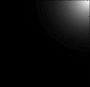
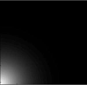
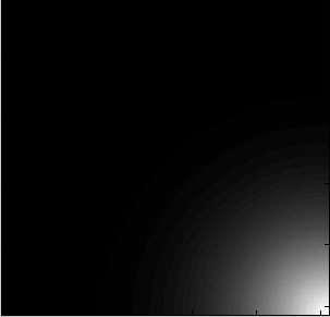
 Non-uniform SNR Great SNR up close Phased-Array Coil Non-uniform SNR SNR generally high Volume Coil Uniform SNR Average SNR")
6 SNR: Surface Coil vs. Volume Coil Surface Coil (single element) Non-uniform SNR Great SNR up close Phased-Array Coil Non-uniform SNR SNR generally high Volume Coil Uniform SNR Average SNR Bottom line: spatial localization of signal depends on PA element location (and size). Critical for pmri. Tx/Rx head 8-ch PA head 32-ch PA head 6


7 Multichannel Coils 8- and 15-channel head coils Multi-element body coils 7
8 Uniformity Tests and PA Coil Concerns ACR Uniformity test was specified during the era of volume coils. Effect of intrinsic non-uniformity of phased-array coils on QA? Uniformity test with 32-channel coil (3T) tests with one element turned off (filter on) Uniformity failures: 0 All elements: PIU=89.6% One missing element: <PIU> N-1 =89.6% s PIU,N-1 =0.5% (SEM~0.1%) PIU N-1,min =88.0%; PIU N-1,max =90.2%
.")
 Uniformity failures: 0 All elements:")
 PIU N-1,min =88.")
9 Uniformity and PA Coils Let s try that again with 15-channel coil (GRU workhorse). 15 tests with one element turned off (filter on) Uniformity failures: 0 All elements: PIU=90.6% One missing element: <PIU> N- 1=90.1% s PIU,N-1 =0.7% (SEM~0.2%) PIU N-1,min =88.7%; PIU N-1,max =91.3%
10 Uniformity and PA Coils One last time with an 8-channel coil (another GRU workhorse). 1 test with one element turned off (symmetry in coil) All elements: PIU=93.6% One missing element: PIU N-1 =91.5% Spatial distribution different, even with filter on.
.")
 All elements: PIU=93.")

11 Uniformity and PA Coils One last time with an 8-channel coil (another GRU workhorse). 1 test with one element turned off (symmetry in coil) All elements: PIU=93.6% One missing element: PIU N-1 =91.5% Spatial distribution different, even with filter on. All elements 7 elements
12 Uniformity and PA Coils Observations: Mean uniformity lower for higher # of elements. Uniformity degradation for coils with broken elements worse with smaller # of elements. Obvious changes in spatial uniformity with 8- channel. What does this suggest for ACR testing of PA coils? Phantom issue? Protocol? Specs? For patient care? pmri performance even more dependent on PA coils.


 Parallel")
13 Use of Phased Array Coil in Parallel Imaging Spatial sensitivity varies for each element can use this in conjunction with undersampling. Conventional use of phased-array (unaliased) Parallel reconstruction of data (aliased)
14 Coil Sensitivity Profiles Different approaches to solving the inverse problem that recovers spatial information. The key information always required to solve this problem is information on the spatial distribution of the RF coils sensitivity. How you collect and use this information different pmri methods.


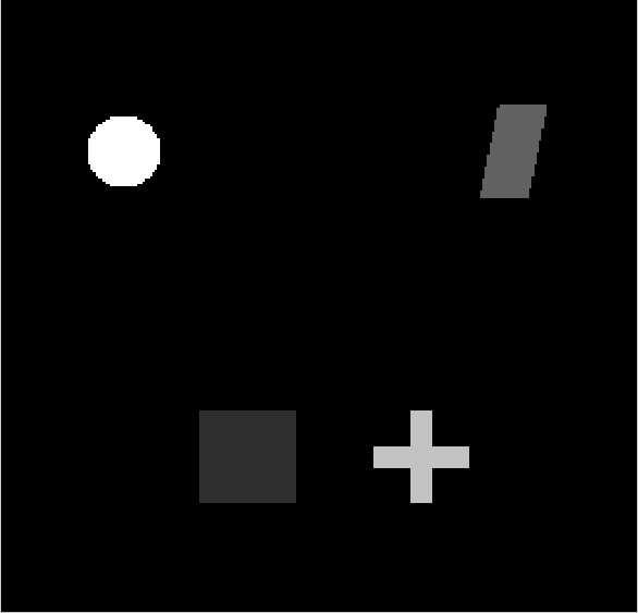
15 Sensitivity Map The spatial sensitivity of each coil element = sensitivity map. A calibration scan may be required to calculate this. s 1 Total s 2 15
16 Using Coil Sensitivity to Un-alias an Image: An Example 16

17 Coil Locations and Sensitivity Maps Object being imaged 17
18 Using Coil Sensitivity to Un-alias an Image 18


19 19
 k-space based: Untangle data to create fully-filled k-space(s), then reconstruct image (SMASH, GRAPPA)")
20 Two Parallel Approaches Image based: Reconstruct images from each element, then untangle (SENSE, ASSET) (our demo) k-space based: Untangle data to create fully-filled k-space(s), then reconstruct image (SMASH, GRAPPA)
21 Image-based pmri: The Encoding Matrix S B p pj j j S p: signal received by the coil, p. j : proton density at the pixel index, j B pj : encoding function that connects the coil response to the proton signal at a location. In matrix notation: or inverting: S = B = B -1 S Thus if B -1 can be calculated, can be determined.


22 A Simplistic SENSE Example a S alias,1 =B 1,a I a + B 1,b I b element 1 I a b S 1 a I b element 2 b S 2 S alias,2 =B 2,a I a + B 2,b I b 22
23 SMASH an Early k-space Based pmri Method Assumes spatial harmonics of phaseencoding gradients can be omitted and emulated by a linear combination of coil sensitivities Coil sensitivity still required (measured in some manner, and complex).


24 A Simplistic SMASH Example Phase encoding modulation of phase as a function of position.
25 Frequency-Domain Basics A B A+B 1D example: complicated wave = sum of simple waves. Need amplitudes/phases to perform the sum. C D E F A+B+C+D A+B+C+D+E+F In this example, we could keep going to create a square wave. Same issue in 2D (here, image = wave ). 25
26 A Simplistic SMASH Example A 1 * +A 2 * +A 3 * + =
27 A Simplistic SMASH Example K-space A + - B + C Rather than preparing all phase modulations, omit some for the sake of time, and use coils to emulate the modulations.
28 A Simplistic SMASH Example K-space A + - B + C Rather than preparing all phase modulations, omit some for the sake of time, and use coils to emulate the modulations.


29 A Simplistic SMASH Example Resultant combinations (spatial harmonics) allow for filling of all lines in a composite k-space.
30 GRAPPA (Griswold, et al. MRM 2002) More general application of SMASH principles. Generate extra lines of k-space via convolution process (similar to weighted sums in SMASH). K-spaces from each coil can be individually reconstructed. How to determine the weights? Use sensitivity information contained in image. Autocalibration: Acquire reference lines (ACS lines) in k-space rather than whole coil sensitivity images (data from center of k-space acts like a sensitivity profile)
31 GRAPPA K-space for each individual element. Weights come from fits to calibration data. W1 W2 W3 0 0 W6 W7 W8
32 Parallel Imaging (Technique Pros/Cons) Image-based reconstruction: More artifacts, but easier to implement the sequence. K-space based reconstruction: Depends more strongly on coil design, less artifacts, but longer to reconstruct. 32
33 Advantages/Uses of pmri 33
34 When Should You Use Parallel MR Imaging? To reduce total scan time To speed up single-shot MRI methods To reduce TE on long echo-train methods To mitigate susceptibility, chemical shift and other artifacts (may cause others) To decrease RF heating (SAR) by minimizing number of RF pulses ( B 2 )
 To decrease RF heating (SAR) by minimizing number of RF pulses Margolis D et al.")
35 Use #1: Body Imaging A: 22 sec acquisition w/ 15 sec breathhold B: 11 sec acquisition w/ 11 sec breathhold + R=2 To reduce total scan time (or eliminate breath holds) To decrease RF heating (SAR) by minimizing number of RF pulses Margolis D et al. Top Magn Reson Imag 2004; 15:

36 Use #2: Spinal Imaging D: non-pmri E: R=2 Image quality is of similar quality for ½ the scan time Noebauer-Huhmann et al. Eur Radiol 2007; 17:

37 Use #3: Reduce T2 Blurring (FSE) Problem #1: Greater ETL lower SNR Problem #2: T2 relaxation during acquisition of ETL results in T2 blurring. pmri: reduce ETL. facilitate reductions in TEeff.


38 Use #3: Reduce T2 Blurring Augustine Me et al. Top Magn Reson Imag 2004; 15:207 Glockner et all. RadioGraphics 2005; 25:

39 Use #4: Susceptibility Artifacts Air Sinuses Regions of air/bone/soft tissue causes local gradients due to differences in magnetic field susceptibility

40 Susceptibility Artifact Reduction with Parallel Imaging Clinical example: remediation of distortion would have been nice in this circumstance.
. EPI-based sequences gain more in general (e.g., DWI, perfusion) Top normal acquisition, Bottom R=2 acceleration")
41 Susceptibility Artifact Reduction with Parallel Imaging Shortening readout window/te helps (must have less phase encodes to do this). EPI-based sequences gain more in general (e.g., DWI, perfusion) Top normal acquisition, Bottom R=2 acceleration
42 Turn Key Parallel Imaging? R=1 R=2.0 R=2.8 R=3.2 R=4.0


43 Turn Key Parallel Imaging? R=1 R=2.0 R=2.8 R=3.2 R=4.0

44 Use #5: Contrast-enhanced MR (MRA) Left: R~ 1.5; Right: non-pmri with reduced FOV Improved spatial resolution for a given scan time. Wilson, et al. Top Magn Reson Imag. 2004; 15:

45 Use #6: Cardiac Imaging Balanced FFE MRI A&B: 11 sec breath holds C&D: 5 sec breath holds + R=2 Van den Brink, et al. Eur. J. Rad. 2003; 46:
46 Drawbacks/Consideration of pmri: SNR Properties & Artifacts 46
 Non-uniformity of noise (pmri) Lower signal from acceleration")
47 SNR SNR is a concern with pmri for three reasons: Non-uniformity of signal (array coils) Non-uniformity of noise (pmri) Lower signal from acceleration (pmri)

48 Non-Uniformity of Noise Larkman DJ et al. Magn Reson Med 2006; 55:
49 Key SNR Parameters in Parallel Imaging SNR depends on number, size and orientation of the coil elements SNR PI i, j, k SNR g i, j, k norm i, j, k R R: acceleration factor g: coil-dependent noise amplification factor (non-uniformity that we observed)
50 Key SNR Parameters in Parallel Imaging SNR depends on number, size and orientation of the coil elements 1 g( r, R) R 2 SNR SNR PI norm ( r ) R: acceleration factor g: coil-dependent noise amplification factor (non-uniformity that we observed)

51 SNR vs. Acceleration Short-axis cardiac images 32-channel coil 1.5 T magnet Reeder SB et al. MRM 54:748, 2005

52 g-factor Calculated Maps 32-channel coil, 1.5 T magnet R=2 R=3 R=2 R=3 R=4 R=5 R=4 R=5 R=6 R=7 R=6 R=7 R-L Phase Encoding g-factor changes with R A-P Phase Encoding Reeder SB et al. MRM 54:748, 2005
 from an 8-channel head array coil conjugated gradient iterative solver after 10 iterations.")
53 2D SENSE (with 3DFT MRI) 2D SENSE reconstruction (2X in L-R and 2X in A-P) from an 8-channel head array coil conjugated gradient iterative solver after 10 iterations. Generally better g-factor with 2D acceleration compared with same acceleration in 1D.

54 Potential Sources of Artifacts Yanasak and Kelly, Radiographics, 2014 (in press)
55 Artifacts Artifacts associated with pmri may or may not be subtle. Similarities to conventional MRI artifacts (aliasing, ghosting). Important to prescribe the acquisition properly, and to avoid movement.


56 Artifact #1: Tissue Outside of FOV (SENSE) Wrap-around artifact Unalias What Undersampling happens with pmri when the FOV is too small? Center region in this example should be unaliased, for acceleration R=2. Normal FOV Smaller FOV Treated as non-aliased tissue during reconstruction.


57 Examples: Phantom and Patient With SENSE-based technique, tissue outside of the FOV yields wrap-into artifact Goldfarb, JMagn Reson Imag Normal SENSE Small FOV

58 Artifact #2: Motion After Calibration Scan (any non-auto-calibrated sequence) Calibration scan must accurately represent tissue position. Small displacement Medium displacement Large displacement

 Affected by")

59 Artifact #2: Motion After Calibration Scan (any non-auto-calibrated sequence) Affected by FOV choice as well. Small FOV Large FOV Not aliasing, folks!


60 Clinical Artifact Examples Pseudo- failure of fat sat: Patient moved between reference and 3D artifact: SENSE faint scans ghost near IAC the structure middle of ghosting FOV that resembles structures located at the edges of scanned volume (nose, ear).

61 Clinical Artifact Examples Thin, bright structures in the periphery of sensitivity map mismatch between sensitivity and anatomy.


62 pmri and Traditional Artifacts Appearance of traditional artifacts may be modified by pmri phantom Susceptibility (artifact not perfectly represented on sensitivity map) Yanasak and Kelly, Radiographics, 2014 (in press) simulation
63 When NOT to use pmri? Regions near metal SNR-starved imaging Small FOV (non-auto-calibrated scans) Patients that move a lot Incapable of holding their breath.
64 Importance of pmri Increases MR imaging speed Is applicable to all MRI sequences Is complimentary to all existing MRI acceleration methods Can often reduce artifacts Alters SNR in MR images
65 Application of pmri pmri offers the promise of high resolution MR imaging at speeds as fast as CT. Applications of parallel imaging include FSE, cardiac MR, diffusion and perfusion EPI brain imaging methods, 3D MRI (and MRA). Parallel imaging is tool for managing RF heating in the body at 3T and higher field strengths. Parallel imaging and dedicated RF coil design are enabling technologies for high B o MRI.
66 Acknowledgments Current and Past Members of TG118 Jason Stafford, Lisa Lemen, Max Amurao, Geoff Clarke, Ron Price, Ishtiaq Bercha, Michael Steckner, Frank Goerner Ed Jackson, Lawrence Wald (MGH), Jerry Allison
What is pmri? Overview. The Need for Speed: A Technical and Clinical Primer for Parallel MR Imaging 8/1/2011
 The Need for Speed: A Technical and Clinical Primer for Parallel MR Imaging Nathan Yanasak, Ph.D. Chair, AAPM TG118 Assistant Professor Department of Radiology Director, Core Imaging Facility for Small
M R I Physics Course
 M R I Physics Course Multichannel Technology & Parallel Imaging Nathan Yanasak, Ph.D. Jerry Allison Ph.D. Tom Lavin, B.S. Department of Radiology Medical College of Georgia References: 1) The Physics of
Parallel Imaging. Marcin.
 Parallel Imaging Marcin m.jankiewicz@gmail.com Parallel Imaging initial thoughts Over the last 15 years, great progress in the development of pmri methods has taken place, thereby producing a multitude
Slide 1. Technical Aspects of Quality Control in Magnetic Resonance Imaging. Slide 2. Annual Compliance Testing. of MRI Systems.
 Slide 1 Technical Aspects of Quality Control in Magnetic Resonance Imaging Slide 2 Compliance Testing of MRI Systems, Ph.D. Department of Radiology Henry Ford Hospital, Detroit, MI Slide 3 Compliance Testing
Accelerated MRI Techniques: Basics of Parallel Imaging and Compressed Sensing
 Accelerated MRI Techniques: Basics of Parallel Imaging and Compressed Sensing Peng Hu, Ph.D. Associate Professor Department of Radiological Sciences PengHu@mednet.ucla.edu 310-267-6838 MRI... MRI has low
Role of Parallel Imaging in High Field Functional MRI
 Role of Parallel Imaging in High Field Functional MRI Douglas C. Noll & Bradley P. Sutton Department of Biomedical Engineering, University of Michigan Supported by NIH Grant DA15410 & The Whitaker Foundation
MR Imaging Artifacts and Parallel Imaging Techniques with Calibration Scanning: A New Twist on Old Problems 1
 IMAGING PHYSICS 532 Note: This copy is for your personal non-commercial use only. To order presentation-ready copies for distribution to your colleagues or clients, contact us at www.rsna.org/rsnarights.
Fast Imaging UCLA. Class Business. Class Business. Daniel B. Ennis, Ph.D. Magnetic Resonance Research Labs. Tuesday (3/7) from 6-9pm HW #1 HW #2
 from 6-9pm HW #1 HW #2") Fast Imaging Daniel B. Ennis, Ph.D. Magnetic Resonance Research Labs Class Business Tuesday (3/7) from 6-9pm 6:00-7:30pm Groups Avanto Sara Said, Yara Azar, April Pan Skyra Timothy Marcum, Diana Lopez,
Fast Imaging Daniel B. Ennis, Ph.D. Magnetic Resonance Research Labs Class Business Tuesday (3/7) from 6-9pm 6:00-7:30pm Groups Avanto Sara Said, Yara Azar, April Pan Skyra Timothy Marcum, Diana Lopez,
Diffusion MRI Acquisition. Karla Miller FMRIB Centre, University of Oxford
 Diffusion MRI Acquisition Karla Miller FMRIB Centre, University of Oxford karla@fmrib.ox.ac.uk Diffusion Imaging How is diffusion weighting achieved? How is the image acquired? What are the limitations,
MRI Physics II: Gradients, Imaging
 MRI Physics II: Gradients, Imaging Douglas C., Ph.D. Dept. of Biomedical Engineering University of Michigan, Ann Arbor Magnetic Fields in MRI B 0 The main magnetic field. Always on (0.5-7 T) Magnetizes
Module 4. K-Space Symmetry. Review. K-Space Review. K-Space Symmetry. Partial or Fractional Echo. Half or Partial Fourier HASTE
 MRES 7005 - Fast Imaging Techniques Module 4 K-Space Symmetry Review K-Space Review K-Space Symmetry Partial or Fractional Echo Half or Partial Fourier HASTE Conditions for successful reconstruction Interpolation
Lab Location: MRI, B2, Cardinal Carter Wing, St. Michael s Hospital, 30 Bond Street
 Lab Location: MRI, B2, Cardinal Carter Wing, St. Michael s Hospital, 30 Bond Street MRI is located in the sub basement of CC wing. From Queen or Victoria, follow the baby blue arrows and ride the CC south
Sparse sampling in MRI: From basic theory to clinical application. R. Marc Lebel, PhD Department of Electrical Engineering Department of Radiology
 Sparse sampling in MRI: From basic theory to clinical application R. Marc Lebel, PhD Department of Electrical Engineering Department of Radiology Objective Provide an intuitive overview of compressed sensing
Scan Acceleration with Rapid Gradient-Echo
 Scan Acceleration with Rapid Gradient-Echo Hsiao-Wen Chung ( 鍾孝文 ), Ph.D., Professor Dept. Electrical Engineering, National Taiwan Univ. Dept. Radiology, Tri-Service General Hospital 1 of 214 The Need
Dynamic Autocalibrated Parallel Imaging Using Temporal GRAPPA (TGRAPPA)
") Magnetic Resonance in Medicine 53:981 985 (2005) Dynamic Autocalibrated Parallel Imaging Using Temporal GRAPPA (TGRAPPA) Felix A. Breuer, 1 * Peter Kellman, 2 Mark A. Griswold, 1 and Peter M. Jakob 1 Current
Magnetic Resonance in Medicine 53:981 985 (2005) Dynamic Autocalibrated Parallel Imaging Using Temporal GRAPPA (TGRAPPA) Felix A. Breuer, 1 * Peter Kellman, 2 Mark A. Griswold, 1 and Peter M. Jakob 1 Current
(a Scrhon5 R2iwd b. P)jc%z 5. ivcr3. 1. I. ZOms Xn,s. 1E IDrAS boms. EE225E/BIOE265 Spring 2013 Principles of MRI. Assignment 8 Solutions
jc%z 5. ivcr3. 1. I. ZOms Xn,s. 1E IDrAS boms. EE225E/BIOE265 Spring 2013 Principles of MRI. Assignment 8 Solutions") EE225E/BIOE265 Spring 2013 Principles of MRI Miki Lustig Assignment 8 Solutions 1. Nishimura 7.1 P)jc%z 5 ivcr3. 1. I Due Wednesday April 10th, 2013 (a Scrhon5 R2iwd b 0 ZOms Xn,s r cx > qs 4-4 8ni6 4
EE225E/BIOE265 Spring 2013 Principles of MRI Miki Lustig Assignment 8 Solutions 1. Nishimura 7.1 P)jc%z 5 ivcr3. 1. I Due Wednesday April 10th, 2013 (a Scrhon5 R2iwd b 0 ZOms Xn,s r cx > qs 4-4 8ni6 4
White Pixel Artifact. Caused by a noise spike during acquisition Spike in K-space <--> sinusoid in image space
 White Pixel Artifact Caused by a noise spike during acquisition Spike in K-space sinusoid in image space Susceptibility Artifacts Off-resonance artifacts caused by adjacent regions with different
MRI. When to use What sequences. Outline 2012/09/19. Sequence: Definition. Basic Principles: Step 2. Basic Principles: Step 1. Govind Chavhan, MD
 MRI When to use What sequences Govind Chavhan, MD Assistant Professor and Staff Radiologist The Hospital For Sick Children, Toronto Planning Acquisition Post processing Interpretation Patient history and
Breast MRI Accreditation Program Clinical Image Quality Guide
 Breast MRI Accreditation Program Clinical Image Quality Guide Introduction This document provides guidance on breast MRI clinical image quality and describes the criteria used by the ACR Breast MRI Accreditation
Module 5: Dynamic Imaging and Phase Sharing. (true-fisp, TRICKS, CAPR, DISTAL, DISCO, HYPR) Review. Improving Temporal Resolution.
 Review. Improving Temporal Resolution.") MRES 7005 - Fast Imaging Techniques Module 5: Dynamic Imaging and Phase Sharing (true-fisp, TRICKS, CAPR, DISTAL, DISCO, HYPR) Review Improving Temporal Resolution True-FISP (I) True-FISP (II) Keyhole
MRES 7005 - Fast Imaging Techniques Module 5: Dynamic Imaging and Phase Sharing (true-fisp, TRICKS, CAPR, DISTAL, DISCO, HYPR) Review Improving Temporal Resolution True-FISP (I) True-FISP (II) Keyhole
Controlled Aliasing in Parallel Imaging Results in Higher Acceleration (CAIPIRINHA)
") www.siemens.com/magnetom-world Controlled Aliasing in Parallel Imaging Results in Higher Acceleration (CAIPIRINHA) Felix Breuer; Martin Blaimer; Mark Griswold; Peter Jakob Answers for life. Controlled
www.siemens.com/magnetom-world Controlled Aliasing in Parallel Imaging Results in Higher Acceleration (CAIPIRINHA) Felix Breuer; Martin Blaimer; Mark Griswold; Peter Jakob Answers for life. Controlled
Use of MRI in Radiotherapy: Technical Consideration
 Use of MRI in Radiotherapy: Technical Consideration Yanle Hu, PhD Department of Radiation Oncology, Mayo Clinic Arizona 04/07/2018 2015 MFMER slide-1 Conflict of Interest: None 2015 MFMER slide-2 Objectives
MRI image formation 8/3/2016. Disclosure. Outlines. Chen Lin, PhD DABR 3. Indiana University School of Medicine and Indiana University Health
 MRI image formation Indiana University School of Medicine and Indiana University Health Disclosure No conflict of interest for this presentation 2 Outlines Data acquisition Spatial (Slice/Slab) selection
Motion Artifacts and Suppression in MRI At a Glance
 Motion Artifacts and Suppression in MRI At a Glance Xiaodong Zhong, PhD MR R&D Collaborations Siemens Healthcare MRI Motion Artifacts and Suppression At a Glance Outline Background Physics Common Motion
Clinical Importance. Aortic Stenosis. Aortic Regurgitation. Ultrasound vs. MRI. Carotid Artery Stenosis
 Clinical Importance Rapid cardiovascular flow quantitation using sliceselective Fourier velocity encoding with spiral readouts Valve disease affects 10% of patients with heart disease in the U.S. Most
Zigzag Sampling for Improved Parallel Imaging
 Magnetic Resonance in Medicine 60:474 478 (2008) Zigzag Sampling for Improved Parallel Imaging Felix A. Breuer, 1 * Hisamoto Moriguchi, 2 Nicole Seiberlich, 3 Martin Blaimer, 1 Peter M. Jakob, 1,3 Jeffrey
Partially Parallel Imaging With Localized Sensitivities (PILS)
") Partially Parallel Imaging With Localized Sensitivities (PILS) Magnetic Resonance in Medicine 44:602 609 (2000) Mark A. Griswold, 1 * Peter M. Jakob, 1 Mathias Nittka, 1 James W. Goldfarb, 2 and Axel Haase
Partially Parallel Imaging With Localized Sensitivities (PILS) Magnetic Resonance in Medicine 44:602 609 (2000) Mark A. Griswold, 1 * Peter M. Jakob, 1 Mathias Nittka, 1 James W. Goldfarb, 2 and Axel Haase
Generalized Autocalibrating Partially Parallel Acquisitions (GRAPPA)
") Magnetic Resonance in Medicine 47:1202 1210 (2002) Generalized Autocalibrating Partially Parallel Acquisitions (GRAPPA) Mark A. Griswold, 1 * Peter M. Jakob, 1 Robin M. Heidemann, 1 Mathias Nittka, 2 Vladimir
Magnetic Resonance in Medicine 47:1202 1210 (2002) Generalized Autocalibrating Partially Parallel Acquisitions (GRAPPA) Mark A. Griswold, 1 * Peter M. Jakob, 1 Robin M. Heidemann, 1 Mathias Nittka, 2 Vladimir
G Practical Magnetic Resonance Imaging II Sackler Institute of Biomedical Sciences New York University School of Medicine. Compressed Sensing
 G16.4428 Practical Magnetic Resonance Imaging II Sackler Institute of Biomedical Sciences New York University School of Medicine Compressed Sensing Ricardo Otazo, PhD ricardo.otazo@nyumc.org Compressed
Non-Cartesian Parallel Magnetic Resonance Imaging
 Non-Cartesian Parallel Magnetic Resonance Imaging Dissertation zur Erlangung des naturwissenschaftlichen Doktorgrades der Bayerischen Julius-Maximilians-Universität Würzburg vorgelegt von Robin Heidemann
K-Space Trajectories and Spiral Scan
 K-Space and Spiral Scan Presented by: Novena Rangwala nrangw2@uic.edu 1 Outline K-space Gridding Reconstruction Features of Spiral Sampling Pulse Sequences Mathematical Basis of Spiral Scanning Variations
Partial k-space Reconstruction
 Chapter 2 Partial k-space Reconstruction 2.1 Motivation for Partial k- Space Reconstruction a) Magnitude b) Phase In theory, most MRI images depict the spin density as a function of position, and hence
VD-AUTO-SMASH Imaging
 Magnetic Resonance in Medicine 45:1066 1074 (2001) VD-AUTO-SMASH Imaging Robin M. Heidemann, Mark A. Griswold, Axel Haase, and Peter M. Jakob* Recently a self-calibrating SMASH technique, AUTO-SMASH, was
Evaluations of k-space Trajectories for Fast MR Imaging for project of the course EE591, Fall 2004
 Evaluations of k-space Trajectories for Fast MR Imaging for project of the course EE591, Fall 24 1 Alec Chi-Wah Wong Department of Electrical Engineering University of Southern California 374 McClintock
Partial k-space Recconstruction
 Partial k-space Recconstruction John Pauly September 29, 2005 1 Motivation for Partial k-space Reconstruction a) Magnitude b) Phase In theory, most MRI images depict the spin density as a function of position,
HST.583 Functional Magnetic Resonance Imaging: Data Acquisition and Analysis Fall 2008
 MIT OpenCourseWare http://ocw.mit.edu HST.583 Functional Magnetic Resonance Imaging: Data Acquisition and Analysis Fall 2008 For information about citing these materials or our Terms of Use, visit: http://ocw.mit.edu/terms.
A Virtual MR Scanner for Education
 A Virtual MR Scanner for Education Hackländer T, Schalla C, Trümper A, Mertens H, Hiltner J, Cramer BM Hospitals of the University Witten/Herdecke, Department of Radiology Wuppertal, Germany Purpose A
Fast Imaging Trajectories: Non-Cartesian Sampling (1)
") Fast Imaging Trajectories: Non-Cartesian Sampling (1) M229 Advanced Topics in MRI Holden H. Wu, Ph.D. 2018.05.03 Department of Radiological Sciences David Geffen School of Medicine at UCLA Class Business
Fast Imaging Trajectories: Non-Cartesian Sampling (1) M229 Advanced Topics in MRI Holden H. Wu, Ph.D. 2018.05.03 Department of Radiological Sciences David Geffen School of Medicine at UCLA Class Business
Constrained Reconstruction of Sparse Cardiac MR DTI Data
 Constrained Reconstruction of Sparse Cardiac MR DTI Data Ganesh Adluru 1,3, Edward Hsu, and Edward V.R. DiBella,3 1 Electrical and Computer Engineering department, 50 S. Central Campus Dr., MEB, University
High Fidelity Brain Connectivity Imaging
 CNI Inauguration Workshop Stanford, March 22 nd, 2012 High Fidelity Brain Connectivity Imaging -Recent Progress on Diffusion Weighted MRI for High Resolution and Low Distortion Allen W. Song, PhD Brain
Orthopedic MRI Protocols. Philips Panorama HFO
 Orthopedic MRI Protocols Philips Panorama HFO 1 2 Prepared in collaboration with Dr. John F. Feller, Medical Director of Desert Medical Imaging, Palm Springs, CA. Desert Medical Imaging will provide the
A Study of Nonlinear Approaches to Parallel Magnetic Resonance Imaging
 University of Wisconsin Milwaukee UWM Digital Commons Theses and Dissertations December 2012 A Study of Nonlinear Approaches to Parallel Magnetic Resonance Imaging Yuchou Chang University of Wisconsin-Milwaukee
Functional MRI in Clinical Research and Practice Preprocessing
 Functional MRI in Clinical Research and Practice Preprocessing fmri Preprocessing Slice timing correction Geometric distortion correction Head motion correction Temporal filtering Intensity normalization
A Novel Iterative Thresholding Algorithm for Compressed Sensing Reconstruction of Quantitative MRI Parameters from Insufficient Data
 A Novel Iterative Thresholding Algorithm for Compressed Sensing Reconstruction of Quantitative MRI Parameters from Insufficient Data Alexey Samsonov, Julia Velikina Departments of Radiology and Medical
divided into structured and unstructured artifacts. Hence unstructured artifact is defined as an random noise, and
 Improving Quality of MR Images Caused by Ghosting and Noise S. Jayaprakash 1, C. Madhubala 2 1 Head of the Department, CSE Dept., Idhaya Engineering College for Women 2 PG Scholar, CSE Department, Idhaya
SPM8 for Basic and Clinical Investigators. Preprocessing
 SPM8 for Basic and Clinical Investigators Preprocessing fmri Preprocessing Slice timing correction Geometric distortion correction Head motion correction Temporal filtering Intensity normalization Spatial
COBRE Scan Information
 COBRE Scan Information Below is more information on the directory structure for the COBRE imaging data. Also below are the imaging parameters for each series. Directory structure: var/www/html/dropbox/1139_anonymized/human:
SPM8 for Basic and Clinical Investigators. Preprocessing. fmri Preprocessing
 SPM8 for Basic and Clinical Investigators Preprocessing fmri Preprocessing Slice timing correction Geometric distortion correction Head motion correction Temporal filtering Intensity normalization Spatial
Field Maps. 1 Field Map Acquisition. John Pauly. October 5, 2005
 Field Maps John Pauly October 5, 25 The acquisition and reconstruction of frequency, or field, maps is important for both the acquisition of MRI data, and for its reconstruction. Many of the imaging methods
Enhao Gong, PhD Candidate, Electrical Engineering, Stanford University Dr. John Pauly, Professor in Electrical Engineering, Stanford University Dr.
 Enhao Gong, PhD Candidate, Electrical Engineering, Stanford University Dr. John Pauly, Professor in Electrical Engineering, Stanford University Dr. Greg Zaharchuk, Associate Professor in Radiology, Stanford
MRI Imaging Options. Frank R. Korosec, Ph.D. Departments of Radiology and Medical Physics University of Wisconsin Madison
 MRI Imaging Options Frank R. Korosec, Ph.D. Departments of Radiolog and Medical Phsics Universit of Wisconsin Madison f.korosec@hosp.wisc.edu As MR imaging becomes more developed, more imaging options
design as a constrained maximization problem. In principle, CODE seeks to maximize the b-value, defined as, where
 Optimal design of motion-compensated diffusion gradient waveforms Óscar Peña-Nogales 1, Rodrigo de Luis-Garcia 1, Santiago Aja-Fernández 1,Yuxin Zhang 2,3, James H. Holmes 2,Diego Hernando 2,3 1 Laboratorio
EPI Data Are Acquired Serially. EPI Data Are Acquired Serially 10/23/2011. Functional Connectivity Preprocessing. fmri Preprocessing
 Functional Connectivity Preprocessing Geometric distortion Head motion Geometric distortion Head motion EPI Data Are Acquired Serially EPI Data Are Acquired Serially descending 1 EPI Data Are Acquired
HST.583 Functional Magnetic Resonance Imaging: Data Acquisition and Analysis Fall 2006
 MIT OpenCourseWare http://ocw.mit.edu HST.583 Functional Magnetic Resonance Imaging: Data Acquisition and Analysis Fall 2006 For information about citing these materials or our Terms of Use, visit: http://ocw.mit.edu/terms.
Compressed Sensing for Rapid MR Imaging
 Compressed Sensing for Rapid Imaging Michael Lustig1, Juan Santos1, David Donoho2 and John Pauly1 1 Electrical Engineering Department, Stanford University 2 Statistics Department, Stanford University rapid
Head motion in diffusion MRI
 Head motion in diffusion MRI Anastasia Yendiki HMS/MGH/MIT Athinoula A. Martinos Center for Biomedical Imaging 11/06/13 Head motion in diffusion MRI 0/33 Diffusion contrast Basic principle of diffusion
Dynamic Contrast enhanced MRA
 Dynamic Contrast enhanced MRA Speaker: Yung-Chieh Chang Date : 106.07.22 Department of Radiology, Taichung Veterans General Hospital, Taichung, Taiwan 1 Outline Basic and advanced principles of Diffusion
Combination of Parallel Imaging and Compressed Sensing for high acceleration factor at 7T
 Combination of Parallel Imaging and Compressed Sensing for high acceleration factor at 7T DEDALE Workshop Nice Loubna EL GUEDDARI (NeuroSPin) Joint work with: Carole LAZARUS, Alexandre VIGNAUD and Philippe
Use of Multicoil Arrays for Separation of Signal from Multiple Slices Simultaneously Excited
 JOURNAL OF MAGNETIC RESONANCE IMAGING 13:313 317 (2001) Technical Note Use of Multicoil Arrays for Separation of Signal from Multiple Slices Simultaneously Excited David J. Larkman, PhD, 1 * Joseph V.
Lucy Phantom MR Grid Evaluation
 Lucy Phantom MR Grid Evaluation Anil Sethi, PhD Loyola University Medical Center, Maywood, IL 60153 November 2015 I. Introduction: The MR distortion grid, used as an insert with Lucy 3D QA phantom, is
SIEMENS MAGNETOM Skyra syngo MR D13
 Page 1 of 8 SIEMENS MAGNETOM Skyra syngo MR D13 \\USER\CIND\StudyProtocols\PTSA\*dm_ep2d_mono70_b0_p2_iso2.0 TA:1:05 PAT:2 Voxel size:2.0 2.0 2.0 mm Rel. SNR:1.00 :epse Properties Routine Prio Recon Load
Steen Moeller Center for Magnetic Resonance research University of Minnesota
 Steen Moeller Center for Magnetic Resonance research University of Minnesota moeller@cmrr.umn.edu Lot of material is from a talk by Douglas C. Noll Department of Biomedical Engineering Functional MRI Laboratory
Classification of Subject Motion for Improved Reconstruction of Dynamic Magnetic Resonance Imaging
 1 CS 9 Final Project Classification of Subject Motion for Improved Reconstruction of Dynamic Magnetic Resonance Imaging Feiyu Chen Department of Electrical Engineering ABSTRACT Subject motion is a significant
fmri Image Preprocessing
 fmri Image Preprocessing Rick Hoge, Ph.D. Laboratoire de neuroimagerie vasculaire (LINeV) Centre de recherche de l institut universitaire de gériatrie de Montréal, Université de Montréal Outline Motion
Basic fmri Design and Analysis. Preprocessing
 Basic fmri Design and Analysis Preprocessing fmri Preprocessing Slice timing correction Geometric distortion correction Head motion correction Temporal filtering Intensity normalization Spatial filtering
8/11/2009. Common Areas of Motion Problem. Motion Compensation Techniques and Applications. Type of Motion. What s your problem
 Common Areas of Motion Problem Motion Compensation Techniques and Applications Abdominal and cardiac imaging. Uncooperative patient, such as pediatric. Dynamic imaging and time series. Chen Lin, PhD Indiana
MR Advance Techniques. Vascular Imaging. Class III
 MR Advance Techniques Vascular Imaging Class III 1 Vascular Imaging There are several methods that can be used to evaluate the cardiovascular systems with the use of MRI. MRI will aloud to evaluate morphology
Abbie M. Diak, PhD Loyola University Medical Center Dept. of Radiation Oncology
 Abbie M. Diak, PhD Loyola University Medical Center Dept. of Radiation Oncology Outline High Spectral and Spatial Resolution MR Imaging (HiSS) What it is How to do it Ways to use it HiSS for Radiation
Removal of EPI Nyquist Ghost Artifacts With Two- Dimensional Phase Correction
 Removal of EPI Nyquist Ghost Artifacts With Two- Dimensional Phase Correction Nan-kuei Chen 1,5 and Alice M. Wyrwicz 4 * Magnetic Resonance in Medicine 51:147 153 (004) Odd even echo inconsistencies result
Collaborative Sparsity and Compressive MRI
 Modeling and Computation Seminar February 14, 2013 Table of Contents 1 T2 Estimation 2 Undersampling in MRI 3 Compressed Sensing 4 Model-Based Approach 5 From L1 to L0 6 Spatially Adaptive Sparsity MRI
Advanced Imaging Trajectories
 Advanced Imaging Trajectories Cartesian EPI Spiral Radial Projection 1 Radial and Projection Imaging Sample spokes Radial out : from k=0 to kmax Projection: from -kmax to kmax Trajectory design considerations
MultiSlice CAIPIRINHA Using View Angle Tilting Technique (CAIPIVAT)
") RESEARCH ARTICLE MultiSlice CAIPIRINHA Using View Angle Tilting Technique (CAIPIVAT) Min-Oh Kim, Taehwa Hong, and Dong-Hyun Kim Department of Electrical and Electronic Engineering, Yonsei University, Korea.
RESEARCH ARTICLE MultiSlice CAIPIRINHA Using View Angle Tilting Technique (CAIPIVAT) Min-Oh Kim, Taehwa Hong, and Dong-Hyun Kim Department of Electrical and Electronic Engineering, Yonsei University, Korea.
Siemens AG, Healthcare Sector. syngo MR D13 0. Supplement - Parameters and image text 0.
 Siemens AG, Healthcare Sector 0 0 n.a. English Cs2 syngo Neuro Operator MR-05014 630 05/2010 01 02 Informatik, Manual D11 Cape syngo MR D13 0.0 Supplement - Parameters and image text 0. syngo MR D13 0.
New Technology Allows Multiple Image Contrasts in a Single Scan
 These images were acquired with an investigational device. PD T2 T2 FLAIR T1 MAP T1 FLAIR PSIR T1 New Technology Allows Multiple Image Contrasts in a Single Scan MR exams can be time consuming. A typical
Correction for EPI Distortions Using Multi-Echo Gradient-Echo Imaging
 Correction for EPI Distortions Using Multi-Echo Gradient-Echo Imaging Nan-kuei Chen and Alice M. Wyrwicz* Magnetic Resonance in Medicine 41:1206 1213 (1999) A novel and effective technique is described
Nuts & Bolts of Advanced Imaging. Image Reconstruction Parallel Imaging
 Nuts & Bolts of Advanced Imaging Image Reconstruction Parallel Imaging Michael S. Hansen, PhD Magnetic Resonance Technology Program National Institutes of Health, NHLBI Declaration of Financial Interests
A Transmission Line Matrix Model for Shielding Effects in Stents
 A Transmission Line Matrix Model for hielding Effects in tents Razvan Ciocan (1), Nathan Ida (2) (1) Clemson University Physics and Astronomy Department Clemson University, C 29634-0978 ciocan@clemon.edu
Functional MRI data preprocessing. Cyril Pernet, PhD
 Functional MRI data preprocessing Cyril Pernet, PhD Data have been acquired, what s s next? time No matter the design, multiple volumes (made from multiple slices) have been acquired in time. Before getting
XI Signal-to-Noise (SNR)
") XI Signal-to-Noise (SNR) Lecture notes by Assaf Tal n(t) t. Noise. Characterizing Noise Noise is a random signal that gets added to all of our measurements. In D it looks like this: while in D
XI Signal-to-Noise (SNR) Lecture notes by Assaf Tal n(t) t. Noise. Characterizing Noise Noise is a random signal that gets added to all of our measurements. In D it looks like this: while in D
HST.583 Functional Magnetic Resonance Imaging: Data Acquisition and Analysis Fall 2008
 MIT OpenCourseWare http://ocw.mit.edu HST.583 Functional Magnetic Resonance Imaging: Data Acquisition and Analysis Fall 2008 For information about citing these materials or our Terms of Use, visit: http://ocw.mit.edu/terms.
Sampling, Ordering, Interleaving
 Sampling, Ordering, Interleaving Sampling patterns and PSFs View ordering Modulation due to transients Temporal modulations Slice interleaving Sequential, Odd/even, bit-reversed Arbitrary Other considerations:
Spiral keyhole imaging for MR fingerprinting
 Spiral keyhole imaging for MR fingerprinting Guido Buonincontri 1, Laura Biagi 1,2, Pedro A Gómez 3,4, Rolf F Schulte 4, Michela Tosetti 1,2 1 IMAGO7 Research Center, Pisa, Italy 2 IRCCS Stella Maris,
Automatic Correction of Echo-Planar Imaging (EPI) Ghosting Artifacts in Real-Time Interactive Cardiac MRI Using Sensitivity Encoding
 Ghosting Artifacts in Real-Time Interactive Cardiac MRI Using Sensitivity Encoding") JOURNAL OF MAGNETIC RESONANCE IMAGING 27:239 245 (2008) Technical Note Automatic Correction of Echo-Planar Imaging (EPI) Ghosting Artifacts in Real-Time Interactive Cardiac MRI Using Sensitivity Encoding
JOURNAL OF MAGNETIC RESONANCE IMAGING 27:239 245 (2008) Technical Note Automatic Correction of Echo-Planar Imaging (EPI) Ghosting Artifacts in Real-Time Interactive Cardiac MRI Using Sensitivity Encoding
Improved Spatial Localization in 3D MRSI with a Sequence Combining PSF-Choice, EPSI and a Resolution Enhancement Algorithm
 Improved Spatial Localization in 3D MRSI with a Sequence Combining PSF-Choice, EPSI and a Resolution Enhancement Algorithm L.P. Panych 1,3, B. Madore 1,3, W.S. Hoge 1,3, R.V. Mulkern 2,3 1 Brigham and
Phase Difference Reconstruction. Outline
 Advanced MRI Phase Difference Reconstruction Faik Can MERAL Outline Introduction Quantitative Description Arctangent operation ATAN2 Phased-Array Multiple Coil Data Correction of Predictable Phase Errors
GE Healthcare CLINICAL GALLERY. Discovery * MR750w 3.0T. This brochure is intended for European healthcare professionals.
 GE Healthcare CLINICAL GALLERY Discovery * MR750w 3.0T This brochure is intended for European healthcare professionals. NEURO PROPELLER delivers high resolution, motion insensitive imaging in all planes.
The SIMRI project A versatile and interactive MRI simulator *
 COST B21 Meeting, Lodz, 6-9 Oct. 2005 The SIMRI project A versatile and interactive MRI simulator * H. Benoit-Cattin 1, G. Collewet 2, B. Belaroussi 1, H. Saint-Jalmes 3, C. Odet 1 1 CREATIS, UMR CNRS
CHAPTER 9: Magnetic Susceptibility Effects in High Field MRI
 Figure 1. In the brain, the gray matter has substantially more blood vessels and capillaries than white matter. The magnified image on the right displays the rich vasculature in gray matter forming porous,
k-space Interpretation of the Rose Model: Noise Limitation on the Detectable Resolution in MRI
 k-space Interpretation of the Rose Model: Noise Limitation on the Detectable Resolution in MRI Richard Watts and Yi Wang* Magnetic Resonance in Medicine 48:550 554 (2002) Noise limitation on the detected
A new single acquisition, two-image difference method for determining MR image SNR
 A new single acquisition, two-image difference method for determining MR image SNR Michael C. Steckner a Toshiba Medical Research Institute USA, Inc., Mayfield Village, Ohio 44143 Bo Liu MR Engineering,
Functional MRI. Jerry Allison, Ph. D. Medical College of Georgia
 Functional MRI Jerry Allison, Ph. D. Medical College of Georgia BOLD Imaging Technique Blood Oxygen Level Dependent contrast can be used to map brain function Right Hand Motor Task Outline fmri BOLD Contrast
6 credits. BMSC-GA Practical Magnetic Resonance Imaging II
 BMSC-GA 4428 - Practical Magnetic Resonance Imaging II 6 credits Course director: Ricardo Otazo, PhD Course description: This course is a practical introduction to image reconstruction, image analysis
TOPICS 2/5/2006 8:17 PM. 2D Acquisition 3D Acquisition
 TOPICS 2/5/2006 8:17 PM 2D Acquisition 3D Acquisition 2D Acquisition Involves two main steps : Slice Selection Slice selection is accomplished by spatially saturating (single or multi slice imaging) or
Parallel magnetic resonance imaging
 IOP PUBLISHING Phys. Med. Biol. 52 (2007) R15 R55 PHYSICS IN MEDICINE AND BIOLOGY doi:10.1088/0031-9155/52/7/r01 INVITED TOPICAL REVIEW Parallel magnetic resonance imaging David J Larkman and Rita G Nunes
MRI Image Quality Assessment
 in partnership with MRI Image Quality Assessment David Collins CR-UK Cancer Imaging Centre, The Institute of Cancer Research Making the discoveries that defeat cancer Overview Current Practice Quality
Sources of Distortion in Functional MRI Data
 Human Brain Mapping 8:80 85(1999) Sources of Distortion in Functional MRI Data Peter Jezzard* and Stuart Clare FMRIB Centre, Department of Clinical Neurology, University of Oxford, Oxford, UK Abstract:
Midterm Review
 Midterm Review - 2017 EE369B Concepts Noise Simulations with Bloch Matrices, EPG Gradient Echo Imaging 1 About the Midterm Monday Oct 30, 2017. CCSR 4107 Up to end of C2 1. Write your name legibly on this
Super-Resolution Reconstruction of Diffusion-Weighted Images from Distortion Compensated Orthogonal Anisotropic Acquisitions.
 Super-Resolution Reconstruction of Diffusion-Weighted Images from Distortion Compensated Orthogonal Anisotropic Acquisitions. Benoit Scherrer Ali Gholipour Simon K. Warfield Children s Hospital Boston,
Blipped-Controlled Aliasing in Parallel Imaging for Simultaneous Multislice Echo Planar Imaging With Reduced g-factor Penalty
 IMAGING METHODOLOGY - Full Papers Magnetic Resonance in Medicine 67:1210 1224 (2012) Blipped-Controlled Aliasing in Parallel Imaging for Simultaneous Multislice Echo Planar Imaging With Reduced g-factor
Image Quality Assessment and Quality Assurance of Advanced Imaging Systems for IGRT. AAPM Penn-Ohio Chapter Sep 25, 2015 Soyoung Lee, PhD
 Image Quality Assessment and Quality Assurance of Advanced Imaging Systems for IGRT AAPM Penn-Ohio Chapter Sep 25, 2015 Soyoung Lee, PhD 1 Outline q Introduction q Imaging performances in 4D-CBCT Image