Anatomic measurement accuracy: CT parameters and 3D rendering effects
|
|
|
- Phebe Perry
- 6 years ago
- Views:
Transcription
1 Anatomic measurement accuracy: CT parameters and 3D rendering effects Brian J Whyms a, E Michael Schimek a, Houri K Vorperian a, Lindell R Gentry b, and Edward T Bersu c University of Wisconsin-Madison a Waisman Center, Vocal Tract Development Lab b UW Hospital and Clinics, Radiology Department c Department of Neuroscience
 Measurement accuracy has been documented for linear measurements, but not volume or surface area How do image acquisition parameters affect")
2 INTRODUCTION Measurements from 3D-CT rendering are used in research and clinical management (treatment planning, patient monitoring, oncology, and diagnostic aid/efficiency) Measurement accuracy has been documented for linear measurements, but not volume or surface area How do image acquisition parameters affect measurements?


3 Purpose: Confirm linear measurement accuracy Investigate accuracy of volume and surface area Assess effect of 3D-CT image resolution on measurements Evaluate effect of 3D-CT rendering techniques on measurements


4 METHODS Scanned: 3 human mandibles a phantom object Phantom Object Prism
5 Independent Variables: CT Scanner Parameters: 1. Reconstruction Algorithm (Boneplus, Soft, Standard) 2. Slice Thickness (1.25mm, 2.5mm) 3. FOV (16x16cm, 18x18cm, 29.9x29.9cm) 3D Rendering Techniques: (Next Slide)


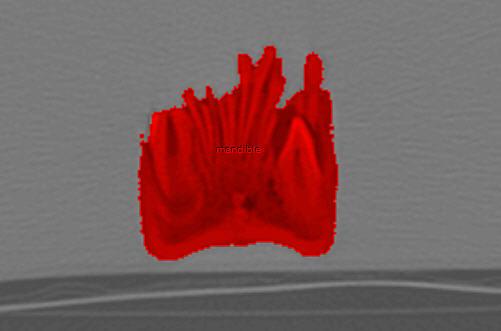
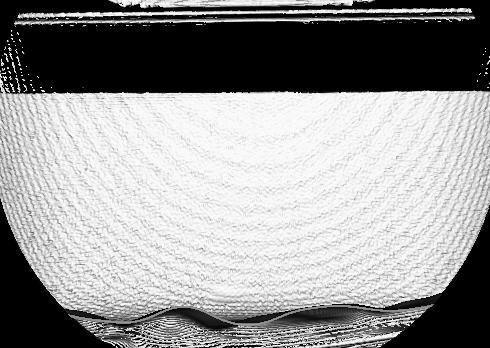
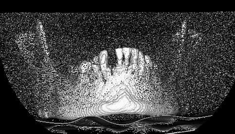
6 3D Rendering Techniques: 1. Volume Render Restricting Density Surface-Shaded Display: 2. Automatically Segmented & 3. Manually Defined g
 were")


7 Linear Measurements (8 linear, 2 angular) were based on landmarks common to published literature Volume & Regional Surface Area Measurements Measurements: Computer models vs. Anatomic Truth
8 RESULTS: 1. Linear 2. Volume 3. Surface Area
9 RESULTS: Linear Measurements

10 RESULTS: Linear Measurements Alg. Linear measurements from 3D-CT are accurate Linear accuracy is not sensitive to the CT scanning parameters or rendering techniques

11 RESULTS: Volume Measurements Alg. Only 1.25mm Slice Thickness for the Mandibles produced accurate group of volumes HOWEVER, all but one of the phantom object parameters produced accurate volumes Therefore, we looked further into the effect of slice thickness

12 RESULTS: Volume Measurements Algorithm 1. Models from 1.25mm original data = (7/9) groups within 0.05 ARE and remaining two approach threshold. 2. Models of 2.5mm slice thickness = NO groups achieve 0.05 ARE.

13 RESULTS: Volume Measurements (*) = group not statistically different from anatomic truth Increase slice thickness = groups of measurements stray farther from anatomic truth
 Volume measurements from thin slices are not sensitive to CT scanning parameters or 3D rendering")
14 To Summarize: Volume measurement accuracy is optimal from thin slices (1.25mm) Volume measurements from thin slices are not sensitive to CT scanning parameters or 3D rendering techniques THIN (1.25mm) vs. THICK (2.5mm)

15 Surface Area Measurements Separated by Rendering Mode Algorithm Surface Area (square cm) Volume Render Mand1 Mand2 Mand3 Auto ROI Manual ROI Volume Render Auto ROI Manual ROI Volume Render Auto ROI Manual ROI Volume Render Auto ROI Manual ROI Alg.

16 Surface area measurements are not accurate All measurements were 25-45% inflated Volume Render = Slice Summation Surface ROI= Stair-Stepping
17 CONCLUSIONS 1. Linear Measurements: ACCURATE NOT sensitive to scanning parameters or rendering techniques 2. Volume : THIN slices MOST ACCURATE NOT sensitive to scanning parameters or rendering techniques 3. Surface Area: NOT ACCURATE Inflated Measurements


18 Potential applications : _ Characterization and grading of bone fractures Orthopedic treatment and Surgical implants 3D-CT alongside 2D images helps increase rate of successful diagnoses Vocal Tract Development Lab subject scans can be used for researching: Speech production Biological basis of development Developmental Disorders
19 References: 1. Guitton, T. G., H. J. van der Werf, et al. (2010). "Quantitative three-dimensional computed tomography measurement of radial head fractures." J Shoulder Elbow Surg 19(7): Ravenel, J. G., W. M. Leue, et al. (2008). "Pulmonary nodule volume: effects of reconstruction parameters on automated measurements--a phantom study." Radiology 247(2): Ball, G., D. Woodside, et al. (2011). "Relationship between cervical vertebral maturation and mandibular growth." Am J Orthod Dentofacial Orthop 139(5): e El-Zanaty, H. M., A. R. El-Beialy, et al. (2010). "Three-dimensional dental measurements: An alternative to plaster models." Am J Orthod Dentofacial Orthop 137(2): Alkadhi, H., S. Wildermuth, et al. (2004). "Accuracy and time efficiency for the detection of thoracic cage fractures: volume rendering compared with transverse computed tomography images." J Comput Assist Tomogr 28(3): Ringl, H., R. Schernthaner, et al. (2009). "Three-dimensional fracture visualisation of multidetector CT of the skull base in trauma patients: comparison of three reconstruction algorithms." Eur Radiol 19(10):

20 QUESTIONS? Process of Volume Render Segmentation: Acknowledgements This work was supported by National Institute on Deafness and Other Communication Disorders NIH-NIDCD Grant R01 DC6282 (MRI and CT Studies of the Developing Vocal Tract)
NIH Public Access Author Manuscript Oral Surg Oral Med Oral Pathol Oral Radiol. Author manuscript; available in PMC 2014 May 01.
 NIH Public Access Author Manuscript Oral Surg Oral Med Oral Pathol Oral Radiol. Author manuscript; available in PMC 2014 May 01. Published in final edited form as: Oral Surg Oral Med Oral Pathol Oral Radiol.
Statement of Clinical Relevance
 The effect of computed tomographic scanner parameters and 3-dimensional volume rendering techniques on the accuracy of linear, angular, and volumetric measurements of the mandible Brian J. Whyms, BS, a
[PDR03] RECOMMENDED CT-SCAN PROTOCOLS
![[PDR03] RECOMMENDED CT-SCAN PROTOCOLS](/thumbs/72/66454100.jpg "[PDR03] RECOMMENDED CT-SCAN PROTOCOLS") SURGICAL & PROSTHETIC DESIGN [PDR03] RECOMMENDED CT-SCAN PROTOCOLS WORK-INSTRUCTIONS DOCUMENT (CUSTOMER) RECOMMENDED CT-SCAN PROTOCOLS [PDR03_V1]: LIVE 1 PRESCRIBING SURGEONS Patient-specific implants,
SURGICAL & PROSTHETIC DESIGN [PDR03] RECOMMENDED CT-SCAN PROTOCOLS WORK-INSTRUCTIONS DOCUMENT (CUSTOMER) RECOMMENDED CT-SCAN PROTOCOLS [PDR03_V1]: LIVE 1 PRESCRIBING SURGEONS Patient-specific implants,
Shadow casting. What is the problem? Cone Beam Computed Tomography THE OBJECTIVES OF DIAGNOSTIC IMAGING IDEAL DIAGNOSTIC IMAGING STUDY LIMITATIONS
 Cone Beam Computed Tomography THE OBJECTIVES OF DIAGNOSTIC IMAGING Reveal pathology Reveal the anatomic truth Steven R. Singer, DDS srs2@columbia.edu IDEAL DIAGNOSTIC IMAGING STUDY Provides desired diagnostic
Optimisation of Toshiba Aquilion ONE Volume Imaging
 Optimisation of Toshiba Aquilion ONE Volume Imaging Jane Edwards, RPRSG Royal Free London NHS Foundation Trust Dr Mufudzi Maviki, Plymouth Hospitals NHS Trust Background In 2011/12 Radiology at RFH was
Qualitative Comparison of Conventional and Oblique MRI for Detection of Herniated Spinal Discs
 Qualitative Comparison of Conventional and Oblique MRI for Detection of Herniated Spinal Discs Doug Dean Final Project Presentation ENGN 2500: Medical Image Analysis May 16, 2011 Outline Review of the
Metal Artifact Reduction CT Techniques. Tobias Dietrich University Hospital Balgrist University of Zurich Switzerland
 Metal Artifact Reduction CT Techniques R S S S Tobias Dietrich University Hospital Balgrist University of Zurich Switzerland N. 1 v o 4 1 0 2. Postoperative CT Metal Implants CT is accurate for assessment
Virtual Phantoms for IGRT QA
 TM Virtual Phantoms for IGRT QA Why ImSimQA? ImSimQA was developed to overcome the limitations of physical phantoms for testing modern medical imaging and radiation therapy software systems, when there
Imaging protocols for navigated procedures
 9732379 G02 Rev. 1 2015-11 Imaging protocols for navigated procedures How to use this document This document contains imaging protocols for navigated cranial, DBS and stereotactic, ENT, and spine procedures
Optimization of CT Simulation Imaging. Ingrid Reiser Dept. of Radiology The University of Chicago
 Optimization of CT Simulation Imaging Ingrid Reiser Dept. of Radiology The University of Chicago Optimization of CT imaging Goal: Achieve image quality that allows to perform the task at hand (diagnostic
Biomet PMI. Patient-Matched Implants. CT Protocols
 CT Protocols One Surgeon. One Patient. Over 1 million times per year, Biomet helps one surgeon provide personalized care to one patient. The science and art of medical care is to provide the right solution
Background. Outline. Radiographic Tomosynthesis: Image Quality and Artifacts Reduction 1 / GE /
 Radiographic Tomosynthesis: Image Quality and Artifacts Reduction Baojun Li, Ph.D Department of Radiology Boston University Medical Center 2012 AAPM Annual Meeting Background Linear Trajectory Tomosynthesis
Volume assessment accuracy in computed tomography: a phantom study
 JOURNAL OF APPLIED CLINICAL MEDICAL PHYSICS, VOLUME 11, NUMBER 2, SPRING 2010 Volume assessment accuracy in computed tomography: a phantom study Nicolas D. Prionas, a Shonket Ray, John M. Boone Department
CT Protocol Clinical Graphics Move Forward 3D motion analysis service
 CT Protocol Clinical Graphics Move Forward 3D motion analysis service Version 1.4 Description of contents This document describes the CT protocol of scans for Clinical Graphics Move Forward 3D Motion Simulation
MODELLING OF PROSTHETIC HIP JOINT GENERATED FROM CT SCAN DATA Mahender Koduri 1, G Krishna Teja 2, O Rajender 3 1,2,3
 MODELLING OF PROSTHETIC HIP JOINT GENERATED FROM CT SCAN DATA Mahender Koduri 1, G Krishna Teja 2, O Rajender 3 1,2,3 Asst. Professor, Dept. of Mech. Engg. AGI ABSTRACT Total hip arthroplasty is a surgical
Evaluation of 1D, 2D and 3D nodule size estimation by radiologists for spherical and non-spherical nodules through CT thoracic phantom imaging
 Evaluation of 1D, 2D and 3D nodule size estimation by radiologists for spherical and non-spherical nodules through CT thoracic phantom imaging Nicholas Petrick, Hyun J. Grace Kim, David Clunie, Kristin
Integrated System for Planning Peripheral Bronchoscopic Procedures
 Integrated System for Planning Peripheral Bronchoscopic Procedures Jason D. Gibbs, Michael W. Graham, Kun-Chang Yu, and William E. Higgins Penn State University Dept. of Electrical Engineering University
HHS Public Access Author manuscript Med Image Comput Comput Assist Interv. Author manuscript; available in PMC 2018 March 20.
 Online Statistical Inference for Large-Scale Binary Images Moo K. Chung 1,2, Ying Ji Chuang 2, and Houri K. Vorperian 2 1 Department of Biostatistics and Medical Informatics, University of Wisconsin, Madison,
Automated Image Analysis Software for Quality Assurance of a Radiotherapy CT Simulator
 Automated Image Analysis Software for Quality Assurance of a Radiotherapy CT Simulator Andrew J Reilly Imaging Physicist Oncology Physics Edinburgh Cancer Centre Western General Hospital EDINBURGH EH4
Ch. 4 Physical Principles of CT
 Ch. 4 Physical Principles of CT CLRS 408: Intro to CT Department of Radiation Sciences Review: Why CT? Solution for radiography/tomography limitations Superimposition of structures Distinguishing between
Spiral CT. Protocol Optimization & Quality Assurance. Ge Wang, Ph.D. Department of Radiology University of Iowa Iowa City, Iowa 52242, USA
 Spiral CT Protocol Optimization & Quality Assurance Ge Wang, Ph.D. Department of Radiology University of Iowa Iowa City, Iowa 52242, USA Spiral CT Protocol Optimization & Quality Assurance Protocol optimization
Heat Kernel Smoothing Using Laplace-Beltrami Eigenfunctions
 Heat Kernel Smoothing Using Laplace-Beltrami Eigenfunctions Seongho Seo 1, Moo K. Chung 1,2,3, and Houri K. Vorperian 4 1 Department of Brain and Cognitive Sciences Seoul National University, Korea 2 Department
Human craniofacial patterns were first analyzed
 ORIGINAL ARTICLE Three-dimensional accuracy of measurements made with software on cone-beam computed tomography images Manuel O. Lagravère, a Jason Carey, b Roger W. Toogood, c and Paul W. Major d Edmonton,
Motion artifact detection in four-dimensional computed tomography images
 Motion artifact detection in four-dimensional computed tomography images G Bouilhol 1,, M Ayadi, R Pinho, S Rit 1, and D Sarrut 1, 1 University of Lyon, CREATIS; CNRS UMR 5; Inserm U144; INSA-Lyon; University
Anatomic Growth Modelling of Cleft Palate Shape. S.K. Chua*, S.H. Ong*, K.W.C. Foong**
 ABSTRACT Anatomic Growth Modelling of Cleft Palate Shape S.K. Chua*, S.H. Ong*, K.W.C. Foong** *Dept of Electrical and Computer Engineering, **Dept of Preventive Dentistry, National University of Singapore
Head and Neck Lymph Node Region Delineation with Auto-segmentation and Image Registration
 Head and Neck Lymph Node Region Delineation with Auto-segmentation and Image Registration Chia-Chi Teng Department of Electrical Engineering University of Washington 1 Outline Introduction Related Work
3/27/2012 WHY SPECT / CT? SPECT / CT Basic Principles. Advantages of SPECT. Advantages of CT. Dr John C. Dickson, Principal Physicist UCLH
 3/27/212 Advantages of SPECT SPECT / CT Basic Principles Dr John C. Dickson, Principal Physicist UCLH Institute of Nuclear Medicine, University College London Hospitals and University College London john.dickson@uclh.nhs.uk
Towards full-body X-ray images
 Towards full-body X-ray images Christoph Luckner 1,2, Thomas Mertelmeier 2, Andreas Maier 1, Ludwig Ritschl 2 1 Pattern Recognition Lab, FAU Erlangen-Nuernberg 2 Siemens Healthcare GmbH, Forchheim christoph.luckner@fau.de
CT NOISE POWER SPECTRUM FOR FILTERED BACKPROJECTION AND ITERATIVE RECONSTRUCTION
 CT NOISE POWER SPECTRUM FOR FILTERED BACKPROJECTION AND ITERATIVE RECONSTRUCTION Frank Dong, PhD, DABR Diagnostic Physicist, Imaging Institute Cleveland Clinic Foundation and Associate Professor of Radiology
Manuscript Information. Manuscript Files
 Manuscript Information Journal name: Journal of computer assisted tomography NIHMS ID: NIHMS Manuscript Title:A novel registration-based semi-automatic mandible segmentation pipeline using computed tomography
Profound understanding of anatomy
 ENGLISH Profound understanding of anatomy The unique Planmeca ProMax 3D product family offers equipment for all maxillofacial imaging. All volume sizes from the smallest special cases to whole head images
COMPREHENSIVE QUALITY CONTROL OF NMR TOMOGRAPHY USING 3D PRINTED PHANTOM
 COMPREHENSIVE QUALITY CONTROL OF NMR TOMOGRAPHY USING 3D PRINTED PHANTOM Mažena MACIUSOVIČ *, Marius BURKANAS *, Jonas VENIUS *, ** * Medical Physics Department, National Cancer Institute, Vilnius, Lithuania
8/2/2016. Measures the degradation/distortion of the acquired image (relative to an ideal image) using a quantitative figure-of-merit
 using a quantitative figure-of-merit") Ke Li Assistant Professor Department of Medical Physics and Department of Radiology School of Medicine and Public Health, University of Wisconsin-Madison This work is partially supported by an NIH Grant
Ke Li Assistant Professor Department of Medical Physics and Department of Radiology School of Medicine and Public Health, University of Wisconsin-Madison This work is partially supported by an NIH Grant
Tomographic Reconstruction
 Tomographic Reconstruction 3D Image Processing Torsten Möller Reading Gonzales + Woods, Chapter 5.11 2 Overview Physics History Reconstruction basic idea Radon transform Fourier-Slice theorem (Parallel-beam)
Automatic 3D Registration of Lung Surfaces in Computed Tomography Scans
 Automatic 3D Registration of Lung Surfaces in Computed Tomography Scans Margrit Betke, PhD 1, Harrison Hong, BA 1, and Jane P. Ko, MD 2 1 Computer Science Department Boston University, Boston, MA 02215,
Conference Biomedical Engineering
 Automatic Medical Image Analysis for Measuring Bone Thickness and Density M. Kovalovs *, A. Glazs Image Processing and Computer Graphics Department, Riga Technical University, Latvia * E-mail: mihails.kovalovs@rtu.lv
Contrast Enhancement with Dual Energy CT for the Assessment of Atherosclerosis
 Contrast Enhancement with Dual Energy CT for the Assessment of Atherosclerosis Stefan C. Saur 1, Hatem Alkadhi 2, Luca Regazzoni 1, Simon Eugster 1, Gábor Székely 1, Philippe Cattin 1,3 1 Computer Vision
Digital Image Processing
 Digital Image Processing SPECIAL TOPICS CT IMAGES Hamid R. Rabiee Fall 2015 What is an image? 2 Are images only about visual concepts? We ve already seen that there are other kinds of image. In this lecture
Acknowledgments. High Performance Cone-Beam CT of Acute Traumatic Brain Injury
 A. Sisniega et al. (presented at RSNA 214) High Performance Cone-Beam CT of Acute Traumatic Brain Injury A. Sisniega 1 W. Zbijewski 1, H. Dang 1, J. Xu 1 J. W. Stayman 1, J. Yorkston 2, N. Aygun 3 V. Koliatsos
Thresholding technique for accurate analysis of density and geometry in QCT, pqct and ÌCT images
 J Musculoskelet Neuronal Interact 2007; 7(1):9-16 2 nd International Conference on Osteoporosis and Bone Research, October 19-23, 2005 Hylonome Thresholding technique for accurate analysis of density and
QIBA PET Amyloid BC March 11, Agenda
 QIBA PET Amyloid BC March 11, 2016 - Agenda 1. QIBA Round 6 Funding a. Deadlines b. What projects can be funded, what cannot c. Discussion of projects Mechanical phantom and DRO Paul & John? Any Profile
TomoTherapy Related Projects. An image guidance alternative on Tomo Low dose MVCT reconstruction Patient Quality Assurance using Sinogram
 TomoTherapy Related Projects An image guidance alternative on Tomo Low dose MVCT reconstruction Patient Quality Assurance using Sinogram Development of A Novel Image Guidance Alternative for Patient Localization
Quantitative Image Analysis and 3-D Digital Reconstruction of Aortic Valve Leaflet
 Quantitative Image Analysis and 3-D Digital Reconstruction of Aortic Valve Leaflet Chi Zheng 1 Mentors: John A. Stella 2 and Michael S. Sacks, Ph. D. 2 1 Bioengineering and Bioinformatics Summer Institute,
Magnetic Resonance Elastography (MRE) of Liver Disease
 of Liver Disease") Magnetic Resonance Elastography (MRE) of Liver Disease Authored by: Jennifer Dolan Fox, PhD VirtualScopics Inc. jennifer_fox@virtualscopics.com 1-585-249-6231 1. Overview of MRE Imaging MRE is a magnetic
Magnetic Resonance Elastography (MRE) of Liver Disease Authored by: Jennifer Dolan Fox, PhD VirtualScopics Inc. jennifer_fox@virtualscopics.com 1-585-249-6231 1. Overview of MRE Imaging MRE is a magnetic
RADIOLOGY AND DIAGNOSTIC IMAGING
 Day 2 part 2 RADIOLOGY AND DIAGNOSTIC IMAGING Dr hab. Zbigniew Serafin, MD, PhD serafin@cm.umk.pl 2 3 4 5 CT technique CT technique 6 CT system Kanal K: RSNA/AAPM web module: CT Systems & CT Image Quality
Automated Model-Based Rib Cage Segmentation and Labeling in CT Images
 Automated Model-Based Rib Cage Segmentation and Labeling in CT Images Tobias Klinder 1,2,CristianLorenz 2,JensvonBerg 2, Sebastian P.M. Dries 2, Thomas Bülow 2,andJörn Ostermann 1 1 Institut für Informationsverarbeitung,
CLASS HOURS: 4 CREDIT HOURS: 4 LABORATORY HOURS: 0
 Revised 10/10 COURSE SYLLABUS TM 220 COMPUTED TOMOGRAPHY PHYSICS CLASS HOURS: 4 CREDIT HOURS: 4 LABORATORY HOURS: 0 CATALOG COURSE DESCRIPTION: This course is one of a three course set in whole body Computed
Michael Speiser, Ph.D.
 IMPROVED CT-BASED VOXEL PHANTOM GENERATION FOR MCNP MONTE CARLO Michael Speiser, Ph.D. Department of Radiation Oncology UT Southwestern Medical Center Dallas, TX September 1 st, 2012 CMPWG Workshop Medical
MDCT and 3D Workstations
 MDCT and 3D Workstations Scott A. Lipson, MD Associate Director of Imaging, Long Beach Memorial Medical Center, Long Beach, California MDCT and 3D Workstations A Practical How-To Guide and Teaching File
Online Statistical Inference for Quantifying Mandible Growth in CT Images
 Online Statistical Inference for Quantifying Mandible Growth in CT Images Moo K. Chung 1,2, Ying Ji Chuang 2, Houri K. Vorperian 2 1 Department of Biostatistics and Medical Informatics 2 Vocal Tract Development
icatvision Quick Reference
 icatvision Quick Reference Navigating the i-cat Interface This guide shows how to: View reconstructed images Use main features and tools to optimize an image. REMINDER Images are displayed as if you are
VieW 3D. 3D Post-Processing WorKstation THE THIRD DIMENSION. Version 3.1
 VieW 3D 3D Post-Processing WorKstation THE THIRD DIMENSION Version 3.1 iq-view 3D THE FULLY-FEATURED 3D MEDICAL IMAGING SOLUTION FOR RADIOLOGISTS iq-view 3D contains all components of iq-view with the
A closer look at CT scanning
 Vet Times The website for the veterinary profession https://www.vettimes.co.uk A closer look at CT scanning Author : Charissa Lee, Natalie Webster Categories : General, Vets Date : April 3, 2017 A basic
CT vs. VolumeScope: image quality and dose comparison
 CT vs. VolumeScope: image quality and dose comparison V.N. Vasiliev *a, A.F. Gamaliy **b, M.Yu. Zaytsev b, K.V. Zaytseva ***b a Russian Sci. Center of Roentgenology & Radiology, 86, Profsoyuznaya, Moscow,
Reduction of Metal Artifacts in Computed Tomographies for the Planning and Simulation of Radiation Therapy
 Reduction of Metal Artifacts in Computed Tomographies for the Planning and Simulation of Radiation Therapy T. Rohlfing a, D. Zerfowski b, J. Beier a, P. Wust a, N. Hosten a, R. Felix a a Department of
Computer-Tomography I: Principles, History, Technology
 Computer-Tomography I: Principles, History, Technology Prof. Dr. U. Oelfke DKFZ Heidelberg Department of Medical Physics (E040) Im Neuenheimer Feld 280 69120 Heidelberg, Germany u.oelfke@dkfz.de History
Online Statistical Inference for Large-Scale Binary Images
 Online Statistical Inference for Large-Scale Binary Images Moo K. Chung 1,2(B),YingJiChuang 2, and Houri K. Vorperian 2 1 Department of Biostatistics and Medical Informatics, University of Wisconsin, Madison,
Computed Tomography Imaging: CT Protocol Management. Caveat 8/3/2017
 Computed Tomography Imaging: CT Protocol Management Mark P. Supanich, Ph.D., DABR AAPM Annual Meeting 3 rd August, 2017 Slides at: goo.gl/k8n8jf Rush is an academic health system comprising Rush University
NON OB ULTRASOUND MORPHOMETRICS AS BIOMARKERS
 NON OB ULTRASOUND MORPHOMETRICS AS BIOMARKERS Brian S Garra Washington DC VA Medical Center and Division of Imaging & Applied Mathematics, OSEL, CDRH, FDA GOALS Review Major Types of Clinical Ultrasonic
Tracked surgical drill calibration
 Tracked surgical drill calibration An acetabular fracture is a break in the socket portion of the "ball-and-socket" hip joint. The majority of acetabular fractures are caused by some type of highenergy
Machine Learning for Medical Image Analysis. A. Criminisi
 Machine Learning for Medical Image Analysis A. Criminisi Overview Introduction to machine learning Decision forests Applications in medical image analysis Anatomy localization in CT Scans Spine Detection
2D-3D Registration using Gradient-based MI for Image Guided Surgery Systems
 2D-3D Registration using Gradient-based MI for Image Guided Surgery Systems Yeny Yim 1*, Xuanyi Chen 1, Mike Wakid 1, Steve Bielamowicz 2, James Hahn 1 1 Department of Computer Science, The George Washington
The theory and practical aspects of proton imaging proton radiography (prad) and proton tomography (pct)
 and proton tomography (pct)") The theory and practical aspects of proton imaging proton radiography (prad) and proton tomography (pct) Fritz DeJongh, ProtonVDA Inc August 30 2018 Stay away from negative people. They have a problem
The theory and practical aspects of proton imaging proton radiography (prad) and proton tomography (pct) Fritz DeJongh, ProtonVDA Inc August 30 2018 Stay away from negative people. They have a problem
Volumetric accuracy of cone-beam computed tomography
 Imaging Science in Dentistry 207; 47: 65-74 https://doi.org/0.5624/isd.207.47.3.65 Volumetric accuracy of cone-beam computed tomography Cheol-Woo Park, Jin-ho Kim, Yu-Kyeong Seo, Sae-Rom Lee, Ju-Hee Kang,
Automatic Lung Surface Registration Using Selective Distance Measure in Temporal CT Scans
 Automatic Lung Surface Registration Using Selective Distance Measure in Temporal CT Scans Helen Hong 1, Jeongjin Lee 2, Kyung Won Lee 3, and Yeong Gil Shin 2 1 School of Electrical Engineering and Computer
MINIMIZATION OF VOLUMETRIC ERRORS IN CAD MEDICAL MODELS USING 64 SLICE SPIRAL CT SCANNER. L. Krishnanand, A. Manmadhachary and Y.
 MINIMIZATION OF VOLUMETRIC ERRORS IN CAD MEDICAL MODELS USING 64 SLICE SPIRAL CT SCANNER L. Krishnanand, A. Manmadhachary and Y. Ravi Kumar Department of Mechanical Engineering,National Institute of Technology
Limitations of Projection Radiography. Stereoscopic Breast Imaging. Limitations of Projection Radiography. 3-D Breast Imaging Methods
 Stereoscopic Breast Imaging Andrew D. A. Maidment, Ph.D. Chief, Physics Section Department of Radiology University of Pennsylvania Limitations of Projection Radiography Mammography is a projection imaging
Comparison of Quality of Multiplanar Reconstructions and Direct Coronal Multidetector CT Scans of the Lung
 Osamu Honda 1 Takeshi Johkoh 1 Shuji Yamamoto 2 Mitsuhiro Koyama 1 Noriyuki Tomiyama 1 Takenori Kozuka 1 Seiki Hamada 1 Naoki Mihara 1 Hironobu Nakamura 1 Nestor L. Müller 3 Received January 2, 22; accepted
PURE. ViSION Edition PET/CT. Patient Comfort Put First.
 PURE ViSION Edition PET/CT Patient Comfort Put First. 2 System features that put patient comfort and safety first. Oncology patients deserve the highest levels of safety and comfort during scans. Our Celesteion
Extraction and recognition of the thoracic organs based on 3D CT images and its application
 1 Extraction and recognition of the thoracic organs based on 3D CT images and its application Xiangrong Zhou, PhD a, Takeshi Hara, PhD b, Hiroshi Fujita, PhD b, Yoshihiro Ida, RT c, Kazuhiro Katada, MD
AC : APPLICATION OF PARAMETRIC SOLID MODELING FOR ORTHOPEDIC STUDIES OF THE HUMAN SPINE
 AC 2011-2785: APPLICATION OF PARAMETRIC SOLID MODELING FOR ORTHOPEDIC STUDIES OF THE HUMAN SPINE Jorge Rodriguez, Western Michigan University Jorge Rodriguez is an Associate Professor in the Department
Visualisation : Lecture 1. So what is visualisation? Visualisation
 So what is visualisation? UG4 / M.Sc. Course 2006 toby.breckon@ed.ac.uk Computer Vision Lab. Institute for Perception, Action & Behaviour Introducing 1 Application of interactive 3D computer graphics to
1. Deployment of a framework for drawing a correspondence between simple figure of merits (FOM) and quantitative imaging performance in CT.
 and quantitative imaging performance in CT.") Progress report: Development of assessment and predictive metrics for quantitative imaging in chest CT Subaward No: HHSN6801000050C (4a) PI: Ehsan Samei Reporting Period: month 1-18 Deliverables: 1. Deployment
Progress report: Development of assessment and predictive metrics for quantitative imaging in chest CT Subaward No: HHSN6801000050C (4a) PI: Ehsan Samei Reporting Period: month 1-18 Deliverables: 1. Deployment
Fiber Selection from Diffusion Tensor Data based on Boolean Operators
 Fiber Selection from Diffusion Tensor Data based on Boolean Operators D. Merhof 1, G. Greiner 2, M. Buchfelder 3, C. Nimsky 4 1 Visual Computing, University of Konstanz, Konstanz, Germany 2 Computer Graphics
Chapter 48 3D video reconstructions of normal and abnormal human vocal folds
 405 48 3D video reconstructions of normal and abnormal human vocal folds Chapter 48 3D video reconstructions of normal and abnormal human vocal folds Jarosław Sova, Joanna Cieszyńska, Jarosław Kijewski
An Accuracy Approach to Robotic Microsurgery in the Ear
 An Accuracy Approach to Robotic Microsurgery in the Ear B. Bell¹,J.Salzmann 1, E.Nielsen 3, N.Gerber 1, G.Zheng 1, L.Nolte 1, C.Stieger 4, M.Caversaccio², S. Weber 1 ¹ Institute for Surgical Technologies
Computer Aided Diagnosis and Treatment Planning for Developmental Dysplasia of the Hip
 Computer Aided Diagnosis and Treatment Planning for Developmental Dysplasia of the Hip Bin Li *a, Hongbing Lu b, Wenli Cai c, Xiang Li d, Jie Meng e and Zhengrong Liang a a Department of Radiology, State
Reduction of metal streak artifacts in x-ray computed tomography using a transmission maximum a posteriori algorithm
 Reduction of metal streak artifacts in x-ray computed tomography using a transmission maximum a posteriori algorithm B. De Man 1, Student Member, IEEE, J. Nuyts 2, Member, IEEE,. Dupont 2, G. Marchal 3,
Spectral analysis of non-stationary CT noise
 Spectral analysis of non-stationary CT noise Kenneth M. Hanson Los Alamos Scientific Laboratory Int. Symposium and Course on Computed Tomography, Las Vegas, April 7-11, 1980 This presentation available
CT is the imaging technique of choice for the evaluation of
 ORIGINAL RESEARCH HEAD & NECK Performance of Iterative Image Reconstruction in CT of the Paranasal Sinuses: A Phantom Study B. Schulz, M. Beeres, B. Bodelle, R. Bauer, F. Al-Butmeh, A. Thalhammer, T.J.
By choosing to view this document, you agree to all provisions of the copyright laws protecting it.
 Copyright 2009 IEEE. Reprinted from 31 st Annual International Conference of the IEEE Engineering in Medicine and Biology Society, 2009. EMBC 2009. Sept. 2009. This material is posted here with permission
Scaling Calibration in the ATRACT Algorithm
 Scaling Calibration in the ATRACT Algorithm Yan Xia 1, Andreas Maier 1, Frank Dennerlein 2, Hannes G. Hofmann 1, Joachim Hornegger 1,3 1 Pattern Recognition Lab (LME), Friedrich-Alexander-University Erlangen-Nuremberg,
Planmeca ProMax 3D Max CBCT unit
 D00107623 1(6) Planmeca ProMax 3D Max CBCT unit Introduction The Planmeca ProMax 3D Max X-ray unit uses cone beam computerized tomography (CBCT) to produce threedimensional X-ray images. Panoramic and
RADIOMICS: potential role in the clinics and challenges
 27 giugno 2018 Dipartimento di Fisica Università degli Studi di Milano RADIOMICS: potential role in the clinics and challenges Dr. Francesca Botta Medical Physicist Istituto Europeo di Oncologia (Milano)
1 Introduction Goal of the study Study and thesis outline Background General MRI MRI Parameters...
 1 ABSTRACT Objectives: In current head and neck oncology practice, three-dimensional (3D) virtual planning of resection and reconstruction followed by guided surgery are standard of care. Multimodality
Medical Imaging Projects
 NSF REU MedIX Summer 2006 Medical Imaging Projects Daniela Stan Raicu, PhD http://facweb.cs.depaul.edu/research draicu@cs.depaul.edu Outline Medical Informatics Imaging Modalities Computed Tomography Medical
Lab Location: MRI, B2, Cardinal Carter Wing, St. Michael s Hospital, 30 Bond Street
 Lab Location: MRI, B2, Cardinal Carter Wing, St. Michael s Hospital, 30 Bond Street MRI is located in the sub basement of CC wing. From Queen or Victoria, follow the baby blue arrows and ride the CC south
Proton dose calculation algorithms and configuration data
 Proton dose calculation algorithms and configuration data Barbara Schaffner PTCOG 46 Educational workshop in Wanjie, 20. May 2007 VARIAN Medical Systems Agenda Broad beam algorithms Concept of pencil beam
Computed tomography of simple objects. Related topics. Principle. Equipment TEP Beam hardening, artefacts, and algorithms
 Related topics Beam hardening, artefacts, and algorithms Principle The CT principle is demonstrated with the aid of simple objects. In the case of very simple targets, only a few images need to be taken
Reproducibility of interactive registration of 3D CT and MR pediatric treatment planning head images
 JOURNAL OF APPLIED CLINICAL MEDICAL PHYSICS, VOLUME 2, NUMBER 3, SUMMER 2001 Reproducibility of interactive registration of 3D CT and MR pediatric treatment planning head images Jaap Vaarkamp* Department
Medical rapid prototyping technologies: state of the art and current limitations for application in oral and maxillofacial surgery
 Loughborough University Institutional Repository Medical rapid prototyping technologies: state of the art and current limitations for application in oral and maxillofacial surgery This item was submitted
Improvement of contrast using reconstruction of 3D Image by PET /CT combination system
 Available online at www.pelagiaresearchlibrary.com Advances in Applied Science Research, 2013, 4(1):285-290 ISSN: 0976-8610 CODEN (USA): AASRFC Improvement of contrast using reconstruction of 3D Image
Evaluation of surface and volume rendering in 3D-CT of facial fractures
 (2006) 35, 227 231 q 2006 The British Institute of Radiology http://dmfr.birjournals.org RESEARCH Evaluation of surface and volume rendering in 3D-CT of facial fractures T Rodt*,1, SO Bartling 2, JE Zajaczek
Determination of rotations in three dimensions using two-dimensional portal image registration
 Determination of rotations in three dimensions using two-dimensional portal image registration Anthony E. Lujan, a) James M. Balter, and Randall K. Ten Haken Department of Nuclear Engineering and Radiological
Physical bases of X-ray diagnostics
 Physical bases of X-ray diagnostics Dr. István Voszka Possibilities of X-ray production (X-ray is produced, when charged particles of high velocity are stopped) X-ray tube: Relatively low accelerating
Frequency split metal artifact reduction (FSMAR) in computed tomography
 in computed tomography") The Johns Hopkins University Advanced Computer Integrated Surgery Group 4 Metal Artifact Removal in C-arm Cone-Beam CT Paper Seminar Critical Review of Frequency split metal artifact reduction (FSMAR)
The Johns Hopkins University Advanced Computer Integrated Surgery Group 4 Metal Artifact Removal in C-arm Cone-Beam CT Paper Seminar Critical Review of Frequency split metal artifact reduction (FSMAR)
Dolphin 3D Imaging 11.7 beta
 Dolphin 3D Imaging 11.7 beta The Dolphin 3D software module is a powerful tool that makes processing 3D data extremely simple, enabling dental specialists from a wide variety of disciplines to diagnose,
Slide 1. Technical Aspects of Quality Control in Magnetic Resonance Imaging. Slide 2. Annual Compliance Testing. of MRI Systems.
 Slide 1 Technical Aspects of Quality Control in Magnetic Resonance Imaging Slide 2 Compliance Testing of MRI Systems, Ph.D. Department of Radiology Henry Ford Hospital, Detroit, MI Slide 3 Compliance Testing
Process to Convert DICOM Data to 3D Printable STL Files
 HOW-TO GUIDE Process to Convert DICOM Data to 3D Printable STL Files Mac Cameron, Application Engineer Anatomical models have several applications in the medical space from patient-specific models used
Dosimetric Analysis Report
 RT-safe 48, Artotinis str 116 33, Athens Greece +30 2107563691 info@rt-safe.com Dosimetric Analysis Report SAMPLE, for demonstration purposes only Date of report: ----------- Date of irradiation: -----------