Bioimage Informatics
|
|
|
- Gervais Underwood
- 6 years ago
- Views:
Transcription
1 Bioimage Informatics Lecture 10, Spring 2012 Bioimage Data Analysis (II): Applications of Point Feature Detection Techniques: Super Resolution Fluorescence Microscopy Bioimage Data Analysis (III): Edge Detection Lecture 10 February 20,
2 List of Groups Group ID Group Member 01 Group Member 02 Group Member 03 1 Fangming Lu Wei Zhou Ruiqin Li 2 Xiaoyi Fu Martin Jaszewski Frank Yeh 3 Sombeet Sahu Madhumitha Raghu Arun Sampath 4 Divya Kagoo Sowmya Aggarwal Hao-Chih Lee 5 Samantha Horvath Akanksha Bhardwaj Jaclyn Brackett 6 Yutong Li Dolsarit Somseang Weilong Wang 2
3 Outline Overview of super-resolution fluorescence microscopy (SRFM) SRFM by random fluorophore switching SRFM by deterministic fluorophore switching SRFM by structural illumination Introduction to edge detection 3
4 Overview of super-resolution fluorescence microscopy (SRFM) SRFM by random fluorophore switching SRFM by deterministic fluorophore switching SRFM by structural illumination Introduction to edge detection 4

5 Why We Need Super Resolution Fluorescence Microscopy (I) Crystallography, NMR, spectroscopy - Resolution: 1 nm - Live/physiological condition: NO Samples must be specially prepared Electron microscopy - Resolution: between 1nm &100nm - Live/physiological condition: NO Samples must be fixed; limited specificity Light microscopy - Resolution: 100nm - Live/physiological condition: YES 5
6 Why We Need Super Resolution Fluorescence Microscopy (II) Most cellular components are smaller than the diffraction limit of visible light. Fluorescence microscopy allows the visualization of cellular processes live under physiological conditions. - Not available under electron microscopy Fluorescence microscopy can achieve high specificity. - Not available under electron microscopy Fluorescence microscopy can achieve multiplexity (i.e. simultaneous multi-color imaging). - Not available under electron microscopy 6
 NAn sin n: refractive index of the medium between the lens and the specimen : half of the angular aperture - Water")
7 Performance Metrics of a Fluorescence Microscope Resolution: - Rayleigh limit: D 061. NA - Sparrow limit: D 047. NA Numerical aperture (NA) NAn sin n: refractive index of the medium between the lens and the specimen : half of the angular aperture - Water n= Immersion oil n=1.51
8 There are Important Exceptions Well separated particles can be localized with an accuracy much smaller than the diffraction limit. s a /12 8 s b N N a N i i si 2.2 NA a: pixel size b: background (in photons) N: total number of photons 8
: Fluorophore")
9 Basic Ideas of SRFM (I): Fluorophore Switching

10 Microscope Image Formation Microscope image formation can be modeled as a convolution with the PSF. I x,y O x,y psf x,y F I x,y F O x,y F psf x,y 10


11 Basic Ideas of SRFM (II): Structural Illumination
12 Overview of super-resolution fluorescence microscopy (SRFM) SRFM by random fluorophore switching SRFM by deterministic fluorophore switching SRFM by structural illumination Introduction to edge detection 12


13 Stochastic Optical Reconstruction Microscopy (STORM) Huang et al, Three-Dimensional Super-Resolution Imaging by Stochastic Optical Reconstruction Microscopy, Science, 319: , 2008 M. J. Rust et al, Sub-diffraction-limit imaging by stochastic optical reconstruction microscopy (STORM). Nature Methods, 10: , 2006.
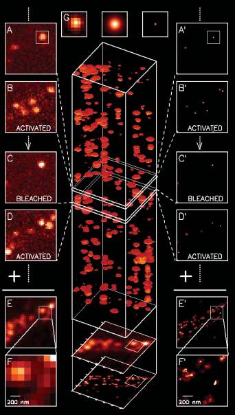
14 Photoactivation Localization Microscopy (PALM) 14
15 Overview of super-resolution fluorescence microscopy (SRFM) SRFM by random fluorophore switching SRFM by deterministic fluorophore switching SRFM by structural illumination Introduction to edge detection 15


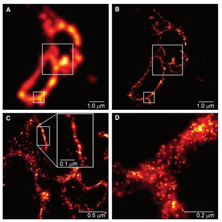
16 Stimulated Emission Depletion (I) T. A. Klar et al, Fluorescence microscopy with diffraction resolution barrier broken by stimulated emission, PNAS, 97: , 2000.


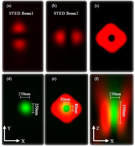
17 Stimulated Emission Depletion (II)
18 Comments STED - Photodamage - Deterministic; Live imaging possible STORM/PALM - Less photodamage - Stochastic; Live imaging challenging - Possible artifacts New possibilities for quantitative image analysis
19 Overview of super-resolution fluorescence microscopy (SRFM) SRFM by random fluorophore switching SRFM by deterministic fluorophore switching SRFM by structural illumination Introduction to edge detection 19


20 Moire Pattern Subdiffraction Multicolor Imaging of the Nuclear Periphery with 3D Structured Illumination Microscopy, Lothar Schermelleh, et al, Science Vol. 320, 1332 (2008 ) 20
21 3D Structural Illumination Microscopy The basic idea is to collect more information by extending the support in frequency domain. Use structural illumination Use three illumination angles Combination of imaging and computation Subdiffraction Multicolor Imaging of the Nuclear Periphery with 3D Structured Illumination Microscopy, Lothar Schermelleh, et al, Science Vol. 320, 1332 (2008 )

22 3D Structural Illumination Microscopy To use structural illumination to capture image information at higher-spatial frequency. Three illumination angles; Five frames per angle. Final images are generated by computational reconstruction. Subdiffraction Multicolor Imaging of the Nuclear Periphery with 3D Structured Illumination Microscopy, Lothar Schermelleh, et al, Science Vol. 320, 1332 (2008 )


23 Structural Illumination Scheme Three-Dimensional Resolution Doubling in Wide-Field Fluorescence Microscopy by Structured Illumination, Mats G. Gustafsson, et al. Biophysical Journal Vol. 94, , 2008


24 3D Structural Illumination Microscopy Three-Dimensional Resolution Doubling in Wide-Field Fluorescence Microscopy by Structured Illumination Mats G. Gustafsson, et al. Biophysical Journal Vol. 94, , 2008
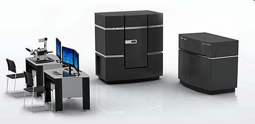

25 Commercial System From AppliedPrecision
26 References Sedat Lab Agard Lab Gustafsson Lab


27 4 Pi Microscopy (I) A technique to improve axial resolution by several folds. Egner & Hell, Fluorescence microscopy with super-resolved optical sections, Trends in Cell Biology, 15: , 2005.

28 4 Pi Microscopy (II) 28


29 4 Pi Microscopy (III) A technique to improve axial resolution by several folds.


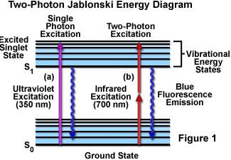
30 4 Pi Microscopy (IV) Absorption of two photons must happen within sec. c E hvh Maria Göppert-Mayer


31 4 Pi Microscopy (V) Nagorni, M. & Hell, S.W. 4Pi-confocal microscopy provides three-dimensional images of the microtubule network with 100- to 150-nm resolution. J. Struct. Biol. 123, (1998).
32 Advantages 4 Pi Microscopy (VI) - Substantial improvement in axial resolution Disadvantages - No improvement in lateral resolution - Artifacts due to side lobes - Complexity, sample restriction, cost


33 I 5 M Microscopy (I) Shao et al, I 5 S: wide-field light microscopy with 100-nm-scale resolution in three dimensions,biophysical Journal, 94: , 2008.


34 I5M Microscopy (II) Shao et al, I5S: wide-field light microscopy with 100-nm-scale resolution in three. dimensions, Biophysical Journal, 94: , 2008
![Some References [1] S. W. Hell, Microscopy and its focal switch, Nature Methods, vol. 6, no.1, pp. 24-32, 2009. [2] J.](/docs-images/77/76310076/images/35-0.jpg "Lippincott-Schwartz & S. Manley, Putting super-resolution fluorescence microscopy to work, Nature Methods, vol.6, no.1, pp.")
35 Some References [1] S. W. Hell, Microscopy and its focal switch, Nature Methods, vol. 6, no.1, pp , [2] J. Lippincott-Schwartz & S. Manley, Putting super-resolution fluorescence microscopy to work, Nature Methods, vol.6, no.1, pp , 2009.
36 Overview of super-resolution fluorescence microscopy (SRFM) SRFM by random fluorophore switching SRFM by deterministic fluorophore switching SRFM by structural illumination Introduction to edge detection 36
37 What is an edge? Edge Detection An edge point, or an edge, is a pixel at or around which the image values undergo a sharp change. 37
38 Type I: Steps Typical Cases of Edges Type II: Ridges The edge can be thought of as a 1D signal when observed under the normal direction. 38

39 Edge Detection Procedure Step I: noise suppression Step II: edge enhancement Step III: Edge localization 39
40 Noise Suppression Low pass filter using a Gaussian kernel Canny, IEEE TPAMI, ,
41 Canny Edge Enhancement Step I: For each pixel I(x 0,y 0 ), calculate the gradient I x I y xx y y 0 0 I y Step II: Estimate edge strength I x 2 2 E x,y I x,y I x,y s 0 0 x 0 0 y 0 0 Step III: Estimation edge direction E x,y o 0 0 x I x,y arctan y I x,y
42 Combination of Noise Suppression and Gradient Estimation (I) A basic property of convolution G I DEF G G I DEF G GI x I GI y I x x y y 2 2 E x,y GI x, y GI x, y s 0 0 x 0 0 y 0 0 E x,y o 0 0 arctan GI GI y x x, y 0 0 x, y
43 Combination of Noise Suppression and Gradient Estimation (II) Gaussian kernel in 1D G x First order derivative Second order derivative x 2 2 e x x 2 2 Gx e G x e x 2 x x Zero-crossing 43


44 Image Gradient y
45 Non-Maximum Suppression For each pixel I(x 0,y 0 ),compare the edge strength along the direction perpendicular to the edge I y An edge point must have its edge strength no less than its two neighbors. 45
46 Hysteresis Thresholding The main purpose is to link detected edge points while minimizing breakage. Basic idea - Using two thresholds T L and T H - Starting from a point where edge gradient magnitude higher than T H - Link to neighboring edge points with edge gradient magnitude higher than T L 46
, Handbook on Computer Vision and Applications, volume 2, pp 239--274, Academic Press.")
47 Influence of Scale Selection on Edge Detection Lindeberg (1999) `` Principles for automatic scale selection'', in: B. J"ahne (et al., eds.), Handbook on Computer Vision and Applications, volume 2, pp , Academic Press. 47
48 Comments Gradient-based edge detection is effective and provides good localization accuracy. Gradient-based edge detection is sensitive to noise. How to achieve robust detection - Set robustness as a main goal in algorithm design - Integrate information at different scales - Integrate information from different sources 48
49 Questions? 49
Bioimage Informatics. Lecture 8, Spring Bioimage Data Analysis (II): Point Feature Detection
: Point Feature Detection") Bioimage Informatics Lecture 8, Spring 2012 Bioimage Data Analysis (II): Point Feature Detection Lecture 8 February 13, 2012 1 Outline Project assignment 02 Comments on reading assignment 01 Review: pixel
Bioimage Informatics Lecture 8, Spring 2012 Bioimage Data Analysis (II): Point Feature Detection Lecture 8 February 13, 2012 1 Outline Project assignment 02 Comments on reading assignment 01 Review: pixel
Optical Sectioning. Bo Huang. Pharmaceutical Chemistry
 Optical Sectioning Bo Huang Pharmaceutical Chemistry Approaches to 3D imaging Physical cutting Technical difficulty Highest resolution Highest sensitivity Optical sectioning Simple sample prep. No physical
Bioimage Informatics
 Bioimage Informatics Lecture 12, Spring 2012 Bioimage Data Analysis (III): Line/Curve Detection Bioimage Data Analysis (IV) Image Segmentation (part 1) Lecture 12 February 27, 2012 1 Outline Review: Line/curve
COLOCALISATION. Alexia Ferrand. Imaging Core Facility Biozentrum Basel
 COLOCALISATION Alexia Ferrand Imaging Core Facility Biozentrum Basel OUTLINE Introduction How to best prepare your samples for colocalisation How to acquire the images for colocalisation How to analyse
Image restoration by deconvolution
 Image restoration by deconvolution chong.zhang@bioquant.uni-heidelberg.de 17/12/2014 (part) Slides courtesy: Sébastien Tosi (IRB Barcelona) A few concepts related to the topic Convolution Deconvolution
Nonlinear optics and two photon microscopy. Table of contents. Sam Whiteley and Seth Parker PHYS 173/BGGN 266 July 13, 2014
 Nonlinear optics and two photon microscopy Sam Whiteley and Seth Parker PHYS 173/BGGN 266 July 13, 2014 Table of contents 1. Introduction 2. Optical setup 3. Initial images and troubleshooting 4. Determining
Super-Resolved Spatial Light Interference Microscopy
 BOSTON UNIVERSITY Department of Electrical and Computer Engineering RPC Report Super-Resolved Spatial Light Interference Microscopy by OGUZHAN AVCI B.S., Bilkent University, 2012 Submitted in partial fulfillment
DeltaVision OMX SR super-resolution microscope. Super-resolution doesn t need to be complicated
 DeltaVision OMX SR super-resolution microscope Super-resolution doesn t need to be complicated Live-cell super-resolution microscopy DeltaVision OMX SR is a compact super-resolution microscope system optimized
Thomas Abraham, PhD
 Thomas Abraham, PhD (tabraham1@hmc.psu.edu) What is Deconvolution? Deconvolution, also termed as Restoration or Deblurring is an image processing technique used in a wide variety of fields from 1D spectroscopy
Edge Detection. CMPUT 206: Introduction to Digital Image Processing. Nilanjan Ray. Source:
 Edge Detection CMPUT 206: Introduction to Digital Image Processing Nilanjan Ray Source: www.imagingbook.com What are edges? Are image positions where local image intensity changes significantly along a
COLOCALISATION. Alexia Loynton-Ferrand. Imaging Core Facility Biozentrum Basel
 COLOCALISATION Alexia Loynton-Ferrand Imaging Core Facility Biozentrum Basel OUTLINE Introduction How to best prepare your samples for colocalisation How to acquire the images for colocalisation How to
Chuang, Chin-Jung (2011) Proximity projection grating structured illumination microscopy. PhD thesis, University of Nottingham.
 Proximity projection grating structured illumination microscopy. PhD thesis, University of Nottingham.") Chuang, Chin-Jung (2011) Proximity projection grating structured illumination microscopy. PhD thesis, University of Nottingham. Access from the University of Nottingham repository: http://eprints.nottingham.ac.uk/12262/1/pgsim_chinjung_chuang.pdf
Chuang, Chin-Jung (2011) Proximity projection grating structured illumination microscopy. PhD thesis, University of Nottingham. Access from the University of Nottingham repository: http://eprints.nottingham.ac.uk/12262/1/pgsim_chinjung_chuang.pdf
Depth-variant blind restoration with pupil-phase constraints for 3D confocal microscopy
 Journal of Physics: Conference Series OPEN ACCESS Depth-variant blind restoration with pupil-phase constraints for 3D confocal microscopy To cite this article: Saima Ben Hadj et al 2013 J. Phys.: Conf.
Supporting Information for Azimuthal Polarization Filtering for Accurate, Precise, and Robust Single-Molecule Localization Microscopy
 Nano Letters Supporting Information for Azimuthal Polarization Filtering for Accurate, Precise, and Robust Single-Molecule Localization Microscopy Matthew D. Lew, and W. E. Moerner *, Departments of Chemistry
SPATIAL DENSITY ESTIMATION BASED SEGMENTATION OF SUPER-RESOLUTION LOCALIZATION MICROSCOPY IMAGES
 SPATIAL DENSITY ESTIMATION ASED SEGMENTATION OF SUPER-RESOLUTION LOCALIZATION MICROSCOPY IMAGES Kuan-Chieh Jackie Chen 1,2,3, Ge Yang 1,2, and Jelena Kovačević 3,1,2 1 Dept. of iomedical Eng., 2 Center
Introductory Guide to Light Microscopy - Biomedical Confocal Microscopy
 Introductory Guide to Light Microscopy - Biomedical Confocal Microscopy 7 May 2007 Michael Hooker Microscopy Facility Michael Chua microscopy@unc.edu 843-3268 6007 Thurston Bowles Wendy Salmon wendy_salmon@med.unc.edu
Single particle tracking: principles and applications
 Single particle tracking: principles and applications Valeria Levi Laboratorio de Dinámica Intracelular Facultad de Ciencias Exactas y Naturales Universidad de Buenos Aires vlevi1@gmail.com Why single
A SUPER-RESOLUTION MICROSCOPY WITH STANDING EVANESCENT LIGHT AND IMAGE RECONSTRUCTION METHOD
 A SUPER-RESOLUTION MICROSCOPY WITH STANDING EVANESCENT LIGHT AND IMAGE RECONSTRUCTION METHOD Hiroaki Nishioka, Satoru Takahashi Kiyoshi Takamasu Department of Precision Engineering, The University of Tokyo,
Edge Detection. CSE 576 Ali Farhadi. Many slides from Steve Seitz and Larry Zitnick
 Edge Detection CSE 576 Ali Farhadi Many slides from Steve Seitz and Larry Zitnick Edge Attneave's Cat (1954) Origin of edges surface normal discontinuity depth discontinuity surface color discontinuity
Chapter 4 Microscopy
 Chapter 4 Microscopy Gabriel Popescu University of Illinois at Urbana Champaign Beckman Institute Quantitative Light Imaging Laboratory http://light.ece.uiuc.edu Principles of Optical Imaging Electrical
Leica TCS STED CW. The Fast Track to Superresolution. Leica TCS STED CW
 The Fast Track to Superresolution content Motivation Concept Realisation Applications - why do researchers need the TCS STED CW? - what is the TCS STED CW based on? - how does the TCS STED CW work? - what
Detection of Sub-resolution Dots in Microscopy Images
 Detection of Sub-resolution Dots in Microscopy Images Karel Štěpka, 2012 Centre for Biomedical Image Analysis, FI MU supervisor: prof. RNDr. Michal Kozubek, Ph.D. Outline Introduction Existing approaches
Lecture 7: Most Common Edge Detectors
 #1 Lecture 7: Most Common Edge Detectors Saad Bedros sbedros@umn.edu Edge Detection Goal: Identify sudden changes (discontinuities) in an image Intuitively, most semantic and shape information from the
Vutara 350. Innovation with Integrity. The Fastest, Super-Resolution Microscope Deep 3D Imaging on Live Cells, Quickly and Easily
 Vutara 350 The Fastest, Super-Resolution Microscope Deep 3D Imaging on Live Cells, Quickly and Easily Innovation with Integrity Fluorescence Microscopy Vutara 350 Don t Get Left Behind Bruker s Vutara
Bioimage Informatics
 Bioimage Informatics Lecture 13, Spring 2012 Bioimage Data Analysis (IV) Image Segmentation (part 2) Lecture 13 February 29, 2012 1 Outline Review: Steger s line/curve detection algorithm Intensity thresholding
NDD FLIM Systems for Leica SP2 MP and SP5 MP Multiphoton Microscopes
 NDD FLIM Systems for Leica SP2 MP and SP5 MP Multiphoton Microscopes bh FLIM systems for the confocal and the multiphoton versions of the Leica SP2 and SP5 microscopes are available since 2002 [4]. These
Digital Image Processing COSC 6380/4393
 Digital Image Processing COSC 6380/4393 Lecture 21 Nov 16 th, 2017 Pranav Mantini Ack: Shah. M Image Processing Geometric Transformation Point Operations Filtering (spatial, Frequency) Input Restoration/
Live-cell 3D super-resolution imaging in thick biological samples
 Nature Methods Live-cell 3D super-resolution imaging in thick biological samples Francesca Cella Zanacchi, Zeno Lavagnino, Michela Perrone Donnorso, Alessio Del Bue, Lauria Furia, Mario Faretta & Alberto
CS5670: Computer Vision
 CS5670: Computer Vision Noah Snavely Lecture 2: Edge detection From Sandlot Science Announcements Project 1 (Hybrid Images) is now on the course webpage (see Projects link) Due Wednesday, Feb 15, by 11:59pm
FLUOVIEW FV1000/FV1200
 FLUOVIEW FV1000/FV1200 UPGRADE TO 3D NANOIMAGING AND SINGLE MOLECULE TRACKING FOR OLYMPUS FLUOVIEW FV1000/FV1200 Within the past few years, several methods have been devised in order to obtain images with
Generalized Hough Transform, line fitting
 Generalized Hough Transform, line fitting Introduction to Computer Vision CSE 152 Lecture 11-a Announcements Assignment 2: Due today Midterm: Thursday, May 10 in class Non-maximum suppression For every
Last class. This class. Single molecule imaging Deconvolution. FLIM Confocal
 FLIM, Confocal Last class Single molecule imaging Deconvolution This class FLIM Confocal FLIM Fluorescence Lifetime IMaging FLIM Fluorescence lifetime imaging Easier and more accurate quantitation Much
PH880 Topics in Physics
 PH880 Topics in Physics Modern Optical Imaging (Fall 2010) Overview of week 4 Monday PSF, OTF Bright field microscopy Resolution/NA Deconvolution Wednesday : holiday Impulse response (PSF) in imaging system
SURVEY ON IMAGE PROCESSING IN THE FIELD OF DE-NOISING TECHNIQUES AND EDGE DETECTION TECHNIQUES ON RADIOGRAPHIC IMAGES
 SURVEY ON IMAGE PROCESSING IN THE FIELD OF DE-NOISING TECHNIQUES AND EDGE DETECTION TECHNIQUES ON RADIOGRAPHIC IMAGES 1 B.THAMOTHARAN, 2 M.MENAKA, 3 SANDHYA VAIDYANATHAN, 3 SOWMYA RAVIKUMAR 1 Asst. Prof.,
Edge detection. Goal: Identify sudden. an image. Ideal: artist s line drawing. object-level knowledge)
") Edge detection Goal: Identify sudden changes (discontinuities) in an image Intuitively, most semantic and shape information from the image can be encoded in the edges More compact than pixels Ideal: artist
Edge detection Goal: Identify sudden changes (discontinuities) in an image Intuitively, most semantic and shape information from the image can be encoded in the edges More compact than pixels Ideal: artist
3-D Reconstruction and Measurement of Microtubules from Multiple Angle-Total Internal Reflection Fluorescence Microscopy
 3-D Reconstruction and Measurement of Microtubules from Multiple Angle-Total Internal Reflection Fluorescence Microscopy Qian Yang Alexander Karpikov Derek Toomre James Duncan Yale University New Haven,
Identify Curvilinear Structure Based on Oriented Phase Congruency in Live Cell Images
 International Journal of Information and Computation Technology. ISSN 0974-2239 Volume 3, Number 4 (2013), pp. 335-340 International Research Publications House http://www. irphouse.com /ijict.htm Identify
1. ABOUT INSTALLATION COMPATIBILITY SURESIM WORKFLOWS a. Workflow b. Workflow SURESIM TUTORIAL...
 SuReSim manual 1. ABOUT... 2 2. INSTALLATION... 2 3. COMPATIBILITY... 2 4. SURESIM WORKFLOWS... 2 a. Workflow 1... 3 b. Workflow 2... 4 5. SURESIM TUTORIAL... 5 a. Import Data... 5 b. Parameter Selection...
Edge Detection. Announcements. Edge detection. Origin of Edges. Mailing list: you should have received messages
 Announcements Mailing list: csep576@cs.washington.edu you should have received messages Project 1 out today (due in two weeks) Carpools Edge Detection From Sandlot Science Today s reading Forsyth, chapters
convolution shift invariant linear system Fourier Transform Aliasing and sampling scale representation edge detection corner detection
 COS 429: COMPUTER VISON Linear Filters and Edge Detection convolution shift invariant linear system Fourier Transform Aliasing and sampling scale representation edge detection corner detection Reading:
CFIM MICROSCOPY COURSE TIMETABLE PRINCIPLES OF MICROSCOPY MONDAY 6 TH OF JANUARY 2014 FRIDAY 10 TH OF JANUARY 2014
 MICROSCOPY COURSE TIMETABLE PRINCIPLES OF MICROSCOPY MONDAY 6 TH OF JANUARY 2014 FRIDAY 10 TH OF JANUARY 2014 CONFOCAL AND FLUORESCENCE MICROSCOPY MONDAY 20 TH OF JANUARY 2014 FRIDAY 24 TH OF JANUARY 2014
Exam Microscopic Measurement Techniques 4T th of April, 2008
 Exam Microscopic Measurement Techniques 4T300 29 th of April, 2008 Name / Initials: Ident. #: Education: This exam consists of 5 questions. Questions and sub questions will be rewarded with the amount
Super-resolution 3D Reconstruction of thick biological samples: a computer vision perspective
 2013 IEEE International Conference on Computer Vision Workshops Super-resolution 3D Reconstruction of thick biological samples: a computer vision perspective Alessio Del Bue, Francesca Cella Zanacchi and
Coherent Diffraction Imaging with Nano- and Microbeams
 Diffraction Imaging with Nano- and Microbeams Why does lensless need? Mark A Pfeifer Cornell High Energy Synchrotron Source Cornell University Ithaca, NY 14850 map322@cornell.edu XLD 2011 June 28, 2011
R. Heintzmann Max-Planck-Institute for Biophysical Chemistry D Göttingen, Germany
 Journal of Biomedical Optics 6(3), 292 299 (July 2001) Spatially modulated illumination microscopy: online visualization of intensity distribution and prediction of nanometer precision of axial distance
Image Processing
 Image Processing 159.731 Canny Edge Detection Report Syed Irfanullah, Azeezullah 00297844 Danh Anh Huynh 02136047 1 Canny Edge Detection INTRODUCTION Edges Edges characterize boundaries and are therefore
Filtering Images. Contents
 Image Processing and Data Visualization with MATLAB Filtering Images Hansrudi Noser June 8-9, 010 UZH, Multimedia and Robotics Summer School Noise Smoothing Filters Sigmoid Filters Gradient Filters Contents
Edge Detection. Ziv Yaniv School of Engineering and Computer Science The Hebrew University, Jerusalem, Israel.
 Edge Detection Ziv Yaniv School of Engineering and Computer Science The Hebrew University, Jerusalem, Israel. This lecture summary deals with the low level image processing task of edge detection. Edges
Principles of Light Microscopy
 Monday 8 August 2011 Principles of Light Microscopy 09:00 09:30 Introduction 09:30 10:15 The story of the microscope / 10:15 Coffee 10:30 12:45 Limitations of the eye. Resolution, contrast, magnification.
12/7/2012. Biomolecular structure. Diffraction, X-ray crystallography, light- and electron microscopy. CD spectroscopy, mass spectrometry
 phase difference at a given distance constructive/destructive interference Biomolecular structure. Diffraction, X-ray crystallography, light- and electron microscopy. CD spectroscopy, mass spectrometry
California at San Francisco, San Francisco, CA Howard Hughes Medical Institute. University, Princeton, NJ
 A Parallel Product-Convolution approach for representing the depth Point Spread Function in 3D widefield microscopy based on principal component analysis Muthuvel Arigovindan 1, Joshua Shaevitz 2, John
Edge Detection. CSC320: Introduction to Visual Computing Michael Guerzhoy. René Magritte, Decalcomania. Many slides from Derek Hoiem, Robert Collins
 Edge Detection René Magritte, Decalcomania Many slides from Derek Hoiem, Robert Collins CSC320: Introduction to Visual Computing Michael Guerzhoy Discontinuities in Intensity Source: Robert Collins Origin
Samples Carolina sample slides (pollen, algae, ). Clean off oil with lens paper then OpticPad around lens (metal not glass) when done.
. Clean off oil with lens paper then OpticPad around lens (metal not glass) when done.") Bi/BE 227 Winter 2018 Assignment #1 Widefield and confocal laser scanning microscopy Schedule: Jan 3: Lecture Jan 3-12: Students get trained on how to use scopes, start on assignment Jan 3-17: Carrying
Bi/BE 227 Winter 2018 Assignment #1 Widefield and confocal laser scanning microscopy Schedule: Jan 3: Lecture Jan 3-12: Students get trained on how to use scopes, start on assignment Jan 3-17: Carrying
Independent Resolution Test of
 Independent Resolution Test of as conducted and published by Dr. Adam Puche, PhD University of Maryland June 2005 as presented by (formerly Thales Optem Inc.) www.qioptiqimaging.com Independent Resolution
MICROTUBULE FILAMENT TRACING AND ESTIMATION. Rohan Chabukswar. Department of Electrical and Computer Engineering Carnegie Mellon University
 MICROTUBULE FILAMENT TRACING AND ESTIMATION Rohan Chabukswar Department of Electrical and Computer Engineering Carnegie Mellon University ABSTRACT The project explores preprocessing of images to facilitate
Edge Detection. CS664 Computer Vision. 3. Edges. Several Causes of Edges. Detecting Edges. Finite Differences. The Gradient
 Edge Detection CS664 Computer Vision. Edges Convert a gray or color image into set of curves Represented as binary image Capture properties of shapes Dan Huttenlocher Several Causes of Edges Sudden changes
Edge Detection Lecture 03 Computer Vision
 Edge Detection Lecture 3 Computer Vision Suggested readings Chapter 5 Linda G. Shapiro and George Stockman, Computer Vision, Upper Saddle River, NJ, Prentice Hall,. Chapter David A. Forsyth and Jean Ponce,
INFINITY-CORRECTED TUBE LENSES
 INFINITY-CORRECTED TUBE LENSES For use with Infinity-Corrected Objectives Available in Focal Lengths Used by Thorlabs, Nikon, Leica, Olympus, and Zeiss Designs for Widefield and Laser Scanning Applications
University, Princeton, NJ-08544, USA.
 A Parallel Product-Convolution approach for representing depth varying Point Spread Functions in 3D widefield microscopy based on principal component analysis Muthuvel Arigovindan 1 *, Joshua Shaevitz
LIGHT SCATTERING THEORY
 LIGHT SCATTERING THEORY Laser Diffraction (Static Light Scattering) When a Light beam Strikes a Particle Some of the light is: Diffracted Reflected Refracted Absorbed and Reradiated Reflected Refracted
Comparison between Various Edge Detection Methods on Satellite Image
 Comparison between Various Edge Detection Methods on Satellite Image H.S. Bhadauria 1, Annapurna Singh 2, Anuj Kumar 3 Govind Ballabh Pant Engineering College ( Pauri garhwal),computer Science and Engineering
Effect of OTF attenuation on single-slice SR-SIM reconstruction
 Effect of OTF attenuation on single-slice SR-SIM reconstruction Supplementary Figure 1: Actin filaments in U2OS cells, labelled with Phalloidin-Atto488, measured on a DeltaVision OMX, excited at 488 nm
Biometrics Technology: Image Processing & Pattern Recognition (by Dr. Dickson Tong)
") Biometrics Technology: Image Processing & Pattern Recognition (by Dr. Dickson Tong) References: [1] http://homepages.inf.ed.ac.uk/rbf/hipr2/index.htm [2] http://www.cs.wisc.edu/~dyer/cs540/notes/vision.html
Biometrics Technology: Image Processing & Pattern Recognition (by Dr. Dickson Tong) References: [1] http://homepages.inf.ed.ac.uk/rbf/hipr2/index.htm [2] http://www.cs.wisc.edu/~dyer/cs540/notes/vision.html
CS534: Introduction to Computer Vision Edges and Contours. Ahmed Elgammal Dept. of Computer Science Rutgers University
 CS534: Introduction to Computer Vision Edges and Contours Ahmed Elgammal Dept. of Computer Science Rutgers University Outlines What makes an edge? Gradient-based edge detection Edge Operators Laplacian
Biomedical Image Analysis. Point, Edge and Line Detection
 Biomedical Image Analysis Point, Edge and Line Detection Contents: Point and line detection Advanced edge detection: Canny Local/regional edge processing Global processing: Hough transform BMIA 15 V. Roth
Review of Filtering. Filtering in frequency domain
 Review of Filtering Filtering in frequency domain Can be faster than filtering in spatial domain (for large filters) Can help understand effect of filter Algorithm: 1. Convert image and filter to fft (fft2
2, the coefficient of variation R 2, and properties of the photon counts traces
 Supplementary Figure 1 Quality control of FCS traces. (a) Typical trace that passes the quality control (QC) according to the parameters shown in f. The QC is based on thresholds applied to fitting parameters
Single-particle electron microscopy (cryo-electron microscopy) CS/CME/BioE/Biophys/BMI 279 Nov. 16 and 28, 2017 Ron Dror
 CS/CME/BioE/Biophys/BMI 279 Nov. 16 and 28, 2017 Ron Dror") Single-particle electron microscopy (cryo-electron microscopy) CS/CME/BioE/Biophys/BMI 279 Nov. 16 and 28, 2017 Ron Dror 1 Last month s Nobel Prize in Chemistry Awarded to Jacques Dubochet, Joachim Frank
Single-particle electron microscopy (cryo-electron microscopy) CS/CME/BioE/Biophys/BMI 279 Nov. 16 and 28, 2017 Ron Dror 1 Last month s Nobel Prize in Chemistry Awarded to Jacques Dubochet, Joachim Frank
Edge Detection Techniques in Processing Digital Images: Investigation of Canny Algorithm and Gabor Method
 World Applied Programming, Vol (3), Issue (3), March 013. 116-11 ISSN: -510 013 WAP journal. www.waprogramming.com Edge Detection Techniques in Processing Digital Images: Investigation of Canny Algorithm
EPFL SV PTBIOP BIOP COURSE 2015 OPTICAL SLICING METHODS
 BIOP COURSE 2015 OPTICAL SLICING METHODS OPTICAL SLICING METHODS Scanning Methods Wide field Methods Point Scanning Deconvolution Line Scanning Multiple Beam Scanning Single Photon Multiple Photon Total
CS334: Digital Imaging and Multimedia Edges and Contours. Ahmed Elgammal Dept. of Computer Science Rutgers University
 CS334: Digital Imaging and Multimedia Edges and Contours Ahmed Elgammal Dept. of Computer Science Rutgers University Outlines What makes an edge? Gradient-based edge detection Edge Operators From Edges
A New Technique of Extraction of Edge Detection Using Digital Image Processing
 International OPEN ACCESS Journal Of Modern Engineering Research (IJMER) A New Technique of Extraction of Edge Detection Using Digital Image Processing Balaji S.C.K 1 1, Asst Professor S.V.I.T Abstract:
PH880 Topics in Physics
 PH880 Topics in Physics Modern Optical Imaging (Fall 2010) Overview of week 8 Monday Nonlinear Microscopy Wednesday No class (Mid term week) Quantum Optics Electro- magnetic Optics Wave Optics Ray Optics
EDGE BASED REGION GROWING
 EDGE BASED REGION GROWING Rupinder Singh, Jarnail Singh Preetkamal Sharma, Sudhir Sharma Abstract Image segmentation is a decomposition of scene into its components. It is a key step in image analysis.
Edge and Texture. CS 554 Computer Vision Pinar Duygulu Bilkent University
 Edge and Texture CS 554 Computer Vision Pinar Duygulu Bilkent University Filters for features Previously, thinking of filtering as a way to remove or reduce noise Now, consider how filters will allow us
9 Super-Resolution Microscopy: Interference and Pattern Techniques
 345 9 Super-Resolution Microscopy: Interference and Pattern Techniques Gerrit Best, Roman Amberger, and Christoph Cremer 9.1 Introduction In many scientific fields such as biology, medicine, and material
TN425: A study of fluorescence standards confirms that OptiGrid confocal images are suitable for quantitative microscopy
 TN425: A study of fluorescence standards confirms that OptiGrid confocal images are suitable for quantitative microscopy Introduction The OptiGrid converts the illumination system of a conventional wide
Edges, interpolation, templates. Nuno Vasconcelos ECE Department, UCSD (with thanks to David Forsyth)
") Edges, interpolation, templates Nuno Vasconcelos ECE Department, UCSD (with thanks to David Forsyth) Gradients and edges edges are points of large gradient magnitude edge detection strategy 1. determine
Edges, interpolation, templates Nuno Vasconcelos ECE Department, UCSD (with thanks to David Forsyth) Gradients and edges edges are points of large gradient magnitude edge detection strategy 1. determine
Straight Lines and Hough
 09/30/11 Straight Lines and Hough Computer Vision CS 143, Brown James Hays Many slides from Derek Hoiem, Lana Lazebnik, Steve Seitz, David Forsyth, David Lowe, Fei-Fei Li Project 1 A few project highlights
45 µm polystyrene bead embedded in scattering tissue phantom. (a,b) raw images under oblique
 raw images under oblique") Phase gradient microscopy in thick tissue with oblique back-illumination Tim N Ford, Kengyeh K Chu & Jerome Mertz Supplementary Figure 1: Comparison of added versus subtracted raw OBM images 45 µm polystyrene
Phase gradient microscopy in thick tissue with oblique back-illumination Tim N Ford, Kengyeh K Chu & Jerome Mertz Supplementary Figure 1: Comparison of added versus subtracted raw OBM images 45 µm polystyrene
An Algorithm for Blurred Thermal image edge enhancement for security by image processing technique
 An Algorithm for Blurred Thermal image edge enhancement for security by image processing technique Vinay Negi 1, Dr.K.P.Mishra 2 1 ECE (PhD Research scholar), Monad University, India, Hapur 2 ECE, KIET,
Edge detection. Stefano Ferrari. Università degli Studi di Milano Elaborazione delle immagini (Image processing I)
") Edge detection Stefano Ferrari Università degli Studi di Milano stefano.ferrari@unimi.it Elaborazione delle immagini (Image processing I) academic year 2011 2012 Image segmentation Several image processing
Edge detection Stefano Ferrari Università degli Studi di Milano stefano.ferrari@unimi.it Elaborazione delle immagini (Image processing I) academic year 2011 2012 Image segmentation Several image processing
Outline 7/2/201011/6/
 Outline Pattern recognition in computer vision Background on the development of SIFT SIFT algorithm and some of its variations Computational considerations (SURF) Potential improvement Summary 01 2 Pattern
APPROACHES IN QUANTITATIVE, MULTI-DIMENSIONAL MICROSCOPY
 APPROACHES IN QUANTITATIVE, MULTI-DIMENSIONAL MICROSCOPY Profs. Zvi Kam and Benjamin Geiger Department of Molecular Cell Biology The Weizmann Institute of Science Rehovot, Israel In this presentation we
UNIT VI OPTICS ALL THE POSSIBLE FORMULAE
 58 UNIT VI OPTICS ALL THE POSSIBLE FORMULAE Relation between focal length and radius of curvature of a mirror/lens, f = R/2 Mirror formula: Magnification produced by a mirror: m = - = - Snell s law: 1
Edge Detection. Today s reading. Cipolla & Gee on edge detection (available online) From Sandlot Science
 From Sandlot Science") Edge Detection From Sandlot Science Today s reading Cipolla & Gee on edge detection (available online) Project 1a assigned last Friday due this Friday Last time: Cross-correlation Let be the image, be
Edge Detection From Sandlot Science Today s reading Cipolla & Gee on edge detection (available online) Project 1a assigned last Friday due this Friday Last time: Cross-correlation Let be the image, be
Assignment 3: Edge Detection
 Assignment 3: Edge Detection - EE Affiliate I. INTRODUCTION This assignment looks at different techniques of detecting edges in an image. Edge detection is a fundamental tool in computer vision to analyse
Computational Image Analysis Techniques for Super-resolution Localization Microscopy
 Computational Image Analysis Techniques for Super-resolution Localization Microscopy Kuan-Chieh Jackie Chen bimagiclab Dept. of Biomedical Engineering Carnegie Mellon University Computational Image Analysis
EECS490: Digital Image Processing. Lecture #19
 Lecture #19 Shading and texture analysis using morphology Gray scale reconstruction Basic image segmentation: edges v. regions Point and line locators, edge types and noise Edge operators: LoG, DoG, Canny
PERFORMANCE ANALYSIS OF CANNY AND OTHER COMMONLY USED EDGE DETECTORS Sandeep Dhawan Director of Technology, OTTE, NEW YORK
 International Journal of Science, Environment and Technology, Vol. 3, No 5, 2014, 1759 1766 ISSN 2278-3687 (O) PERFORMANCE ANALYSIS OF CANNY AND OTHER COMMONLY USED EDGE DETECTORS Sandeep Dhawan Director
High-Resolution 3D Reconstruction of the Surface of Live Early-Stage Toad Embryo
 BIOL 507 PROJECT REPORT High-Resolution 3D Reconstruction of the Surface of Live Early-Stage Toad Embryo PLEDGE As my honor, I pledge that I have neither received nor given help in this homework. SIGNATURE
International Journal of Advance Engineering and Research Development
 Scientific Journal of Impact Factor (SJIF): 4.72 International Journal of Advance Engineering and Research Development Volume 4, Issue 11, November -2017 e-issn (O): 2348-4470 p-issn (P): 2348-6406 Comparative
COMPARATIVE STUDY OF IMAGE EDGE DETECTION ALGORITHMS
 COMPARATIVE STUDY OF IMAGE EDGE DETECTION ALGORITHMS Shubham Saini 1, Bhavesh Kasliwal 2, Shraey Bhatia 3 1 Student, School of Computing Science and Engineering, Vellore Institute of Technology, India,
SURVEY PROCESS MODEL ON PALM PRINT AND PALM VEIN USING BIOMETRIC SYSTEM
 Volume 118 No. 18 2018, 1557-1563 ISSN: 1311-8080 (printed version); ISSN: 1314-3395 (on-line version) url: http://www.ijpam.eu ijpam.eu SURVEY PROCESS MODEL ON PALM PRINT AND PALM VEIN USING BIOMETRIC
Prof. Feng Liu. Winter /15/2019
 Prof. Feng Liu Winter 2019 http://www.cs.pdx.edu/~fliu/courses/cs410/ 01/15/2019 Last Time Filter 2 Today More on Filter Feature Detection 3 Filter Re-cap noisy image naïve denoising Gaussian blur better
Supplementary Figure 1
 Supplementary Figure 1 Experimental unmodified 2D and astigmatic 3D PSFs. (a) The averaged experimental unmodified 2D PSF used in this study. PSFs at axial positions from -800 nm to 800 nm are shown. The
Coherent Diffraction Imaging of Biological Materials
 Coherent Diffraction Imaging of Biological Materials Jianwei Miao Dept. of Physics and Astronomy & California NanoSystems Institute UCLA Workshop on Science with Free Electron Lasers SINAP, Shanghai, Aug.
Lecture: Edge Detection
 CMPUT 299 Winter 2007 Lecture: Edge Detection Irene Cheng Overview. What is a pixel in an image? 2. How does Photoshop, + human assistance, detect an edge in a picture/photograph? 3. Behind Photoshop -
Digital Image Processing. Image Enhancement - Filtering
 Digital Image Processing Image Enhancement - Filtering Derivative Derivative is defined as a rate of change. Discrete Derivative Finite Distance Example Derivatives in 2-dimension Derivatives of Images
Faster STORM using compressed sensing. Supplementary Software. Nature Methods. Nature Methods: doi: /nmeth.1978
 Nature Methods Faster STORM using compressed sensing Lei Zhu, Wei Zhang, Daniel Elnatan & Bo Huang Supplementary File Supplementary Figure 1 Supplementary Figure 2 Supplementary Figure 3 Supplementary