Digital Processing of Medical Images. First Medical Image. Development of Computed Tomography
|
|
|
- Roger Fowler
- 6 years ago
- Views:
Transcription


1 Digital Processing of Medical Images Hervé Delingette INRIA Route des Lucioles, Sophia-Antipolis, France Herve.Delingette@sophia.inria.fr First Medical Image Roentgen, 1895 Development of Computed Tomography 1

2 Characteristics of medical images (1) Intensity values are related to physical tissue characteristics which in turn may relate to a physiological phenomenon Physics Anatomy Physiology Main Imaging Modalities MRI CT-Scanner Density and structure of Protons Density of X-Ray absorption Scintigraphy Ultrasound Density of injected isotopes Variations of Acoustic Impedance Common Structure of 3-D Images Voxel Representation M(i,j,k) = I (x,y,z) I (x,y,z) measures physical properties of a volume element centered around (x,y,z). 2
 Sagittal")


")


")


3 Magnetic Resonance Imagery (MRI) Sagittal Coronal Axial I(x,y,z) measures a function of the density and structure of protons Millimeter Resolution 16 millions of points CT-scan (Scanner) Size: 512 x 512 x 128 Resolution: 0.5 x 0.5 x 1 mm Other 3-D Modalities Functional MRI (fmri), DT MRI Interventional MRI (imri) MR Angiographies (MRA) Spectroscopic MRI US Angiographies, Perfusion US, Magneto-EncephaloGraphies (MEG) Electro-EncephaloGraphies (EEG) 3

4 Visual Examination Difficult task, mainly qualitative Digital Image Analysis To improve diagnosis quantitative and objective measurements Fusion and comparison of images, patients To improve therapy planning before control during evaluation after Intensive Use of Medical Imaging DIAGNOSIS THERAPY Pre-operative Patient Patient Post-operative robot CT MRI US NM SIMULATION Virtual Patient US X-Ray Video imri Per-operative 4


5 Digital Image Analysis : Classes of Generic Problems 1. Enhancement 2. Visualization 3. Segmentation 4. Compression 5. Registration 6. Statistics 7. Morphometry 8. Motion 9. Simulation 10. Robotics,... Segmentation 1. Introduction Image Segmentation Rayons XIsolate a Region IRM of Interest in a Medical Gated-SPECT Image 2D 3D 4D (3D+T) 5
6 Segmentation Task Large number of available algorithms Possible classifications : Generic vs task-oriented Bottom-up vs Top-down approaches Boundary vs Region approaches Explicit vs Implicit A priori knowledge Validation No Universal Segmentation Algorithm A segmentation algorithm has a limited range of application Example : deformable models Bony Structures in CT Hepatic Parenchyma in CT Bony Structures in MR High Contrast Low Contrast Lesions Gray Matter Liver Non typical Shape Typical Shape Bottom-up Approach Medical Image Feature Extraction Feature Grouping Region/Boundary Extraction 6

7 Region vs Boundary Methods Image Region-based segmentation Boundary-based segmentation Computational vs Explicit A priori knowledge A priori knowledge about the structure to segment is the key to enhance robustness Computational knowledge : statistical analysis Statistical classifier Neural Networks Image + structure Database Principal Component Analysis.. Training Explicit knowledge Explicit knowledge : expert system Define rules of delineation from expert Translate predicate into high/low level image processing Combine rules in a probabilistic framework 7
 Difficult to simulate artefacts")

 or when")
8 Validation of Segmentation Algorithm Intrinsic Validation : comparison against Observation of Physical Phantoms Difficult and expensive to build May not be representative of real data Simulated images (MNI Brain Atlas, ) Difficult to simulate artefacts Segmentation of experts Large inter and intra variability of segmentation across experts May not be representation of population variability How to judge segmentations of the peripheral zone? 0.5T MR of prostate Peripheral zone and segmentations Validation of Segmentation Algorithm (2) Extrinsic Validation : comparison against other segmentation algorithms Only possibility when no ground truth exists (Inter-patient registration of images) or when it not available Estimate consistency, repeatability and size of convergence basin 8
9 Two Segmentation Methods Focus on 2 segmentation methods : Bottom-up : Thresholding /Classification Top-down :3D and 4D deformable models Thresholding /Classification Deformable Models Markov Random Field Shape Information None Important local Intensity Information Essential Important Important Boundary / Region Region Boundary Region Segmentation 2. Thresholding and Classification Thresholding and classification Basic idea : a structure is uniquely characterized by its intensity values in the image Valid for highly contrasted structures Basic thresholding algorithm : Thresholding between two grey-levels (windowing) Mathematical morphology operations [Serra82] Erosion and Dilation Closure and Opening Connected components extraction 9

 Isosurface")


10 Thresholding Example (1) Interactive thresholding Abdominal CT scan Image Thresholded Image Thresholding Example (2) Isosurface After mathematical morphology operations Isosurfacing (Marching Cube) + decimation algorithm Thresholding + mathematical morphology + connected components 10
 Use of classification methods Classification Method It is often not valid to consider that a voxel belongs to a single tissue type.")
11 Limitation of thresholding Thresholding : Choice of threshold can be computed from grey-level histogram Does not assume any spatial correlation of voxel intensity Does not take into account the effect of partial volume effect (PVE) Use of classification methods Classification Method It is often not valid to consider that a voxel belongs to a single tissue type. It is therefore reasonable to estimate that each voxelxhas a probability p k (x) of belonging to a tissue class k (1 k K) K k= 1 p k ( x) = 1 CT scan image of the Liver with 3 tissue classes Classification Method (2) Various classification methods : Fuzzy c-means General classification approach Non parametric EM Algorithm Parametric approach (mixture of Gaussians) Can take into account bias field Curve fitting Use a hierarchical approach Non-linear optimization 11
12 Brain Tissue Classification Typical application : use MR cerebral image Courtesy of D. Vandermeulen Cerebro-spinal fluid Grey matter White matter Optimisation (3) Use the EM algorithm [Dempster77,Wells94] : Expectation-Maximisation classification p k (x) distribution estimation φ κ (I) Stage 1: classification data distribution φk ( I( x)) pk ( x) = φ ( I( x)) k k Courtesy of D. Vandermeulen classification 12


 x p k ( x )")
 x 2")

")


13 Stage 2: Distribution Estimation classification Courtesy of D. Vandermeulen data σ µ 2 k k = = distribution p k ( x ) I ( x ) x p k ( x ) x p k ( x ) ( I ( x ) µ k ) x p k ( x ) x 2 Initialisation issue Use a computed digital atlas with p k (x) T1 template gray matter white matter csf Affine registration between the image and atlas to initialize p k (x) Courtesy of D. Vandermeulen Fuzzy C-means (Exemple) 13
14 Results without bias correction with bias correction white matter surface Courtesy of D. Vandermeulen gray matter surface Segmentation 3. Deformable Models Deformable model Snake / active contours Minimisation of a two/three terms energy: E( v( s)) Ein( v( s)) + Eim( v( s)) + Econ( v( s)) ds E con imin E ( v( s)) ( = v( s α )) ( s) = v ( s ) I ( x+, yβ )( s) v ( s) in 1 = 0 con im in : external internal other (a energy priori, etc) (regularisation (image) regularisation) E im s 2 2 ss 2 14
15 Deformable Model Segmentation A deformable model is a container of prior knowledge about the Shape and Appearance of anatomical structures in medical images Two levels of prior knowledge : Shape Appearance Weak Prior C1 or C2 continuity constraint Initialize with generic shape (sphere, ) Use gradient, edge or region information StrongPrior Shape continuity constraint Initialize with mean shape Use intensity profile or block matching information Weak prior deformable model Valid for highly contrasted structrures May require user interaction Segmentation: endocranium CT scan image, Bony structures Time of convergence : 13,8 s model: cm mold: cm 15
16 Strong prior deformable model Valid a given structure and a given image modality More robust except with abnormal shapes Segmentation: MRI Deformed 4D model volume Model intersection with image time Deformable Model Geometry (3) Deformable Models Continuous Models Discrete Models Explicit Representation Implicit Representation Discrete Meshes Particle Systems Deformable Modal Templates Decomposition Finite-Elements Level-Sets Algebraic Curves Simplex Mesh Spring-Mass Models [Montagnat2001] 16

17 Deformable models Level-sets Curve/Surface C (in R 2 / R 3 ) that corresponds to an isolevel of a surface/hypersurface (in R 3 / R 4 ) C( t) = {( x, y) u( x, y, t) = 0} u t + F u = 0 Implicit representation Main difficulties in segmentation algorithms Ill-posed problem Boundaries between structures may not be seen on images Strong variability between experts for validation Most algorithms are dependent on the acquisition protocole and image modality Robustness required in the presence of pathologies Use of Image Segmentation Software Segmentation software is not widely available in current medical practice : Diagnosis (low demand): Currently almost no quantitative analysis in performed even in oncology Therapy planning (high demand) Bottleneck stage in radiotherapy or surgery planning 17
18 Perspectives (1) Current trends in medical imaging Number of image modalities is exploding Image resolution is increasing Image quality is improving IT is invading hospitals (PACS) More patients less doctors Perspectives (2) Applications of segmentation : Diagnosis demand for very fast and automated algorithms with degree of confidence Planning - Prediction -Prevention demand for accurate but potentially not fully automated algorithms combined with high quality meshing Clinical Research demand for automated and accurate algorithm for use with large database (grid computing) Perspectives (3) Segmentation techniques is more and more split between : Registration techniques : registration with a anatomical/physical/physiological model registration with a set of images (data fusion) Low-level techniques : anisotropic filtering, watershed, mathematical morphology Need to define a unifying framework 18
19 Registration 1. Introduction A central problem Registration Survey by Maintz and Viergever in Medical Image Analysis journal (MedIA) (300 references) [vol 2, No 1, pages 1-36, 1998] Objective of Registration Find the best geometric transformation T which superimposes homologous points between two 3-D images Image 1 Image 2 x T x = T(x) 19


20 Main Applications Temporal Evolution Fusion of multimodal images Inter-patients comparaison Atlas Superposition Temporal Evolution Precise comparison of images of a given patient, taken at different times. 12 coronal One must suppress the apparent motion of the patient. sagittal axial Fusion of Multimodal Images IRM TEP TDM US anatomical Visible Man 20

21 Atlas Superposition Classes of Problems Mono- or multimodal images Intra- or Inter-patients Rigid or Deformable Classes of Problems vs. Applications Temporal Evolution Fusion multimodal images Inter-patients comparaison Atlas Superposition Intra Patient - Monomodal Intra Patient - Multimodal Inter Patients - Monomodal Inter Patients - Multimodal Intra Patient : Rigid or Non-Rigid Inter Patients : Non-Rigid 21
22 Classes of Solutions Geometric Registration (or feature-based) Iconic Registration (or intensity-based) Registration 2. Geometric Approaches Principle of Geometric (Feature-Based) Approaches Extract geometric landmarks Find correspondences and best transformation T 22
 Rigid, Mono-patient, Monomodal coronal coronal Time 1 Time 2 sagittal axial sagittal axial Choosing a Class of Transformations In the case of brain images of the same patient, one can restrict the")
23 Temporal Evolution (To Start!) Rigid, Mono-patient, Monomodal coronal coronal Time 1 Time 2 sagittal axial sagittal axial Choosing a Class of Transformations In the case of brain images of the same patient, one can restrict the geometric transformation to the group of rigid transformations ( 3-D displacements) Combination of Rotation and Translation (6 parameters) Issues Not a one to one mapping between images (occlusions) For an accurate solution, one must find explicitly correspondences (matches) between images High computational complexity 23
 Crest Lines and Extremal Points Defined from differential properties of")
24 Artificial Landmarks Stereotactic Frame Invasive External markers Brain motion Limited period Anatomical Landmarks Search for geometric invariants to characterize a limited number of singular points and lines on anatomical surfaces Generalization of edges and vertices on differentiable surfaces (Monga-Ayache-Sander, Thirion) Crest Lines and Extremal Points Defined from differential properties of the anatomical surfaces; Correspond to extremal values of one or two principal curvatures 24
25 Stage 1: Anatomical Surfaces f ( x, y, z) = I 2 f ( x, y, z) = 0 Iso-surfaces defined by an implicit equation Zero-crossings of the Laplacian of the intensity Stage 2: Crest Lines Maximum principal curvature (in absolute value) must be extremal in the associated principal direction (not defined at umbilics) e 1 = k1. t1 = 0 Stage 3: Extremal Points Second principal curvature is also extremal in the second principal direction. e 1 = k1. t1 = 0 e = k2. t2 2 = 0 25


 These algorithms use additional invariants computed along")
26 Skull Crest Lines on Cortex (IRM) Compact description Invariant by displacement Rigid Registration Geometric Hashing algorithms establish correspondences between homologous points and the best rigid transformation between the 2 images (Rigoutsos-Wolfson, Guéziec-Pennec-Ayache-IEEE Trans. Computers) These algorithms use additional invariants computed along crest lines and on the underlying anatomical surface 26
27 Geometric Invariants on Crest Lines Curvature, Torsion, and angles between Frénet frame and local surface frame Registration by Geometric Hashing 5-D hash table P1 P1 κ, τ, θ, φ, α P2 P2 Guéziec-Ayache Geometric Hashing (2) P1 6-D Hash table P2 R, t R, t The sought transformation is the most represented one Robust to occlusions and small local deformations Fully automatic, low computational complexity, high accuracy 27


28 Application of Rigid Registration to Multiple Sclerosis Evolution 24 3-D images before registration Same patient followed during one year Brigham & Women s Hospital Harvard Medical School After Rigid Registration (24 times) 24 3-D images after rigid registration J.P. Thirion IJCV 98 Thanks to rigid registration, one can follow the temporal evolution in any arbitrary 2-D plane Geometric Registration of Surfaces Collaboration Epidaure-Robotvis Feldmar-Devernay 28


29 Augmented Reality (Fiction) Feldmar-Devernay Augmented Reality (Real) Active Stereovision Brigham & Women s Hospital- MIT E. Grimson et al. Non Rigid Geometric Registration Some crest lines are anatomical invariants Court Cutting et al. 29
")


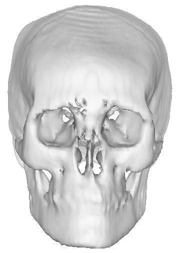


30 Skull (Scanner) X. Pennec Average Morphology of Skull average G. Subsol pathological comparison Skull Growth trough Aging 1 mois 8 mois 4 ans G. Subsol Andresen & Nielsen 30
31 Geometric Registration Can be applied to surface registration in augmented reality problems Fuse per-operative images with pre-operative images Limitations of Geometric Registration Previous geometric invariants not valid in general to compare multimodal images, or arbitrary homologous structures between different patients (e.g. brain) Problems with low-resolution or noisy images. Distribution of geometric invariants might be too sparse to handle local deformations Registration 3. Iconic Approaches 31
32 Principle of Iconic (Intensity-Based) Registration Use all voxels and their intensity to guide the registration process Energy minimization between registered images Energy Minimization Energy with two components: W ( T ) = x, y, z 2 f ( I, J ot ) dxdydz + W ( T ) f : Measure of intensity similarity between homologous points; W d : Measure of deformation to insure a regular solution (Tikhonov, linear elasticity, viscous fluid, etc.). Bajcsy, Christensen, Bro-Nielsen, Thirion, Pennec, Cachier, Ourselin... d Minimization Algorithms Non Convex Energy Convergence towards a Local Minimum Important Stages: Good initialization (rigid registration) Multi-scale analysis Hierarchy of deformations similitude, affine, polynomial, free-form, etc. 32
33 Classification of similarity measures Assumed relationship Identity Image I Adapted measures Image J 2 Sum of Squared Differences SSD( I, J ) = ( I k J k ) k Sum of Absolute Differences Measures based on image difference (Buzug et al, 1997) Classification of similarity measures Assumed relationship Affine Image I Adapted measures Correlation Coefficient ρ( I, J ) = cov( I, J ) var( I) var( J ) Image J Classification of similarity measures Assumed relationship Functional Image I Adapted measures Woods criterion (Woods et al, 1993) Robust Woods criterion (Nikou et al, 1997) Correlation Ratio (Roche et al, 1998) 2 var( I ˆ( φ J )) η ( I, J ) = 1 var( I) Image J 33
34 Classification of similarity measures Assumed relationship Statistical Image I Adapted measures Image J Joint Entropy (Hill et al., 94) Mutual Information (Viola & Wells, 1997; Maes et al, 1997) Normalised Mutual Information (Studholme, 1998) p( i, j) MI ( I, J ) = H ( I ) H ( I / J ) = p( i, j)log p( i) p( j) i j Measure of Intensity Similarity sum of squared differences correlation coefficient correlation ratio mutual information Image I A common framework? Image J A General Framework [Roche-Malandain-Ayache-Prima, MICCAI 99, pp ] A Dependence Model between images and a Maximum Likelihood approach Following the pioneering work of (Costa et al, 1993), (Viola, 1995), (Leventon & Grimson, 1998), (Bansal et al, 1998) 34

 more realistic model (-) many parameters PET /")

35 Choosing the right similarity measure Requires a good knowledge of the physics of image formation Choosing the model with the lowest number of parameters tends to lead to higher robustness Experimental Comparison Correlation Ratio (-) simplistic model (+) few parameters vs. Accuracy study CT / MR rigid registration Mutual information (+) more realistic model (-) many parameters PET / MR rigid registration Robustness study Ultrasound / MR rigid registration Some Applications of Iconic Registration 1. Detection and Measure of Lesions 2. Inter-patient comparisons 3. Superposition of an Atlas 4. Measure of Asymmetry 5. Superposition MRI-Ultrasounds 6. Stress-Rest Comparisons 35


36 1. Detection and Measure of Multiple Sclerosis Lesions Time 1 Time2 J.P Thirion Medical Image Analysis 2:3, 1998 Deformation Field Time 1 Time 2 Analysis of deformations Automatic extraction of regions with an apparent variation of volume. (Jacobian operator) Segmentation of evolving lesions Robust to small errors of rigid registration D. Rey, G. Subsol, H. Delingette,N.Ayache IPMI 99 36


37 Evolution of Lesions (3 times) Frontal Sagittal Rey-Le Brun-Chanalet -Ayache-Chatel 2000 Axial Digital Microscope Time 1 Time 2 Time 3 Rey-Subsol- Delingette-Ayache IPMI 99 European project Biomorph 2. Application to inter-patient comparison Patient 1 Patient 2 Morphometry: average subjects/patients, variations around average Would Require BOTH Geometric and Iconic Methods J.P. Thirion, X. Pennec, G. Subsol, A.Guimond, Application to the Superposition of a Brain Atlas Harvard Medical School Collaboration Pitié-Salpétrière, Parkinson (S. Ourselin) Centre Antoine Lacassagne. Radiotherapy (Bondiau-Malandain) 37


38 4. Quantitative Measure of Brain Asymmetry European Project Biomorph Application to Schizophrenia Prima-Subsol- Thirion-Roberts Miccai 98 Blue: larger than on opposite side Red: smaller than on opposite side Roboscope Image-Guided Manipulator-Assisted Neuro-Endoscopy MMIT ROBOSIM 3DUS Probe Controller Robot GUI 3D Ultrasound Courtesy Brian Davies Manipulator Steady Hand Motion Compensation Active Motion Constraints QuickTime and a Intel Indeo Video 5.0 decompressor are needed to see this picture. Courtesy B. Davies & S. Starkie 38


39 Multimodal Image Fusion T Rob / MR MR coordinate system US coordinate system T MR0 / MR1 T MR/US Robot / patient coordinate system MR 0 with surgical plan Rigid matching tools for MR MR 1 Virtual MR 2 Brain deformation over time US 1 US 2 MR / US registration US tracking of brain deformations Virtual MR n US n Typical MR-US Images Pre - Operative MR Image axial Per - Operative US Image axial coronal sagittal coronal sagittal Acquisition of images : L. & D. Auer, M. Rudolf Roche-Pennec- Ayache-Malandain MICCAI 99 MR Image 5. MRI-Ultrasound Registration US image, manual init. Registered US image Acquisition of images : L. & D. Auer, M. Rudolf 39

40 6. Application to 3-D Stress-Rest comparison (Gated Spect) Rest Stress Rest Stress Model Perfusion of cardiac muscle lateral ischemia CardioMatch (Focus Imaging) Declerck-Feldmar Conclusion Medical Imaging is nearing maturity New image modalities across scale and function Validation of algorithms sometimes impossible always difficult Growing availability of large image databases 40
Nonrigid Registration using Free-Form Deformations
 Nonrigid Registration using Free-Form Deformations Hongchang Peng April 20th Paper Presented: Rueckert et al., TMI 1999: Nonrigid registration using freeform deformations: Application to breast MR images
Filtering, reconstruction and registration of 3D Ultrasound Images
 Filtering, reconstruction and registration of 3D Ultrasound Images Hervé Delingette Projet Epidaure Herve.Delingette Delingette@inria.fr Acknowledgement Johan Montagnat Alexis Roche Karl Krissian Jérémie
Medical Image Analysis
 Computer assisted Image Analysis VT04 29 april 2004 Medical Image Analysis Lecture 10 (part 1) Xavier Tizon Medical Image Processing Medical imaging modalities XRay,, CT Ultrasound MRI PET, SPECT Generic
Computational Neuroanatomy
 Computational Neuroanatomy John Ashburner john@fil.ion.ucl.ac.uk Smoothing Motion Correction Between Modality Co-registration Spatial Normalisation Segmentation Morphometry Overview fmri time-series kernel
Multiple Sclerosis Brain MRI Segmentation Workflow deployment on the EGEE grid
 Multiple Sclerosis Brain MRI Segmentation Workflow deployment on the EGEE grid Erik Pernod 1, Jean-Christophe Souplet 1, Javier Rojas Balderrama 2, Diane Lingrand 2, Xavier Pennec 1 Speaker: Grégoire Malandain
Image Registration I
 Image Registration I Comp 254 Spring 2002 Guido Gerig Image Registration: Motivation Motivation for Image Registration Combine images from different modalities (multi-modality registration), e.g. CT&MRI,
Multi-Modal Volume Registration Using Joint Intensity Distributions
 Multi-Modal Volume Registration Using Joint Intensity Distributions Michael E. Leventon and W. Eric L. Grimson Artificial Intelligence Laboratory, Massachusetts Institute of Technology, Cambridge, MA leventon@ai.mit.edu
Medical Image Registration
 Medical Image Registration Submitted by NAREN BALRAJ SINGH SB ID# 105299299 Introduction Medical images are increasingly being used within healthcare for diagnosis, planning treatment, guiding treatment
Volumetric Deformable Models for Simulation of Laparoscopic Surgery
 Volumetric Deformable Models for Simulation of Laparoscopic Surgery S. Cotin y, H. Delingette y, J.M. Clément z V. Tassetti z, J. Marescaux z, N. Ayache y y INRIA, Epidaure Project 2004, route des Lucioles,
Methods for data preprocessing
 Methods for data preprocessing John Ashburner Wellcome Trust Centre for Neuroimaging, 12 Queen Square, London, UK. Overview Voxel-Based Morphometry Morphometry in general Volumetrics VBM preprocessing
Automatic Generation of Training Data for Brain Tissue Classification from MRI
 MICCAI-2002 1 Automatic Generation of Training Data for Brain Tissue Classification from MRI Chris A. Cocosco, Alex P. Zijdenbos, and Alan C. Evans McConnell Brain Imaging Centre, Montreal Neurological
Methodological progress in image registration for ventilation estimation, segmentation propagation and multi-modal fusion
 Methodological progress in image registration for ventilation estimation, segmentation propagation and multi-modal fusion Mattias P. Heinrich Julia A. Schnabel, Mark Jenkinson, Sir Michael Brady 2 Clinical
HST.582J / 6.555J / J Biomedical Signal and Image Processing Spring 2007
 MIT OpenCourseWare http://ocw.mit.edu HST.582J / 6.555J / 16.456J Biomedical Signal and Image Processing Spring 2007 For information about citing these materials or our Terms of Use, visit: http://ocw.mit.edu/terms.
MR IMAGE SEGMENTATION
 MR IMAGE SEGMENTATION Prepared by : Monil Shah What is Segmentation? Partitioning a region or regions of interest in images such that each region corresponds to one or more anatomic structures Classification
Technical aspects of SPECT and SPECT-CT. John Buscombe
 Technical aspects of SPECT and SPECT-CT John Buscombe What does the clinician need to know? For SPECT What factors affect SPECT How those factors should be sought Looking for artefacts For SPECT-CT Issues
Where are we now? Structural MRI processing and analysis
 Where are we now? Structural MRI processing and analysis Pierre-Louis Bazin bazin@cbs.mpg.de Leipzig, Germany Structural MRI processing: why bother? Just use the standards? SPM FreeSurfer FSL However:
Introduction to Medical Image Processing
 Introduction to Medical Image Processing Δ Essential environments of a medical imaging system Subject Image Analysis Energy Imaging System Images Image Processing Feature Images Image processing may be
Performance evaluation of registration (and segmentation?) algorithms in the absence of Gold Standard
 algorithms in the absence of Gold Standard") Performance evaluation of registration (and segmentation?) algorithms in the absence of Gold Standard ;3HQQHF EPIDAURE Project 2004, route des Lucioles B.P. 93 06902 Sophia Antipolis Cedex (France) November
Performance evaluation of registration (and segmentation?) algorithms in the absence of Gold Standard ;3HQQHF EPIDAURE Project 2004, route des Lucioles B.P. 93 06902 Sophia Antipolis Cedex (France) November
Automated segmentation methods for liver analysis in oncology applications
 University of Szeged Department of Image Processing and Computer Graphics Automated segmentation methods for liver analysis in oncology applications Ph. D. Thesis László Ruskó Thesis Advisor Dr. Antal
2 Michael E. Leventon and Sarah F. F. Gibson a b c d Fig. 1. (a, b) Two MR scans of a person's knee. Both images have high resolution in-plane, but ha
 Two MR scans of a person's knee. Both images have high resolution in-plane, but ha") Model Generation from Multiple Volumes using Constrained Elastic SurfaceNets Michael E. Leventon and Sarah F. F. Gibson 1 MIT Artificial Intelligence Laboratory, Cambridge, MA 02139, USA leventon@ai.mit.edu
Model Generation from Multiple Volumes using Constrained Elastic SurfaceNets Michael E. Leventon and Sarah F. F. Gibson 1 MIT Artificial Intelligence Laboratory, Cambridge, MA 02139, USA leventon@ai.mit.edu
A Study of Medical Image Analysis System
 Indian Journal of Science and Technology, Vol 8(25), DOI: 10.17485/ijst/2015/v8i25/80492, October 2015 ISSN (Print) : 0974-6846 ISSN (Online) : 0974-5645 A Study of Medical Image Analysis System Kim Tae-Eun
Preprocessing II: Between Subjects John Ashburner
 Preprocessing II: Between Subjects John Ashburner Pre-processing Overview Statistics or whatever fmri time-series Anatomical MRI Template Smoothed Estimate Spatial Norm Motion Correct Smooth Coregister
Assessing Accuracy Factors in Deformable 2D/3D Medical Image Registration Using a Statistical Pelvis Model
 Assessing Accuracy Factors in Deformable 2D/3D Medical Image Registration Using a Statistical Pelvis Model Jianhua Yao National Institute of Health Bethesda, MD USA jyao@cc.nih.gov Russell Taylor The Johns
An Introduction to Statistical Methods of Medical Image Registration
 his is page 1 Printer: Opaque this An Introduction to Statistical Methods of Medical Image Registration Lilla Zöllei, John Fisher, William Wells ABSRAC After defining the medical image registration problem,
Overview of Proposed TG-132 Recommendations
 Overview of Proposed TG-132 Recommendations Kristy K Brock, Ph.D., DABR Associate Professor Department of Radiation Oncology, University of Michigan Chair, AAPM TG 132: Image Registration and Fusion Conflict
Image Registration + Other Stuff
 Image Registration + Other Stuff John Ashburner Pre-processing Overview fmri time-series Motion Correct Anatomical MRI Coregister m11 m 21 m 31 m12 m13 m14 m 22 m 23 m 24 m 32 m 33 m 34 1 Template Estimate
Clinical Prospects and Technological Challenges for Multimodality Imaging Applications in Radiotherapy Treatment Planning
 Clinical Prospects and Technological Challenges for Multimodality Imaging Applications in Radiotherapy Treatment Planning Issam El Naqa, PhD Assistant Professor Department of Radiation Oncology Washington
Spatio-Temporal Registration of Biomedical Images by Computational Methods
 Spatio-Temporal Registration of Biomedical Images by Computational Methods Francisco P. M. Oliveira, João Manuel R. S. Tavares tavares@fe.up.pt, www.fe.up.pt/~tavares Outline 1. Introduction 2. Spatial
Biomedical Imaging Registration Trends and Applications. Francisco P. M. Oliveira, João Manuel R. S. Tavares
 Biomedical Imaging Registration Trends and Applications Francisco P. M. Oliveira, João Manuel R. S. Tavares tavares@fe.up.pt, www.fe.up.pt/~tavares Outline 1. Introduction 2. Spatial Registration of (2D
Object Identification in Ultrasound Scans
 Object Identification in Ultrasound Scans Wits University Dec 05, 2012 Roadmap Introduction to the problem Motivation Related Work Our approach Expected Results Introduction Nowadays, imaging devices like
Basic principles of MR image analysis. Basic principles of MR image analysis. Basic principles of MR image analysis
 Basic principles of MR image analysis Basic principles of MR image analysis Julien Milles Leiden University Medical Center Terminology of fmri Brain extraction Registration Linear registration Non-linear
MEDICAL IMAGE ANALYSIS
 SECOND EDITION MEDICAL IMAGE ANALYSIS ATAM P. DHAWAN g, A B IEEE Engineering in Medicine and Biology Society, Sponsor IEEE Press Series in Biomedical Engineering Metin Akay, Series Editor +IEEE IEEE PRESS
An Introduction To Automatic Tissue Classification Of Brain MRI. Colm Elliott Mar 2014
 An Introduction To Automatic Tissue Classification Of Brain MRI Colm Elliott Mar 2014 Tissue Classification Tissue classification is part of many processing pipelines. We often want to classify each voxel
Neuroimaging and mathematical modelling Lesson 2: Voxel Based Morphometry
 Neuroimaging and mathematical modelling Lesson 2: Voxel Based Morphometry Nivedita Agarwal, MD Nivedita.agarwal@apss.tn.it Nivedita.agarwal@unitn.it Volume and surface morphometry Brain volume White matter
Quantitative Study of Brain Anatomy
 Quantitative Study of Brain Anatomy Mei Chen, Takeo Kanade, Henry A. Rowley, Dean Pomerleau CMU-RI-TR-98-5 The Robotics Institute Carnegie Mellon University Pittsburgh, Pennsylvania 15213 March, 1998 c
Image Registration. Prof. Dr. Lucas Ferrari de Oliveira UFPR Informatics Department
 Image Registration Prof. Dr. Lucas Ferrari de Oliveira UFPR Informatics Department Introduction Visualize objects inside the human body Advances in CS methods to diagnosis, treatment planning and medical
Automatic MS Lesion Segmentation by Outlier Detection and Information Theoretic Region Partitioning Release 0.00
 Automatic MS Lesion Segmentation by Outlier Detection and Information Theoretic Region Partitioning Release 0.00 Marcel Prastawa 1 and Guido Gerig 1 Abstract July 17, 2008 1 Scientific Computing and Imaging
Anomaly Detection through Registration
 Anomaly Detection through Registration Mei Chen Takeo Kanade Henry A. Rowley Dean Pomerleau meichen@cs.cmu.edu tk@cs.cmu.edu har@cs.cmu.edu pomerlea@cs.cmu.edu School of Computer Science, Carnegie Mellon
Norbert Schuff VA Medical Center and UCSF
 Norbert Schuff Medical Center and UCSF Norbert.schuff@ucsf.edu Medical Imaging Informatics N.Schuff Course # 170.03 Slide 1/67 Objective Learn the principle segmentation techniques Understand the role
Free Form Deformations Guided by Gradient Vector Flow: a Surface Registration Method in Thoracic and Abdominal PET-CT Applications
 Free Form Deformations Guided by Gradient Vector Flow: a Surface Registration Method in Thoracic and Abdominal PET-CT Applications Oscar Camara, Gaspar Delso, and Isabelle Bloch Ecole Nationale Supérieure
Probabilistic Registration of 3-D Medical Images
 Probabilistic Registration of 3-D Medical Images Mei Chen, Takeo Kanade, Dean Pomerleau, Jeff Schneider CMU-RI-TR-99-16 The Robotics Institute Carnegie Mellon University Pittsburgh, Pennsylvania 1513 July,
Knowledge-Based Deformable Matching for Pathology Detection
 Knowledge-Based Deformable Matching for Pathology Detection Thesis Proposal Mei Chen CMU-RI-TR-97-20 The Robotics Institute Carnegie Mellon University Pittsburgh, Pennsylvania 15213 May 1997 c 1997 Carnegie
Good Morning! Thank you for joining us
 Good Morning! Thank you for joining us Deformable Registration, Contour Propagation and Dose Mapping: 101 and 201 Marc Kessler, PhD, FAAPM The University of Michigan Conflict of Interest I receive direct
1 Introduction Motivation and Aims Functional Imaging Computational Neuroanatomy... 12
 Contents 1 Introduction 10 1.1 Motivation and Aims....... 10 1.1.1 Functional Imaging.... 10 1.1.2 Computational Neuroanatomy... 12 1.2 Overview of Chapters... 14 2 Rigid Body Registration 18 2.1 Introduction.....
Efficient population registration of 3D data
 Efficient population registration of 3D data Lilla Zöllei 1, Erik Learned-Miller 2, Eric Grimson 1, William Wells 1,3 1 Computer Science and Artificial Intelligence Lab, MIT; 2 Dept. of Computer Science,
Comparison Study of Clinical 3D MRI Brain Segmentation Evaluation
 Comparison Study of Clinical 3D MRI Brain Segmentation Evaluation Ting Song 1, Elsa D. Angelini 2, Brett D. Mensh 3, Andrew Laine 1 1 Heffner Biomedical Imaging Laboratory Department of Biomedical Engineering,
Image Acquisition Systems
 Image Acquisition Systems Goals and Terminology Conventional Radiography Axial Tomography Computer Axial Tomography (CAT) Magnetic Resonance Imaging (MRI) PET, SPECT Ultrasound Microscopy Imaging ITCS
NIH Public Access Author Manuscript Proc Soc Photo Opt Instrum Eng. Author manuscript; available in PMC 2014 October 07.
 NIH Public Access Author Manuscript Published in final edited form as: Proc Soc Photo Opt Instrum Eng. 2014 March 21; 9034: 903442. doi:10.1117/12.2042915. MRI Brain Tumor Segmentation and Necrosis Detection
Multimodal Brain Warping Using the Demons Algorithm and Adaptative Intensity Corrections
 Multimodal Brain Warping Using the Demons Algorithm and Adaptative Intensity Corrections Alexandre Guimond, Alexis Roche, Nicholas Ayache, Jean Meunier To cite this version: Alexandre Guimond, Alexis Roche,
Anomaly Detection through Registration
 Anomaly Detection through Registration Mei Chen, Takeo Kanade, Henry A. Rowley, Dean Pomerleau CMU-RI-TR-97-41 The Robotics Institute Carnegie Mellon University Pittsburgh, Pennsylvania 15213 November,
Automatic segmentation of the cortical grey and white matter in MRI using a Region Growing approach based on anatomical knowledge
 Automatic segmentation of the cortical grey and white matter in MRI using a Region Growing approach based on anatomical knowledge Christian Wasserthal 1, Karin Engel 1, Karsten Rink 1 und André Brechmann
Annales UMCS Informatica AI 1 (2003) UMCS. Registration of CT and MRI brain images. Karol Kuczyński, Paweł Mikołajczak
 UMCS. Registration of CT and MRI brain images. Karol Kuczyński, Paweł Mikołajczak") Annales Informatica AI 1 (2003) 149-156 Registration of CT and MRI brain images Karol Kuczyński, Paweł Mikołajczak Annales Informatica Lublin-Polonia Sectio AI http://www.annales.umcs.lublin.pl/ Laboratory
Annales Informatica AI 1 (2003) 149-156 Registration of CT and MRI brain images Karol Kuczyński, Paweł Mikołajczak Annales Informatica Lublin-Polonia Sectio AI http://www.annales.umcs.lublin.pl/ Laboratory
Retrospective Evaluation of Inter-subject Brain Registration
 Retrospective Evaluation of Inter-subject Brain Registration P. Hellier 1, C. Barillot 1, I. Corouge 1,B.Gibaud 2, G. Le Goualher 2,3, L. Collins 3, A. Evans 3, G. Malandain 4, and N. Ayache 4 1 Projet
Segmenting Glioma in Multi-Modal Images using a Generative Model for Brain Lesion Segmentation
 Segmenting Glioma in Multi-Modal Images using a Generative Model for Brain Lesion Segmentation Bjoern H. Menze 1,2, Koen Van Leemput 3, Danial Lashkari 4 Marc-André Weber 5, Nicholas Ayache 2, and Polina
Biomedical Image Analysis based on Computational Registration Methods. João Manuel R. S. Tavares
 Biomedical Image Analysis based on Computational Registration Methods João Manuel R. S. Tavares tavares@fe.up.pt, www.fe.up.pt/~tavares Outline 1. Introduction 2. Methods a) Spatial Registration of (2D
Fundamentals of medical imaging registration I - II. Olivier Clatz Ph.D.
 Fundamentals of medical imaging registration I - II Olivier Clatz Ph.D. oclatz@bwh.harvard.edu 1 What is registration? The process of aligning a target image to a source image More generally, determining
Hybrid Formulation of the Model-Based Non-rigid Registration Problem to Improve Accuracy and Robustness
 Hybrid Formulation of the Model-Based Non-rigid Registration Problem to Improve Accuracy and Robustness Olivier Clatz 1,2,Hervé Delingette 1, Ion-Florin Talos 2, Alexandra J. Golby 2,RonKikinis 2, Ferenc
CHAPTER 2. Morphometry on rodent brains. A.E.H. Scheenstra J. Dijkstra L. van der Weerd
 CHAPTER 2 Morphometry on rodent brains A.E.H. Scheenstra J. Dijkstra L. van der Weerd This chapter was adapted from: Volumetry and other quantitative measurements to assess the rodent brain, In vivo NMR
Computer-Aided Diagnosis in Abdominal and Cardiac Radiology Using Neural Networks
 Computer-Aided Diagnosis in Abdominal and Cardiac Radiology Using Neural Networks Du-Yih Tsai, Masaru Sekiya and Yongbum Lee Department of Radiological Technology, School of Health Sciences, Faculty of
STIC AmSud Project. Graph cut based segmentation of cardiac ventricles in MRI: a shape-prior based approach
 STIC AmSud Project Graph cut based segmentation of cardiac ventricles in MRI: a shape-prior based approach Caroline Petitjean A joint work with Damien Grosgeorge, Pr Su Ruan, Pr JN Dacher, MD October 22,
Non-Rigid Registration of Medical Images: Theory, Methods and Applications
 Non-Rigid Registration of Medical Images: Theory, Methods and Applications Daniel Rueckert Paul Aljabar Medical mage registration [1] plays an increasingly important role in many clinical applications
Image Segmentation and Registration
 Image Segmentation and Registration Dr. Christine Tanner (tanner@vision.ee.ethz.ch) Computer Vision Laboratory, ETH Zürich Dr. Verena Kaynig, Machine Learning Laboratory, ETH Zürich Outline Segmentation
Registration-Based Segmentation of Medical Images
 School of Computing National University of Singapore Graduate Research Paper Registration-Based Segmentation of Medical Images by Li Hao under guidance of A/Prof. Leow Wee Kheng July, 2006 Abstract Medical
Segmenting Glioma in Multi-Modal Images using a Generative-Discriminative Model for Brain Lesion Segmentation
 Segmenting Glioma in Multi-Modal Images using a Generative-Discriminative Model for Brain Lesion Segmentation Bjoern H. Menze 1,2, Ezequiel Geremia 2, Nicholas Ayache 2, and Gabor Szekely 1 1 Computer
Hybrid Spline-based Multimodal Registration using a Local Measure for Mutual Information
 Hybrid Spline-based Multimodal Registration using a Local Measure for Mutual Information Andreas Biesdorf 1, Stefan Wörz 1, Hans-Jürgen Kaiser 2, Karl Rohr 1 1 University of Heidelberg, BIOQUANT, IPMB,
TG 132: Use of Image Registration and Fusion in RT
 TG 132: Use of Image Registration and Fusion in RT Kristy K Brock, PhD, DABR, FAAPM Associate Professor Department of Radiation Oncology, University of Michigan Chair, AAPM TG 132: Image Registration and
Non-Rigid Image Registration III
 Non-Rigid Image Registration III CS6240 Multimedia Analysis Leow Wee Kheng Department of Computer Science School of Computing National University of Singapore Leow Wee Kheng (CS6240) Non-Rigid Image Registration
Retrospective evaluation of inter-subject brain registration
 Retrospective evaluation of inter-subject brain registration P. Hellier 1, C. Barillot 1, I. Corouge 1,B.Gibaud 2, G. Le Goualher 2,3, L. Collins 3, A. Evans 3, G. Malandain 4, N. Ayache 4 1 Projet Vista,
Ischemic Stroke Lesion Segmentation Proceedings 5th October 2015 Munich, Germany
 0111010001110001101000100101010111100111011100100011011101110101101012 Ischemic Stroke Lesion Segmentation www.isles-challenge.org Proceedings 5th October 2015 Munich, Germany Preface Stroke is the second
REAL-TIME ADAPTIVITY IN HEAD-AND-NECK AND LUNG CANCER RADIOTHERAPY IN A GPU ENVIRONMENT
 REAL-TIME ADAPTIVITY IN HEAD-AND-NECK AND LUNG CANCER RADIOTHERAPY IN A GPU ENVIRONMENT Anand P Santhanam Assistant Professor, Department of Radiation Oncology OUTLINE Adaptive radiotherapy for head and
Deformable Registration Using Scale Space Keypoints
 Deformable Registration Using Scale Space Keypoints Mehdi Moradi a, Purang Abolmaesoumi a,b and Parvin Mousavi a a School of Computing, Queen s University, Kingston, Ontario, Canada K7L 3N6; b Department
Medical Image Registration by Maximization of Mutual Information
 Medical Image Registration by Maximization of Mutual Information EE 591 Introduction to Information Theory Instructor Dr. Donald Adjeroh Submitted by Senthil.P.Ramamurthy Damodaraswamy, Umamaheswari Introduction
Image Segmentation. Ross Whitaker SCI Institute, School of Computing University of Utah
 Image Segmentation Ross Whitaker SCI Institute, School of Computing University of Utah What is Segmentation? Partitioning images/volumes into meaningful pieces Partitioning problem Labels Isolating a specific
The Effect of Segmentation Method on the Performance of Point Based Registration of Intra-Ultrasound with Pre-MR Images
 The Effect of Segmentation Method on the Performance of Point Based Registration of Intra-Ultrasound with Pre-MR Images P.Farnia, A. Ahmadian,*, A. Khoshnevisan Tehran University of Medical Sciences, Biomedical
Supplementary methods
 Supplementary methods This section provides additional technical details on the sample, the applied imaging and analysis steps and methods. Structural imaging Trained radiographers placed all participants
Performance Issues in Shape Classification
 Performance Issues in Shape Classification Samson J. Timoner 1, Pollina Golland 1, Ron Kikinis 2, Martha E. Shenton 3, W. Eric L. Grimson 1, and William M. Wells III 1,2 1 MIT AI Laboratory, Cambridge
SIGMI Meeting ~Image Fusion~ Computer Graphics and Visualization Lab Image System Lab
 SIGMI Meeting ~Image Fusion~ Computer Graphics and Visualization Lab Image System Lab Introduction Medical Imaging and Application CGV 3D Organ Modeling Model-based Simulation Model-based Quantification
Learning-based Neuroimage Registration
 Learning-based Neuroimage Registration Leonid Teverovskiy and Yanxi Liu 1 October 2004 CMU-CALD-04-108, CMU-RI-TR-04-59 School of Computer Science Carnegie Mellon University Pittsburgh, PA 15213 Abstract
Non-Rigid Multimodal Medical Image Registration using Optical Flow and Gradient Orientation
 M. HEINRICH et al.: MULTIMODAL REGISTRATION USING GRADIENT ORIENTATION 1 Non-Rigid Multimodal Medical Image Registration using Optical Flow and Gradient Orientation Mattias P. Heinrich 1 mattias.heinrich@eng.ox.ac.uk
Machine Learning for Medical Image Analysis. A. Criminisi
 Machine Learning for Medical Image Analysis A. Criminisi Overview Introduction to machine learning Decision forests Applications in medical image analysis Anatomy localization in CT Scans Spine Detection
Abstract. 1. Introduction
 A New Automated Method for Three- Dimensional Registration of Medical Images* P. Kotsas, M. Strintzis, D.W. Piraino Department of Electrical and Computer Engineering, Aristotelian University, 54006 Thessaloniki,
SCIENCE & TECHNOLOGY
 Pertanika J. Sci. & Technol. 26 (1): 309-316 (2018) SCIENCE & TECHNOLOGY Journal homepage: http://www.pertanika.upm.edu.my/ Application of Active Contours Driven by Local Gaussian Distribution Fitting
Incorporating Statistical Measures of Anatomical Variability in Atlas-to-Subject Registration for Conformal Brain Radiotherapy
 Incorporating Statistical Measures of Anatomical Variability in Atlas-to-Subject Registration for Conformal Brain Radiotherapy Olivier Commowick 1,2, Radu Stefanescu 1, Pierre Fillard 1, Vincent Arsigny
Surgery Simulation and Planning
 Surgery Simulation and Planning S. H. Martin Roth Dr. Rolf M. Koch Daniel Bielser Prof. Dr. Markus Gross Facial surgery project in collaboration with Prof. Dr. Dr. H. Sailer, University Hospital Zurich,
Introduction to Medical Image Registration
 Introduction to Medical Image Registration Sailesh Conjeti Computer Aided Medical Procedures (CAMP), Technische Universität München, Germany sailesh.conjeti@tum.de Partially adapted from slides by: 1.
Classification of Subject Motion for Improved Reconstruction of Dynamic Magnetic Resonance Imaging
 1 CS 9 Final Project Classification of Subject Motion for Improved Reconstruction of Dynamic Magnetic Resonance Imaging Feiyu Chen Department of Electrical Engineering ABSTRACT Subject motion is a significant
A Registration-Based Atlas Propagation Framework for Automatic Whole Heart Segmentation
 A Registration-Based Atlas Propagation Framework for Automatic Whole Heart Segmentation Xiahai Zhuang (PhD) Centre for Medical Image Computing University College London Fields-MITACS Conference on Mathematics
3D Surface Reconstruction of the Brain based on Level Set Method
 3D Surface Reconstruction of the Brain based on Level Set Method Shijun Tang, Bill P. Buckles, and Kamesh Namuduri Department of Computer Science & Engineering Department of Electrical Engineering University
INRIA s Research Centers
 Statistical computing on manifolds and data assimilation: from medical images to anatomical and physiological models http://www-sop.inria.fr/asclepios/cours/mva/module2/index.htm Centrale OMA MIA - MVA
Atlas Based Segmentation of the prostate in MR images
 Atlas Based Segmentation of the prostate in MR images Albert Gubern-Merida and Robert Marti Universitat de Girona, Computer Vision and Robotics Group, Girona, Spain {agubern,marly}@eia.udg.edu Abstract.
Multimodal Elastic Image Matching
 Research results based on my diploma thesis supervised by Prof. Witsch 2 and in cooperation with Prof. Mai 3. 1 February 22 nd 2011 1 Karlsruhe Institute of Technology (KIT) 2 Applied Mathematics Department,
Non-Rigid Registration of Medical Images: Theory, Methods and Applications
 Non-Rigid Registration of Medical Images: Theory, Methods and Applications Daniel Rueckert Paul Aljabar Medical image registration [1] plays an increasingly important role in many clinical applications
Medical Images Analysis and Processing
 Medical Images Analysis and Processing - 25642 Emad Course Introduction Course Information: Type: Graduated Credits: 3 Prerequisites: Digital Image Processing Course Introduction Reference(s): Insight
STATISTICAL ATLAS-BASED SUB-VOXEL SEGMENTATION OF 3D BRAIN MRI
 STATISTICA ATAS-BASED SUB-VOXE SEGMENTATION OF 3D BRAIN MRI Marcel Bosc 1,2, Fabrice Heitz 1, Jean-Paul Armspach 2 (1) SIIT UMR-7005 CNRS / Strasbourg I University, 67400 Illkirch, France (2) IPB UMR-7004
The Anatomical Equivalence Class Formulation and its Application to Shape-based Computational Neuroanatomy
 The Anatomical Equivalence Class Formulation and its Application to Shape-based Computational Neuroanatomy Sokratis K. Makrogiannis, PhD From post-doctoral research at SBIA lab, Department of Radiology,
A Generation Methodology for Numerical Phantoms with Statistically Relevant Variability of Geometric and Physical Properties
 A Generation Methodology for Numerical Phantoms with Statistically Relevant Variability of Geometric and Physical Properties Steven Dolly 1, Eric Ehler 1, Yang Lou 2, Mark Anastasio 2, Hua Li 2 (1) University
Mutual Information Based Methods to Localize Image Registration
 Mutual Information Based Methods to Localize Image Registration by Kathleen P. Wilkie A thesis presented to the University of Waterloo in fulfilment of the thesis requirement for the degree of Master of
Automatic Subthalamic Nucleus Targeting for Deep Brain Stimulation. A Validation Study
 Automatic Subthalamic Nucleus Targeting for Deep Brain Stimulation. A Validation Study F. Javier Sánchez Castro a, Claudio Pollo a,b, Jean-Guy Villemure b, Jean-Philippe Thiran a a École Polytechnique
Computational Medical Imaging Analysis Chapter 5: Processing and Analysis
 Computational Medical Imaging Analysis Chapter 5: Processing and Analysis Jun Zhang Laboratory for Computational Medical Imaging & Data Analysis Department of Computer Science University of Kentucky Lexington,
Whole Body MRI Intensity Standardization
 Whole Body MRI Intensity Standardization Florian Jäger 1, László Nyúl 1, Bernd Frericks 2, Frank Wacker 2 and Joachim Hornegger 1 1 Institute of Pattern Recognition, University of Erlangen, {jaeger,nyul,hornegger}@informatik.uni-erlangen.de