Technical aspects of SPECT and SPECT-CT. John Buscombe
|
|
|
- Olivia Chapman
- 5 years ago
- Views:
Transcription

1 Technical aspects of SPECT and SPECT-CT John Buscombe
2 What does the clinician need to know? For SPECT What factors affect SPECT How those factors should be sought Looking for artefacts For SPECT-CT Issues of registration
3 Factors that effect SPECT Uniformity CFOV <2.5% variation in uniformity (extrinsic) Should be tested by flood source Intrinsic uniformity done with point source and no collimator Centre of rotation Linerarity Energy correction maps (for different isotopes) Using the correct collimator SPECT resolution
4 Needs a QC programme Typical programme Daily Flood for uniformity Monthly COR Linearity 3 Monthly Flood corrections for less used isotopes Resolution

5 Note flood tank on gamma camera Uniformity

6 Uniformity

7 What is wrong on this triple headed gamma camera?

8 Linearity Tungsten cut out shield used with flood tank
9 COR Use capillary tube of activity placed in the centre of rotation normally 10 cm long Acquire SPECT Reconstruct image
10 COR Perfect single head poor COR 2 heads both with good COR but heads misaligned


11 SPECT resolution Jaszack SPECT phantom Images (filtered and unfiltered
12 Then all you need to do Reconstruct image correctly Back projection Interative Correct filtering Some filters more count dependent that others Though called the same different manufacturers filters often very different


13 Filtering Can use predefined protocols However filter count related Maybe best to use a manual filter Check images are not over filtered
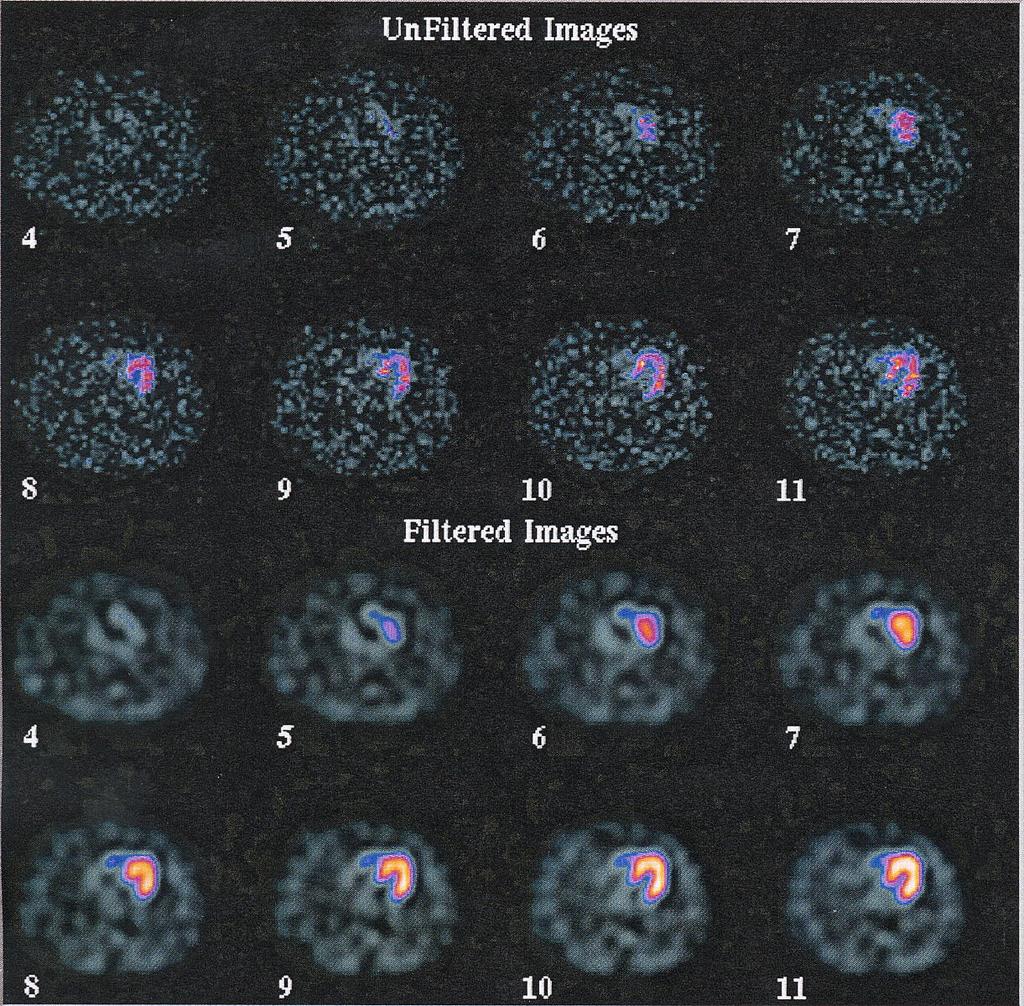
14 Post filtering-correct"

15 Over smoothed"

16 Over filtered"

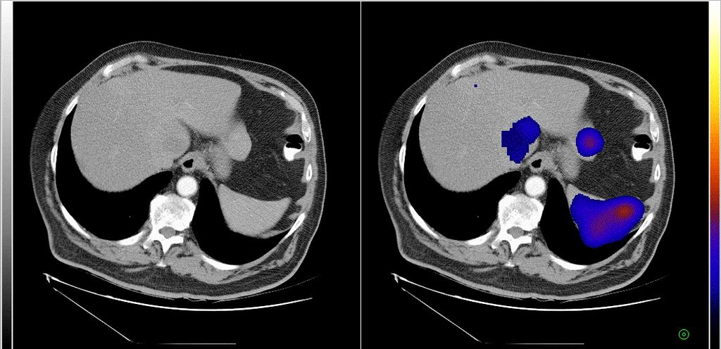
17 Image Fusion
18 Image Registration Process of spatially aligning images from the same or matching modalities CT/MR/NM
19 Image Registration Intra/Inter modality Two approaches Dedicated dual modality hardware Software algorithms
20 Why? Allows: Fusion of molecular and anatomical information Maximise potential of the complementary information Precise localisation of organs and lesions Comparison of studies performed at different times
21 Registration vs Fusion Registration = Technical manipulations applied to align images Fusion = Use of the registered images; most often superimposed images in a single display
22 Registration
23 Fusion
24 How? Software used to identify the necessary transformation required, so that when it is applied to one study, the result is the best possible spatial alignment with another study Many different software packages available Many different ways to register images
25 Transformations Rigid (Linear) position or orientation different between two image sets simple translation or rotation required used to register scans from the same patient at different times
26 Transformations Affine (Linear) - additional scaling/zooming and shearing - spatial transformation of one co-ordinate system (x,y,z) to another (u,v,w) p z p y p x p w p z p y p x p v p z p y p x p u = = = p z p y p x p w p z p y p x p v p z p y p x p u = = =
27 Transformations Non-linear - usually applied after linear techniques - correct for differences in shape that cannot be accounted for by affine normalisation - warping of the image
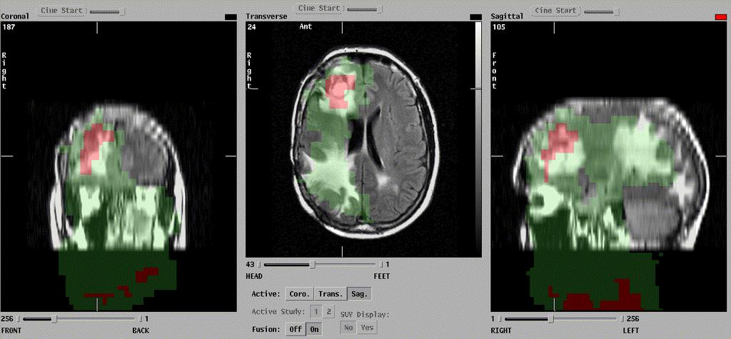
28 Initial guess at transformation parameters Floater reorientated Similarity compared Parameters updated Continued until preset termination point reached
29 Measures of Similarity Require a quantitative measure of alignment between the two images Similarity measure will reach a maximum when the images are most closely aligned Best match ambiguous Large variety been proposed
30 Measures of Similarity Point based methods - extrinsic markers (prospective) - intrinsic markers (retrospective) Comparison of voxel intensity values Edge/Surface based

31 Wrist Registration Splint with cobalt markers UV sensitive Digital DP film Registered with bone scan

32 Intrinsic Markers

33 Intrinsic Markers

34 Intrinsic Markers

35 Layout of fused images
36 Image Display Fusion Display - Grey scale for CT/MR - Colour for PET/SPECT - New colour values computed for each pixel, simulating the transparency effect - Artificial increase in the perceived intensity of colour images
37 Image Display Multimodality Display - Display both modalities side by side - Synchronized cursors displayed simultaneously on both images - Advantage no information lost

38 Fused Display
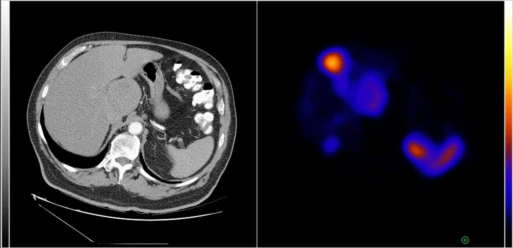
39 Multimodality Display
40
41 Oncology - Clinical Applications Registration MR, CT, SPECT and PET Excellent tool in radiotherapy planning Biopsy guidance Surgical planning SPECT (with CEA, prostacint scans, targeted therapy, accurate lesion localisation)
42
43
44 SRS Registered
45 SRS Fused
46 Pitfalls and problems Not unique to SPECT-CT but may be worse because of longer acquisition times Respiration and mis-registration and ACerrors in lower chest, liver and spleen Over correction around high density objects in iterative recon-suggest use back-projection
 lung movementdiaphragm d) lung movement chest wall e) correct image")
47 Effects of movement of chest McQuaid and Hutton UCL, EANMMI a) cardiac mismatch b) heart and liver overlap c) lung movementdiaphragm d) lung movement chest wall e) correct image
48 Is it being used? Despite undoubted advantages, clinical applications limited Limitations - requires knowledge of registration methods - can be misused and is time consuming - very mathematical (involve a physicist) - easier with automated systems - movement (subject, diaphragm, cardiac) - organ changes (GB, bladder)
49 In Summary Powerful, versatile and available tool Does have limitations and must be used with caution Clinical use of dedicated hardware and software registration will increase Software registration will remain important for hardware devices
3/27/2012 WHY SPECT / CT? SPECT / CT Basic Principles. Advantages of SPECT. Advantages of CT. Dr John C. Dickson, Principal Physicist UCLH
 3/27/212 Advantages of SPECT SPECT / CT Basic Principles Dr John C. Dickson, Principal Physicist UCLH Institute of Nuclear Medicine, University College London Hospitals and University College London john.dickson@uclh.nhs.uk
VALIDATION OF DIR. Raj Varadhan, PhD, DABMP Minneapolis Radiation Oncology
 VALIDATION OF DIR Raj Varadhan, PhD, DABMP Minneapolis Radiation Oncology Overview Basics: Registration Framework, Theory Discuss Validation techniques Using Synthetic CT data & Phantoms What metrics to
multimodality image processing workstation Visualizing your SPECT-CT-PET-MRI images
 multimodality image processing workstation Visualizing your SPECT-CT-PET-MRI images FUSION FUSION is a new visualization and evaluation software developed by Mediso built on state of the art technology,
Philips SPECT/CT Systems
 Philips SPECT/CT Systems Ling Shao, PhD Director, Imaging Physics & System Analysis Nuclear Medicine, Philips Healthcare June 14, 2008 *Presented SNM08 Categorical Seminar - Quantitative SPECT and PET
Conflicts of Interest Nuclear Medicine and PET physics reviewer for the ACR Accreditation program
 James R Halama, PhD Loyola University Medical Center Conflicts of Interest Nuclear Medicine and PET physics reviewer for the ACR Accreditation program Learning Objectives 1. Be familiar with recommendations
SPECT QA and QC. Bruce McBride St. Vincent s Hospital Sydney.
 SPECT QA and QC Bruce McBride St. Vincent s Hospital Sydney. SPECT QA and QC What is needed? Why? How often? Who says? QA and QC in Nuclear Medicine QA - collective term for all the efforts made to produce
Mediso AnyScan S Single-Head and Dual-Head SPECT
 Mediso AnyScan S Single-Head and Dual-Head SPECT ANYSCAN S (SINGLE-HEAD & DUAL-HEAD SPECT) Low Cost of Ownership Small Footprint Absolute Imaging Solutions tm is the exclusive U.S. source for Mediso Medical
CT Basics Principles of Spiral CT Dose. Always Thinking Ahead.
 1 CT Basics Principles of Spiral CT Dose 2 Who invented CT? 1963 - Alan Cormack developed a mathematical method of reconstructing images from x-ray projections Sir Godfrey Hounsfield worked for the Central
Nonrigid Registration using Free-Form Deformations
 Nonrigid Registration using Free-Form Deformations Hongchang Peng April 20th Paper Presented: Rueckert et al., TMI 1999: Nonrigid registration using freeform deformations: Application to breast MR images
Mediso AnyScan. Single-Head and Dual-Head SPECT
 Mediso AnyScan S Single-Head and Dual-Head SPECT ANYSCAN S (SINGLE-HEAD & DUAL-HEAD SPECT) Low Cost of Ownership Associated Imaging Services is the exclusive U.S. source in the Midwest for Mediso Medical
James R Halama, PhD Loyola University Medical Center
 James R Halama, PhD Loyola University Medical Center Conflicts of Interest Nuclear Medicine and PET physics reviewer for the ACR Accreditation program Learning Objectives Be familiar with the tests recommended
Medical Image Registration by Maximization of Mutual Information
 Medical Image Registration by Maximization of Mutual Information EE 591 Introduction to Information Theory Instructor Dr. Donald Adjeroh Submitted by Senthil.P.Ramamurthy Damodaraswamy, Umamaheswari Introduction
Image Acquisition Systems
 Image Acquisition Systems Goals and Terminology Conventional Radiography Axial Tomography Computer Axial Tomography (CAT) Magnetic Resonance Imaging (MRI) PET, SPECT Ultrasound Microscopy Imaging ITCS
Medical Image Analysis
 Computer assisted Image Analysis VT04 29 april 2004 Medical Image Analysis Lecture 10 (part 1) Xavier Tizon Medical Image Processing Medical imaging modalities XRay,, CT Ultrasound MRI PET, SPECT Generic
Joint CI-JAI advanced accelerator lecture series Imaging and detectors for medical physics Lecture 1: Medical imaging
 Joint CI-JAI advanced accelerator lecture series Imaging and detectors for medical physics Lecture 1: Medical imaging Dr Barbara Camanzi barbara.camanzi@stfc.ac.uk Course layout Day AM 09.30 11.00 PM 15.30
Image Registration. Prof. Dr. Lucas Ferrari de Oliveira UFPR Informatics Department
 Image Registration Prof. Dr. Lucas Ferrari de Oliveira UFPR Informatics Department Introduction Visualize objects inside the human body Advances in CS methods to diagnosis, treatment planning and medical
Utilizing Salient Region Features for 3D Multi-Modality Medical Image Registration
 Utilizing Salient Region Features for 3D Multi-Modality Medical Image Registration Dieter Hahn 1, Gabriele Wolz 2, Yiyong Sun 3, Frank Sauer 3, Joachim Hornegger 1, Torsten Kuwert 2 and Chenyang Xu 3 1
Is deformable image registration a solved problem?
 Is deformable image registration a solved problem? Marcel van Herk On behalf of the imaging group of the RT department of NKI/AVL Amsterdam, the Netherlands DIR 1 Image registration Find translation.deformation
Basic principles of MR image analysis. Basic principles of MR image analysis. Basic principles of MR image analysis
 Basic principles of MR image analysis Basic principles of MR image analysis Julien Milles Leiden University Medical Center Terminology of fmri Brain extraction Registration Linear registration Non-linear
Automated Image Analysis Software for Quality Assurance of a Radiotherapy CT Simulator
 Automated Image Analysis Software for Quality Assurance of a Radiotherapy CT Simulator Andrew J Reilly Imaging Physicist Oncology Physics Edinburgh Cancer Centre Western General Hospital EDINBURGH EH4
Methods for data preprocessing
 Methods for data preprocessing John Ashburner Wellcome Trust Centre for Neuroimaging, 12 Queen Square, London, UK. Overview Voxel-Based Morphometry Morphometry in general Volumetrics VBM preprocessing
Medical Image Registration
 Medical Image Registration Submitted by NAREN BALRAJ SINGH SB ID# 105299299 Introduction Medical images are increasingly being used within healthcare for diagnosis, planning treatment, guiding treatment
Introduction to Medical Image Registration
 Introduction to Medical Image Registration Sailesh Conjeti Computer Aided Medical Procedures (CAMP), Technische Universität München, Germany sailesh.conjeti@tum.de Partially adapted from slides by: 1.
RADIOMICS: potential role in the clinics and challenges
 27 giugno 2018 Dipartimento di Fisica Università degli Studi di Milano RADIOMICS: potential role in the clinics and challenges Dr. Francesca Botta Medical Physicist Istituto Europeo di Oncologia (Milano)
Implementation of Advanced Image Guided Radiation Therapy
 Image Acquisition Course Outline Principles, characteristics& applications of the available modalities Image Processing in the T x room Image guided treatment delivery What can / can t we do in the room
Venus Explorer Processing Technical specifications
 24, rue du champ de l Alouette F 75013 Paris, France Venus Explorer Processing Technical specifications Venus Explorer Processing Server software Web base application sever; tomcat Apache technology. Include
Introduction to Positron Emission Tomography
 Planar and SPECT Cameras Summary Introduction to Positron Emission Tomography, Ph.D. Nuclear Medicine Basic Science Lectures srbowen@uw.edu System components: Collimator Detector Electronics Collimator
Fits you like no other
 Fits you like no other BrightView X and XCT specifications The new BrightView X system is a fully featured variableangle camera that is field-upgradeable to BrightView XCT without any increase in room
Medical Image Processing: Image Reconstruction and 3D Renderings
 Medical Image Processing: Image Reconstruction and 3D Renderings 김보형 서울대학교컴퓨터공학부 Computer Graphics and Image Processing Lab. 2011. 3. 23 1 Computer Graphics & Image Processing Computer Graphics : Create,
7/31/2011. Learning Objective. Video Positioning. 3D Surface Imaging by VisionRT
 CLINICAL COMMISSIONING AND ACCEPTANCE TESTING OF A 3D SURFACE MATCHING SYSTEM Hania Al-Hallaq, Ph.D. Assistant Professor Radiation Oncology The University of Chicago Learning Objective Describe acceptance
Assessing Accuracy Factors in Deformable 2D/3D Medical Image Registration Using a Statistical Pelvis Model
 Assessing Accuracy Factors in Deformable 2D/3D Medical Image Registration Using a Statistical Pelvis Model Jianhua Yao National Institute of Health Bethesda, MD USA jyao@cc.nih.gov Russell Taylor The Johns
Non-Rigid Multimodal Medical Image Registration using Optical Flow and Gradient Orientation
 M. HEINRICH et al.: MULTIMODAL REGISTRATION USING GRADIENT ORIENTATION 1 Non-Rigid Multimodal Medical Image Registration using Optical Flow and Gradient Orientation Mattias P. Heinrich 1 mattias.heinrich@eng.ox.ac.uk
Methodological progress in image registration for ventilation estimation, segmentation propagation and multi-modal fusion
 Methodological progress in image registration for ventilation estimation, segmentation propagation and multi-modal fusion Mattias P. Heinrich Julia A. Schnabel, Mark Jenkinson, Sir Michael Brady 2 Clinical
3D Voxel-Based Volumetric Image Registration with Volume-View Guidance
 3D Voxel-Based Volumetric Image Registration with Volume-View Guidance Guang Li*, Huchen Xie, Holly Ning, Deborah Citrin, Jacek Copala, Barbara Arora, Norman Coleman, Kevin Camphausen, and Robert Miller
Using Probability Maps for Multi organ Automatic Segmentation
 Using Probability Maps for Multi organ Automatic Segmentation Ranveer Joyseeree 1,2, Óscar Jiménez del Toro1, and Henning Müller 1,3 1 University of Applied Sciences Western Switzerland (HES SO), Sierre,
Quantitative imaging for clinical dosimetry
 Quantitative imaging for clinical dosimetry Irène Buvat Laboratoire d Imagerie Fonctionnelle U678 INSERM - UPMC CHU Pitié-Salpêtrière, Paris buvat@imed.jussieu.fr http://www.guillemet.org/irene Methodology
Clinical Prospects and Technological Challenges for Multimodality Imaging Applications in Radiotherapy Treatment Planning
 Clinical Prospects and Technological Challenges for Multimodality Imaging Applications in Radiotherapy Treatment Planning Issam El Naqa, PhD Assistant Professor Department of Radiation Oncology Washington
Use of image registration and fusion algorithms and techniques in radiotherapy: Report of the AAPM Radiation Therapy Committee Task Group No.
 Use of image registration and fusion algorithms and techniques in radiotherapy: Report of the AAPM Radiation Therapy Committee Task Group No. 132 Kristy K. Brock a) Department of Imaging Physics, The University
radiotherapy Andrew Godley, Ergun Ahunbay, Cheng Peng, and X. Allen Li NCAAPM Spring Meeting 2010 Madison, WI
 GPU-Accelerated autosegmentation for adaptive radiotherapy Andrew Godley, Ergun Ahunbay, Cheng Peng, and X. Allen Li agodley@mcw.edu NCAAPM Spring Meeting 2010 Madison, WI Overview Motivation Adaptive
Michael Speiser, Ph.D.
 IMPROVED CT-BASED VOXEL PHANTOM GENERATION FOR MCNP MONTE CARLO Michael Speiser, Ph.D. Department of Radiation Oncology UT Southwestern Medical Center Dallas, TX September 1 st, 2012 CMPWG Workshop Medical
Fits you like no other
 Fits you like no other Philips BrightView X and XCT specifications The new BrightView X system is a fully featured variableangle camera that is field-upgradeable to BrightView XCT without any increase
ADVANCING CANCER TREATMENT
 3 ADVANCING CANCER TREATMENT SUPPORTING CLINICS WORLDWIDE RaySearch is advancing cancer treatment through pioneering software. We believe software has un limited potential, and that it is now the driving
GPU applications in Cancer Radiation Therapy at UCSD. Steve Jiang, UCSD Radiation Oncology Amit Majumdar, SDSC Dongju (DJ) Choi, SDSC
 Choi, SDSC") GPU applications in Cancer Radiation Therapy at UCSD Steve Jiang, UCSD Radiation Oncology Amit Majumdar, SDSC Dongju (DJ) Choi, SDSC Conventional Radiotherapy SIMULATION: Construciton, Dij Days PLANNING:
GPU applications in Cancer Radiation Therapy at UCSD Steve Jiang, UCSD Radiation Oncology Amit Majumdar, SDSC Dongju (DJ) Choi, SDSC Conventional Radiotherapy SIMULATION: Construciton, Dij Days PLANNING:
Image Co-Registration II: TG132 Quality Assurance for Image Registration. Image Co-Registration II: TG132 Quality Assurance for Image Registration
 Image Co-Registration II: TG132 Quality Assurance for Image Registration Preliminary Recommendations from TG 132* Kristy Brock, Sasa Mutic, Todd McNutt, Hua Li, and Marc Kessler *Recommendations are NOT
Use of Deformable Image Registration in Radiation Therapy. Colin Sims, M.Sc. Accuray Incorporated 1
 Use of Deformable Image Registration in Radiation Therapy Colin Sims, M.Sc. Accuray Incorporated 1 Overview of Deformable Image Registration (DIR) Algorithms that can deform one dataset to another have
Computational Neuroanatomy
 Computational Neuroanatomy John Ashburner john@fil.ion.ucl.ac.uk Smoothing Motion Correction Between Modality Co-registration Spatial Normalisation Segmentation Morphometry Overview fmri time-series kernel
Introduction to Emission Tomography
 Introduction to Emission Tomography Gamma Camera Planar Imaging Robert Miyaoka, PhD University of Washington Department of Radiology rmiyaoka@u.washington.edu Gamma Camera: - collimator - detector (crystal
Advanced Targeting Using Image Deformation. Justin Keister, MS DABR Aurora Health Care Kenosha, WI
 Advanced Targeting Using Image Deformation Justin Keister, MS DABR Aurora Health Care Kenosha, WI History of Targeting The advance of IMRT and CT simulation has changed how targets are identified in radiation
Good Morning! Thank you for joining us
 Good Morning! Thank you for joining us Deformable Registration, Contour Propagation and Dose Mapping: 101 and 201 Marc Kessler, PhD, FAAPM The University of Michigan Conflict of Interest I receive direct
Overview of Proposed TG-132 Recommendations
 Overview of Proposed TG-132 Recommendations Kristy K Brock, Ph.D., DABR Associate Professor Department of Radiation Oncology, University of Michigan Chair, AAPM TG 132: Image Registration and Fusion Conflict
Ch. 4 Physical Principles of CT
 Ch. 4 Physical Principles of CT CLRS 408: Intro to CT Department of Radiation Sciences Review: Why CT? Solution for radiography/tomography limitations Superimposition of structures Distinguishing between
Mutual Information Based Methods to Localize Image Registration
 Mutual Information Based Methods to Localize Image Registration by Kathleen P. Wilkie A thesis presented to the University of Waterloo in fulfilment of the thesis requirement for the degree of Master of
Validation of GEANT4 for Accurate Modeling of 111 In SPECT Acquisition
 Validation of GEANT4 for Accurate Modeling of 111 In SPECT Acquisition Bernd Schweizer, Andreas Goedicke Philips Technology Research Laboratories, Aachen, Germany bernd.schweizer@philips.com Abstract.
8/3/2017. Contour Assessment for Quality Assurance and Data Mining. Objective. Outline. Tom Purdie, PhD, MCCPM
 Contour Assessment for Quality Assurance and Data Mining Tom Purdie, PhD, MCCPM Objective Understand the state-of-the-art in contour assessment for quality assurance including data mining-based techniques
TG 132: Use of Image Registration and Fusion in RT
 TG 132: Use of Image Registration and Fusion in RT Kristy K Brock, PhD, DABR, FAAPM Associate Professor Department of Radiation Oncology, University of Michigan Chair, AAPM TG 132: Image Registration and
Respiratory Motion Estimation using a 3D Diaphragm Model
 Respiratory Motion Estimation using a 3D Diaphragm Model Marco Bögel 1,2, Christian Riess 1,2, Andreas Maier 1, Joachim Hornegger 1, Rebecca Fahrig 2 1 Pattern Recognition Lab, FAU Erlangen-Nürnberg 2
Department of ECE, SCSVMV University, Kanchipuram
 Medical Image Registering: A Matlab Based Approach [1] Dr.K.Umapathy, [2] D.Vedasri, [3] H.Vaishnavi [1] Associate Professor, [2][3] UG Student, Department of ECE, SCSVMV University, Kanchipuram Abstract:-
Fmri Spatial Processing
 Educational Course: Fmri Spatial Processing Ray Razlighi Jun. 8, 2014 Spatial Processing Spatial Re-alignment Geometric distortion correction Spatial Normalization Smoothing Why, When, How, Which Why is
Multimodality Imaging for Tumor Volume Definition in Radiation Oncology
 81 There are several commercial and academic software tools that support different segmentation algorithms. In general, commercial software packages have better implementation (with a user-friendly interface
Fundamentals of CT imaging
 SECTION 1 Fundamentals of CT imaging I History In the early 1970s Sir Godfrey Hounsfield s research produced the first clinically useful CT scans. Original scanners took approximately 6 minutes to perform
Biomedical Image Analysis based on Computational Registration Methods. João Manuel R. S. Tavares
 Biomedical Image Analysis based on Computational Registration Methods João Manuel R. S. Tavares tavares@fe.up.pt, www.fe.up.pt/~tavares Outline 1. Introduction 2. Methods a) Spatial Registration of (2D
The Emory Reconstruction Toolbox Version 1.0
 The Emory Reconstruction Toolbox Version 1.0 Operating Instructions Revision 02 (April, 2008) Operating Instructions The Emory Reconstruction Toolbox Application Copyrights, Trademarks, Restrictions
ADVANCING CANCER TREATMENT
 The RayPlan treatment planning system makes proven, innovative RayStation technology accessible to clinics that need a cost-effective and streamlined solution. Fast, efficient and straightforward to use,
Spatio-temporal Analysis of Biomedical Images based on Automated Methods of Image Registration
 Spatio-temporal Analysis of Biomedical Images based on Automated Methods of Image Registration João Manuel R. S. Tavares tavares@fe.up.pt, www.fe.up.pt/~tavares Outline 1. Introduction 2. Methods a) Spatial
UvA-DARE (Digital Academic Repository) Motion compensation for 4D PET/CT Kruis, M.F. Link to publication
 Motion compensation for 4D PET/CT Kruis, M.F. Link to publication") UvA-DARE (Digital Academic Repository) Motion compensation for 4D PET/CT Kruis, M.F. Link to publication Citation for published version (APA): Kruis, M. F. (2014). Motion compensation for 4D PET/CT General
UvA-DARE (Digital Academic Repository) Motion compensation for 4D PET/CT Kruis, M.F. Link to publication Citation for published version (APA): Kruis, M. F. (2014). Motion compensation for 4D PET/CT General
Image Registration + Other Stuff
 Image Registration + Other Stuff John Ashburner Pre-processing Overview fmri time-series Motion Correct Anatomical MRI Coregister m11 m 21 m 31 m12 m13 m14 m 22 m 23 m 24 m 32 m 33 m 34 1 Template Estimate
Respiratory Motion Compensation for Simultaneous PET/MR Based on Strongly Undersampled Radial MR Data
 Respiratory Motion Compensation for Simultaneous PET/MR Based on Strongly Undersampled Radial MR Data Christopher M Rank 1, Thorsten Heußer 1, Andreas Wetscherek 1, and Marc Kachelrieß 1 1 German Cancer
Introduction to fmri. Pre-processing
 Introduction to fmri Pre-processing Tibor Auer Department of Psychology Research Fellow in MRI Data Types Anatomical data: T 1 -weighted, 3D, 1/subject or session - (ME)MPRAGE/FLASH sequence, undistorted
PET-CT in Radiation Treatment Planning
 PET-CT in Radiation Treatment Planning TA van de Water, Radiotherapeutic Institute Friesland, Leeuwarden JA van Dalen, Isala, Zwolle ACKNOWLEDGEMENTS A. van der Schaaf (University of Groningen, University
Motion artifact detection in four-dimensional computed tomography images
 Motion artifact detection in four-dimensional computed tomography images G Bouilhol 1,, M Ayadi, R Pinho, S Rit 1, and D Sarrut 1, 1 University of Lyon, CREATIS; CNRS UMR 5; Inserm U144; INSA-Lyon; University
SIGMI. ISL & CGV Joint Research Proposal ~Image Fusion~
 SIGMI ISL & CGV Joint Research Proposal ~Image Fusion~ Introduction Research Diagram What CGV Lab is interested in What ISL is interested in Research Plan Research Diagram Medical Imaging and Application
Optimisation of Toshiba Aquilion ONE Volume Imaging
 Optimisation of Toshiba Aquilion ONE Volume Imaging Jane Edwards, RPRSG Royal Free London NHS Foundation Trust Dr Mufudzi Maviki, Plymouth Hospitals NHS Trust Background In 2011/12 Radiology at RFH was
GPU implementation for rapid iterative image reconstruction algorithm
 GPU implementation for rapid iterative image reconstruction algorithm and its applications in nuclear medicine Jakub Pietrzak Krzysztof Kacperski Department of Medical Physics, Maria Skłodowska-Curie Memorial
Tomographic Reconstruction
 Tomographic Reconstruction 3D Image Processing Torsten Möller Reading Gonzales + Woods, Chapter 5.11 2 Overview Physics History Reconstruction basic idea Radon transform Fourier-Slice theorem (Parallel-beam)
Limitations of Projection Radiography. Stereoscopic Breast Imaging. Limitations of Projection Radiography. 3-D Breast Imaging Methods
 Stereoscopic Breast Imaging Andrew D. A. Maidment, Ph.D. Chief, Physics Section Department of Radiology University of Pennsylvania Limitations of Projection Radiography Mammography is a projection imaging
Help Guide. mm Copyright Mirada Medical Ltd, Mirada Medical RTx 1
 Help Guide mm3237-1.6-1 Copyright Mirada Medical Ltd, 2000-2014. Mirada Medical RTx 1 Contents Help Guide... 1 Contents... 2 Introduction to RTx... 4 Regulatory Statement... 6 Notes... 15 Data Supported...
Manual image registration in BrainVoyager QX Table of Contents
 Manual image registration in BrainVoyager QX Table of Contents Manual image registration in BrainVoyager QX......1 Performing manual alignment for functional to anatomical images......2 Step 1: preparation......2
REAL-TIME ADAPTIVITY IN HEAD-AND-NECK AND LUNG CANCER RADIOTHERAPY IN A GPU ENVIRONMENT
 REAL-TIME ADAPTIVITY IN HEAD-AND-NECK AND LUNG CANCER RADIOTHERAPY IN A GPU ENVIRONMENT Anand P Santhanam Assistant Professor, Department of Radiation Oncology OUTLINE Adaptive radiotherapy for head and
Lucy Phantom MR Grid Evaluation
 Lucy Phantom MR Grid Evaluation Anil Sethi, PhD Loyola University Medical Center, Maywood, IL 60153 November 2015 I. Introduction: The MR distortion grid, used as an insert with Lucy 3D QA phantom, is
Adaptive Local Multi-Atlas Segmentation: Application to Heart Segmentation in Chest CT Scans
 Adaptive Local Multi-Atlas Segmentation: Application to Heart Segmentation in Chest CT Scans Eva M. van Rikxoort, Ivana Isgum, Marius Staring, Stefan Klein and Bram van Ginneken Image Sciences Institute,
Leksell SurgiPlan Overview. Powerful planning for surgical success
 Leksell SurgiPlan Overview Powerful planning for surgical success Making a Difference in Surgical Planning Leksell SurgiPlan Leksell SurgiPlan is an advanced image-based neuro surgical planning software,
Image Registration I
 Image Registration I Comp 254 Spring 2002 Guido Gerig Image Registration: Motivation Motivation for Image Registration Combine images from different modalities (multi-modality registration), e.g. CT&MRI,
Virtual Phantoms for IGRT QA
 TM Virtual Phantoms for IGRT QA Why ImSimQA? ImSimQA was developed to overcome the limitations of physical phantoms for testing modern medical imaging and radiation therapy software systems, when there
Public Comment Form for QIBA Documents
 Public Comment Form for QIBA Documents Notes: 1. identify the commenter to facilitate clarification of the issue and/or communication of the resolution.. : ow. Typo or other minor correction that an editor
Mutual information based CT registration of the lung at exhale and inhale breathing states using thin-plate splines
 Mutual information based CT registration of the lung at exhale and inhale breathing states using thin-plate splines Martha M. Coselmon, a) James M. Balter, Daniel L. McShan, and Marc L. Kessler Department
Spatio-Temporal Registration of Biomedical Images by Computational Methods
 Spatio-Temporal Registration of Biomedical Images by Computational Methods Francisco P. M. Oliveira, João Manuel R. S. Tavares tavares@fe.up.pt, www.fe.up.pt/~tavares Outline 1. Introduction 2. Spatial
Symbia E and S System Specifications Answers for life.
 www.siemens.com/mi Symbia E and S System Specifications Answers for life. Symbia E: Work with confidence. What does it mean to work with confidence? It is clarity not only image clarity but the clarity
Brilliance CT Big Bore.
 1 2 2 There are two methods of RCCT acquisition in widespread clinical use: cine axial and helical. In RCCT with cine axial acquisition, repeat CT images are taken each couch position while recording respiration.
Knowledge-Based Deformable Matching for Pathology Detection
 Knowledge-Based Deformable Matching for Pathology Detection Thesis Proposal Mei Chen CMU-RI-TR-97-20 The Robotics Institute Carnegie Mellon University Pittsburgh, Pennsylvania 15213 May 1997 c 1997 Carnegie
Neuroimaging and mathematical modelling Lesson 2: Voxel Based Morphometry
 Neuroimaging and mathematical modelling Lesson 2: Voxel Based Morphometry Nivedita Agarwal, MD Nivedita.agarwal@apss.tn.it Nivedita.agarwal@unitn.it Volume and surface morphometry Brain volume White matter
INTRODUCTION TO MEDICAL IMAGING- 3D LOCALIZATION LAB MANUAL 1. Modifications for P551 Fall 2013 Medical Physics Laboratory
 INTRODUCTION TO MEDICAL IMAGING- 3D LOCALIZATION LAB MANUAL 1 Modifications for P551 Fall 2013 Medical Physics Laboratory Introduction Following the introductory lab 0, this lab exercise the student through
Introduction. Biomedical Image Analysis. Contents. Prof. Dr. Philippe Cattin. MIAC, University of Basel. Feb 22nd, of
 Introduction Prof. Dr. Philippe Cattin MIAC, University of Basel Contents Abstract 1 Varia About Me About these Slides 2 My Research 2.1 Segmentation Segmentation of Facial Soft Tissues Segmentation of
Automated segmentation methods for liver analysis in oncology applications
 University of Szeged Department of Image Processing and Computer Graphics Automated segmentation methods for liver analysis in oncology applications Ph. D. Thesis László Ruskó Thesis Advisor Dr. Antal
Medical Imaging Projects
 NSF REU MedIX Summer 2006 Medical Imaging Projects Daniela Stan Raicu, PhD http://facweb.cs.depaul.edu/research draicu@cs.depaul.edu Outline Medical Informatics Imaging Modalities Computed Tomography Medical
Initial Clinical Experience with 3D Surface Image Guidance
 Initial Clinical Experience with 3D Surface Image Guidance Amanda Havnen-Smith, Ph.D. Minneapolis Radiation Oncology Ridges Radiation Therapy Center Burnsville, MN April 20 th, 2012 Non-funded research
Medicale Image Analysis
 Medicale Image Analysis Registration Validation Prof. Dr. Philippe Cattin MIAC, University of Basel Prof. Dr. Philippe Cattin: Registration Validation Contents 1 Validation 1.1 Validation of Registration
The IORT Treatment Planning System. radiance. GMV, 2012 Property of GMV All rights reserved
 The IORT Treatment Planning System radiance Property of GMV All rights reserved WHY RADIANCE? JUSTIFICATION Property of GMV All rights reserved ADVANTAGES OF IORT PRECISION: RT guided by direct vision.
Preprocessing II: Between Subjects John Ashburner
 Preprocessing II: Between Subjects John Ashburner Pre-processing Overview Statistics or whatever fmri time-series Anatomical MRI Template Smoothed Estimate Spatial Norm Motion Correct Smooth Coregister
CT NOISE POWER SPECTRUM FOR FILTERED BACKPROJECTION AND ITERATIVE RECONSTRUCTION
 CT NOISE POWER SPECTRUM FOR FILTERED BACKPROJECTION AND ITERATIVE RECONSTRUCTION Frank Dong, PhD, DABR Diagnostic Physicist, Imaging Institute Cleveland Clinic Foundation and Associate Professor of Radiology
The Insight Toolkit. Image Registration Algorithms & Frameworks
 The Insight Toolkit Image Registration Algorithms & Frameworks Registration in ITK Image Registration Framework Multi Resolution Registration Framework Components PDE Based Registration FEM Based Registration