GPU Based Region Growth and Vessel Tracking. Supratik Moulik M.D. Jason Walsh
|
|
|
- Millicent Harrison
- 5 years ago
- Views:
Transcription
1 GPU Based Region Growth and Vessel Tracking Supratik Moulik M.D. Jason Walsh
2 Conflict of Interest Dr. Supratik Moulik does not have a significant financial stake in any company, nor does he receive financial support or grants from any corporate or government entity.
3 Overview Introduction Traditional Region Growth Algorithms Simple GPU based Region Growth Enhanced Threshold Based Techniques Conclusions
4 Overview Introduction Traditional Region Growth Algorithms Simple GPU based Region Growth Enhanced Threshold Based Techniques Conclusions
5 The Presenter Who am I Fellowship trained cardiovascular radiologist. Background in engineering and physics CUDA developer since ~2007 (v1.0) What kind of work do I do 50% clinical 50% programming What to expect from the talk GPU specific region growth and vessel tracking algorithms What not to expect Optimized algorithm with perfectly coalesced memory access Comprehensive analysis of traditional CPU algorithms
6 General Principles General Philosophical Principles Code should adaptive to input All algorithms fail in the situations where it is actually needed Importance of Error Checking Primary Goal - Not showing incorrect result Secondary Goal - Showing correct result Focus of talk Various region growth and segmentation algorithms Application to medical imaging. Novel GPU vessel tracking techniques
7 Overview Introduction Traditional Region Growth Algorithms Simple GPU based Region Growth Enhanced Threshold Based Techniques Conclusions
8 Who is Jason Walsh? Full time student Studying CS Plans for BS and MS Informatics work at Hospital of the University of Pennsylvania (HUP) Creating a secure online patient record system Interested in research opportunities in medical imaging or GPU Development
9 General Principles Simple image segmentation process Assign points to groups based on set of criteria All point determined to be in or out of the region Points in region connected spatially and by membership criteria Utilizes an initial seed point(s) Seed points are provided either manually or auto generated Serve as starting point for region Provide parameters for initial region membership criteria Boundaries determined by processes such as thresholding Based on differences in specific pixel properties Can be used with RGB or BW images
10 Target Lesion with Various Red Values
11 Initial Growth Threshold Set Narrow Initial region growth threshold -set narrowing/specific -only deepest red values
12 Progressively Widen Threshold Widened threshold -includes more of the lesion
13 Progressively Widen Threshold

14 Progressively Widen Threshold
15 Region Growth in Medical Imaging Used for partitioning volumetric data Identify individual organs Remove extraneous data Segmenting Thin Section or Volumetric Studies CT Angiograms Cardiac MRI Multiphase CT scans Region Growth Often Combined with Volume Rendering Provides information regarding region volume Aids in visualizing spatial relationships
16 Major Uses in medical imaging Vessel Segmentation Bone Segmentation Targeted Lesion Segmentation
17 Vessel Segmentation Isolation of the vessels Aorta and major 1 st and 2 nd order branches Dependant on degree of vascular contrast Aids treatment planning by providing accurate 3 dimensional path length of vessels. Airway Segmentation Similar to vessel tracking in terms of algorithms Virtual Bronchoscopy for identifying tumors

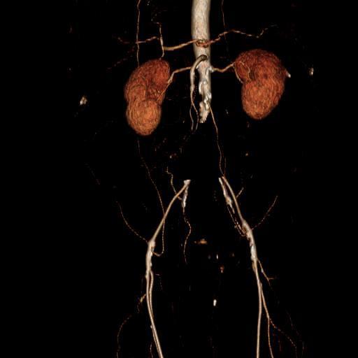
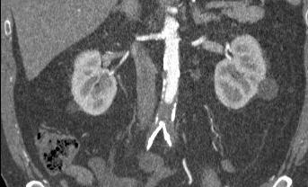
18 Volume Rendered Image from CTA Aorta Renal Artery
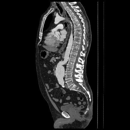
19 Vascular Stent Treatment Length Aortic Stent
20 Virtual Bronchoscopy
21 Major uses in medical imaging Vessel Segmentation Bone Segmentation Targeted Lesion Segmentation
22 Bone Segmentation Primary use: Modeling fractures for operative planning Excluding bone points to aid in the accuracy of vessel tracking Individual bone segmentation for lesion localization Bone has overlap of density values with numerous other structures Vascular calcium Densely enhanced vessels Enteric contrast Small metallic structures

23 Region Growth of the Pelvis

24 Individual Bone Growth Sacrum Iliac Femur

25 Not all that is white is bone Intravenous Contrast Vascular Calcium Calcium in Bone
26 Major Uses in medical imaging Vessel Segmentation Bone Segmentation Targeted Lesion Segmentation
27 Target Lesion Segmentation The most critical role of region growth in medical imaging is in the isolation of target lesions Measure size of primary and metastatic tumors Change of lesion size and morphology over time Critical to incorporate edge detection data as well as dynamic thresholds

28 Length Area Width

29 Tumor Left Atrium

30 Primary Development Workstation
31 Overview Introduction Traditional Region Growth Algorithms Simple GPU based Region Growth Enhanced Threshold Based Techniques Conclusions
32 Translating Region Growth to the GPU Calculate Gradients Well suited in general for GPU implementation. 2D or 3D gradients Region Growth Loop Major difference between CPU and GPU implementation. Result of i-th iteration depends on result of iteration (i-1) results from earlier threads Race condition can cause variability in results when number of iterations is limited. Speed Maintain a single data set that is modified and changes during a single kernel execution RAW (result from one thread effect other thread in warp or block) WAR (missed opportunity for region growth) Reproducibility Main data set and Result data Determining Completion Typically achieved when all points are classified Not always a straight forward task with large 3D data sets
33 How We Think it Happens

34 What Really Happens
35 Code Path User Input P (x,y,z) Establish Region Criteria Process Image -Gradient Calculation -Initial Threshold Apply Selection Criteria to Boundary Elements Apply Result Region Growth Loop

36 Host Code Initial Region Assignment And Propagation Region Growth Loops
37 Device Code Check for seed point Check Criteria for Immediately Surrounding Points Memory Access Not Globally Convergent
38 Initial Seed Points
39 Results The quality of a region growth algorithm judged by: Speed of completion Number of seed points Limit iterations on non-edge points Sensitivity Specificity Reproducibility In general only possible with the use of memory result buffer How to determine failure Non-target organs included in region Large sections of bone omitted.
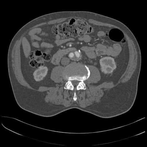
40 Pelvic Subsegmentation By Contextual Analysis

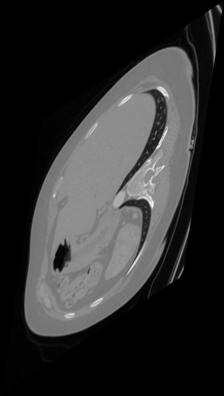
41 Image Through Lower Chest
42 Growth Error Including Hypogastrics

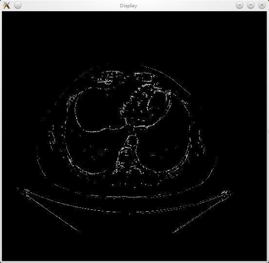
43 Closer look at boundary problems
44 Distinct Structures, But How Do You Know?
45 Context Clearly Matters More than Density

46 Cooling A Hexa-GPU System After Hours
47 Overview Introduction Traditional Region Growth Algorithms Simple GPU based Region Growth Enhanced Threshold Based Techniques Conclusions
48 Methods for Guiding Region Growth General Edge/Gradient based Established method for constraining growth Perform an initial edge detection Use derivative in N directions for edges/bounding Work poorly for noisy or low contrast images Problem Specific Geometric Features of structures used to guide region growth. Avoid overly rigid geometries that prevent accommodating variations in anatomy Not generally useful in structures that have significant variations. Several key geometries are useful in the human body Spherical Symmetry Cylindrical Symmetry Model based geometry Other
49 Guided Methods Spherical Symmetry for Small Vessel Tracking Cylindrical Symmetry for Large Vessel Tracking Variable Threshold for Small Vessel Tracking
50 Guided Methods Spherical Symmetry for Small Vessel Tracking Cylindrical Symmetry for Large Vessel Tracking Variable Threshold for Small Vessel Tracking
51 Spherical Coordinate in Vessel Tracking Very simply coordinate system to manipulate Provides an intrinsic symmetry when analyzing structures which would otherwise require multiple coordinate transformations Auto compensates for rotation about long axis Well Suited for specific tasks Identifying Initial segments of vessels Maintaining luminal axis
52 Vessel Tracking With Spherical Symmetry What is most important Continuity is one of the hallmarks of vessels Relative density is important Exploiting spherical symmetry requires appropriately positioned seed point To track vessels you can use concentric spheres Isolate potential points for region growth Vastly improves subsequent growth by limiting extraneous analysis
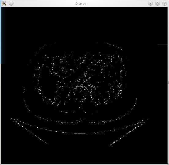
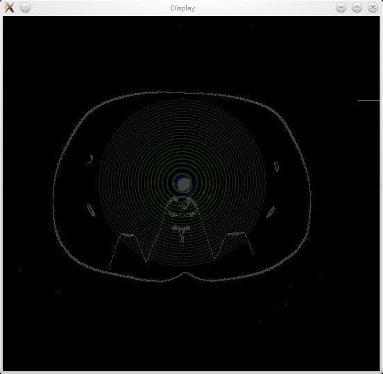
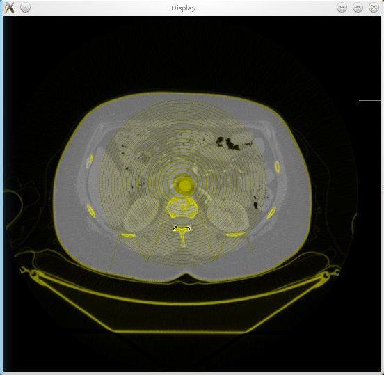
53 Applying Concentric Sphere Model
54 Device Code Calculate Point on Sphere Surface Global Memory Access Determine if Point Meets Criteria
55 Device Code (2) Global Memory Access to Retreive Sphere Points on Adjacent Shells Restrict to Points that Passed Previous Stage Check Point Between Shells Record Results
56 Limitations Vessels that deviate significantly from spherical symmetry Lumen follows contour of sphere shell Significant obliquity of course No straight forward way to limit inclusion of non-vascular points Only an initial/ pre-processing step for more guided region growth Requires appropriate positioning of initial seed point Typically near the origin of vessel Works best for short segments of small straight-ish vessels
57 Guided Methods Spherical Symmetry for Small Vessel Tracking Cylindrical Symmetry for Large Vessel Tracking Variable Threshold for Small Vessel Tracking
58 Principles of Large Vessel Tracking Numerous structures in human body have intrinsic cylindrical symmetry Scale on which the symmetry is maintained varies Aorta is not a true cylinders Coordinate system matters Aorta tends toward a specific direction Starts near midline and ends near midline Not necessarily predictable in between Massively threaded code allows for cooperative behavior One thread can motivate but not restrict other threads. Numerous paths exist which satisfy a set of conditions The tendency is for true paths to outnumber stray paths


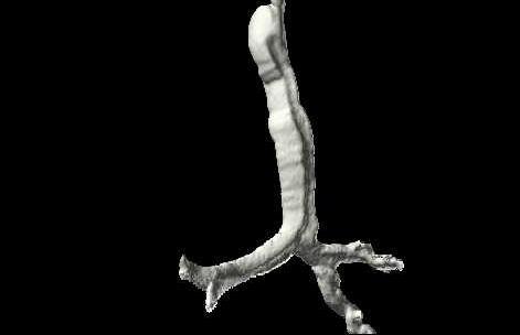
59 Defying Density with Brute GPU force
60 Highly computationally intensive algorithm Initial slice search grid (256 * 256) Distal slice target points (64 * 64) Distance between slices (128) Total ~ 270 million combinations (128 points sampled per combination) Longer path length Increases specificity. Decreases sensitivity Location matters Regardless of path length, IVC contamination is always an issue Spine needs to be avoided

61 Host Code MemCopy Run kernel Statistical Analysis of Results
62 Device Code Thread and Block Indexing Use Shared Memory Convert thd/blk index to spatial positioning Global Memory Access Converge Shared Data Compare Result to Existing Max
63
64
65 Limitations Vessels which vary significantly in directionality Very common, though not on scales typically used by algorithm Surgical aorta poses additional problems Significant variations in contrast density over length of vessel analysis Fortunately uncommon Significant variants in vessel size Common and important to recognize Discontinuity of vessel Uncommon though important to recognize Extra-vascular contrast Uncommon though important to recognize
66
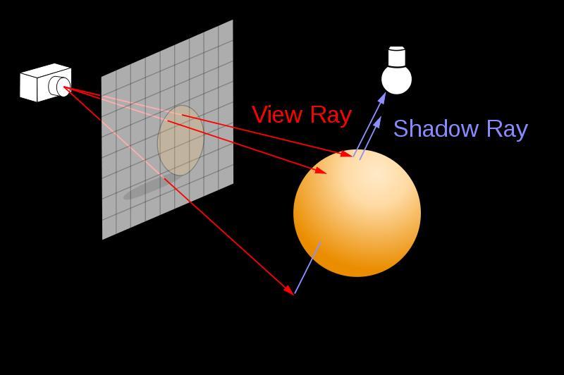
67 Roots in ray tracing This algorithm inspired by ray tracing Typical Ray tracing provides structures with attributes Reflection, refraction, and shadow Acceleration structures Way to efficiently traverse static scene GPU implementation exist (OptiX) Vessel tracking implementation Continuous medium Acceleration structure dynamically changing Fractional transmission rather than refraction Ray Trace Image courtesy of wikipedia

68
69
70 Ray Tracing/Mean Free Path Each ray made up of multiple points Various density values Separate average and standard deviation Potential Fate of Rays Reflection/Refraction Not a possible outcome in this algorithm True absorbed rays Average or standard deviation outside range Significant acute drop in density (edge) Partial transmission Results from fluctuations in density (e.g. image noise, metal artifact, heart failure) Excluded unless no other choice Successful rays All points meet criteria Low variance in density with average within range Limited fluctuations (>95% transmission)
71 Guided Methods Spherical Symmetry for Small Vessel Tracking Cylindrical Symmetry for Large Vessel Tracking Variable Threshold for Small Vessel Tracking
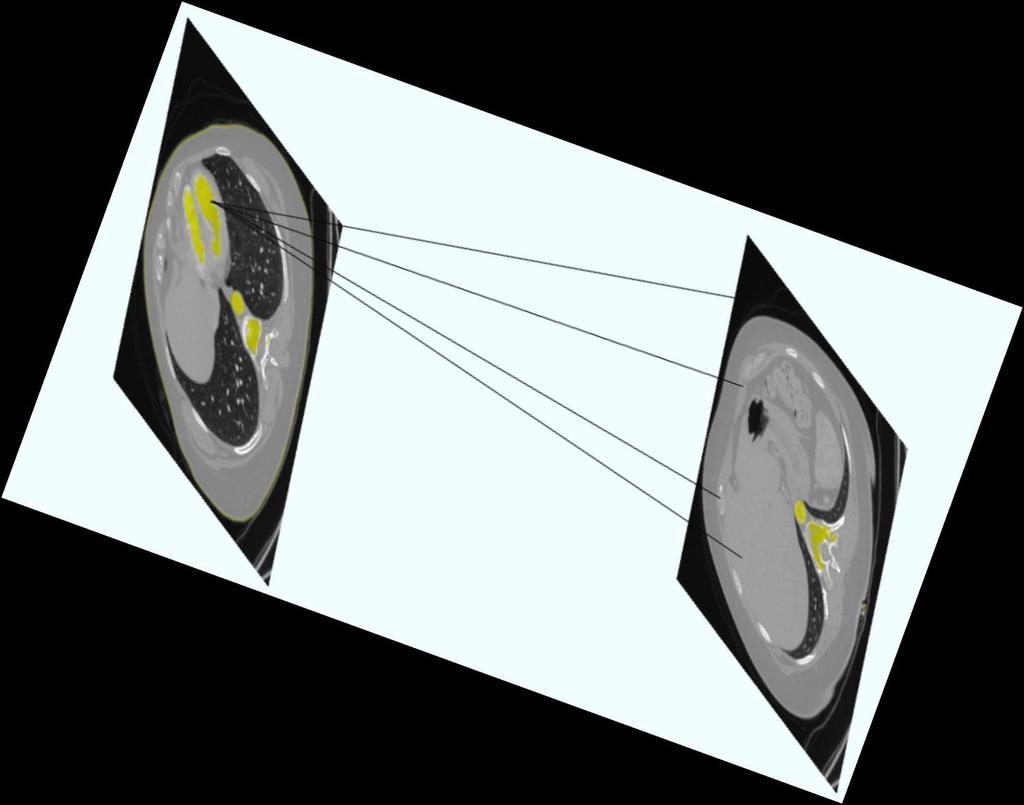
72 Variable Threshold for Small Vessels Small vessel tracking Significantly more challenging than aortic tracking No reasonable expectation of linearity Vessel width varies greatly inter and intra scan Quality of contrast enhancement Varies greatly between scans. Scanner can outruns the contrast bolus Minimizing the density window is desirable Artery / vein differentiation often relies on very small differences Artery/ bone differentiation relies critically on boundary point which have intermediate values
 -Arteries -Veins")
73 Points that fall in window ( HU) -Arteries -Veins -Organs

74 Intra vessel variations in average HU


75 Dynamic Variable Threshold Requires partitioning a region into sub-regions Sub-regions have their own parameters Overall constraint on parameters Dynamically computed Connection Sub-region with overall 1 region Not necessarily more computationally intensive -Level 1 Avoid unnecessary -Width 1 computations Easier to identify aberrant paths. Each thread block defines separate sub-region Cooperative interaction within sub-region analysis Sub-region 2 Keeps threshold and window appropriately narrow Global memory -Width for communication 2 between sub-regions -Level 2 Prevents backtracking Allows low level coordination and communication Entire Vessel is Region
76 Host Code MemCopy Run kernel Check Results
77 Device Code Thread and Block Indexing Adjust Level Perform Adjacent Point Analysis Converge Shared Data and Check Bounds Write Results to Global Memory

78 First Server Running Alpha Build of Software
79 Overview Introduction Traditional Region Growth Algorithms Simple GPU based Region Growth Enhanced Threshold Based Techniques Conclusions
80 Conclusions Basic Principles of Region Growth Utilization of Region Growth in Medical Imaging Basic GPU Implementation of Region Growth Algorithm Direct translation of CPU code problematic Doesn t utilize full potential of massively parallel GPU Guiding region growth Large vessel tracking with modified ray tracing Small vessel tracking Spherical Variable threshold

81 ???QUESTIONS???
FINDING THE TRUE EDGE IN CTA
 FINDING THE TRUE EDGE IN CTA by: John A. Rumberger, PhD, MD, FACC Your patient has chest pain. The Cardiac CT Angiography shows plaque in the LAD. You adjust the viewing window trying to evaluate the stenosis
Human Heart Coronary Arteries Segmentation
 Human Heart Coronary Arteries Segmentation Qian Huang Wright State University, Computer Science Department Abstract The volume information extracted from computed tomography angiogram (CTA) datasets makes
Norbert Schuff VA Medical Center and UCSF
 Norbert Schuff Medical Center and UCSF Norbert.schuff@ucsf.edu Medical Imaging Informatics N.Schuff Course # 170.03 Slide 1/67 Objective Learn the principle segmentation techniques Understand the role
Modeling and preoperative planning for kidney surgery
 Modeling and preoperative planning for kidney surgery Refael Vivanti Computer Aided Surgery and Medical Image Processing Lab Hebrew University of Jerusalem, Israel Advisor: Prof. Leo Joskowicz Clinical
How to Adjust the 16-Bit CLUT (Color Look Up Table) Editor in OsiriX
 Editor in OsiriX") How to Adjust the 16-Bit CLUT (Color Look Up Table) Editor in OsiriX 1. Import the series you are interested in into the 2D/3D Viewer. I m using the OBELIX data set, slices 1-395, available for download
How to Adjust the 16-Bit CLUT (Color Look Up Table) Editor in OsiriX 1. Import the series you are interested in into the 2D/3D Viewer. I m using the OBELIX data set, slices 1-395, available for download
CT Basics Principles of Spiral CT Dose. Always Thinking Ahead.
 1 CT Basics Principles of Spiral CT Dose 2 Who invented CT? 1963 - Alan Cormack developed a mathematical method of reconstructing images from x-ray projections Sir Godfrey Hounsfield worked for the Central
Norbert Schuff Professor of Radiology VA Medical Center and UCSF
 Norbert Schuff Professor of Radiology Medical Center and UCSF Norbert.schuff@ucsf.edu 2010, N.Schuff Slide 1/67 Overview Definitions Role of Segmentation Segmentation methods Intensity based Shape based
Refraction Corrected Transmission Ultrasound Computed Tomography for Application in Breast Imaging
 Refraction Corrected Transmission Ultrasound Computed Tomography for Application in Breast Imaging Joint Research With Trond Varslot Marcel Jackowski Shengying Li and Klaus Mueller Ultrasound Detection
MR IMAGE SEGMENTATION
 MR IMAGE SEGMENTATION Prepared by : Monil Shah What is Segmentation? Partitioning a region or regions of interest in images such that each region corresponds to one or more anatomic structures Classification
RSNA, /rg
 RSNA, 2015 10.1148/rg.2015140320 Appendix As noted in the main article, DICOM image files cannot be used directly for 3D printing; further steps are necessary to make them readable by 3D printers. The
Computational Medical Imaging Analysis Chapter 4: Image Visualization
 Computational Medical Imaging Analysis Chapter 4: Image Visualization Jun Zhang Laboratory for Computational Medical Imaging & Data Analysis Department of Computer Science University of Kentucky Lexington,
Classification of Subject Motion for Improved Reconstruction of Dynamic Magnetic Resonance Imaging
 1 CS 9 Final Project Classification of Subject Motion for Improved Reconstruction of Dynamic Magnetic Resonance Imaging Feiyu Chen Department of Electrical Engineering ABSTRACT Subject motion is a significant
Medical Image Processing: Image Reconstruction and 3D Renderings
 Medical Image Processing: Image Reconstruction and 3D Renderings 김보형 서울대학교컴퓨터공학부 Computer Graphics and Image Processing Lab. 2011. 3. 23 1 Computer Graphics & Image Processing Computer Graphics : Create,
CT NOISE POWER SPECTRUM FOR FILTERED BACKPROJECTION AND ITERATIVE RECONSTRUCTION
 CT NOISE POWER SPECTRUM FOR FILTERED BACKPROJECTION AND ITERATIVE RECONSTRUCTION Frank Dong, PhD, DABR Diagnostic Physicist, Imaging Institute Cleveland Clinic Foundation and Associate Professor of Radiology
Automated segmentation methods for liver analysis in oncology applications
 University of Szeged Department of Image Processing and Computer Graphics Automated segmentation methods for liver analysis in oncology applications Ph. D. Thesis László Ruskó Thesis Advisor Dr. Antal
Chapter 4. Clustering Core Atoms by Location
 Chapter 4. Clustering Core Atoms by Location In this chapter, a process for sampling core atoms in space is developed, so that the analytic techniques in section 3C can be applied to local collections
8/3/2017. Contour Assessment for Quality Assurance and Data Mining. Objective. Outline. Tom Purdie, PhD, MCCPM
 Contour Assessment for Quality Assurance and Data Mining Tom Purdie, PhD, MCCPM Objective Understand the state-of-the-art in contour assessment for quality assurance including data mining-based techniques
Ch. 4 Physical Principles of CT
 Ch. 4 Physical Principles of CT CLRS 408: Intro to CT Department of Radiation Sciences Review: Why CT? Solution for radiography/tomography limitations Superimposition of structures Distinguishing between
Programmable Shaders for Deformation Rendering
 Programmable Shaders for Deformation Rendering Carlos D. Correa, Deborah Silver Rutgers, The State University of New Jersey Motivation We present a different way of obtaining mesh deformation. Not a modeling,
A Generic Lie Group Model for Computer Vision
 A Generic Lie Group Model for Computer Vision Within this research track we follow a generic Lie group approach to computer vision based on recent physiological research on how the primary visual cortex
[PDR03] RECOMMENDED CT-SCAN PROTOCOLS
![[PDR03] RECOMMENDED CT-SCAN PROTOCOLS](/thumbs/72/66454100.jpg "[PDR03] RECOMMENDED CT-SCAN PROTOCOLS") SURGICAL & PROSTHETIC DESIGN [PDR03] RECOMMENDED CT-SCAN PROTOCOLS WORK-INSTRUCTIONS DOCUMENT (CUSTOMER) RECOMMENDED CT-SCAN PROTOCOLS [PDR03_V1]: LIVE 1 PRESCRIBING SURGEONS Patient-specific implants,
SURGICAL & PROSTHETIC DESIGN [PDR03] RECOMMENDED CT-SCAN PROTOCOLS WORK-INSTRUCTIONS DOCUMENT (CUSTOMER) RECOMMENDED CT-SCAN PROTOCOLS [PDR03_V1]: LIVE 1 PRESCRIBING SURGEONS Patient-specific implants,
CHAPTER 3 RETINAL OPTIC DISC SEGMENTATION
 60 CHAPTER 3 RETINAL OPTIC DISC SEGMENTATION 3.1 IMPORTANCE OF OPTIC DISC Ocular fundus images provide information about ophthalmic, retinal and even systemic diseases such as hypertension, diabetes, macular
Color Space Invariance for Various Edge Types in Simple Images. Geoffrey Hollinger and Dr. Bruce Maxwell Swarthmore College Summer 2003
 Color Space Invariance for Various Edge Types in Simple Images Geoffrey Hollinger and Dr. Bruce Maxwell Swarthmore College Summer 2003 Abstract This paper describes a study done to determine the color
Image Analysis, Geometrical Modelling and Image Synthesis for 3D Medical Imaging
 Image Analysis, Geometrical Modelling and Image Synthesis for 3D Medical Imaging J. SEQUEIRA Laboratoire d'informatique de Marseille - FRE CNRS 2246 Faculté des Sciences de Luminy, 163 avenue de Luminy,
Corso di laurea in Fisica A.A Fisica Medica 4 TC
 Corso di laurea in Fisica A.A. 2007-2008 Fisica Medica 4 TC Computed Tomography Principles 1. Projection measurement 2. Scanner systems 3. Scanning modes Basic Tomographic Principle The internal structure
EE795: Computer Vision and Intelligent Systems
 EE795: Computer Vision and Intelligent Systems Spring 2012 TTh 17:30-18:45 WRI C225 Lecture 04 130131 http://www.ee.unlv.edu/~b1morris/ecg795/ 2 Outline Review Histogram Equalization Image Filtering Linear
Three-dimensional graphics system of medical images on the Web for patients
 Three-dimensional graphics system of medical images on the Web for patients Koji Kobayashi *1, Koichi Ito *2, Takafumi Aoki *2 *1 Vocsis Corporation *2 Graduate School of Information Sciences, Tohoku University
Vessel Explorer: a tool for quantitative measurements in CT and MR angiography
 Clinical applications Vessel Explorer: a tool for quantitative measurements in CT and MR angiography J. Oliván Bescós J. Sonnemans R. Habets J. Peters H. van den Bosch T. Leiner Healthcare Informatics/Patient
Engineering Problem and Goal
 Engineering Problem and Goal Engineering Problem: Traditional active contour models can not detect edges or convex regions in noisy images. Engineering Goal: The goal of this project is to design an algorithm
Dynamic digital phantoms
 Dynamic digital phantoms In radiation research the term phantom is used to describe an inanimate object or system used to tune the performance of radiation imaging or radiotherapeutic devices. A wide range
Lecture 12 Level Sets & Parametric Transforms. sec & ch. 11 of Machine Vision by Wesley E. Snyder & Hairong Qi
 Lecture 12 Level Sets & Parametric Transforms sec. 8.5.2 & ch. 11 of Machine Vision by Wesley E. Snyder & Hairong Qi Spring 2017 16-725 (CMU RI) : BioE 2630 (Pitt) Dr. John Galeotti The content of these
Scalar Data. Visualization Torsten Möller. Weiskopf/Machiraju/Möller
 Scalar Data Visualization Torsten Möller Weiskopf/Machiraju/Möller Overview Basic strategies Function plots and height fields Isolines Color coding Volume visualization (overview) Classification Segmentation
Segmentation of Bony Structures with Ligament Attachment Sites
 Segmentation of Bony Structures with Ligament Attachment Sites Heiko Seim 1, Hans Lamecker 1, Markus Heller 2, Stefan Zachow 1 1 Visualisierung und Datenanalyse, Zuse-Institut Berlin (ZIB), 14195 Berlin
INDUSTRIAL SYSTEM DEVELOPMENT FOR VOLUMETRIC INTEGRITY
 INDUSTRIAL SYSTEM DEVELOPMENT FOR VOLUMETRIC INTEGRITY VERIFICATION AND ANALYSIS M. L. Hsiao and J. W. Eberhard CR&D General Electric Company Schenectady, NY 12301 J. B. Ross Aircraft Engine - QTC General
Probabilistic Tracking and Model-based Segmentation of 3D Tubular Structures
 Probabilistic Tracking and Model-based Segmentation of 3D Tubular Structures Stefan Wörz, William J. Godinez, Karl Rohr University of Heidelberg, BIOQUANT, IPMB, and DKFZ Heidelberg, Dept. Bioinformatics
Digital Image Processing
 Digital Image Processing SPECIAL TOPICS CT IMAGES Hamid R. Rabiee Fall 2015 What is an image? 2 Are images only about visual concepts? We ve already seen that there are other kinds of image. In this lecture
Case TAVR080: Aortic Valve step-by-step guide
 Case TAVR080: Aortic Valve step-by-step guide 3D printing the aortic valve M-0635B Case Airways - Learning objectives At the end of the session, the student will be able to: Use and refine automatic segmentation.
AORTA CTA VPMC-12419
 AORTA CTA VPMC-12419 Workflow Overview: The Aorta can be post-processed in various ways. Auto Bone Removal and Vessel Pick provide a quick overview of the entire Aorta. Vessel Probe creates a centerline
GE Healthcare. Vivid 7 Dimension 08 Cardiovascular ultrasound system
 GE Healthcare Vivid 7 Dimension 08 Cardiovascular ultrasound system ltra Definition. Technology. Performance. Start with a system that s proven its worth in LV quantification and 4D imaging. Then add even
Tomographic Reconstruction
 Tomographic Reconstruction 3D Image Processing Torsten Möller Reading Gonzales + Woods, Chapter 5.11 2 Overview Physics History Reconstruction basic idea Radon transform Fourier-Slice theorem (Parallel-beam)
A Fast GPU-Based Approach to Branchless Distance-Driven Projection and Back-Projection in Cone Beam CT
 A Fast GPU-Based Approach to Branchless Distance-Driven Projection and Back-Projection in Cone Beam CT Daniel Schlifske ab and Henry Medeiros a a Marquette University, 1250 W Wisconsin Ave, Milwaukee,
Solid Modeling. Thomas Funkhouser Princeton University C0S 426, Fall Represent solid interiors of objects
 Solid Modeling Thomas Funkhouser Princeton University C0S 426, Fall 2000 Solid Modeling Represent solid interiors of objects Surface may not be described explicitly Visible Human (National Library of Medicine)
The Near Future in Cardiac CT Image Reconstruction
 SCCT 2010 The Near Future in Cardiac CT Image Reconstruction Marc Kachelrieß Institute of Medical Physics (IMP) Friedrich-Alexander Alexander-University Erlangen-Nürnberg rnberg www.imp.uni-erlangen.de
Point Cloud Filtering using Ray Casting by Eric Jensen 2012 The Basic Methodology
 Point Cloud Filtering using Ray Casting by Eric Jensen 01 The Basic Methodology Ray tracing in standard graphics study is a method of following the path of a photon from the light source to the camera,
K-Means Clustering Using Localized Histogram Analysis
 K-Means Clustering Using Localized Histogram Analysis Michael Bryson University of South Carolina, Department of Computer Science Columbia, SC brysonm@cse.sc.edu Abstract. The first step required for many
Fundamentals of CT imaging
 SECTION 1 Fundamentals of CT imaging I History In the early 1970s Sir Godfrey Hounsfield s research produced the first clinically useful CT scans. Original scanners took approximately 6 minutes to perform
Implementation of Advanced Image Guided Radiation Therapy
 Image Acquisition Course Outline Principles, characteristics& applications of the available modalities Image Processing in the T x room Image guided treatment delivery What can / can t we do in the room
Previously... contour or image rendering in 2D
 Volume Rendering Visualisation Lecture 10 Taku Komura Institute for Perception, Action & Behaviour School of Informatics Volume Rendering 1 Previously... contour or image rendering in 2D 2D Contour line
The organization of the human cerebral cortex estimated by intrinsic functional connectivity
 1 The organization of the human cerebral cortex estimated by intrinsic functional connectivity Journal: Journal of Neurophysiology Author: B. T. Thomas Yeo, et al Link: https://www.ncbi.nlm.nih.gov/pubmed/21653723
Scattering/Wave Terminology A few terms show up throughout the discussion of electron microscopy:
 1. Scattering and Diffraction Scattering/Wave Terology A few terms show up throughout the discussion of electron microscopy: First, what do we mean by the terms elastic and inelastic? These are both related
Isosurface Rendering. CSC 7443: Scientific Information Visualization
 Isosurface Rendering What is Isosurfacing? An isosurface is the 3D surface representing the locations of a constant scalar value within a volume A surface with the same scalar field value Isosurfaces form
CS179: GPU Programming Recitation 5: Rendering Fractals
 CS179: GPU Programming Recitation 5: Rendering Fractals Rendering Fractals Volume data vs. texture memory Creating and using CUDA arrays Using PBOs for screen output Quaternion Julia Sets Rendering volume
Segmentation and Modeling of the Spinal Cord for Reality-based Surgical Simulator
 Segmentation and Modeling of the Spinal Cord for Reality-based Surgical Simulator Li X.C.,, Chui C. K.,, and Ong S. H.,* Dept. of Electrical and Computer Engineering Dept. of Mechanical Engineering, National
CPSC GLOBAL ILLUMINATION
 CPSC 314 21 GLOBAL ILLUMINATION Textbook: 20 UGRAD.CS.UBC.CA/~CS314 Mikhail Bessmeltsev ILLUMINATION MODELS/ALGORITHMS Local illumination - Fast Ignore real physics, approximate the look Interaction of
Prostate Detection Using Principal Component Analysis
 Prostate Detection Using Principal Component Analysis Aamir Virani (avirani@stanford.edu) CS 229 Machine Learning Stanford University 16 December 2005 Introduction During the past two decades, computed
Image Acquisition Systems
 Image Acquisition Systems Goals and Terminology Conventional Radiography Axial Tomography Computer Axial Tomography (CAT) Magnetic Resonance Imaging (MRI) PET, SPECT Ultrasound Microscopy Imaging ITCS
Introduction to Medical Image Processing
 Introduction to Medical Image Processing Δ Essential environments of a medical imaging system Subject Image Analysis Energy Imaging System Images Image Processing Feature Images Image processing may be
CHAPTER 3 TUMOR DETECTION BASED ON NEURO-FUZZY TECHNIQUE
 32 CHAPTER 3 TUMOR DETECTION BASED ON NEURO-FUZZY TECHNIQUE 3.1 INTRODUCTION In this chapter we present the real time implementation of an artificial neural network based on fuzzy segmentation process
Scalar Data. CMPT 467/767 Visualization Torsten Möller. Weiskopf/Machiraju/Möller
 Scalar Data CMPT 467/767 Visualization Torsten Möller Weiskopf/Machiraju/Möller Overview Basic strategies Function plots and height fields Isolines Color coding Volume visualization (overview) Classification
Exploiting Typical Clinical Imaging Constraints for 3D Outer Bone Surface Segmentation
 Exploiting Typical Clinical Imaging Constraints for 3D Outer Bone Surface Segmentation Chris Mack, Vishali Mogallapu, Andrew Willis, Thomas P. Weldon UNC Charlotte, Department of Electrical and Computer
2 Michael E. Leventon and Sarah F. F. Gibson a b c d Fig. 1. (a, b) Two MR scans of a person's knee. Both images have high resolution in-plane, but ha
 Two MR scans of a person's knee. Both images have high resolution in-plane, but ha") Model Generation from Multiple Volumes using Constrained Elastic SurfaceNets Michael E. Leventon and Sarah F. F. Gibson 1 MIT Artificial Intelligence Laboratory, Cambridge, MA 02139, USA leventon@ai.mit.edu
Model Generation from Multiple Volumes using Constrained Elastic SurfaceNets Michael E. Leventon and Sarah F. F. Gibson 1 MIT Artificial Intelligence Laboratory, Cambridge, MA 02139, USA leventon@ai.mit.edu
Volume Rendering. Computer Animation and Visualisation Lecture 9. Taku Komura. Institute for Perception, Action & Behaviour School of Informatics
 Volume Rendering Computer Animation and Visualisation Lecture 9 Taku Komura Institute for Perception, Action & Behaviour School of Informatics Volume Rendering 1 Volume Data Usually, a data uniformly distributed
Volume Illumination. Visualisation Lecture 11. Taku Komura. Institute for Perception, Action & Behaviour School of Informatics
 Volume Illumination Visualisation Lecture 11 Taku Komura Institute for Perception, Action & Behaviour School of Informatics Taku Komura Volume Illumination & Vector Vis. 1 Previously : Volume Rendering
VieW 3D. 3D Post-Processing WorKstation THE THIRD DIMENSION. Version 3.1
 VieW 3D 3D Post-Processing WorKstation THE THIRD DIMENSION Version 3.1 iq-view 3D THE FULLY-FEATURED 3D MEDICAL IMAGING SOLUTION FOR RADIOLOGISTS iq-view 3D contains all components of iq-view with the
6.837 Introduction to Computer Graphics Final Exam Tuesday, December 20, :05-12pm Two hand-written sheet of notes (4 pages) allowed 1 SSD [ /17]
![6.837 Introduction to Computer Graphics Final Exam Tuesday, December 20, :05-12pm Two hand-written sheet of notes (4 pages) allowed 1 SSD [ /17]](/thumbs/87/95541269.jpg "6.837 Introduction to Computer Graphics Final Exam Tuesday, December 20, :05-12pm Two hand-written sheet of notes (4 pages) allowed 1 SSD [ /17]") 6.837 Introduction to Computer Graphics Final Exam Tuesday, December 20, 2011 9:05-12pm Two hand-written sheet of notes (4 pages) allowed NAME: 1 / 17 2 / 12 3 / 35 4 / 8 5 / 18 Total / 90 1 SSD [ /17]
6.837 Introduction to Computer Graphics Final Exam Tuesday, December 20, 2011 9:05-12pm Two hand-written sheet of notes (4 pages) allowed NAME: 1 / 17 2 / 12 3 / 35 4 / 8 5 / 18 Total / 90 1 SSD [ /17]
Assessing Accuracy Factors in Deformable 2D/3D Medical Image Registration Using a Statistical Pelvis Model
 Assessing Accuracy Factors in Deformable 2D/3D Medical Image Registration Using a Statistical Pelvis Model Jianhua Yao National Institute of Health Bethesda, MD USA jyao@cc.nih.gov Russell Taylor The Johns
Constrained Diffusion Limited Aggregation in 3 Dimensions
 Constrained Diffusion Limited Aggregation in 3 Dimensions Paul Bourke Swinburne University of Technology P. O. Box 218, Hawthorn Melbourne, Vic 3122, Australia. Email: pdb@swin.edu.au Abstract Diffusion
Segmentation tools and workflows in PerGeos
 Segmentation tools and workflows in PerGeos 1. Introduction Segmentation typically consists of a complex workflow involving multiple algorithms at multiple steps. Smart denoising and morphological filters
Case TAVR080: Aortic Valve Step-by-Step Guide
 Case TAVR080: Aortic Valve Step-by-Step Guide 3D printing the aortic valve The 3D printed anatomical model is not for diagnostic use. M-06235C 208 Vital Images, Inc. www.vitalimages.com Case Airways -
Clinical Importance. Aortic Stenosis. Aortic Regurgitation. Ultrasound vs. MRI. Carotid Artery Stenosis
 Clinical Importance Rapid cardiovascular flow quantitation using sliceselective Fourier velocity encoding with spiral readouts Valve disease affects 10% of patients with heart disease in the U.S. Most
Module 7 VIDEO CODING AND MOTION ESTIMATION
 Module 7 VIDEO CODING AND MOTION ESTIMATION Lesson 20 Basic Building Blocks & Temporal Redundancy Instructional Objectives At the end of this lesson, the students should be able to: 1. Name at least five
diffuse diffuse reflection refraction diffuse mapping diffuse reflection reflection filter mapping mapping reflection
 Matières 1 2 3 mapping diffuse reflection diffuse transparency reflection refraction diffuse mapping diffuse reflection diffuse reflection filter mapping bump mapping mapping mapping diffuse reflection
Shape representation by skeletonization. Shape. Shape. modular machine vision system. Feature extraction shape representation. Shape representation
 Shape representation by skeletonization Kálmán Palágyi Shape It is a fundamental concept in computer vision. It can be regarded as the basis for high-level image processing stages concentrating on scene
Basics of treatment planning II
 Basics of treatment planning II Sastry Vedam PhD DABR Introduction to Medical Physics III: Therapy Spring 2015 Dose calculation algorithms! Correction based! Model based 1 Dose calculation algorithms!
radiotherapy Andrew Godley, Ergun Ahunbay, Cheng Peng, and X. Allen Li NCAAPM Spring Meeting 2010 Madison, WI
 GPU-Accelerated autosegmentation for adaptive radiotherapy Andrew Godley, Ergun Ahunbay, Cheng Peng, and X. Allen Li agodley@mcw.edu NCAAPM Spring Meeting 2010 Madison, WI Overview Motivation Adaptive
C a t p h a n / T h e P h a n t o m L a b o r a t o r y
 C a t p h a n 5 0 0 / 6 0 0 T h e P h a n t o m L a b o r a t o r y C a t p h a n 5 0 0 / 6 0 0 Internationally recognized for measuring the maximum obtainable performance of axial, spiral and multi-slice
Segmenting Lesions in Multiple Sclerosis Patients James Chen, Jason Su
 Segmenting Lesions in Multiple Sclerosis Patients James Chen, Jason Su Radiologists and researchers spend countless hours tediously segmenting white matter lesions to diagnose and study brain diseases.
US 1.
 US 1 Sample image: Normal pancreas seen on sonogram. Looking up from abdomen toward the head of the patient. The liver is in front of the pancreas. A vein draining the spleen is behind the pancreas http://www.radiologyinfo.org/photocat/photos.cfm?image=abdo-us-pancr.jpg&&subcategory=abdomen&&stop=9
Volume Illumination and Segmentation
 Volume Illumination and Segmentation Computer Animation and Visualisation Lecture 13 Institute for Perception, Action & Behaviour School of Informatics Overview Volume illumination Segmentation Volume
Volume visualization. Volume visualization. Volume visualization methods. Sources of volume visualization. Sources of volume visualization
 Volume visualization Volume visualization Volumes are special cases of scalar data: regular 3D grids of scalars, typically interpreted as density values. Each data value is assumed to describe a cubic
Robust PDF Table Locator
 Robust PDF Table Locator December 17, 2016 1 Introduction Data scientists rely on an abundance of tabular data stored in easy-to-machine-read formats like.csv files. Unfortunately, most government records
RADIOLOGY AND DIAGNOSTIC IMAGING
 Day 2 part 2 RADIOLOGY AND DIAGNOSTIC IMAGING Dr hab. Zbigniew Serafin, MD, PhD serafin@cm.umk.pl 2 3 4 5 CT technique CT technique 6 CT system Kanal K: RSNA/AAPM web module: CT Systems & CT Image Quality
2D image segmentation based on spatial coherence
 2D image segmentation based on spatial coherence Václav Hlaváč Czech Technical University in Prague Center for Machine Perception (bridging groups of the) Czech Institute of Informatics, Robotics and Cybernetics
Cross-Hardware GPGPU implementation of Acceleration Structures for Ray Tracing using OpenCL
 The Open University of Israel Department of Mathematics and Computer Science Cross-Hardware GPGPU implementation of Acceleration Structures for Ray Tracing using OpenCL Advanced Project in Computer Science
Computer Graphics (CS 543) Lecture 13b Ray Tracing (Part 1) Prof Emmanuel Agu. Computer Science Dept. Worcester Polytechnic Institute (WPI)
 Lecture 13b Ray Tracing (Part 1) Prof Emmanuel Agu. Computer Science Dept. Worcester Polytechnic Institute (WPI)") Computer Graphics (CS 543) Lecture 13b Ray Tracing (Part 1) Prof Emmanuel Agu Computer Science Dept. Worcester Polytechnic Institute (WPI) Raytracing Global illumination-based rendering method Simulates
Computer Graphics (CS 543) Lecture 13b Ray Tracing (Part 1) Prof Emmanuel Agu Computer Science Dept. Worcester Polytechnic Institute (WPI) Raytracing Global illumination-based rendering method Simulates
MEDICAL IMAGE COMPUTING (CAP 5937) LECTURE 9: Medical Image Segmentation (III) (Fuzzy Connected Image Segmentation)
 LECTURE 9: Medical Image Segmentation (III) (Fuzzy Connected Image Segmentation)") SPRING 2017 1 MEDICAL IMAGE COMPUTING (CAP 5937) LECTURE 9: Medical Image Segmentation (III) (Fuzzy Connected Image Segmentation) Dr. Ulas Bagci HEC 221, Center for Research in Computer Vision (CRCV),
SPRING 2017 1 MEDICAL IMAGE COMPUTING (CAP 5937) LECTURE 9: Medical Image Segmentation (III) (Fuzzy Connected Image Segmentation) Dr. Ulas Bagci HEC 221, Center for Research in Computer Vision (CRCV),
Lucy Phantom MR Grid Evaluation
 Lucy Phantom MR Grid Evaluation Anil Sethi, PhD Loyola University Medical Center, Maywood, IL 60153 November 2015 I. Introduction: The MR distortion grid, used as an insert with Lucy 3D QA phantom, is
Advanced Image Reconstruction Methods for Photoacoustic Tomography
 Advanced Image Reconstruction Methods for Photoacoustic Tomography Mark A. Anastasio, Kun Wang, and Robert Schoonover Department of Biomedical Engineering Washington University in St. Louis 1 Outline Photoacoustic/thermoacoustic
Range Imaging Through Triangulation. Range Imaging Through Triangulation. Range Imaging Through Triangulation. Range Imaging Through Triangulation
 Obviously, this is a very slow process and not suitable for dynamic scenes. To speed things up, we can use a laser that projects a vertical line of light onto the scene. This laser rotates around its vertical
Chapter 10 Computation Culling with Explicit Early-Z and Dynamic Flow Control
 Chapter 10 Computation Culling with Explicit Early-Z and Dynamic Flow Control Pedro V. Sander ATI Research John R. Isidoro ATI Research Jason L. Mitchell ATI Research Introduction In last year s course,
Extraction and recognition of the thoracic organs based on 3D CT images and its application
 1 Extraction and recognition of the thoracic organs based on 3D CT images and its application Xiangrong Zhou, PhD a, Takeshi Hara, PhD b, Hiroshi Fujita, PhD b, Yoshihiro Ida, RT c, Kazuhiro Katada, MD
Design and performance characteristics of a Cone Beam CT system for Leksell Gamma Knife Icon
 Design and performance characteristics of a Cone Beam CT system for Leksell Gamma Knife Icon WHITE PAPER Introduction Introducing an image guidance system based on Cone Beam CT (CBCT) and a mask immobilization
An Automated Image-based Method for Multi-Leaf Collimator Positioning Verification in Intensity Modulated Radiation Therapy
 An Automated Image-based Method for Multi-Leaf Collimator Positioning Verification in Intensity Modulated Radiation Therapy Chenyang Xu 1, Siemens Corporate Research, Inc., Princeton, NJ, USA Xiaolei Huang,
Motion Analysis. Motion analysis. Now we will talk about. Differential Motion Analysis. Motion analysis. Difference Pictures
 Now we will talk about Motion Analysis Motion analysis Motion analysis is dealing with three main groups of motionrelated problems: Motion detection Moving object detection and location. Derivation of
Arbitrary cut planes Slab control with slab thickness Projection plane adjustment Box cropping Mandible detection MPR cross-section linked views
 Provides unparalleled quality and performance no need to sacrifice one for the other Enables high fidelity viewing with interactive deep supersampling Requires no GPUs or hardware accelerators a true,
Image Segmentation and Registration
 Image Segmentation and Registration Dr. Christine Tanner (tanner@vision.ee.ethz.ch) Computer Vision Laboratory, ETH Zürich Dr. Verena Kaynig, Machine Learning Laboratory, ETH Zürich Outline Segmentation
Cerner SkyVue Cardiology Remote Review with NVIDIA and VMware Horizon
 Cerner SkyVue Cardiology Remote Review with NVIDIA and VMware Horizon Stuart Jackson Sr. Technology Architect Agenda Meet Cerner Cardiology Solution Overview Workflow Challenges Workflow Requirements Testing
Quantitative IntraVascular UltraSound (QCU)
") Quantitative IntraVascular UltraSound (QCU) Authors: Jouke Dijkstra, Ph.D. and Johan H.C. Reiber, Ph.D., Leiden University Medical Center, Dept of Radiology, Leiden, The Netherlands Introduction: For decades,
Quantitative IntraVascular UltraSound (QCU) Authors: Jouke Dijkstra, Ph.D. and Johan H.C. Reiber, Ph.D., Leiden University Medical Center, Dept of Radiology, Leiden, The Netherlands Introduction: For decades,
Image Post-Processing, Workflow, & Interpretation
 Image Post-Processing, Workflow, & Interpretation Richard L. Hallett, MD Chief, Cardiovascular Imaging Northwest Radiology Network Indianapolis, IN Adjunct Assistant Professor Stanford University Stanford,
Deviceless respiratory motion correction in PET imaging exploring the potential of novel data driven strategies
 g Deviceless respiratory motion correction in PET imaging exploring the potential of novel data driven strategies Presented by Adam Kesner, Ph.D., DABR Assistant Professor, Division of Radiological Sciences,