Computational Neuroanatomy
|
|
|
- Collin Franklin
- 5 years ago
- Views:
Transcription

1 Computational Neuroanatomy Bruce Fischl MGH ATHINOULA A. MARTINOS CENTER Harvard Medical School MIT CSAIL
2 Talk Outline 1. Cortical Analysis. 2. Subcortical Analysis. 3. Sources of distortion in MRI. 4. Linking micro structure to macro structure.
3 Talk Outline 1. Cortical Analysis. 2. Subcortical Analysis. 3. Sources of distortion in MRI. 4. Linking micro structure to macro structure.

4 Why Is a Model of the Cortical Surface Useful? Local functional organization of cortex is largely 2- dimensional! From (Sereno et al, 1995, Science).
5 Why Is Constructing a Model of The Cortical Surface Difficult? The cortex is highly folded! Partial voluming. Subject motion. Susceptibility artifacts. Bias field. Tissue inhomogeneities. Intensity of a tissue class varies as a function of spatial location
6 Sources of within-class intensity variation Partial voluming a single voxel may contain more than one tissue type. Bias field effective flip angle or sensitivity of receive coil may vary across space. Tissue inhomogeneities even within tissue type (e.g. cortical gray matter), intrinsic properties such as T1, PD can vary (up to 20%).

7 T1 weighted MR volume Assigning tissue classes to voxels can be difficult
8 Which Surface to Reconstruct? Pial surface is ultimate goal, but pretty much impossible to directly generate a representation of from MRI images (many have tried!). Alternative: construct an interim representation of the interface between gray matter and white matter, and use it to infer the location of the true cortical surface (Dale and Sereno, 1993).


9 Skull Stripping and building of Boundary Element Models


10 MRI Segmentation and Surface Reconstruction
11 Topology Correction The true topology of the cortical ribbon is that of a sheet (Euler number=1). We would like the reconstructed gray/white boundary to have spherical topology (Euler number=2), but errors in the segmentation and non-cortical anatomical features of the white matter cause departures from spherical topology ( defects ).

12 Typical Defects Cut Fornix Fill Pallidum and Putamen Cortical Defects Fill Ventricles and Caudate spackle hippocampus
13 Topological Defects
14 Standard method*: shrink wrapping start with a surface S (e.g. sphere) of known topology find a mapping M:SaC of it to the cortex C that doesn t change its topology (e.g. Davatzikos, 1996; Macdonald 2000) *newer volumetric work (Shattuck and Leahy, 2001; Han et al., 2002)
15 How to maintain geometric accuracy? Problems: 1. The initial surface S is typically much smoother than the target surface C. The energy functionals for finding M are therefore highly non-convex. 2. Local errors that would have given rise to inaccurate segmentation if the topology were not constrained, can cause large scale geometric inaccuracies in the surfaces.

16 What Surface Would a Shrink- Wrapping Algorithm Result in?

17 Solution: Manifold Surgery. Generate C and find a mapping M -1 from C to S that is invertible over as much of C as possible. Noninvertible regions contain defects!
18 Manifold Surgery: Equations 0.1 Energy Functional: E = F i= 1 log(1 + e k kr i ) det(j) R i jacobian at the i th face in tessellation F number of faces in tessellation k positive real constant negative semi-definite area (%) iteration #
19 Detection of non-homeomorphic Regions
20 Detection of non-homeomorphic Regions

21 Detection of non-homeomorphic Regions

22 Detection of non-homeomorphic Regions
23 (Florent Ségonne currently working on using genetic algorithm to evaluate different potential retessellations, but the space is huge!) Manifold Surgery: Retessellation 1. Mark all triangles that have any edge overlapping any other edge in the tessellation. 2. Discard all faces and edges in marked triangles. 3. Sort all possible edges by image likelihood (they should go through MR values between gray and white). 4. Use a greedy retessellation algorithm: keep adding edges between all vertices in defects until no more can be added without causing an intersection with an existing edge on the sphere.


24 Manifold Surgery: Results BEFORE AFTER

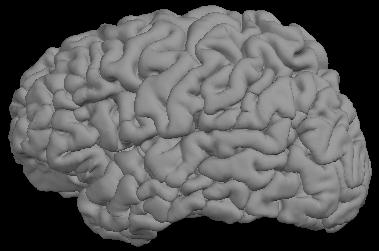
25 Surface Inflation


26 White matter and pial surfaces Gray-white boundary Pial surface


27 Surface Flattening Whole Hemisphere Inflated surface with cuts central anterior sylvian superior temporal posterior calcarine Metrically optimal flat map
 , 1999)")
28 Borrowed from (Halgren et al., 1999) Borrowed from (Halgren et al., 1999)
29 Talairach Coordinates Can mean many things, but most common is linear transform to align input image with a target image that is average of many individuals aligned with the atlas of Talairach and Tournoux (1988). Not Good For Cortex! 1. Typical transform is too low dimensional to account for variability in cortical folds. 2. Landmarks are subcortical (and far from much of cortex). 3. Implicit assumption that 3D metric is appropriate one.


30 Talairach averaging Average of 40 Single subject


31 How to align different cortical surfaces?
32 Surface-Based Coordinate System Establish a 2-D coordinate system on cortical surface - Every point in cortex should have a (unique) coordinate - Every coordinate should refer to a point in cortex Inter-subject alignment of cortical folding patterns Improve alignment of functional areas

33 A Surface-Based Coordinate System

34 Maximally Isometric Spherical Mapping Inflated Surface Transformed Surface
35 Spherical Morphing: Equations Energy Functional: J c +λ d J d +λ T J T J c: Correlation error (aligns folding patterns) J d : Metric distortion (constrains allowable shape differences) J T: Topology term (forces mapping to be invertible)
36 Spherical Morphing: Equations ), ( 1 ), ( 1 θ ϕ θ ϕ = = N i C i N C Average Folding Pattern: 2 )), ( ), ( 1 ( 1 1 ), ( 2 θ ϕ θ ϕ θ ϕ σ C N i C N i = = Variance of Folding 2 1 ) )) ( ), ( ( ))) ( ), ( ( *( ( 2 1 = = V v v c v v v v C C G V J θ φ σ θ φ α Maximum Likelihood Term: d d T T c J J J J λ + λ + = Complete Energy Functional:


37 Inter-Subject Morphing Individual Subject Average (Target)

38 Surface-Based Averaging Average surface created from 30 subjects
39 Applications Increased statistical power for inter-subject averaging Automatic functional/anatomical labeling Inter-subject averaging of morphometric properties Statistical analysis of morphometric properties aging neurodegenerative diseases longitudinal studies of structural changes hemispheric asymmetry
40 Applications Increased statistical power for inter-subject averaging Automatic functional/anatomical labeling Inter-subject averaging of morphometric properties Statistical analysis of morphometric properties aging neurodegenerative diseases longitudinal studies of structural changes hemispheric asymmetry


41 Inter-Subject Averaging of Activations Talairach Average Spherical Average
42 Applications Increased statistical power for inter-subject averaging Automatic functional/anatomical labeling Inter-subject averaging of morphometric properties Statistical analysis of morphometric properties aging neurodegenerative diseases longitudinal studies of structural changes hemispheric asymmetry
43 Applications Increased statistical power for inter-subject averaging Automatic functional/anatomical labeling Inter-subject averaging of morphometric properties Statistical analysis of morphometric properties aging neurodegenerative diseases longitudinal studies of structural changes hemispheric asymmetry

44 Cortical Parcellation: Manual vs. Automated Manual Parcellation Automatic Parcellation Thanks to Christophe Destrieux for this slide.
45 Applications Increased statistical power for inter-subject averaging Automatic functional/anatomical labeling Inter-subject averaging of morphometric properties Statistical analysis of morphometric properties aging neurodegenerative diseases longitudinal studies of structural changes hemispheric asymmetry
46 Applications Increased statistical power for inter-subject averaging Automatic functional/anatomical labeling Inter-subject averaging of morphometric properties Statistical analysis of morphometric properties aging neurodegenerative diseases longitudinal studies of structural changes hemispheric asymmetry

47 Lateral Selective Regional Thinning in AD Aging Central Sulcus/ Precentral Gyrus AD Parietal Cortex Prefrontal Cortex Medial Posterior Parahippocampal Entorhinal Cortex Ventral Dorsal Parietal Cortex Courtesy of Drs. Randy Buckner and David Salat
48 Talk Outline 1. Cortical Analysis. 2. Subcortical Analysis. 3. Sources of distortion in MRI. 4. Linking micro structure to macro structure.
49 Talk Outline 1. Cortical Analysis. 2. Subcortical Analysis. 3. Sources of distortion in MRI. 4. Linking micro structure to macro structure.
50 Whole-Brain Segmentation Goal: Segment T1-weighted MRI into anatomically and semantically meaningful structures (e.g. caudate, putamen, etc ). Requirements: Insensitive to pathology. Insensitive to varying pulse sequences. Prerequisite: registration with anatomically meaningful space (e.g. Talairach)
51 Why Segmentation is Hard! % voxels in label WM GM lv Th Ca Pu Pa Hp Am intensity
52 Inter-subject Registration Goal: align functionally homologous points across subjects (e.g. hippocampus with hippocampus, amygdala with amygdala, etc ). Problem: this information is in general unavailable Typical solution: align image intensities and hope this results in alignment of function/structure as well.
53 What does Mean-Squared Error Estimation mean from a Probabilistic Perspective? Find f that minimizes ( I( f ( r)) T ( r)) (T is target image, I is input image, r is spatial coordinate) 2 dr Assume log( p( I f, T )) = ( I( f ( r)) T ( r)) 2 dr Then: p( I f, T ) = e ( I ( f ( r)) T ( r)) 2 f is the maximum likelihood solution assuming the image noise can be modeled as a set of IID random variables with means T(r) and equal (unit) variances.


54 Mean-squared Error Registration: Low Quality Data I(r) T(r) I(Lr)

55 Mean-squared Error Registration Anatomy is variable, particularly in cases of pathology* A given spatial location may contain a different tissue class in different types of subjects! * Thanks to Marilyn Albert and Ron Killiany for providing this data.
56 Segmentation-based Registration Find the transformation that maximizes the probability that each point in the individual is drawn from one of the tissue classes in the template. Find the L that maximizes the probability of observing image I given the segmentation C: L = arg max p( I L, C) How do we find the segmentation C?
57 Segmentation-based Registration Problem of finding L is highly overdetermined (many, many more data points than parameters to solve for). Can assume C in certain atlas locations (a few thousand) where prior probabilities are high, and use them to find L using a global search (local minima/maxima not a problem).


58 Atlas Points After Registration


59 Segmentation-based Registration: Results Normal AD

60 Segmentation Results: CMA Labeling
61 Tissue Segmentation Given a transform f into an atlas space, C can be estimated using a Maximum a Posteriori (MAP) approach: what is the most likely tissue classification C given the observed image I, the transformation f, and prior information about C? C = arg max p( C I, f ) C p( C I, f ) ~ p( I C, f ) p( C ) What prior information p(c) can we use to constrain the allowable segmentations?
62 Tissue Segmentation The probability distribution of each voxel is modeled as an independent nonstationary Gaussian (because it is a function of r): ) ), (, ) ( ( ), ( r r r r C f I p C f I p R = Forward Model of Image Formation: ) )) ( ( ))) ( ( ) ( ( exp( 2 )) ( ( 1 ), ) (, ) ( ( 2 2 r r r r r r r f f I f c C f I p c c c σ µ π σ = = Note: can make µ c (f(r)) a function of MR parameters and embed physics of imaging into forward model (more later)

63 Gibbs Priors: Motivation What is the probability that cortical gray matter occurs inferior to hippocampus?
64 Markov Random Fields Modeling the segmentation as a Markov Random Field (MRF) means: p(c(r) the rest of the labels) = p(c(r) labels in a neighborhood around r)
65 Segmentation: MRF Problem: the segmentation is fractured because no spatial smoothness constraints are encoded in model. Solution: incorporate prior probability of one tissue class being the neighbor of another into model: R N i i i i C C p C p C p r r r r r r r r r ) ( ),, ), ( ) ( ( ) ) ( ( ) (
66 Segmentation: MRF p(c(r i ) C(r), I, r,r i ) encodes the probability that tissue class C(r i ) occurs at spatial location r i when tissue class C(r) occurred at r. The segmentation is thus modeled as an anisotropic nonstationary MRF. C(r 6 ) C(r 5 ) C(r 1 ) C(r) C(r 4 ) C(r 2 ) C(r 3 )

67 Segmentation: MRF Preliminary Segmentation Final Segmentation
68 Segmentation with MRF: Fly Through
69 Volume Differences Predictive of AD 2.5 lateral-ventricle volume (pct brain) LH RH blue=ctrl (25), cyan=questbl (71), y=converters (21), red=ad (17) Data courtesy of Drs Marilyn Albert Ron Killiany
70 Optimizing CNR for Segmentation Problem: CNR varies over brain because of intrinsic tissue property inhomogeneities, bias fields, etc. Global CNR dramatically underestimates true CNR. Need a local measure that reflects the true difficulty of the segmentation problem. Solution: Use atlas to compute local segmentation ambiguity and integrate across brain.

 ( k ( r ) α )")
 α ) e 1 e TE")
71 Estimation of Intrinsic Tissue Parameters Inverse Bloch Eq. Estimated T 1 values S ( TR, TE, α, T P sin k ( r ) ( k ( r ) α ) TR / T1 * 1, T2, P ) = ( ) TR / T 1 1 e cos ( k ( r ) α ) e 1 e TE / T * 2

72 Pulse Sequence Independent Segmentation Test-retest structure volumes measured from three separate datasets (dark, medium and light bars) acquired on the same subject. Each dataset had different acquisition parameters (LV=lateral ventricle, HP=hippocampus, TH=thalamus, CA=caudate, PU=putamen, PA=pallidum, AM=amygdala).
73 Sequence Optimization )), ( ), ( ( 2 1 ) ( min arg M c c p M c c p M A M c c c + = = r I I d M c I p M c I p c p M c c p ), ( ), ( ) ( ), ( = )) ( ˆ ), ( ˆ ( ~ ), ( M M N M c I p c µ c Σ ), (, ( ) ( ˆ training predicted c c M M S M µ β µ = Id J J J J M T c c λ + Σ = Σ + + T predicted training training predicted ) ( ) ( ˆ Where J i is the Jacobian of S(M i,β), and A + and A T are the pseudoinverse and transpose of A respectively.
74 Predicting Means and Covariances I training S -1 (β,m training ) Tissue Parameters β (Τ1/Τ2 /PD) I predicted S(β,M predicted ) Σˆ c ˆ µ ( M ) = S( M, β ( M, µ ) c ( M ) = predicted training + T J predicted ( J trainingσcj training ) J + T predicted c + λid Where M i are the MR pulse parameters used in acquisition i.
75 Ambiguity for 2 flip angles (2 flash scans, TR=20ms, TE=3ms, 18 subjects)
 TR=20 msec, 2ms esp,")

76 Multi-Echo Flash (Andre van der Kouwe and Anders Dale) TR=20 msec, 2ms esp, flip angle=5 o, BW=651 Hz/voxel TR=20 msec, flip angle=30 o, BW=651 Hz/voxel
77 T2* decay at 5 0 (left) and 30 0 (right) FLASH Simulation, TR=20ms, TE=3ms GM T2 * 75 ms (dotted), WM T2* 55 ms (solid)


78 T2* Decay with Echo Time (5 o left, 30 o right, 65ms echo train)
79 Talk Outline 1. Cortical Analysis. 2. Subcortical Analysis. 3. Sources of Variance in MRI. 4. Linking macro and microstructure.
80 Talk Outline 1. Cortical Analysis. 2. Subcortical Analysis. 3. Sources of Variance in MRI. 4. Linking macro and microstructure.
81 Sources of Variance in MRI 1. Slice positioning 2. Subject motion 3. MRI distortions - Gradient Nonlinearities - Bias fields - Susceptibility effects 4. Hardware/Software changes.
82 Longitudinal Analysis (baseline)

83 Longitudinal Analysis (600 days)


84 Auto Slice Registration: prescribing where slices should be placed without human intervention 5 scans separate localizer scans, pre-registration Low-resolution localizers, post-registration Dale / Van der Kouwe / Schmitt MGH / Cortechs / Siemens
")
 Thanks to Andre van der Kouwe")
85 Real-Time Motion Correction with Cloverleaf Navigators Average No motion correction Average (equal weight) Real-time motion corr. Average (MSE weighted) Real-time motion corr. Thanks to Andre van der Kouwe for this slide.
86 Gradient Distortions: Between Scanner Variance Without Correction (Scanner A) Brain Morphometry BIRN: MGH, BWH, Duke, UCSD, UCI, UCLA,JHU
 Brain Morphometry BIRN: MGH, BWH, Duke, UCSD, UCI,")
87 Gradient Distortions: Between Scanner Variance Without Correction (Scanner B) Brain Morphometry BIRN: MGH, BWH, Duke, UCSD, UCI, UCLA,JHU
 Pulse-Sequence Dependence (S/I readout")
88 Spatial Distortion due to Magnetic Susceptibility (B 0 ) Pulse-Sequence Dependence (S/I readout direction)
 Pulse-Sequence Dependence (I/S readout")
89 Spatial Distortion due to Magnetic Susceptibility (B 0 ) Pulse-Sequence Dependence (I/S readout direction)
90 Talk Outline 1. Cortical Analysis. 2. Subcortical Analysis. 3. Sources of distortion in MRI. 4. Linking micro structure to macro structure.
91 Talk Outline 1. Cortical Analysis. 2. Subcortical Analysis. 3. Sources of distortion in MRI. 4. Linking micro structure to macro structure.


92 Histology in Alzheimer s disease CONTROL AD Nissl Stain thioflavin S (neurofibrillary tangles and neuritic plaques) Thanks to Brad Hyman and Jean Augustinack for this slide.
 Temporal Lobe")
93 100µm isotropic MR, 7T, TR=20msec, TE=7.8msec, α=23 o (synthesized) Temporal Lobe Fly-Through S M L I 1mm


94 1mm Entorhinal Islands with MRI!
95 c.f. (Schleicher et al, 1999) Areal border detection


96 Towards MR Histology and Stereology MRI Block Face Nissl Stain Joint work with Jean Augustinack, Larry Wald, Matt Frosch, Megan Blackwell, Chris Wiggins, David Salat and Andre van der Kouwe


97 Probabilistic Atlas of Cytoarchitectonic Boundaries (EC) spherical mapping me


98 Joint work with Mona Spiridon, Nancy Kanwisher, Jean Augustinack, Becca Schwarzlose, Megan Blackwell and Brian T. Quinn. Using In-vivo Functional Data to Look for Histological Boundaries FFA probability map Predicted FFA border


99 Acknowledgements & Disclosures MGH Andre van der Kouwe David Salat Stephan Heckers Diana Rosas Larry Wald Graham Wiggins Jorge Jovicich David Kennedy Chris Wiggins Nikos Makris Megan Blackwell Jean Augustinack UC San Diego Anders Dale Marty Sereno Johns Hopkins University Marilyn Albert Washington University Randy Buckner Siemens Medical Systems Franz Schmitt Cortechs Labs, Inc Gen-Nan Chen Mukund Balasubramanian Georgetown University Tom Zeffiro Guinevere Eden Peter E. Turkeltaub MIT Florent Ségonne Polina Golland Peng Yu Nancy Kanwisher Mona Spiridon
SURFACE RECONSTRUCTION OF EX-VIVO HUMAN V1 THROUGH IDENTIFICATION OF THE STRIA OF GENNARI USING MRI AT 7T
 SURFACE RECONSTRUCTION OF EX-VIVO HUMAN V1 THROUGH IDENTIFICATION OF THE STRIA OF GENNARI USING MRI AT 7T Oliver P. Hinds 1, Jonathan R. Polimeni 2, Megan L. Blackwell 3, Christopher J. Wiggins 3, Graham
Surface-based Analysis: Inter-subject Registration and Smoothing
 Surface-based Analysis: Inter-subject Registration and Smoothing Outline Exploratory Spatial Analysis Coordinate Systems 3D (Volumetric) 2D (Surface-based) Inter-subject registration Volume-based Surface-based
Lilla Zöllei A.A. Martinos Center, MGH; Boston, MA
 Lilla Zöllei lzollei@nmr.mgh.harvard.edu A.A. Martinos Center, MGH; Boston, MA Bruce Fischl Gheorghe Postelnicu Jean Augustinack Anastasia Yendiki Allison Stevens Kristen Huber Sita Kakonoori + the FreeSurfer
The organization of the human cerebral cortex estimated by intrinsic functional connectivity
 1 The organization of the human cerebral cortex estimated by intrinsic functional connectivity Journal: Journal of Neurophysiology Author: B. T. Thomas Yeo, et al Link: https://www.ncbi.nlm.nih.gov/pubmed/21653723
ADAPTIVE GRAPH CUTS WITH TISSUE PRIORS FOR BRAIN MRI SEGMENTATION
 ADAPTIVE GRAPH CUTS WITH TISSUE PRIORS FOR BRAIN MRI SEGMENTATION Abstract: MIP Project Report Spring 2013 Gaurav Mittal 201232644 This is a detailed report about the course project, which was to implement
MR IMAGE SEGMENTATION
 MR IMAGE SEGMENTATION Prepared by : Monil Shah What is Segmentation? Partitioning a region or regions of interest in images such that each region corresponds to one or more anatomic structures Classification
FreeSurfer: Troubleshooting surfer.nmr.mgh.harvard.edu
 FreeSurfer: Troubleshooting surfer.nmr.mgh.harvard.edu 1 Hard and Soft Failures Categories of errors: Hard & Soft Failures Hard = recon-all quits before it finishes Soft = recon-all finishes but results
Shape-based Diffeomorphic Registration on Hippocampal Surfaces Using Beltrami Holomorphic Flow
 Shape-based Diffeomorphic Registration on Hippocampal Surfaces Using Beltrami Holomorphic Flow Abstract. Finding meaningful 1-1 correspondences between hippocampal (HP) surfaces is an important but difficult
Introduc)on to FreeSurfer h0p://surfer.nmr.mgh.harvard.edu. Jenni Pacheco.
on to FreeSurfer h0p://surfer.nmr.mgh.harvard.edu. Jenni Pacheco.") Introduc)on to FreeSurfer h0p://surfer.nmr.mgh.harvard.edu Jenni Pacheco jpacheco@mail.utexas.edu Outline FreeSurfer ROI Terminology ROI Sta)s)cs Files ROI Studies Volumetric/Area Intensity FreeSurfer
Introduc)on to FreeSurfer h0p://surfer.nmr.mgh.harvard.edu Jenni Pacheco jpacheco@mail.utexas.edu Outline FreeSurfer ROI Terminology ROI Sta)s)cs Files ROI Studies Volumetric/Area Intensity FreeSurfer
THE identification and delineation of brain structures from
 IEEE TRANSACTIONS ON MEDICAL IMAGING, VOL. 26, NO. 4, APRIL 2007 479 Atlas Renormalization for Improved Brain MR Image Segmentation Across Scanner Platforms Xiao Han and Bruce Fischl* Abstract Atlas-based
The Anatomical Equivalence Class Formulation and its Application to Shape-based Computational Neuroanatomy
 The Anatomical Equivalence Class Formulation and its Application to Shape-based Computational Neuroanatomy Sokratis K. Makrogiannis, PhD From post-doctoral research at SBIA lab, Department of Radiology,
Structural Segmentation
 Structural Segmentation FAST tissue-type segmentation FIRST sub-cortical structure segmentation FSL-VBM voxelwise grey-matter density analysis SIENA atrophy analysis FAST FMRIB s Automated Segmentation
Fmri Spatial Processing
 Educational Course: Fmri Spatial Processing Ray Razlighi Jun. 8, 2014 Spatial Processing Spatial Re-alignment Geometric distortion correction Spatial Normalization Smoothing Why, When, How, Which Why is
Supplementary methods
 Supplementary methods This section provides additional technical details on the sample, the applied imaging and analysis steps and methods. Structural imaging Trained radiographers placed all participants
HHS Public Access Author manuscript IEEE Trans Med Imaging. Author manuscript; available in PMC 2015 June 14.
 Cortical Surface Shape Analysis Based on Spherical Wavelet Transformation Peng Yu 1, Xiao Han 2, Florent Ségonne 3,4, Rudolph Pienaar 3, Randy L. Buckner 3,5,6, Polina Golland 4, P. Ellen Grant 3,7, and
A combined Surface And VOlumetric Registration (SAVOR) framework to study cortical biomarkers and volumetric imaging data
 framework to study cortical biomarkers and volumetric imaging data") A combined Surface And VOlumetric Registration (SAVOR) framework to study cortical biomarkers and volumetric imaging data Eli Gibson 1, Ali R. Khan 1, and Mirza Faisal Beg 1 School of Engineering Science,
A combined Surface And VOlumetric Registration (SAVOR) framework to study cortical biomarkers and volumetric imaging data Eli Gibson 1, Ali R. Khan 1, and Mirza Faisal Beg 1 School of Engineering Science,
Simultaneous Cortical Surface Labeling and Sulcal Curve Extraction
 Simultaneous Cortical Surface Labeling and Sulcal Curve Extraction Zhen Yang a, Aaron Carass a, Chen Chen a, Jerry L Prince a,b a Electrical and Computer Engineering, b Biomedical Engineering, Johns Hopkins
Quantitative MRI of the Brain: Investigation of Cerebral Gray and White Matter Diseases
 Quantities Measured by MR - Quantitative MRI of the Brain: Investigation of Cerebral Gray and White Matter Diseases Static parameters (influenced by molecular environment): T, T* (transverse relaxation)
Norbert Schuff VA Medical Center and UCSF
 Norbert Schuff Medical Center and UCSF Norbert.schuff@ucsf.edu Medical Imaging Informatics N.Schuff Course # 170.03 Slide 1/67 Objective Learn the principle segmentation techniques Understand the role
Structural Segmentation
 Structural Segmentation FAST tissue-type segmentation FIRST sub-cortical structure segmentation FSL-VBM voxelwise grey-matter density analysis SIENA atrophy analysis FAST FMRIB s Automated Segmentation
Subvoxel Segmentation and Representation of Brain Cortex Using Fuzzy Clustering and Gradient Vector Diffusion
 Subvoxel Segmentation and Representation of Brain Cortex Using Fuzzy Clustering and Gradient Vector Diffusion Ming-Ching Chang Xiaodong Tao GE Global Research Center {changm, taox} @ research.ge.com SPIE
FreeSurfer Register Download Platforms: Linux, Mac, Windows.
 FreeSurfer http://surfer.nmr.mgh.harvard.edu Register Download Platforms: Linux, Mac, Windows freesurfer@nmr.mgh.harvard.edu Big Picture Input: T1-weighted (MPRAGE,SPGR) 1mm 3 resolution (.dcm) Output:
Geometrically-Accurate Topology Simplification of Triangulated Cortical Surfaces Using Non-Separating Loops
 Geometrically-Accurate Topology Simplification of Triangulated Cortical Surfaces Using Non-Separating Loops Florent Ségonne, Jenni Pacheco, and Bruce Fischl Research Report 06-24 September 2006 CERTIS,
NIH Public Access Author Manuscript Neuroimage. Author manuscript; available in PMC 2009 February 15.
 NIH Public Access Author Manuscript Published in final edited form as: Neuroimage. 2008 February 15; 39(4): 1585 1599. Accurate prediction of V1 location from cortical folds in a surface coordinate system
Spline-Based Probabilistic Model for Anatomical Landmark Detection
 Spline-Based Probabilistic Model for Anatomical Landmark Detection Camille Izard 1,2, Bruno Jedynak 1,2, and Craig E.L. Stark 3 1 Laboratoire Paul Painlevé, Université des Sciences et Technologies de Lille,
HST.583 Functional Magnetic Resonance Imaging: Data Acquisition and Analysis Fall 2008
 MIT OpenCourseWare http://ocw.mit.edu HST.583 Functional Magnetic Resonance Imaging: Data Acquisition and Analysis Fall 2008 For information about citing these materials or our Terms of Use, visit: http://ocw.mit.edu/terms.
Head motion in diffusion MRI
 Head motion in diffusion MRI Anastasia Yendiki HMS/MGH/MIT Athinoula A. Martinos Center for Biomedical Imaging 11/06/13 Head motion in diffusion MRI 0/33 Diffusion contrast Basic principle of diffusion
This exercise uses one anatomical data set (ANAT1) and two functional data sets (FUNC1 and FUNC2).
 and two functional data sets (FUNC1 and FUNC2).") Exploring Brain Anatomy This week s exercises will let you explore the anatomical organization of the brain to learn some of its basic properties, as well as the location of different structures. The human
Exploring Brain Anatomy This week s exercises will let you explore the anatomical organization of the brain to learn some of its basic properties, as well as the location of different structures. The human
UNC 4D Infant Cortical Surface Atlases, from Neonate to 6 Years of Age
 UNC 4D Infant Cortical Surface Atlases, from Neonate to 6 Years of Age Version 1.0 UNC 4D infant cortical surface atlases from neonate to 6 years of age contain 11 time points, including 1, 3, 6, 9, 12,
An Introduction To Automatic Tissue Classification Of Brain MRI. Colm Elliott Mar 2014
 An Introduction To Automatic Tissue Classification Of Brain MRI Colm Elliott Mar 2014 Tissue Classification Tissue classification is part of many processing pipelines. We often want to classify each voxel
Graphical Models, Bayesian Method, Sampling, and Variational Inference
 Graphical Models, Bayesian Method, Sampling, and Variational Inference With Application in Function MRI Analysis and Other Imaging Problems Wei Liu Scientific Computing and Imaging Institute University
Caret5 Tutorial Segmentation, Flattening, and Registration
 Caret5 Tutorial Segmentation, Flattening, and Registration John Harwell, Donna Dierker, Heather A. Drury, and David C. Van Essen Version 5.5 September 2006 Copyright 1995-2006 Washington University Washington
Neuroimaging and mathematical modelling Lesson 2: Voxel Based Morphometry
 Neuroimaging and mathematical modelling Lesson 2: Voxel Based Morphometry Nivedita Agarwal, MD Nivedita.agarwal@apss.tn.it Nivedita.agarwal@unitn.it Volume and surface morphometry Brain volume White matter
CHAPTER 2. Morphometry on rodent brains. A.E.H. Scheenstra J. Dijkstra L. van der Weerd
 CHAPTER 2 Morphometry on rodent brains A.E.H. Scheenstra J. Dijkstra L. van der Weerd This chapter was adapted from: Volumetry and other quantitative measurements to assess the rodent brain, In vivo NMR
Open Topology: A Toolkit for Brain Isosurface Correction
 Open Topology: A Toolkit for Brain Isosurface Correction Sylvain Jaume 1, Patrice Rondao 2, and Benoît Macq 2 1 National Institute of Research in Computer Science and Control, INRIA, France, sylvain@mit.edu,
Automatic segmentation of the cortical grey and white matter in MRI using a Region Growing approach based on anatomical knowledge
 Automatic segmentation of the cortical grey and white matter in MRI using a Region Growing approach based on anatomical knowledge Christian Wasserthal 1, Karin Engel 1, Karsten Rink 1 und André Brechmann
Computational Neuroanatomy
 Computational Neuroanatomy John Ashburner john@fil.ion.ucl.ac.uk Smoothing Motion Correction Between Modality Co-registration Spatial Normalisation Segmentation Morphometry Overview fmri time-series kernel
Preprocessing II: Between Subjects John Ashburner
 Preprocessing II: Between Subjects John Ashburner Pre-processing Overview Statistics or whatever fmri time-series Anatomical MRI Template Smoothed Estimate Spatial Norm Motion Correct Smooth Coregister
Cortical Surface Shape Analysis Based on Spherical Wavelet Transformation
 Cortical Surface Shape Analysis Based on Spherical Wavelet ransformation Peng Yu 1, Xiao Han 2, Florent Ségonne 3, 4, Rudolph Pienaar 3, Randy L. Buckner 3, 5, 6 Polina Golland 4, P. Ellen Grant 3, 7 1,
Basic fmri Design and Analysis. Preprocessing
 Basic fmri Design and Analysis Preprocessing fmri Preprocessing Slice timing correction Geometric distortion correction Head motion correction Temporal filtering Intensity normalization Spatial filtering
Methods for data preprocessing
 Methods for data preprocessing John Ashburner Wellcome Trust Centre for Neuroimaging, 12 Queen Square, London, UK. Overview Voxel-Based Morphometry Morphometry in general Volumetrics VBM preprocessing
Structural MRI analysis
 Structural MRI analysis volumetry and voxel-based morphometry cortical thickness measurements structural covariance network mapping Boris Bernhardt, PhD Department of Social Neuroscience, MPI-CBS bernhardt@cbs.mpg.de
HST.583 Functional Magnetic Resonance Imaging: Data Acquisition and Analysis Fall 2008
 MIT OpenCourseWare http://ocw.mit.edu HST.583 Functional Magnetic Resonance Imaging: Data Acquisition and Analysis Fall 2008 For information about citing these materials or our Terms of Use, visit: http://ocw.mit.edu/terms.
Diffusion model fitting and tractography: A primer
 Diffusion model fitting and tractography: A primer Anastasia Yendiki HMS/MGH/MIT Athinoula A. Martinos Center for Biomedical Imaging 03/18/10 Why n how Diffusion model fitting and tractography 0/18 Why
Image Registration + Other Stuff
 Image Registration + Other Stuff John Ashburner Pre-processing Overview fmri time-series Motion Correct Anatomical MRI Coregister m11 m 21 m 31 m12 m13 m14 m 22 m 23 m 24 m 32 m 33 m 34 1 Template Estimate
Functional MRI data preprocessing. Cyril Pernet, PhD
 Functional MRI data preprocessing Cyril Pernet, PhD Data have been acquired, what s s next? time No matter the design, multiple volumes (made from multiple slices) have been acquired in time. Before getting
Where are we now? Structural MRI processing and analysis
 Where are we now? Structural MRI processing and analysis Pierre-Louis Bazin bazin@cbs.mpg.de Leipzig, Germany Structural MRI processing: why bother? Just use the standards? SPM FreeSurfer FSL However:
for Images A Bayesian Deformation Model
 Statistics in Imaging Workshop July 8, 2004 A Bayesian Deformation Model for Images Sining Chen Postdoctoral Fellow Biostatistics Division, Dept. of Oncology, School of Medicine Outline 1. Introducing
Sereno, M. I., A. M. Dale, et al. (1995). Borders of multiple visual areas in humans revealed by functional magnetic resonance imaging.
. Borders of multiple visual areas in humans revealed by functional magnetic resonance imaging.") Sereno, M. I., A. M. Dale, et al. (1995). Borders of multiple visual areas in humans revealed by functional magnetic resonance imaging. Science 268(May 12): 889-893. Squire, L. R., J. G. Ojemann, et al.
Sereno, M. I., A. M. Dale, et al. (1995). Borders of multiple visual areas in humans revealed by functional magnetic resonance imaging. Science 268(May 12): 889-893. Squire, L. R., J. G. Ojemann, et al.
Spatial Distribution of Temporal Lobe Subregions in First Episode Schizophrenia: Statistical Probability Atlas Approach
 Spatial Distribution of Temporal Lobe Subregions in First Episode Schizophrenia: Statistical Probability Atlas Approach Hae-Jeong Park, Martha E. Shenton, James Levitt, Dean Salisbury, Marek Kubicki, Ferenc
3D Visualization of FreeSurfer Data
 3D Visualization of FreeSurfer Data Sonia Pujol, Ph.D. Silas Mann, B.Sc. Randy Gollub, MD., Ph.D. Surgical Planning Laboratory Athinoula A. Martinos Center Harvard University Acknowledgements NIH U54EB005149
HST.583 Functional Magnetic Resonance Imaging: Data Acquisition and Analysis Fall 2006
 MIT OpenCourseWare http://ocw.mit.edu HST.583 Functional Magnetic Resonance Imaging: Data Acquisition and Analysis Fall 2006 For information about citing these materials or our Terms of Use, visit: http://ocw.mit.edu/terms.
mritc: A Package for MRI Tissue Classification
 mritc: A Package for MRI Tissue Classification Dai Feng 1 Luke Tierney 2 1 Merck Research Labratories 2 University of Iowa July 2010 Feng & Tierney (Merck & U of Iowa) MRI Tissue Classification July 2010
ARTICLE IN PRESS. Accurate prediction of V1 location from cortical folds in a surface coordinate system. Introduction
 YNIMG-05001; No. of pages: 15; 4C: 4, 8, 9 www.elsevier.com/locate/ynimg NeuroImage xx (2007) xxx xxx Accurate prediction of V1 location from cortical folds in a surface coordinate system Oliver P. Hinds,
A Generative Model for Image Segmentation Based on Label Fusion Mert R. Sabuncu*, B. T. Thomas Yeo, Koen Van Leemput, Bruce Fischl, and Polina Golland
 1714 IEEE TRANSACTIONS ON MEDICAL IMAGING, VOL. 29, NO. 10, OCTOBER 2010 A Generative Model for Image Segmentation Based on Label Fusion Mert R. Sabuncu*, B. T. Thomas Yeo, Koen Van Leemput, Bruce Fischl,
Towards Learning-Based Holistic Brain Image Segmentation
 Towards Learning-Based Holistic Brain Image Segmentation Zhuowen Tu Lab of Neuro Imaging University of California, Los Angeles in collaboration with (A. Toga, P. Thompson et al.) Supported by (NIH CCB
Discriminative, Semantic Segmentation of Brain Tissue in MR Images
 Discriminative, Semantic Segmentation of Brain Tissue in MR Images Zhao Yi 1, Antonio Criminisi 2, Jamie Shotton 2, and Andrew Blake 2 1 University of California, Los Angeles, USA. zyi@ucla.edu. 2 Microsoft
Analysis of Functional MRI Timeseries Data Using Signal Processing Techniques
 Analysis of Functional MRI Timeseries Data Using Signal Processing Techniques Sea Chen Department of Biomedical Engineering Advisors: Dr. Charles A. Bouman and Dr. Mark J. Lowe S. Chen Final Exam October
SPM8 for Basic and Clinical Investigators. Preprocessing. fmri Preprocessing
 SPM8 for Basic and Clinical Investigators Preprocessing fmri Preprocessing Slice timing correction Geometric distortion correction Head motion correction Temporal filtering Intensity normalization Spatial
1 Introduction Motivation and Aims Functional Imaging Computational Neuroanatomy... 12
 Contents 1 Introduction 10 1.1 Motivation and Aims....... 10 1.1.1 Functional Imaging.... 10 1.1.2 Computational Neuroanatomy... 12 1.2 Overview of Chapters... 14 2 Rigid Body Registration 18 2.1 Introduction.....
Norbert Schuff Professor of Radiology VA Medical Center and UCSF
 Norbert Schuff Professor of Radiology Medical Center and UCSF Norbert.schuff@ucsf.edu 2010, N.Schuff Slide 1/67 Overview Definitions Role of Segmentation Segmentation methods Intensity based Shape based
FreeSurfer Register Download Platforms: Linux, Mac, Windows.
 FreeSurfer http://surfer.nmr.mgh.harvard.edu Register Download Platforms: Linux, Mac, Windows freesurfer@nmr.mgh.harvard.edu Analyzing the Individual Subject in FreeSurfer What happens? How do I do that?
Reliability in multi-site structural MRI studies: Effects of gradient non-linearity correction on phantom and human data
 www.elsevier.com/locate/ynimg NeuroImage 30 (2006) 436 443 Reliability in multi-site structural MRI studies: Effects of gradient non-linearity correction on phantom and human data Jorge Jovicich, a, *
EVIDENCE suggests that morphological changes of neuroanatomical
 582 IEEE TRANSACTIONS ON MEDICAL IMAGING, VOL. 26, NO. 4, APRIL 2007 Cortical Surface Shape Analysis Based on Spherical Wavelets Peng Yu, Student Member, IEEE, P. Ellen Grant, Yuan Qi, Xiao Han, Florent
Knowledge-Based Segmentation of Brain MRI Scans Using the Insight Toolkit
 Knowledge-Based Segmentation of Brain MRI Scans Using the Insight Toolkit John Melonakos 1, Ramsey Al-Hakim 1, James Fallon 2 and Allen Tannenbaum 1 1 Georgia Institute of Technology, Atlanta GA 30332,
Electrical Engineering, Vanderbilt University, Nashville, TN, USA b. Computer Science, Vanderbilt University, Nashville, TN, USA c
 Improved Stability of Whole Brain Surface Parcellation with Multi-Atlas Segmentation Yuankai Huo* a, Shunxing Bao b, Prasanna Parvathaneni a, Bennett A. Landman a,b,c a Electrical Engineering, Vanderbilt
Learning-based Neuroimage Registration
 Learning-based Neuroimage Registration Leonid Teverovskiy and Yanxi Liu 1 October 2004 CMU-CALD-04-108, CMU-RI-TR-04-59 School of Computer Science Carnegie Mellon University Pittsburgh, PA 15213 Abstract
Functional MRI in Clinical Research and Practice Preprocessing
 Functional MRI in Clinical Research and Practice Preprocessing fmri Preprocessing Slice timing correction Geometric distortion correction Head motion correction Temporal filtering Intensity normalization
A Model-Independent, Multi-Image Approach to MR Inhomogeneity Correction
 Tina Memo No. 2007-003 Published in Proc. MIUA 2007 A Model-Independent, Multi-Image Approach to MR Inhomogeneity Correction P. A. Bromiley and N.A. Thacker Last updated 13 / 4 / 2007 Imaging Science and
Attention modulates spatial priority maps in human occipital, parietal, and frontal cortex
 Attention modulates spatial priority maps in human occipital, parietal, and frontal cortex Thomas C. Sprague 1 and John T. Serences 1,2 1 Neuroscience Graduate Program, University of California San Diego
HST.583 Functional Magnetic Resonance Imaging: Data Acquisition and Analysis Fall 2008
 MIT OpenCourseWare http://ocw.mit.edu HST.583 Functional Magnetic Resonance Imaging: Data Acquisition and Analysis Fall 2008 For information about citing these materials or our Terms of Use, visit: http://ocw.mit.edu/terms.
Automatic Generation of Training Data for Brain Tissue Classification from MRI
 Automatic Generation of Training Data for Brain Tissue Classification from MRI Chris A. COCOSCO, Alex P. ZIJDENBOS, and Alan C. EVANS http://www.bic.mni.mcgill.ca/users/crisco/ McConnell Brain Imaging
Topology Correction for Brain Atlas Segmentation using a Multiscale Algorithm
 Topology Correction for Brain Atlas Segmentation using a Multiscale Algorithm Lin Chen and Gudrun Wagenknecht Central Institute for Electronics, Research Center Jülich, Jülich, Germany Email: l.chen@fz-juelich.de
Regional Manifold Learning for Deformable Registration of Brain MR Images
 Regional Manifold Learning for Deformable Registration of Brain MR Images Dong Hye Ye, Jihun Hamm, Dongjin Kwon, Christos Davatzikos, and Kilian M. Pohl Department of Radiology, University of Pennsylvania,
Caret Tutorial Contours and Sections April 17, 2007
 Caret Tutorial Contours and Sections April 17, 2007 John Harwell, Donna Dierker, and David C. Van Essen Washington University School of Medicine Department of Anatomy and Neurobiology Saint Louis, Missouri,
Probabilistic Registration of 3-D Medical Images
 Probabilistic Registration of 3-D Medical Images Mei Chen, Takeo Kanade, Dean Pomerleau, Jeff Schneider CMU-RI-TR-99-16 The Robotics Institute Carnegie Mellon University Pittsburgh, Pennsylvania 1513 July,
Automatic Registration-Based Segmentation for Neonatal Brains Using ANTs and Atropos
 Automatic Registration-Based Segmentation for Neonatal Brains Using ANTs and Atropos Jue Wu and Brian Avants Penn Image Computing and Science Lab, University of Pennsylvania, Philadelphia, USA Abstract.
Segmenting Glioma in Multi-Modal Images using a Generative Model for Brain Lesion Segmentation
 Segmenting Glioma in Multi-Modal Images using a Generative Model for Brain Lesion Segmentation Bjoern H. Menze 1,2, Koen Van Leemput 3, Danial Lashkari 4 Marc-André Weber 5, Nicholas Ayache 2, and Polina
Automatic Optimization of Segmentation Algorithms Through Simultaneous Truth and Performance Level Estimation (STAPLE)
") Automatic Optimization of Segmentation Algorithms Through Simultaneous Truth and Performance Level Estimation (STAPLE) Mahnaz Maddah, Kelly H. Zou, William M. Wells, Ron Kikinis, and Simon K. Warfield
Automatic Optimization of Segmentation Algorithms Through Simultaneous Truth and Performance Level Estimation (STAPLE) Mahnaz Maddah, Kelly H. Zou, William M. Wells, Ron Kikinis, and Simon K. Warfield
EPI Data Are Acquired Serially. EPI Data Are Acquired Serially 10/23/2011. Functional Connectivity Preprocessing. fmri Preprocessing
 Functional Connectivity Preprocessing Geometric distortion Head motion Geometric distortion Head motion EPI Data Are Acquired Serially EPI Data Are Acquired Serially descending 1 EPI Data Are Acquired
Norbert Schuff Professor of Radiology VA Medical Center and UCSF
 Norbert Schuff Professor of Radiology Medical Center and UCSF Norbert.schuff@ucsf.edu Slide 1/67 Overview Definitions Role of Segmentation Segmentation methods Intensity based Shape based Texture based
RIGID IMAGE REGISTRATION
 RIGID IMAGE REGISTRATION Duygu Tosun-Turgut, Ph.D. Center for Imaging of Neurodegenerative Diseases Department of Radiology and Biomedical Imaging duygu.tosun@ucsf.edu What is registration? Image registration
Appendix E1. Supplementary Methods. MR Image Acquisition. MR Image Analysis
 RSNA, 2015 10.1148/radiol.2015150532 Appendix E1 Supplementary Methods MR Image Acquisition By using a 1.5-T system (Avanto, Siemens Medical, Erlangen, Germany) under a program of regular maintenance (no
Evaluation of multiple voxel-based morphometry approaches and applications in the analysis of white matter changes in temporal lobe epilepsy
 Evaluation of multiple voxel-based morphometry approaches and applications in the analysis of white matter changes in temporal lobe epilepsy Wenjing Li a, Huiguang He a, Jingjing Lu b, Bin Lv a, Meng Li
Histograms. h(r k ) = n k. p(r k )= n k /NM. Histogram: number of times intensity level rk appears in the image
 = n k. p(r k )= n k /NM. Histogram: number of times intensity level rk appears in the image") Histograms h(r k ) = n k Histogram: number of times intensity level rk appears in the image p(r k )= n k /NM normalized histogram also a probability of occurence 1 Histogram of Image Intensities Create
Histograms h(r k ) = n k Histogram: number of times intensity level rk appears in the image p(r k )= n k /NM normalized histogram also a probability of occurence 1 Histogram of Image Intensities Create
HHS Public Access Author manuscript Proc IEEE Int Conf Comput Vis. Author manuscript; available in PMC 2015 June 14.
 What Data to Co-register for Computing Atlases B.T. Thomas Yeo 1, Mert Sabuncu 1, Hartmut Mohlberg 4, Katrin Amunts 4,5, Karl Zilles 3,4, Polina Golland 1, and Bruce Fischl 1,2 B.T. Thomas Yeo: ythomas@mit.edu;
Topology Preserving Brain Tissue Segmentation Using Graph Cuts
 Topology Preserving Brain Tissue Segmentation Using Graph Cuts Xinyang Liu 1, Pierre-Louis Bazin 2, Aaron Carass 3, and Jerry Prince 3 1 Brigham and Women s Hospital, Boston, MA 1 xinyang@bwh.harvard.edu
Measuring longitudinal brain changes in humans and small animal models. Christos Davatzikos
 Measuring longitudinal brain changes in humans and small animal models Christos Davatzikos Section of Biomedical Image Analysis University of Pennsylvania (Radiology) http://www.rad.upenn.edu/sbia Computational
Introduction to fmri. Pre-processing
 Introduction to fmri Pre-processing Tibor Auer Department of Psychology Research Fellow in MRI Data Types Anatomical data: T 1 -weighted, 3D, 1/subject or session - (ME)MPRAGE/FLASH sequence, undistorted
FSL Pre-Processing Pipeline
 The Art and Pitfalls of fmri Preprocessing FSL Pre-Processing Pipeline Mark Jenkinson FMRIB Centre, University of Oxford FSL Pre-Processing Pipeline Standard pre-processing: Task fmri Resting-state fmri
Neuroimage Processing
 Neuroimage Processing Instructor: Moo K. Chung mkchung@wisc.edu Lecture 2-3. General Linear Models (GLM) Voxel-based Morphometry (VBM) September 11, 2009 What is GLM The general linear model (GLM) is a
Introduc)on to FreeSurfer h0p://surfer.nmr.mgh.harvard.edu. Jenni Pacheco.
on to FreeSurfer h0p://surfer.nmr.mgh.harvard.edu. Jenni Pacheco.") Introduc)on to FreeSurfer h0p://surfer.nmr.mgh.harvard.edu Jenni Pacheco jpacheco@mail.utexas.edu Analyzing the individual subject What happens? How do I do that? Now what? Tools you will use recon all
Introduc)on to FreeSurfer h0p://surfer.nmr.mgh.harvard.edu Jenni Pacheco jpacheco@mail.utexas.edu Analyzing the individual subject What happens? How do I do that? Now what? Tools you will use recon all
HST.583 Functional Magnetic Resonance Imaging: Data Acquisition and Analysis Fall 2008
 MIT OpenCourseWare http://ocw.mit.edu HST.583 Functional Magnetic Resonance Imaging: Data Acquisition and Analysis Fall 2008 For information about citing these materials or our Terms of Use, visit: http://ocw.mit.edu/terms.
Medical Image Synthesis Methods and Applications
 MR Intensity Scale is Arbitrary This causes problems in most postprocessing methods Inconsistency or algorithm failure 11/5/2015 2 Joint Histogram 1.5 T GE SPGR 3 T Philips MPRAGE 11/5/2015 3 Problem With
Anatomical Analysis with FreeSurfer surfer.nmr.mgh.harvard.edu
 Anatomical Analysis with FreeSurfer surfer.nmr.mgh.harvard.edu 1 Processing Stream Overview T1 Weighted Input Skull Stripping Volumetric Labeling Intensity Normalization Gyral Labeling Surface Extraction
Parametric Response Surface Models for Analysis of Multi-Site fmri Data
 Parametric Response Surface Models for Analysis of Multi-Site fmri Data Seyoung Kim 1, Padhraic Smyth 1, Hal Stern 1, Jessica Turner 2, and FIRST BIRN 1 Bren School of Information and Computer Sciences,
MEDICAL IMAGE COMPUTING (CAP 5937) LECTURE 4: Pre-Processing Medical Images (II)
 LECTURE 4: Pre-Processing Medical Images (II)") SPRING 2016 1 MEDICAL IMAGE COMPUTING (CAP 5937) LECTURE 4: Pre-Processing Medical Images (II) Dr. Ulas Bagci HEC 221, Center for Research in Computer Vision (CRCV), University of Central Florida (UCF),
SPRING 2016 1 MEDICAL IMAGE COMPUTING (CAP 5937) LECTURE 4: Pre-Processing Medical Images (II) Dr. Ulas Bagci HEC 221, Center for Research in Computer Vision (CRCV), University of Central Florida (UCF),
This Time. fmri Data analysis
 This Time Reslice example Spatial Normalization Noise in fmri Methods for estimating and correcting for physiologic noise SPM Example Spatial Normalization: Remind ourselves what a typical functional image
NeuroImage (in press) PDF - 6/14/05
 PDF - 6/14/05") 1 NeuroImage (in press) PDF - 6/14/05 A Population-Average, Landmark- and Surface-based (PALS) Atlas of Human Cerebral Cortex David C. Van Essen Department of Anatomy and Neurobiology Washington University
1 NeuroImage (in press) PDF - 6/14/05 A Population-Average, Landmark- and Surface-based (PALS) Atlas of Human Cerebral Cortex David C. Van Essen Department of Anatomy and Neurobiology Washington University
The Allen Human Brain Atlas offers three types of searches to allow a user to: (1) obtain gene expression data for specific genes (or probes) of
 obtain gene expression data for specific genes (or probes) of") Microarray Data MICROARRAY DATA Gene Search Boolean Syntax Differential Search Mouse Differential Search Search Results Gene Classification Correlative Search Download Search Results Data Visualization
Microarray Data MICROARRAY DATA Gene Search Boolean Syntax Differential Search Mouse Differential Search Search Results Gene Classification Correlative Search Download Search Results Data Visualization