Lilla Zöllei A.A. Martinos Center, MGH; Boston, MA
|
|
|
- Tracy McBride
- 5 years ago
- Views:
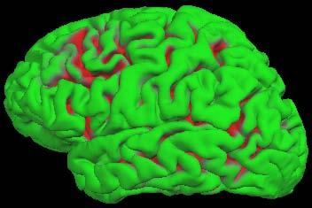
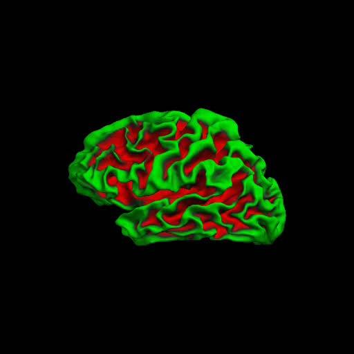
Transcription

1 Lilla Zöllei A.A. Martinos Center, MGH; Boston, MA

2 Bruce Fischl Gheorghe Postelnicu Jean Augustinack Anastasia Yendiki Allison Stevens Kristen Huber Sita Kakonoori + the FreeSurfer team KEPAF
3 Pre-processing step Surgical planning, radiation treatment, Intra- and inter-subject comparison Multi-modality fusion / comparison / validation Longitudinal analysis (development, aging, drug treatment) Atlas building Etc KEPAF


4 PDw MRI T2w MRI unknown ground truth transformation The goal is: KEPAF

5 Affine transform of surfaces from one subject mapped to another. KEPAF

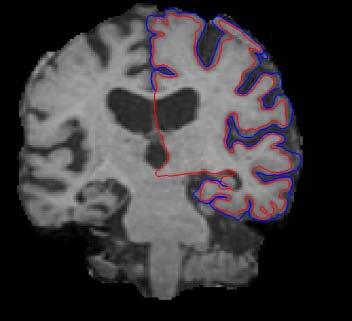
6 Template Affine Nonlinear pial surface WM surface 6


7 Local functional organization of cortex is largely 2-dimensional! (Sereno et al, 1995, Science). 7

8 D.J. Felleman and D.C. Van Essen, CC, 1991 KEPAF
9 ultimate goal: pial (GM CSF) surface BUT: impossible to directly generate representation from MRI (many have tried!) alternative: construct interim representation of the interface between GM and WM infer the location of the true cortical surface (Dale and Sereno, 1993) KEPAF


10 10

11 Courtesy of B. Fischl 11
12 spherical registration for each of the hemispheres Template Source Sulcal pattern projected on the sphere Registered Source KEPAF
13 Motivation: Surface-based (2D) registration does an excellent job of aligning cortical folds, but does not apply to non-cortical structures (e.g. basal ganglia). Volumetric (3D) registration applies to the entire brain but does not in general align folding patterns. Goal: integrate them KEPAF

14 Our goal: automated and accurate registration of both cortical folding patterns and non-cortical regions in 3D Previous work: Peckar, JMIV99 Boundary mapping as a hard constraint for volumetric warp Ferrant TMI01 Volumetric deformation inferred after tracking key surface boundaries; brain shift modellin Collins, ICVBC96 combine the use of intensity measures with automatically extracted sulci Thompson, TMI96 ASM to extract the external cortical surface and the lateral ventricles; volumetric elastic model HAMMER, Shen TMI02 implicitly combines surface and volume information in elastic registration Liu, NeuroImage04 surface-driven registration after HAMMER Joshi, TMI07 surface-based alignment using manually selected sulcal landmarks and inverse-consistent volumetric registration KEPAF
15 Combined volume- and surface-based registration: Automated Surface-based Registration Initialization via an Elastic Transform Intensity-based Volumetric Registration KEPAF
16 accurate, topologically correct reconstructions of the cortical surfaces (L/R white/pial) registration in the spherical space 1-to-1 mapping J= J p + λ J + A A λ d J d Data term Topology preservation Metric distortion KEPAF

17 17
18 to diffuse vector field from the cortical surfaces to the rest of the volume regularity constraint: elastic deformation, i.e. a smooth, orientationpreserving deformation satisfying our choice: Navier operator from the linear elasticity theory + finite element method (FEM) KEPAF

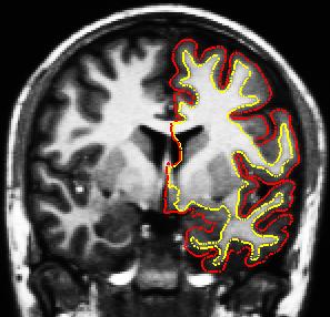
19 Iterate for i=1:n create mesh set up BC s from surface setup linear elastic system solve system solve topology problems Tetrahedral mesh used for one iteration of the elastic solver. Notice how the mesh is denser near the input surfaces KEPAF
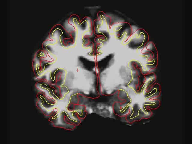
20 20
21 Template Affine HAMMER Elastic KEPAF
22 nonlinear intensity based registration [Fischl, NeuroImage04]: G = G I + λ G + T T λ S G S G I : intensity term G T : topology constraining term G S : smoothness term KEPAF
23 KEPAF

24 Template Elastic CVS Template KEPAF


25 mesh to be warped elastic CVS KEPAF
26 Compare: FLIRT, HAMMER, CVS Template: single, randomly selected scan; rest in the data set is registered to it Accuracy: Jaccard overlap metric KEPAF


27 Template HAMMER FLIRT CVS 27

28 Extended Jaccard Coefficient measures: 20 cortical and 21 sub-cortical labels. The vertical lines represent the standard error of the mean of the measurement.


29
 Cortical")
30 Average Jaccard Coefficient (SE: standard error) Cortical structures
 Sub-cortical")
31 Average Jaccard Coefficient (SE: standard error) Sub-cortical structures

32 Identify fiber bundles in cerebral white matter (WM) Characterizing these WM pathways is important for: Inferring connections b/w brain regions Understanding effects of neurodegenerative diseases, stroke, aging, development From Gray's Anatomy: IX. Neurology KEPAF Courtesy of A. Yendiki 32

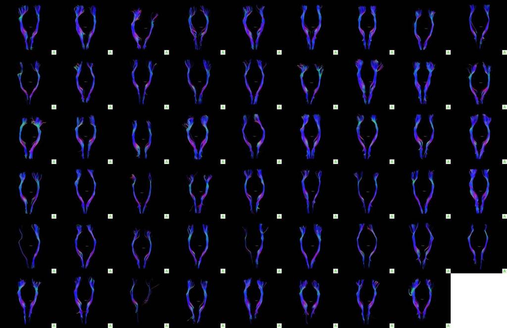
33 Differentiate tissues based on the diffusion (random motion) of water molecules within them Gray matter: Diffusion is unrestricted isotropic White matter: Diffusion is restricted anisotropic KEPAF Courtesy of A. Yendiki 33


34 Magnetic resonance imaging can provide diffusion encoding Magnetic field strength is varied by gradients in different directions Image intensity is attenuated depending on water diffusion in each direction Compare with baseline images to infer on diffusion process No diffusion encoding KEPAF g 4 g 5 Diffusion encoding in direction g 1 g6 g 2 g 3 Courtesy of A. Yendiki
 KEPAF11-2011.")
35 Trackvis and Diffusion Toolkit ( KEPAF
 tracts going through the precentral gyri ROIS")

36 (a) (b) (c) (d) (a) deterministic tractography seeded in the whole brain (b) tracts going through the precentral gyri ROIS (c) tracts going through the brainstem ROI (d) tracts going through both the precentral gyri and brainstem ROIs. KEPAF
37 37
38 38
39 39
40 KEPAF
41 Goal: fiber bundle alignment Study: compare CVS to methods directly aligning DWI-derived scalar volumes Registration methods: FLIRT FA-FLIRT FA-FNIRT CVS KEPAF
")
")
42 (a) FLIRT (b) FA-FLIRT (c) FA-FNIRT (d) CVS 42
")
 CVS")
43 (a) FLIRT (b) FA-FNIRT (c) CVS 43
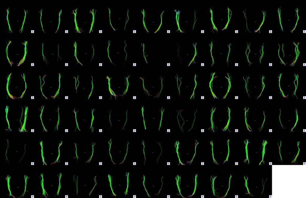
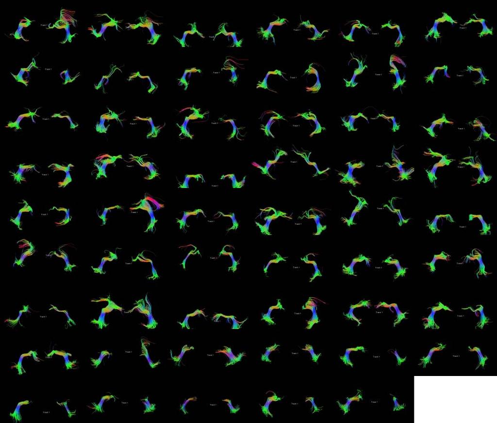
 UNCINATE 44")
44 (a) CST (b) ILF (c) UNCINATE 44
45 CVS outperformed FLIRT, FA-FLIRT and FA-FNIRT for all three tracts and both hemispheres, in a statistically significant manner (p-values were computed using the Student T-test with alpha<.0025) FA-FLIRT was outperformed by all other three methods in a statistically significant manner FLIRT was outperformed also by FA-FNIRT in all cases in a statistically significantly manner except for lh UNC and lh ILF where FLIRT outperformed FA-FNIRT Note: accuracy of the linear registration computed by FLIRT increased substantially when using the structural data (FLIRT) over the DWI data (FA-FLIRT), even though the gold standard we use for assessing accuracy is derived from the DWI data. KEPAF
46 We ran two sets of further experiments: after completing our CVS registration framework we added an additional registration step using FA information from the diffusion images. CVS+FLIRT-FA CVS+FNIRT-FA KEPAF
 UNCINATE 47")
47 (a) CST (b) ILF (c) UNCINATE 47

48 high accuracy cross-subject registration based on structural MRI images can provide improved alignment L. Zöllei, A. Stevens, K. Huber, S. Kakunoori, B. Fischl: "Improved Tractography Alignment Using Combined Volumetric and Surface Registration", NeuroImage 51 (2010), KEPAF
49 Different image acquisition sequences In-vivo MRI scans: T1-weighted magnetization-prepared rapid gradient echo (MP-RAGE) scans were obtained according to the following protocol: two sagittal acquisitions, FOV = 224, Matrix = 256x256, Resolution = 1x1x1.25 mm, TR = 9.7 ms, TE = 4 ms, Flip angle = 10o, TI = 20 ms, TD = 200 ms. Two acquisitions were averaged together to increase the contrast-to-noise ratio Ex-vivo MRI scans: T2*-weighted Multi Echo Flash protocol due to the reduced T1 contrast observed post-mortem Mutual information (MI) replaces the volumetric registration objective function Lilla Zöllei, Allison Stevens, Bruce Fischl: Non-linear registration of intra-subject ex-vivo and in-vivo brain acquisitions ; HBM, 2010 Barcelona KEPAF

50 in vivo ex vivo Courtesy of Xiao Han. 50
")


51 Target (in-vivo) Masked target 2-step CVS CVS with MI 51
")


52 Target (in-vivo) Masked target 2-step CVS CVS with MI 52
")


53 Target (in-vivo) Masked target 2-step CVS CVS with MI 53
")


54 Target (in-vivo) Masked target 2-step CVS CVS with MI 54
55 Manual segmentation of ex-vivo MRI Qualitative validation Increase robustness to scanning artifacts KEPAF
56 KEPAF
Diffusion model fitting and tractography: A primer
 Diffusion model fitting and tractography: A primer Anastasia Yendiki HMS/MGH/MIT Athinoula A. Martinos Center for Biomedical Imaging 03/18/10 Why n how Diffusion model fitting and tractography 0/18 Why
HST.583 Functional Magnetic Resonance Imaging: Data Acquisition and Analysis Fall 2008
 MIT OpenCourseWare http://ocw.mit.edu HST.583 Functional Magnetic Resonance Imaging: Data Acquisition and Analysis Fall 2008 For information about citing these materials or our Terms of Use, visit: http://ocw.mit.edu/terms.
Quantitative MRI of the Brain: Investigation of Cerebral Gray and White Matter Diseases
 Quantities Measured by MR - Quantitative MRI of the Brain: Investigation of Cerebral Gray and White Matter Diseases Static parameters (influenced by molecular environment): T, T* (transverse relaxation)
FROM IMAGE RECONSTRUCTION TO CONNECTIVITY ANALYSIS: A JOURNEY THROUGH THE BRAIN'S WIRING. Francesca Pizzorni Ferrarese
 FROM IMAGE RECONSTRUCTION TO CONNECTIVITY ANALYSIS: A JOURNEY THROUGH THE BRAIN'S WIRING Francesca Pizzorni Ferrarese Pipeline overview WM and GM Segmentation Registration Data reconstruction Tractography
Head motion in diffusion MRI
 Head motion in diffusion MRI Anastasia Yendiki HMS/MGH/MIT Athinoula A. Martinos Center for Biomedical Imaging 11/06/13 Head motion in diffusion MRI 0/33 Diffusion contrast Basic principle of diffusion
UNC 4D Infant Cortical Surface Atlases, from Neonate to 6 Years of Age
 UNC 4D Infant Cortical Surface Atlases, from Neonate to 6 Years of Age Version 1.0 UNC 4D infant cortical surface atlases from neonate to 6 years of age contain 11 time points, including 1, 3, 6, 9, 12,
Advanced MRI Techniques (and Applications)
") Advanced MRI Techniques (and Applications) Jeffry R. Alger, PhD Department of Neurology Ahmanson-Lovelace Brain Mapping Center Brain Research Institute Jonsson Comprehensive Cancer Center University of
Advanced MRI Techniques (and Applications) Jeffry R. Alger, PhD Department of Neurology Ahmanson-Lovelace Brain Mapping Center Brain Research Institute Jonsson Comprehensive Cancer Center University of
HST.583 Functional Magnetic Resonance Imaging: Data Acquisition and Analysis Fall 2008
 MIT OpenCourseWare http://ocw.mit.edu HST.583 Functional Magnetic Resonance Imaging: Data Acquisition and Analysis Fall 2008 For information about citing these materials or our Terms of Use, visit: http://ocw.mit.edu/terms.
Subvoxel Segmentation and Representation of Brain Cortex Using Fuzzy Clustering and Gradient Vector Diffusion
 Subvoxel Segmentation and Representation of Brain Cortex Using Fuzzy Clustering and Gradient Vector Diffusion Ming-Ching Chang Xiaodong Tao GE Global Research Center {changm, taox} @ research.ge.com SPIE
The organization of the human cerebral cortex estimated by intrinsic functional connectivity
 1 The organization of the human cerebral cortex estimated by intrinsic functional connectivity Journal: Journal of Neurophysiology Author: B. T. Thomas Yeo, et al Link: https://www.ncbi.nlm.nih.gov/pubmed/21653723
Neuroimaging and mathematical modelling Lesson 2: Voxel Based Morphometry
 Neuroimaging and mathematical modelling Lesson 2: Voxel Based Morphometry Nivedita Agarwal, MD Nivedita.agarwal@apss.tn.it Nivedita.agarwal@unitn.it Volume and surface morphometry Brain volume White matter
Measuring longitudinal brain changes in humans and small animal models. Christos Davatzikos
 Measuring longitudinal brain changes in humans and small animal models Christos Davatzikos Section of Biomedical Image Analysis University of Pennsylvania (Radiology) http://www.rad.upenn.edu/sbia Computational
NEURO M203 & BIOMED M263 WINTER 2014
 NEURO M203 & BIOMED M263 WINTER 2014 MRI Lab 2: Neuroimaging Connectivity Lab In today s lab we will work with sample diffusion imaging data and the group averaged fmri data collected during your scanning
Supplementary methods
 Supplementary methods This section provides additional technical details on the sample, the applied imaging and analysis steps and methods. Structural imaging Trained radiographers placed all participants
SURFACE RECONSTRUCTION OF EX-VIVO HUMAN V1 THROUGH IDENTIFICATION OF THE STRIA OF GENNARI USING MRI AT 7T
 SURFACE RECONSTRUCTION OF EX-VIVO HUMAN V1 THROUGH IDENTIFICATION OF THE STRIA OF GENNARI USING MRI AT 7T Oliver P. Hinds 1, Jonathan R. Polimeni 2, Megan L. Blackwell 3, Christopher J. Wiggins 3, Graham
Surface-based Analysis: Inter-subject Registration and Smoothing
 Surface-based Analysis: Inter-subject Registration and Smoothing Outline Exploratory Spatial Analysis Coordinate Systems 3D (Volumetric) 2D (Surface-based) Inter-subject registration Volume-based Surface-based
Diffusion-MRI processing for group analysis
 Diffusion-MRI processing for group analysis Felix Renard IRMaGe: Inserm US 17 / CNRS UMS 3552 University Hospital of Grenoble - France 25/09/2015 felixrenard@gmail.com 1 Diffusion-MRI processing for group
Automatic segmentation of the cortical grey and white matter in MRI using a Region Growing approach based on anatomical knowledge
 Automatic segmentation of the cortical grey and white matter in MRI using a Region Growing approach based on anatomical knowledge Christian Wasserthal 1, Karin Engel 1, Karsten Rink 1 und André Brechmann
Brain Extraction, Registration & EPI Distortion Correction
 Brain Extraction, Registration & EPI Distortion Correction What use is Registration? Some common uses of registration: Combining across individuals in group studies: including fmri & diffusion Quantifying
Geometry Driven Volumetric Registration
 Geometry Driven Volumetric Registration Gheorghe Postelnicu, Lilla Zollei, Rahul Desikan, and Bruce Fischl MGH/MIT/HMS Athinoula A. Martinos Center for Biomedical Imaging, Charlestown, MA {postelni,lzollei,rahul,fischl}@nmr.mgh.harvard.edu
UCLA Advanced Neuroimaging Summer Program David W. Shattuck, PhD
 BrainSuite UCLA Advanced Neuroimaging Summer Program Presented 18 July 2013 David W. Shattuck, PhD Associate Professor Department of Neurology David Geffen School of Medicine at UCLA http://www.loni.ucla.edu/~shattuck/
Electrical Engineering, Vanderbilt University, Nashville, TN, USA b. Computer Science, Vanderbilt University, Nashville, TN, USA c
 Improved Stability of Whole Brain Surface Parcellation with Multi-Atlas Segmentation Yuankai Huo* a, Shunxing Bao b, Prasanna Parvathaneni a, Bennett A. Landman a,b,c a Electrical Engineering, Vanderbilt
Methods for data preprocessing
 Methods for data preprocessing John Ashburner Wellcome Trust Centre for Neuroimaging, 12 Queen Square, London, UK. Overview Voxel-Based Morphometry Morphometry in general Volumetrics VBM preprocessing
TRACULA: Troubleshooting, visualization, and group analysis
 TRACULA: Troubleshooting, visualization, and group analysis Anastasia Yendiki HMS/MGH/MIT Athinoula A. Martinos Center for Biomedical Imaging 18/11/13 TRACULA: troubleshooting, visualization, group analysis
MR IMAGE SEGMENTATION
 MR IMAGE SEGMENTATION Prepared by : Monil Shah What is Segmentation? Partitioning a region or regions of interest in images such that each region corresponds to one or more anatomic structures Classification
Reproducibility of Whole-brain Structural Connectivity Networks
 Reproducibility of Whole-brain Structural Connectivity Networks Christopher Parker Thesis for Masters of Research in Medical and Biomedical Imaging Supervised by Prof. Sebastien Ourselin and Dr Jonathan
BrainSuite. presented at the UCLA/NITP Advanced Neuroimaging Summer Program 29 July 2014
 BrainSuite presented at the UCLA/NITP Advanced Neuroimaging Summer Program 29 July 2014 David Shattuck Ahmanson-Lovelace Brain Mapping Center Department of Neurology David Geffen School of Medicine at
Where are we now? Structural MRI processing and analysis
 Where are we now? Structural MRI processing and analysis Pierre-Louis Bazin bazin@cbs.mpg.de Leipzig, Germany Structural MRI processing: why bother? Just use the standards? SPM FreeSurfer FSL However:
BrainSuite Lab Exercises. presented at the UCLA/NITP Advanced Neuroimaging Summer Program 29 July 2014
 BrainSuite Lab Exercises presented at the UCLA/NITP Advanced Neuroimaging Summer Program 29 July 2014 1. Opening and Displaying an MRI Start BrainSuite Drag and drop the T1 image from the native space
Automatic MS Lesion Segmentation by Outlier Detection and Information Theoretic Region Partitioning Release 0.00
 Automatic MS Lesion Segmentation by Outlier Detection and Information Theoretic Region Partitioning Release 0.00 Marcel Prastawa 1 and Guido Gerig 1 Abstract July 17, 2008 1 Scientific Computing and Imaging
CHAPTER 2. Morphometry on rodent brains. A.E.H. Scheenstra J. Dijkstra L. van der Weerd
 CHAPTER 2 Morphometry on rodent brains A.E.H. Scheenstra J. Dijkstra L. van der Weerd This chapter was adapted from: Volumetry and other quantitative measurements to assess the rodent brain, In vivo NMR
ADAPTIVE GRAPH CUTS WITH TISSUE PRIORS FOR BRAIN MRI SEGMENTATION
 ADAPTIVE GRAPH CUTS WITH TISSUE PRIORS FOR BRAIN MRI SEGMENTATION Abstract: MIP Project Report Spring 2013 Gaurav Mittal 201232644 This is a detailed report about the course project, which was to implement
The Insight Toolkit. Image Registration Algorithms & Frameworks
 The Insight Toolkit Image Registration Algorithms & Frameworks Registration in ITK Image Registration Framework Multi Resolution Registration Framework Components PDE Based Registration FEM Based Registration
ACCURATE NONLINEAR MAPPING BETWEEN MNI152/COLIN27 VOLUMETRIC AND FREESURFER SURFACE COORDINATE SYSTEMS
 ACCURATE NONLINEAR MAPPING BETWEEN MNI152/COLIN27 VOLUMETRIC AND FREESURFER SURFACE COORDINATE SYSTEMS WU JIANXIAO (B.Eng. (Hons.), NUS) A THESIS SUBMITTED FOR THE DEGREE OF MASTER OF ENGINEERING DEPARTMENT
Nonrigid Registration using Free-Form Deformations
 Nonrigid Registration using Free-Form Deformations Hongchang Peng April 20th Paper Presented: Rueckert et al., TMI 1999: Nonrigid registration using freeform deformations: Application to breast MR images
ARTICLE IN PRESS. Accurate prediction of V1 location from cortical folds in a surface coordinate system. Introduction
 YNIMG-05001; No. of pages: 15; 4C: 4, 8, 9 www.elsevier.com/locate/ynimg NeuroImage xx (2007) xxx xxx Accurate prediction of V1 location from cortical folds in a surface coordinate system Oliver P. Hinds,
Automatic Registration-Based Segmentation for Neonatal Brains Using ANTs and Atropos
 Automatic Registration-Based Segmentation for Neonatal Brains Using ANTs and Atropos Jue Wu and Brian Avants Penn Image Computing and Science Lab, University of Pennsylvania, Philadelphia, USA Abstract.
A Fast and Accurate Method for Automated Cortical Surface Registration and Labeling
 A Fast and Accurate Method for Automated Cortical Surface Registration and Labeling Anand A Joshi 1, David W Shattuck 2 and Richard M Leahy 1 1 Signal and Image Processing Institute, University of Southern
NIH Public Access Author Manuscript Neuroimage. Author manuscript; available in PMC 2009 February 15.
 NIH Public Access Author Manuscript Published in final edited form as: Neuroimage. 2008 February 15; 39(4): 1585 1599. Accurate prediction of V1 location from cortical folds in a surface coordinate system
Cocozza S., et al. : ALTERATIONS OF FUNCTIONAL CONNECTIVITY OF THE MOTOR CORTEX IN FABRY'S DISEASE: AN RS-FMRI STUDY
 ALTERATIONS OF FUNCTIONAL CONNECTIVITY OF THE MOTOR CORTEX IN FABRY'S DISEASE: AN RS-FMRI STUDY SUPPLEMENTARY MATERIALS Sirio Cocozza, MD 1*, Antonio Pisani, MD, PhD 2, Gaia Olivo, MD 1, Francesco Saccà,
Multiple Cortical Surface Correspondence Using Pairwise Shape Similarity
 Multiple Cortical Surface Correspondence Using Pairwise Shape Similarity Pahal Dalal 1, Feng Shi 2, Dinggang Shen 2, and Song Wang 1 1 Department of Computer Science and Engineering, University of South
Fiber Selection from Diffusion Tensor Data based on Boolean Operators
 Fiber Selection from Diffusion Tensor Data based on Boolean Operators D. Merhof 1, G. Greiner 2, M. Buchfelder 3, C. Nimsky 4 1 Visual Computing, University of Konstanz, Konstanz, Germany 2 Computer Graphics
How to create a head model
 How to create a head model This document describes the command line tools: mri2mesh: Central tool to reconstruct a head model from T1w and T2w data dwi2cond: Reconstruct conductivity tensors for brain
Diffusion Tensor Processing and Visualization
 NA-MIC National Alliance for Medical Image Computing http://na-mic.org Diffusion Tensor Processing and Visualization Guido Gerig University of Utah Martin Styner, UNC NAMIC: National Alliance for Medical
Machine learning improves automated cortical surface reconstruction in human MRI studies
 University of Iowa Iowa Research Online Theses and Dissertations Spring 2017 Machine learning improves automated cortical surface reconstruction in human MRI studies David G. Ellis University of Iowa Copyright
BDP: BrainSuite Diffusion Pipeline. Chitresh Bhushan
 BDP: BrainSuite Diffusion Pipeline Chitresh Bhushan Why diffusion MRI? T 2 weighted MPRAGE FA map Fiber track Quantify microstructural tissue characteristics Structural connectivity Connectome Clinical
Structural Segmentation
 Structural Segmentation FAST tissue-type segmentation FIRST sub-cortical structure segmentation FSL-VBM voxelwise grey-matter density analysis SIENA atrophy analysis FAST FMRIB s Automated Segmentation
RIGID IMAGE REGISTRATION
 RIGID IMAGE REGISTRATION Duygu Tosun-Turgut, Ph.D. Center for Imaging of Neurodegenerative Diseases Department of Radiology and Biomedical Imaging duygu.tosun@ucsf.edu What is registration? Image registration
Atelier 2 : Calcul Haute Performance et Sciences du Vivant Forum er juillet, Paris, France
 From Diffusion MR Image Analysis to Whole Brain Connectivity Simulation Jean-Philippe Thiran EPFL Lausanne, Switzerland EPFL - Lausanne HPC in life sciences at EPFL The Blue Brain project: create a biologically
Reconstruction of Fiber Trajectories via Population-Based Estimation of Local Orientations
 IDEA Reconstruction of Fiber Trajectories via Population-Based Estimation of Local Orientations Pew-Thian Yap, John H. Gilmore, Weili Lin, Dinggang Shen Email: ptyap@med.unc.edu 2011-09-21 Poster: P2-46-
Multimodal Imaging Brain Connectivity Analysis (MIBCA)
") Multimodal Imaging Brain Connectivity Analysis (MIBCA) Andre Santos Ribeiro, Luis Miguel Lacerda, Hugo Ferreira April 23, 2015 Abstract In recent years, connectivity studies using neuroimaging data have
Multimodal Imaging Brain Connectivity Analysis (MIBCA) Andre Santos Ribeiro, Luis Miguel Lacerda, Hugo Ferreira April 23, 2015 Abstract In recent years, connectivity studies using neuroimaging data have
BDP: BrainSuite Diffusion Pipeline. Chitresh Bhushan
 BDP: BrainSuite Diffusion Pipeline Chitresh Bhushan Why diffusion MRI? T 2 weighted MPRAGE FA map Fiber track Quantify microstructural tissue characteristics Structural connectivity Connectome Clinical
Structural MRI analysis
 Structural MRI analysis volumetry and voxel-based morphometry cortical thickness measurements structural covariance network mapping Boris Bernhardt, PhD Department of Social Neuroscience, MPI-CBS bernhardt@cbs.mpg.de
An Introduction To Automatic Tissue Classification Of Brain MRI. Colm Elliott Mar 2014
 An Introduction To Automatic Tissue Classification Of Brain MRI Colm Elliott Mar 2014 Tissue Classification Tissue classification is part of many processing pipelines. We often want to classify each voxel
Comparison Study of Clinical 3D MRI Brain Segmentation Evaluation
 Comparison Study of Clinical 3D MRI Brain Segmentation Evaluation Ting Song 1, Elsa D. Angelini 2, Brett D. Mensh 3, Andrew Laine 1 1 Heffner Biomedical Imaging Laboratory Department of Biomedical Engineering,
Automatic Generation of Training Data for Brain Tissue Classification from MRI
 Automatic Generation of Training Data for Brain Tissue Classification from MRI Chris A. COCOSCO, Alex P. ZIJDENBOS, and Alan C. EVANS http://www.bic.mni.mcgill.ca/users/crisco/ McConnell Brain Imaging
A combined Surface And VOlumetric Registration (SAVOR) framework to study cortical biomarkers and volumetric imaging data
 framework to study cortical biomarkers and volumetric imaging data") A combined Surface And VOlumetric Registration (SAVOR) framework to study cortical biomarkers and volumetric imaging data Eli Gibson 1, Ali R. Khan 1, and Mirza Faisal Beg 1 School of Engineering Science,
A combined Surface And VOlumetric Registration (SAVOR) framework to study cortical biomarkers and volumetric imaging data Eli Gibson 1, Ali R. Khan 1, and Mirza Faisal Beg 1 School of Engineering Science,
Analysis of Functional MRI Timeseries Data Using Signal Processing Techniques
 Analysis of Functional MRI Timeseries Data Using Signal Processing Techniques Sea Chen Department of Biomedical Engineering Advisors: Dr. Charles A. Bouman and Dr. Mark J. Lowe S. Chen Final Exam October
Diffusion Tensor Imaging and Reading Development
 Diffusion Tensor Imaging and Reading Development Bob Dougherty Stanford Institute for Reading and Learning Reading and Anatomy Every brain is different... Not all brains optimized for highly proficient
CHAPTER 9: Magnetic Susceptibility Effects in High Field MRI
 Figure 1. In the brain, the gray matter has substantially more blood vessels and capillaries than white matter. The magnified image on the right displays the rich vasculature in gray matter forming porous,
Structural Segmentation
 Structural Segmentation FAST tissue-type segmentation FIRST sub-cortical structure segmentation FSL-VBM voxelwise grey-matter density analysis SIENA atrophy analysis FAST FMRIB s Automated Segmentation
Appendix E1. Supplementary Methods. MR Image Acquisition. MR Image Analysis
 RSNA, 2015 10.1148/radiol.2015150532 Appendix E1 Supplementary Methods MR Image Acquisition By using a 1.5-T system (Avanto, Siemens Medical, Erlangen, Germany) under a program of regular maintenance (no
Segmentation of the neonatal brain for MR images A REVIEW
 Segmentation of the neonatal brain for MR images A REVIEW Tineke Hoppinga 19-5-2010 Inhoud Abbreviations... 4 Abstract... 5 Introduction... 6 Method... 8 Basic explanations of the methods... 8 Weisenfeld...
Efficient population registration of 3D data
 Efficient population registration of 3D data Lilla Zöllei 1, Erik Learned-Miller 2, Eric Grimson 1, William Wells 1,3 1 Computer Science and Artificial Intelligence Lab, MIT; 2 Dept. of Computer Science,
Network connectivity via inference over curvature-regularizing line graphs
 Network connectivity via inference over curvature-regularizing line graphs Asian Conference on Computer Vision Maxwell D. Collins 1,2, Vikas Singh 2,1, Andrew L. Alexander 3 1 Department of Computer Sciences
Complex Fiber Visualization
 Annales Mathematicae et Informaticae 34 (2007) pp. 103 109 http://www.ektf.hu/tanszek/matematika/ami Complex Fiber Visualization Henrietta Tomán a, Róbert Tornai b, Marianna Zichar c a Department of Computer
GPU-Based Implementation of a Computational Model of Cerebral Cortex Folding
 GPU-Based Implementation of a Computational Model of Cerebral Cortex Folding Jingxin Nie 1, Kaiming 1,2, Gang Li 1, Lei Guo 1, Tianming Liu 2 1 School of Automation, Northwestern Polytechnical University,
Functional MRI data preprocessing. Cyril Pernet, PhD
 Functional MRI data preprocessing Cyril Pernet, PhD Data have been acquired, what s s next? time No matter the design, multiple volumes (made from multiple slices) have been acquired in time. Before getting
Distance Transforms in Multi Channel MR Image Registration
 Distance Transforms in Multi Channel MR Image Registration Min Chen 1, Aaron Carass 1, John Bogovic 1, Pierre-Louis Bazin 2 and Jerry L. Prince 1 1 Image Analysis and Communications Laboratory, 2 The Laboratory
Non-rigid Image Registration using Electric Current Flow
 Non-rigid Image Registration using Electric Current Flow Shu Liao, Max W. K. Law and Albert C. S. Chung Lo Kwee-Seong Medical Image Analysis Laboratory, Department of Computer Science and Engineering,
Multi-atlas labeling with population-specific template and non-local patch-based label fusion
 Multi-atlas labeling with population-specific template and non-local patch-based label fusion Vladimir Fonov, Pierrick Coupé, Simon Eskildsen, Jose Manjon, Louis Collins To cite this version: Vladimir
A Novel Nonrigid Registration Algorithm and Applications
 A Novel Nonrigid Registration Algorithm and Applications J. Rexilius 1, S.K. Warfield 1, C.R.G. Guttmann 1, X. Wei 1, R. Benson 2, L. Wolfson 2, M. Shenton 1, H. Handels 3, and R. Kikinis 1 1 Surgical
Detecting Changes In Non-Isotropic Images
 Detecting Changes In Non-Isotropic Images K.J. Worsley 1, M. Andermann 1, T. Koulis 1, D. MacDonald, 2 and A.C. Evans 2 August 4, 1999 1 Department of Mathematics and Statistics, 2 Montreal Neurological
A Method for Registering Diffusion Weighted Magnetic Resonance Images
 A Method for Registering Diffusion Weighted Magnetic Resonance Images Xiaodong Tao and James V. Miller GE Research, Niskayuna, New York, USA Abstract. Diffusion weighted magnetic resonance (DWMR or DW)
1 Introduction Motivation and Aims Functional Imaging Computational Neuroanatomy... 12
 Contents 1 Introduction 10 1.1 Motivation and Aims....... 10 1.1.1 Functional Imaging.... 10 1.1.2 Computational Neuroanatomy... 12 1.2 Overview of Chapters... 14 2 Rigid Body Registration 18 2.1 Introduction.....
A Multiple-Layer Flexible Mesh Template Matching Method for Nonrigid Registration between a Pelvis Model and CT Images
 A Multiple-Layer Flexible Mesh Template Matching Method for Nonrigid Registration between a Pelvis Model and CT Images Jianhua Yao 1, Russell Taylor 2 1. Diagnostic Radiology Department, Clinical Center,
Human Connectom Project : The minimal processing Pipeline
 Human Connectom Project : The minimal processing Pipeline Human Connectom Project : The minimal processing Pipeline Van Essen DC, The WU-Minn Human Connectome Project: an overview. Neuroimage. 2013 Marcus
A Ray-based Approach for Boundary Estimation of Fiber Bundles Derived from Diffusion Tensor Imaging
 A Ray-based Approach for Boundary Estimation of Fiber Bundles Derived from Diffusion Tensor Imaging M. H. A. Bauer 1,3, S. Barbieri 2, J. Klein 2, J. Egger 1,3, D. Kuhnt 1, B. Freisleben 3, H.-K. Hahn
Diffusion Imaging Models 1: from DTI to HARDI models
 Diffusion Imaging Models 1: from DTI to HARDI models Flavio Dell Acqua, PhD. www.natbrainlab.com flavio.dellacqua@kcl.ac.uk @flaviodellacqua Diffusion Tensor Imaging (DTI) z λ 1 λ 2 The profile of the
FSL Pre-Processing Pipeline
 The Art and Pitfalls of fmri Preprocessing FSL Pre-Processing Pipeline Mark Jenkinson FMRIB Centre, University of Oxford FSL Pre-Processing Pipeline Standard pre-processing: Task fmri Resting-state fmri
FreeSurfer: Troubleshooting surfer.nmr.mgh.harvard.edu
 FreeSurfer: Troubleshooting surfer.nmr.mgh.harvard.edu 1 Hard and Soft Failures Categories of errors: Hard & Soft Failures Hard = recon-all quits before it finishes Soft = recon-all finishes but results
A Binary Entropy Measure to Assess Nonrigid Registration Algorithms
 A Binary Entropy Measure to Assess Nonrigid Registration Algorithms Simon K. Warfield 1, Jan Rexilius 1, Petra S. Huppi 2, Terrie E. Inder 3, Erik G. Miller 1, William M. Wells III 1, Gary P. Zientara
Super-Resolution Reconstruction of Diffusion-Weighted Images from Distortion Compensated Orthogonal Anisotropic Acquisitions.
 Super-Resolution Reconstruction of Diffusion-Weighted Images from Distortion Compensated Orthogonal Anisotropic Acquisitions. Benoit Scherrer Ali Gholipour Simon K. Warfield Children s Hospital Boston,
Form follows func-on. Which one of them can fly? And why? Why would anyone bother about brain structure, when we can do func5onal imaging?
 Why would anyone bother about brain structure, when we can do func5onal imaging? Form follows func-on Which one of them can fly? And why? h;p://animals.na5onalgeographic.com Why would anyone bother about
Simultaneous Cortical Surface Labeling and Sulcal Curve Extraction
 Simultaneous Cortical Surface Labeling and Sulcal Curve Extraction Zhen Yang a, Aaron Carass a, Chen Chen a, Jerry L Prince a,b a Electrical and Computer Engineering, b Biomedical Engineering, Johns Hopkins
Diffusion MRI Acquisition. Karla Miller FMRIB Centre, University of Oxford
 Diffusion MRI Acquisition Karla Miller FMRIB Centre, University of Oxford karla@fmrib.ox.ac.uk Diffusion Imaging How is diffusion weighting achieved? How is the image acquired? What are the limitations,
Slide 1. Technical Aspects of Quality Control in Magnetic Resonance Imaging. Slide 2. Annual Compliance Testing. of MRI Systems.
 Slide 1 Technical Aspects of Quality Control in Magnetic Resonance Imaging Slide 2 Compliance Testing of MRI Systems, Ph.D. Department of Radiology Henry Ford Hospital, Detroit, MI Slide 3 Compliance Testing
Lecture 13 Theory of Registration. ch. 10 of Insight into Images edited by Terry Yoo, et al. Spring (CMU RI) : BioE 2630 (Pitt)
 : BioE 2630 (Pitt)") Lecture 13 Theory of Registration ch. 10 of Insight into Images edited by Terry Yoo, et al. Spring 2018 16-725 (CMU RI) : BioE 2630 (Pitt) Dr. John Galeotti The content of these slides by John Galeotti,
Lecture 13 Theory of Registration ch. 10 of Insight into Images edited by Terry Yoo, et al. Spring 2018 16-725 (CMU RI) : BioE 2630 (Pitt) Dr. John Galeotti The content of these slides by John Galeotti,
HST.582J / 6.555J / J Biomedical Signal and Image Processing Spring 2007
 MIT OpenCourseWare http://ocw.mit.edu HST.582J / 6.555J / 16.456J Biomedical Signal and Image Processing Spring 2007 For information about citing these materials or our Terms of Use, visit: http://ocw.mit.edu/terms.
HST.583 Functional Magnetic Resonance Imaging: Data Acquisition and Analysis Fall 2008
 MIT OpenCourseWare http://ocw.mit.edu HST.583 Functional Magnetic Resonance Imaging: Data Acquisition and Analysis Fall 2008 For information about citing these materials or our Terms of Use, visit: http://ocw.mit.edu/terms.
Multiresolution analysis: theory and applications. Analisi multirisoluzione: teoria e applicazioni
 Multiresolution analysis: theory and applications Analisi multirisoluzione: teoria e applicazioni Course overview Course structure The course is about wavelets and multiresolution Exam Theory: 4 hours
Evaluation of Local Filter Approaches for Diffusion Tensor based Fiber Tracking
 Evaluation of Local Filter Approaches for Diffusion Tensor based Fiber Tracking D. Merhof 1, M. Buchfelder 2, C. Nimsky 3 1 Visual Computing, University of Konstanz, Konstanz 2 Department of Neurosurgery,
syngo.mr Neuro 3D: Your All-In-One Post Processing, Visualization and Reporting Engine for BOLD Functional and Diffusion Tensor MR Imaging Datasets
 syngo.mr Neuro 3D: Your All-In-One Post Processing, Visualization and Reporting Engine for BOLD Functional and Diffusion Tensor MR Imaging Datasets Julien Gervais; Lisa Chuah Siemens Healthcare, Magnetic
Enhao Gong, PhD Candidate, Electrical Engineering, Stanford University Dr. John Pauly, Professor in Electrical Engineering, Stanford University Dr.
 Enhao Gong, PhD Candidate, Electrical Engineering, Stanford University Dr. John Pauly, Professor in Electrical Engineering, Stanford University Dr. Greg Zaharchuk, Associate Professor in Radiology, Stanford
Groupwise Shape Correspondence with Local Features
 Groupwise Shape Correspondence with Local Features İpek Oğuz A dissertation submitted to the faculty of the University of North Carolina at Chapel Hill in partial fulfillment of the requirements for the
AFNI Preprocessing: Outline, Recommendations, and New(ish) Stuff. Robert W Cox SSCC / NIMH & NINDS / NIH / DHHS / USA / EARTH
 Stuff. Robert W Cox SSCC / NIMH & NINDS / NIH / DHHS / USA / EARTH") AFNI Preprocessing: Outline, Recommendations, and New(ish) Stuff Robert W Cox SSCC / NIMH & NINDS / NIH / DHHS / USA / EARTH HBM 2016 As a work of a US Government official, this presentation is not copyrighted
AFNI Preprocessing: Outline, Recommendations, and New(ish) Stuff Robert W Cox SSCC / NIMH & NINDS / NIH / DHHS / USA / EARTH HBM 2016 As a work of a US Government official, this presentation is not copyrighted
SPM8 for Basic and Clinical Investigators. Preprocessing. fmri Preprocessing
 SPM8 for Basic and Clinical Investigators Preprocessing fmri Preprocessing Slice timing correction Geometric distortion correction Head motion correction Temporal filtering Intensity normalization Spatial
TractoR and Other Software
 TractoR and Other Software Jon Clayden DIBS Teaching Seminar, 11 Dec 2015 Photo by José Martín Ramírez Carrasco https://www.behance.net/martini_rc TractoR A set of R packages Additional
Multiresolution analysis: theory and applications. Analisi multirisoluzione: teoria e applicazioni
 Multiresolution analysis: theory and applications Analisi multirisoluzione: teoria e applicazioni Course overview Course structure The course is about wavelets and multiresolution Exam Theory: 4 hours
Detection of Unique Point Landmarks in HARDI Images of the Human Brain
 Detection of Unique Point Landmarks in HARDI Images of the Human Brain Henrik Skibbe and Marco Reisert Department of Radiology, University Medical Center Freiburg, Germany {henrik.skibbe, marco.reisert}@uniklinik-freiburg.de