Measuring longitudinal brain changes in humans and small animal models. Christos Davatzikos
|
|
|
- Randolph Fleming
- 5 years ago
- Views:
Transcription
1 Measuring longitudinal brain changes in humans and small animal models Christos Davatzikos Section of Biomedical Image Analysis University of Pennsylvania (Radiology)
 A reference template (e.g.")
2 Computational Anatomy: Measuring Anatomical Structure 1) A reference template (e.g. an atlas or an average shape) is the unit 2) A shape transformation quantifies the shape characteristics of and individual brain with respect to the template Template Subject Warped template warping of a grid 3) Two shapes are compared by comparing the corresponding transformations

3 The deformation function measures the local deformation of the template: Template Shape 1 Shape 2 Red: Expansion Green: Contraction Deformation 1 Deformation 2 Deformation function measured after adaptation of the template to each shape Local measurements of shape characteristics can be therefore measured by analyzing the deformation functions with standard statistical methodologies Davatzikos et.al., J. Comp. Assist. Tomogr., 1996
4 Problems when using the warping transformation: The target anatomy is assumed to be a diffeomorphism of the template Residual information is discarded A very accurate warping requires time and computational resources Use the pair (Transformation, Residual) to measure shape


5 RAVENS: Mass-preserving shape transformations for morphological analysis RAVENS = Regional Analysis of Volumes Examined in Normalized Space
6
7 Example of Detection of Subtle Change:
8 Example of Detection of Subtle Change:


9 Validation Experiments: Localized atrophy identified via t-maps of the RAVENS images Atrophy detected in the two gyri: PCG and STG T-maps are overlaid on the average WM RAVENS map of 24 subjects Davatzikos et.al., NeuroImage, 2001


10 Average brain from 158 subjects in BLSA project Average Model Average Template HAMMER image warping, by Dinggang Shen (IEEE-TMI, 2001)

11 Significant 4-year GM changes in 107 older adults Resnick et.al., 2001, Cerebral Cortex


12 Finding associations between local atrophy and MCI Gray matter White matter

13 Longitudinal changes in WM/GM MR signal contrast Demyelination or other degenerative processes Scale of t-statistic of longitudinal changes Shape and signal changes with aging were uncorrelated Davatzikos and Resnick, 2000


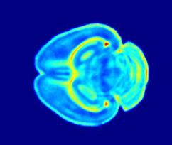
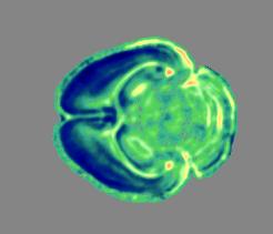
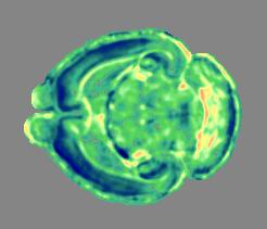
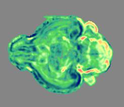
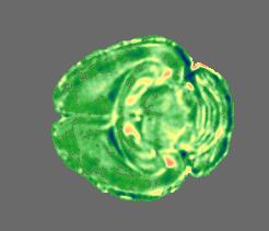
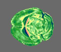
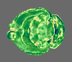
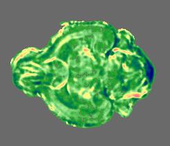
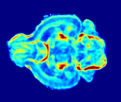
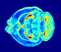
14 Measuring longitudinal change in early postnatal development of the mouse brain Standard T1, T2 imaging is inadequate Æ Diffusion tensor imaging color coded FA map orientation map
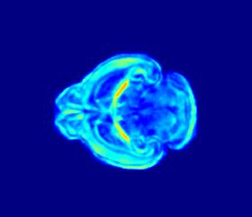
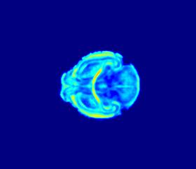
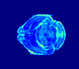
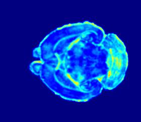
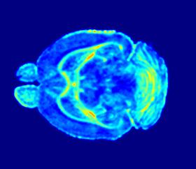
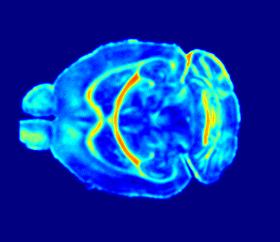
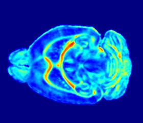
15 Longitudinal change of fractional anisotropy Day 2 Day 5 Day 7 Day 10 Day 15 Day 30 Day 80


16 Correlation of FA and age Cortex Caudate-putamen Internal capsule and ventr/hippo. commisure Corpus callosum Hippocampus Splenium of the corpus callosum Anterior commisure


17 Young mice (days 2 7) Adolescent mice (days 10 30) Old mice (days 45 80) Difference between young and adolescent mice Difference between old and adolescent mice
18 Problem with 3D warping: -- Either independent warping between the template and the individual, for each time-point -- or warping from time t-1 to time t, and from time t to time t+1, etc. Sequence of independent warpings Æ inconsistencies 4D Warping


19 4D template warping All temporal images of the same individual are simultaneously considered in image warping subject Year t-1 Year t Year t+1 template Year t-1 Year t Year t+1 9 Robust anatomical correspondence detection 9 Smooth and temporaly consistent transformations


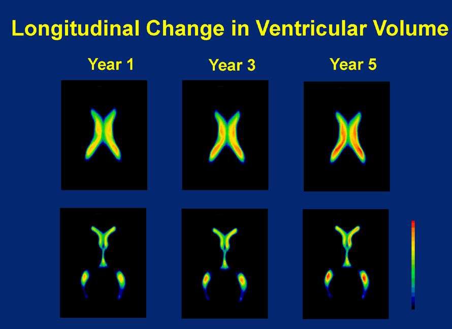
20 Warping consistence - comparison Warped 3D images 3D warping Model 4D warping Year 1 Year 2 Year 3 Year 4 Year 5 Mdl Warped 4D image
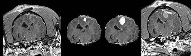
21 Problems when tumors are present: The anatomy is partially obscured by edema Extreme deformations make anatomical matching difficult Part of the tissue has died Need for Biomechanical models of soft tissue deformation Statistical models for estimation of obscured anatomy and for generation of atlas templates that look more like deformed anatomy 3. Deformable registration methods robust to deformations

22 Fundamental Limitation: Estimating the inverse deformation field is a very ill-posed problem Atlas Unknown normal brain Patient s brain deformed by tumor Unknown initial tumor position Edema
23 Training stage Biomechanical simulation Statistical Estimation Estimation stage Deformable registration stage
24 Biomechanical Modeling
 to simulate deformation based on realistic tissue properties and boundary conditions (a)")
25 Biomechanical Modeling Motivation Generate biomechanical simulations of deformations of interest for the purpose of training statistical models used to predict those deformations. Approach Automatically construct biomechanical models from segmented scans Use Finite Element Analysis (FEA) to simulate deformation based on realistic tissue properties and boundary conditions (a) (b) Postprocessing (c) (e) (a) A regular grid of small cubes is cast over the segmented volume. (b) Cubes are tesselated into tetrahedra. (c) Mesh refinement by subdivision of tetrahedra using edge split and LEPP. (d) Making the mesh conform to the geometry of the segmented volume. (e) Post-processing for improvement of quality of elements for FE analysis. (d) A mechanical FEA simulation of growth of a tumor using a mesh generated from a normal brain scan.
26 Training stage Biomechanical simulation Statistical Estimation Estimation stage Deformable registration stage

27 Deformable registration of tumor-occluded brain images Original sequence: 5 scans, total 2 years apart Result of image warping Confidence of matching map Magnitude of deformation
28 Overlay of 2nd time-point: warped original + 2 nd scan Overlay of 5th time-point: warped original + 5th scan
29 Training stage Biomechanical simulation Statistical Estimation Estimation stage Deformable registration stage

30 We will generate training samples, using tumor growth simulation s = [ s 1, s 2 ] Perform a number of forward biomechanical simulations Estimate joint pdf
31 2) MAP estimation framework:
32 s 1 s 2 IEEE Trans. on Med. Imaging, 20(8): , 2001
33 Training stage Biomechanical simulation Statistical Estimation Estimation stage Deformable registration stage
34 7KDQN\RX
35


36 after nonlinear warping to the template (Day 10) Voxel-based or multi-variate analysis of FA changes Day 2 Day 5 Day 7 Day 10 Day 15 Day 30 Day 80
37 Results from 9 BLSA subjects Manual expert: 5.5% 4D HAMMER: 5.7% 3D HAMMER: 2.1% Average Hippo Volumes of 9 Subjects volume (x1.5) D results 3D results year
38
Quantitative MRI of the Brain: Investigation of Cerebral Gray and White Matter Diseases
 Quantities Measured by MR - Quantitative MRI of the Brain: Investigation of Cerebral Gray and White Matter Diseases Static parameters (influenced by molecular environment): T, T* (transverse relaxation)
The Anatomical Equivalence Class Formulation and its Application to Shape-based Computational Neuroanatomy
 The Anatomical Equivalence Class Formulation and its Application to Shape-based Computational Neuroanatomy Sokratis K. Makrogiannis, PhD From post-doctoral research at SBIA lab, Department of Radiology,
Methods for data preprocessing
 Methods for data preprocessing John Ashburner Wellcome Trust Centre for Neuroimaging, 12 Queen Square, London, UK. Overview Voxel-Based Morphometry Morphometry in general Volumetrics VBM preprocessing
Morphological classification of brains via high-dimensional shape transformations and machine learning methods
 Morphological classification of brains via high-dimensional shape transformations and machine learning methods Zhiqiang Lao, a,1 Dinggang Shen, a Zhong Xue, a Bilge Karacali, a Susan M. Resnick, b and
Neuroimaging and mathematical modelling Lesson 2: Voxel Based Morphometry
 Neuroimaging and mathematical modelling Lesson 2: Voxel Based Morphometry Nivedita Agarwal, MD Nivedita.agarwal@apss.tn.it Nivedita.agarwal@unitn.it Volume and surface morphometry Brain volume White matter
Joint Segmentation and Registration for Infant Brain Images
 Joint Segmentation and Registration for Infant Brain Images Guorong Wu 1, Li Wang 1, John Gilmore 2, Weili Lin 1, and Dinggang Shen 1(&) 1 Department of Radiology and BRIC, University of North Carolina,
CLASSIC: Consistent Longitudinal Alignment and Segmentation for Serial Image Computing
 www.elsevier.com/locate/ynimg NeuroImage 30 (2006) 388 399 CLASSIC: Consistent Longitudinal Alignment and Segmentation for Serial Image Computing Zhong Xue,* Dinggang Shen, and Christos Davatzikos Section
CHAPTER 2. Morphometry on rodent brains. A.E.H. Scheenstra J. Dijkstra L. van der Weerd
 CHAPTER 2 Morphometry on rodent brains A.E.H. Scheenstra J. Dijkstra L. van der Weerd This chapter was adapted from: Volumetry and other quantitative measurements to assess the rodent brain, In vivo NMR
Automatic segmentation of the cortical grey and white matter in MRI using a Region Growing approach based on anatomical knowledge
 Automatic segmentation of the cortical grey and white matter in MRI using a Region Growing approach based on anatomical knowledge Christian Wasserthal 1, Karin Engel 1, Karsten Rink 1 und André Brechmann
Accurate and Consistent 4D Segmentation of Serial Infant Brain MR Images
 Accurate and Consistent 4D Segmentation of Serial Infant Brain MR Images Li Wang 1, Feng Shi 1,Pew-ThianYap 1, John H.Gilmore 2, Weili Lin 3, and Dinggang Shen 1, 1 IDEA Lab, Department of Radiology and
GLIRT: Groupwise and Longitudinal Image Registration Toolbox
 Software Release (1.0.1) Last updated: March. 30, 2011. GLIRT: Groupwise and Longitudinal Image Registration Toolbox Guorong Wu 1, Qian Wang 1,2, Hongjun Jia 1, and Dinggang Shen 1 1 Image Display, Enhancement,
Morphological Analysis of Brain Structures Using Spatial Normalization
 Morphological Analysis of Brain Structures Using Spatial Normalization C. Davatzikos 1, M. Vaillant 1, S. Resnick 2, J.L. Prince 3;1, S. Letovsky 1, and R.N. Bryan 1 1 Department of Radiology, Johns Hopkins
Lilla Zöllei A.A. Martinos Center, MGH; Boston, MA
 Lilla Zöllei lzollei@nmr.mgh.harvard.edu A.A. Martinos Center, MGH; Boston, MA Bruce Fischl Gheorghe Postelnicu Jean Augustinack Anastasia Yendiki Allison Stevens Kristen Huber Sita Kakonoori + the FreeSurfer
MARS: Multiple Atlases Robust Segmentation
 Software Release (1.0.1) Last updated: April 30, 2014. MARS: Multiple Atlases Robust Segmentation Guorong Wu, Minjeong Kim, Gerard Sanroma, and Dinggang Shen {grwu, mjkim, gerard_sanroma, dgshen}@med.unc.edu
Preprocessing II: Between Subjects John Ashburner
 Preprocessing II: Between Subjects John Ashburner Pre-processing Overview Statistics or whatever fmri time-series Anatomical MRI Template Smoothed Estimate Spatial Norm Motion Correct Smooth Coregister
This exercise uses one anatomical data set (ANAT1) and two functional data sets (FUNC1 and FUNC2).
 and two functional data sets (FUNC1 and FUNC2).") Exploring Brain Anatomy This week s exercises will let you explore the anatomical organization of the brain to learn some of its basic properties, as well as the location of different structures. The human
Exploring Brain Anatomy This week s exercises will let you explore the anatomical organization of the brain to learn some of its basic properties, as well as the location of different structures. The human
Topology Correction for Brain Atlas Segmentation using a Multiscale Algorithm
 Topology Correction for Brain Atlas Segmentation using a Multiscale Algorithm Lin Chen and Gudrun Wagenknecht Central Institute for Electronics, Research Center Jülich, Jülich, Germany Email: l.chen@fz-juelich.de
Structural Segmentation
 Structural Segmentation FAST tissue-type segmentation FIRST sub-cortical structure segmentation FSL-VBM voxelwise grey-matter density analysis SIENA atrophy analysis FAST FMRIB s Automated Segmentation
Evaluation of multiple voxel-based morphometry approaches and applications in the analysis of white matter changes in temporal lobe epilepsy
 Evaluation of multiple voxel-based morphometry approaches and applications in the analysis of white matter changes in temporal lobe epilepsy Wenjing Li a, Huiguang He a, Jingjing Lu b, Bin Lv a, Meng Li
Classification of Structural Images via High-Dimensional Image Warping, Robust Feature Extraction, and SVM
 Classification of Structural Images via High-Dimensional Image Warping, Robust Feature Extraction, and SVM Yong Fan, Dinggang Shen, and Christos Davatzikos Section of Biomedical Image Analysis, Department
Diffusion Tensor Imaging and Reading Development
 Diffusion Tensor Imaging and Reading Development Bob Dougherty Stanford Institute for Reading and Learning Reading and Anatomy Every brain is different... Not all brains optimized for highly proficient
Structural Segmentation
 Structural Segmentation FAST tissue-type segmentation FIRST sub-cortical structure segmentation FSL-VBM voxelwise grey-matter density analysis SIENA atrophy analysis FAST FMRIB s Automated Segmentation
Automatic Registration-Based Segmentation for Neonatal Brains Using ANTs and Atropos
 Automatic Registration-Based Segmentation for Neonatal Brains Using ANTs and Atropos Jue Wu and Brian Avants Penn Image Computing and Science Lab, University of Pennsylvania, Philadelphia, USA Abstract.
An Intensity Consistent Approach to the Cross Sectional Analysis of Deformation Tensor Derived Maps of Brain Shape
 An Intensity Consistent Approach to the Cross Sectional Analysis of Deformation Tensor Derived Maps of Brain Shape C. Studholme, V. Cardenas, A. Maudsley, and M. Weiner U.C.S.F., Dept of Radiology, VAMC
Simulating deformations of MR brain images for validation of atlas-based segmentation and registration algorithms
 www.elsevier.com/locate/ynimg NeuroImage 33 (2006) 855 866 Simulating deformations of MR brain images for validation of atlas-based segmentation and registration algorithms Zhong Xue, a, Dinggang Shen,
Reconstruction of Fiber Trajectories via Population-Based Estimation of Local Orientations
 IDEA Reconstruction of Fiber Trajectories via Population-Based Estimation of Local Orientations Pew-Thian Yap, John H. Gilmore, Weili Lin, Dinggang Shen Email: ptyap@med.unc.edu 2011-09-21 Poster: P2-46-
Attribute Similarity and Mutual-Saliency Weighting for Registration and Label Fusion
 Attribute Similarity and Mutual-Saliency Weighting for Registration and Label Fusion Yangming Ou, Jimit Doshi, Guray Erus, and Christos Davatzikos Section of Biomedical Image Analysis (SBIA) Department
Appendix E1. Supplementary Methods. MR Image Acquisition. MR Image Analysis
 RSNA, 2015 10.1148/radiol.2015150532 Appendix E1 Supplementary Methods MR Image Acquisition By using a 1.5-T system (Avanto, Siemens Medical, Erlangen, Germany) under a program of regular maintenance (no
Measuring Size and Shape of the Hippocampus in MR Images Using a Deformable Shape Model
 NeuroImage 15, 422 434 (2002) doi:10.1006/nimg.2001.0987, available online at http://www.idealibrary.com on Measuring Size and Shape of the Hippocampus in MR Images Using a Deformable Shape Model Dinggang
Nonrigid Registration using Free-Form Deformations
 Nonrigid Registration using Free-Form Deformations Hongchang Peng April 20th Paper Presented: Rueckert et al., TMI 1999: Nonrigid registration using freeform deformations: Application to breast MR images
Multi-Atlas Segmentation of the Cardiac MR Right Ventricle
 Multi-Atlas Segmentation of the Cardiac MR Right Ventricle Yangming Ou, Jimit Doshi, Guray Erus, and Christos Davatzikos Section of Biomedical Image Analysis (SBIA) Department of Radiology, University
NIH Public Access Author Manuscript Proc Soc Photo Opt Instrum Eng. Author manuscript; available in PMC 2011 September 7.
 NIH Public Access Author Manuscript Published in final edited form as: Proc Soc Photo Opt Instrum Eng. 2011 March ; 7962: 796225-1 796225-7. doi:10.1117/12.878405. Automatic Skull-stripping of Rat MRI/DTI
Regional Manifold Learning for Deformable Registration of Brain MR Images
 Regional Manifold Learning for Deformable Registration of Brain MR Images Dong Hye Ye, Jihun Hamm, Dongjin Kwon, Christos Davatzikos, and Kilian M. Pohl Department of Radiology, University of Pennsylvania,
Supplementary methods
 Supplementary methods This section provides additional technical details on the sample, the applied imaging and analysis steps and methods. Structural imaging Trained radiographers placed all participants
MR IMAGE SEGMENTATION
 MR IMAGE SEGMENTATION Prepared by : Monil Shah What is Segmentation? Partitioning a region or regions of interest in images such that each region corresponds to one or more anatomic structures Classification
Neuroimage Processing
 Neuroimage Processing Instructor: Moo K. Chung mkchung@wisc.edu Lecture 2-3. General Linear Models (GLM) Voxel-based Morphometry (VBM) September 11, 2009 What is GLM The general linear model (GLM) is a
FROM IMAGE RECONSTRUCTION TO CONNECTIVITY ANALYSIS: A JOURNEY THROUGH THE BRAIN'S WIRING. Francesca Pizzorni Ferrarese
 FROM IMAGE RECONSTRUCTION TO CONNECTIVITY ANALYSIS: A JOURNEY THROUGH THE BRAIN'S WIRING Francesca Pizzorni Ferrarese Pipeline overview WM and GM Segmentation Registration Data reconstruction Tractography
Corpus Callosum Subdivision based on a Probabilistic Model of Inter-Hemispheric Connectivity
 Corpus Callosum Subdivision based on a Probabilistic Model of Inter-Hemispheric Connectivity Martin A. Styner 1,2, Ipek Oguz 1, Rachel Gimpel Smith 2, Carissa Cascio 2, and Matthieu Jomier 1 1 Dept. of
EMSegmenter Tutorial (Advanced Mode)
") EMSegmenter Tutorial (Advanced Mode) Dominique Belhachemi Section of Biomedical Image Analysis Department of Radiology University of Pennsylvania 1/65 Overview The goal of this tutorial is to apply the
EMSegmenter Tutorial (Advanced Mode) Dominique Belhachemi Section of Biomedical Image Analysis Department of Radiology University of Pennsylvania 1/65 Overview The goal of this tutorial is to apply the
Elastically Deforming a Three-Dimensional Atlas to Match Anatomical Brain Images
 University of Pennsylvania ScholarlyCommons Technical Reports (CIS) Department of Computer & Information Science May 1993 Elastically Deforming a Three-Dimensional Atlas to Match Anatomical Brain Images
Integrated Approaches to Non-Rigid Registration in Medical Images
 Work. on Appl. of Comp. Vision, pg 102-108. 1 Integrated Approaches to Non-Rigid Registration in Medical Images Yongmei Wang and Lawrence H. Staib + Departments of Electrical Engineering and Diagnostic
Assessment of Reliability of Multi-site Neuroimaging via Traveling Phantom Study
 Assessment of Reliability of Multi-site Neuroimaging via Traveling Phantom Study Sylvain Gouttard 1, Martin Styner 2,3, Marcel Prastawa 1, Joseph Piven 3, and Guido Gerig 1 1 Scientific Computing and Imaging
Deformable Registration of Cortical Structures via Hybrid Volumetric and Surface Warping
 Deformable Registration of Cortical Structures via Hybrid Volumetric and Surface Warping Tianming Liu, Dinggang Shen, and Christos Davatzikos Section of Biomedical Image Analysis, Department of Radiology,
Distance Transforms in Multi Channel MR Image Registration
 Distance Transforms in Multi Channel MR Image Registration Min Chen 1, Aaron Carass 1, John Bogovic 1, Pierre-Louis Bazin 2 and Jerry L. Prince 1 1 Image Analysis and Communications Laboratory, 2 The Laboratory
A Multiple-Layer Flexible Mesh Template Matching Method for Nonrigid Registration between a Pelvis Model and CT Images
 A Multiple-Layer Flexible Mesh Template Matching Method for Nonrigid Registration between a Pelvis Model and CT Images Jianhua Yao 1, Russell Taylor 2 1. Diagnostic Radiology Department, Clinical Center,
A Novel Nonrigid Registration Algorithm and Applications
 A Novel Nonrigid Registration Algorithm and Applications J. Rexilius 1, S.K. Warfield 1, C.R.G. Guttmann 1, X. Wei 1, R. Benson 2, L. Wolfson 2, M. Shenton 1, H. Handels 3, and R. Kikinis 1 1 Surgical
Voxel-Based Morphometry & DARTEL. Ged Ridgway, London With thanks to John Ashburner and the FIL Methods Group
 Zurich SPM Course 2012 Voxel-Based Morphometry & DARTEL Ged Ridgway, London With thanks to John Ashburner and the FIL Methods Group Aims of computational neuroanatomy * Many interesting and clinically
Automatic MS Lesion Segmentation by Outlier Detection and Information Theoretic Region Partitioning Release 0.00
 Automatic MS Lesion Segmentation by Outlier Detection and Information Theoretic Region Partitioning Release 0.00 Marcel Prastawa 1 and Guido Gerig 1 Abstract July 17, 2008 1 Scientific Computing and Imaging
Machine Learning for Medical Image Analysis. A. Criminisi
 Machine Learning for Medical Image Analysis A. Criminisi Overview Introduction to machine learning Decision forests Applications in medical image analysis Anatomy localization in CT Scans Spine Detection
NIH Public Access Author Manuscript Proc SPIE. Author manuscript; available in PMC 2013 December 30.
 NIH Public Access Author Manuscript Published in final edited form as: Proc SPIE. 2013 March 12; 8669:. doi:10.1117/12.2006682. Longitudinal Intensity Normalization of Magnetic Resonance Images using Patches
DRAMMS: Deformable Registration via Attribute Matching and Mutual-Saliency weighting
 DRAMMS: Deformable Registration via Attribute Matching and Mutual-Saliency weighting Yangming Ou, Christos Davatzikos Section of Biomedical Image Analysis (SBIA) University of Pennsylvania Outline 1. Background
Automated Surface Matching using Mutual Information Applied to Riemann Surface Structures
 Automated Surface Matching using Mutual Information Applied to Riemann Surface Structures Yalin Wang 1, Ming-Chang Chiang 2, and Paul M. Thompson 2 1 Mathematics Department, UCLA, Los Angeles, CA 90095,
On Classifying Disease-Induced Patterns in the Brain Using Diffusion Tensor Images
 On Classifying Disease-Induced Patterns in the Brain Using Diffusion Tensor Images Peng Wang 1,2 and Ragini Verma 1 1 Section of Biomedical Image Analysis, Department of Radiology, University of Pennsylvania,
Deformable Segmentation using Sparse Shape Representation. Shaoting Zhang
 Deformable Segmentation using Sparse Shape Representation Shaoting Zhang Introduction Outline Our methods Segmentation framework Sparse shape representation Applications 2D lung localization in X-ray 3D
Non-rigid Image Registration using Electric Current Flow
 Non-rigid Image Registration using Electric Current Flow Shu Liao, Max W. K. Law and Albert C. S. Chung Lo Kwee-Seong Medical Image Analysis Laboratory, Department of Computer Science and Engineering,
Computational Methods in NeuroImage Analysis!
 Computational Methods in NeuroImage Analysis! Instructor: Moo K. Chung" mkchung@wisc.edu" Lecture 8" Geometric computation" October 29, 2010" NOTICE! Final Exam: December 3 9:00-12:00am (35%)" Topics:
Manifold Learning: Applications in Neuroimaging
 Your own logo here Manifold Learning: Applications in Neuroimaging Robin Wolz 23/09/2011 Overview Manifold learning for Atlas Propagation Multi-atlas segmentation Challenges LEAP Manifold learning for
This Time. fmri Data analysis
 This Time Reslice example Spatial Normalization Noise in fmri Methods for estimating and correcting for physiologic noise SPM Example Spatial Normalization: Remind ourselves what a typical functional image
Where are we now? Structural MRI processing and analysis
 Where are we now? Structural MRI processing and analysis Pierre-Louis Bazin bazin@cbs.mpg.de Leipzig, Germany Structural MRI processing: why bother? Just use the standards? SPM FreeSurfer FSL However:
Spatial Normalization of Spine MR Images, for Statistical Correlation. of Lesions with Clinical Symptoms
 Spatial Normalization of Spine MR Images, for Statistical Correlation of Lesions with Clinical Symptoms * Christos Davatzikos 1,2, Ph.D, Dengfeng Liu 3, M.S, Dinggang Shen 1, Ph.D, Edward H. Herskovits
Surface-based Analysis: Inter-subject Registration and Smoothing
 Surface-based Analysis: Inter-subject Registration and Smoothing Outline Exploratory Spatial Analysis Coordinate Systems 3D (Volumetric) 2D (Surface-based) Inter-subject registration Volume-based Surface-based
Anatomical landmark and region mapping based on a template surface deformation for foot bone morphology
 Anatomical landmark and region mapping based on a template surface deformation for foot bone morphology Jaeil Kim 1, Sang Gyo Seo 2, Dong Yeon Lee 2, Jinah Park 1 1 Department of Computer Science, KAIST,
Normalization for clinical data
 Normalization for clinical data Christopher Rorden, Leonardo Bonilha, Julius Fridriksson, Benjamin Bender, Hans-Otto Karnath (2012) Agespecific CT and MRI templates for spatial normalization. NeuroImage
Feature Selection Based on Iterative Canonical Correlation Analysis for Automatic Diagnosis of Parkinson s Disease
 Feature Selection Based on Iterative Canonical Correlation Analysis for Automatic Diagnosis of Parkinson s Disease Luyan Liu 1, Qian Wang 1, Ehsan Adeli 2, Lichi Zhang 1,2, Han Zhang 2, and Dinggang Shen
Subvoxel Segmentation and Representation of Brain Cortex Using Fuzzy Clustering and Gradient Vector Diffusion
 Subvoxel Segmentation and Representation of Brain Cortex Using Fuzzy Clustering and Gradient Vector Diffusion Ming-Ching Chang Xiaodong Tao GE Global Research Center {changm, taox} @ research.ge.com SPIE
NIH Public Access Author Manuscript Proc SPIE. Author manuscript; available in PMC 2014 July 31.
 NIH Public Access Author Manuscript Published in final edited form as: Proc SPIE. 2013 March 13; 8669:. doi:10.1117/12.2006651. Consistent 4D Brain Extraction of Serial Brain MR Images Yaping Wang a,b,
Optimally-Discriminative Voxel-Based Analysis
 Optimally-Discriminative Voxel-Based Analysis Tianhao Zhang and Christos Davatzikos Section of Biomedical Image Analysis, Department of Radiology, University of Pennsylvania, Philadelphia, PA 19104, USA
The organization of the human cerebral cortex estimated by intrinsic functional connectivity
 1 The organization of the human cerebral cortex estimated by intrinsic functional connectivity Journal: Journal of Neurophysiology Author: B. T. Thomas Yeo, et al Link: https://www.ncbi.nlm.nih.gov/pubmed/21653723
Accuracy and Sensitivity of Detection of Activation Foci in the Brain via Statistical Parametric Mapping: A Study Using a PET Simulator
 NeuroImage 13, 176 184 (2001) doi:10.1006/nimg.2000.0655, available online at http://www.idealibrary.com on Accuracy and Sensitivity of Detection of Activation Foci in the Brain via Statistical Parametric
better images mean better results
 better images mean better results A better way for YOU and YOUR patient brought to you by Advanced Neuro analysis with access to studies wherever you need it Advanced Neuro from Invivo Advancements in
Spatial Normalization of Diffusion Tensor Fields
 Magnetic Resonance in Medicine 50:175 182 (2003) Spatial Normalization of Diffusion Tensor Fields Dongrong Xu, 1 * Susumu Mori, 2 Dinggang Shen, 1 Peter C.M. van Zijl, 2 and Christos Davatzikos 1 A method
Automatic Generation of Training Data for Brain Tissue Classification from MRI
 Automatic Generation of Training Data for Brain Tissue Classification from MRI Chris A. COCOSCO, Alex P. ZIJDENBOS, and Alan C. EVANS http://www.bic.mni.mcgill.ca/users/crisco/ McConnell Brain Imaging
ADAPTIVE GRAPH CUTS WITH TISSUE PRIORS FOR BRAIN MRI SEGMENTATION
 ADAPTIVE GRAPH CUTS WITH TISSUE PRIORS FOR BRAIN MRI SEGMENTATION Abstract: MIP Project Report Spring 2013 Gaurav Mittal 201232644 This is a detailed report about the course project, which was to implement
Functional MRI data preprocessing. Cyril Pernet, PhD
 Functional MRI data preprocessing Cyril Pernet, PhD Data have been acquired, what s s next? time No matter the design, multiple volumes (made from multiple slices) have been acquired in time. Before getting
Boundary and Medial Shape Analysis of the Hippocampus in Schizophrenia
 Boundary and Medial Shape Analysis of the Hippocampus in Schizophrenia Martin Styner 1, Jeffrey A. Lieberman 2, and Guido Gerig 2,3 1 M.E. Müller Research Center for Orthopaedic Surgery, Institute for
Structural MRI analysis
 Structural MRI analysis volumetry and voxel-based morphometry cortical thickness measurements structural covariance network mapping Boris Bernhardt, PhD Department of Social Neuroscience, MPI-CBS bernhardt@cbs.mpg.de
IN BIOMEDICAL image matching, a template is warped to
 868 IEEE TRANSACTIONS ON MEDICAL IMAGING, VOL. 23, NO. 7, JULY 2004 Estimating Topology Preserving and Smooth Displacement Fields Bilge Karaçalı, Member, IEEE and Christos Davatzikos, Member, IEEE Abstract
Assessment of Reliability of Multi-site Neuroimaging Via Traveling Phantom Study
 Assessment of Reliability of Multi-site Neuroimaging Via Traveling Phantom Study Sylvain Gouttard 1, Martin Styner 2,3, Marcel Prastawa 1, Joseph Piven 3, and Guido Gerig 1 1 Scientific Computing and Imaging
REAL-TIME ADAPTIVITY IN HEAD-AND-NECK AND LUNG CANCER RADIOTHERAPY IN A GPU ENVIRONMENT
 REAL-TIME ADAPTIVITY IN HEAD-AND-NECK AND LUNG CANCER RADIOTHERAPY IN A GPU ENVIRONMENT Anand P Santhanam Assistant Professor, Department of Radiation Oncology OUTLINE Adaptive radiotherapy for head and
Diffusion Tensor Processing and Visualization
 NA-MIC National Alliance for Medical Image Computing http://na-mic.org Diffusion Tensor Processing and Visualization Guido Gerig University of Utah Martin Styner, UNC NAMIC: National Alliance for Medical
HST.583 Functional Magnetic Resonance Imaging: Data Acquisition and Analysis Fall 2008
 MIT OpenCourseWare http://ocw.mit.edu HST.583 Functional Magnetic Resonance Imaging: Data Acquisition and Analysis Fall 2008 For information about citing these materials or our Terms of Use, visit: http://ocw.mit.edu/terms.
MEDICAL IMAGE COMPUTING (CAP 5937) LECTURE 10: Medical Image Segmentation as an Energy Minimization Problem
 LECTURE 10: Medical Image Segmentation as an Energy Minimization Problem") SPRING 07 MEDICAL IMAGE COMPUTING (CAP 97) LECTURE 0: Medical Image Segmentation as an Energy Minimization Problem Dr. Ulas Bagci HEC, Center for Research in Computer Vision (CRCV), University of Central
SPRING 07 MEDICAL IMAGE COMPUTING (CAP 97) LECTURE 0: Medical Image Segmentation as an Energy Minimization Problem Dr. Ulas Bagci HEC, Center for Research in Computer Vision (CRCV), University of Central
A Method for Registering Diffusion Weighted Magnetic Resonance Images
 A Method for Registering Diffusion Weighted Magnetic Resonance Images Xiaodong Tao and James V. Miller GE Research, Niskayuna, New York, USA Abstract. Diffusion weighted magnetic resonance (DWMR or DW)
Fast Interactive Region of Interest Selection for Volume Visualization
 Fast Interactive Region of Interest Selection for Volume Visualization Dominik Sibbing and Leif Kobbelt Lehrstuhl für Informatik 8, RWTH Aachen, 20 Aachen Email: {sibbing,kobbelt}@informatik.rwth-aachen.de
A Combined Statistical and Biomechanical Model for Estimation of Intra-operative Prostate Deformation
 A Combined Statistical and Biomechanical Model for Estimation of Intra-operative Prostate Deformation Ashraf Mohamed 1,2, Christos Davatzikos 1,2, and Russell Taylor 1 1 CISST NSF Engineering Research
Automated MR Image Analysis Pipelines
 Automated MR Image Analysis Pipelines Andy Simmons Centre for Neuroimaging Sciences, Kings College London Institute of Psychiatry. NIHR Biomedical Research Centre for Mental Health at IoP & SLAM. Neuroimaging
Computational Neuroanatomy
 Computational Neuroanatomy John Ashburner john@fil.ion.ucl.ac.uk Smoothing Motion Correction Between Modality Co-registration Spatial Normalisation Segmentation Morphometry Overview fmri time-series kernel
Non-rigid Image Registration
 Overview Non-rigid Image Registration Introduction to image registration - he goal of image registration - Motivation for medical image registration - Classification of image registration - Nonrigid registration
DEVELOPMENT OF REALISTIC HEAD MODELS FOR ELECTRO- MAGNETIC SOURCE IMAGING OF THE HUMAN BRAIN
 DEVELOPMENT OF REALISTIC HEAD MODELS FOR ELECTRO- MAGNETIC SOURCE IMAGING OF THE HUMAN BRAIN Z. "! #$! Acar, N.G. Gençer Department of Electrical and Electronics Engineering, Middle East Technical University,
An Object-Based Volumetric Deformable Atlas for the Improved Localization of Neuroanatomy in MR Images
 An Object-Based Volumetric Deformable Atlas for the Improved Localization of Neuroanatomy in MR Images Tim McInerney 1,2 and Ron Kikinis 3 1 University of Toronto, Toronto, ON, Canada M5S 3H5 2 Massachusetts
Semi-automated Basal Ganglia Segmentation Using Large Deformation Diffeomorphic Metric Mapping
 Semi-automated Basal Ganglia Segmentation Using Large Deformation Diffeomorphic Metric Mapping Ali Khan 1, Elizabeth Aylward 2, Patrick Barta 3, Michael Miller 4,andM.FaisalBeg 1 1 School of Engineering
An Introduction To Automatic Tissue Classification Of Brain MRI. Colm Elliott Mar 2014
 An Introduction To Automatic Tissue Classification Of Brain MRI Colm Elliott Mar 2014 Tissue Classification Tissue classification is part of many processing pipelines. We often want to classify each voxel
Free-Form B-spline Deformation Model for Groupwise Registration
 Free-Form B-spline Deformation Model for Groupwise Registration The Harvard community has made this article openly available. Please share how this access benefits you. Your story matters Citation Balci
NIH Public Access Author Manuscript Med Image Comput Comput Assist Interv. Author manuscript; available in PMC 2011 January 17.
 NIH Public Access Author Manuscript Med Image Comput Comput Assist Interv. Author manuscript; available in PMC 2011 January 17. Published in final edited form as: Med Image Comput Comput Assist Interv.
Correspondence Detection Using Wavelet-Based Attribute Vectors
 Correspondence Detection Using Wavelet-Based Attribute Vectors Zhong Xue, Dinggang Shen, and Christos Davatzikos Section of Biomedical Image Analysis, Department of Radiology University of Pennsylvania,
Consistent 4D Cortical Thickness Measurement for Longitudinal Neuroimaging Study
 Consistent 4D Cortical Thickness Measurement for Longitudinal Neuroimaging Study Yang Li 1, Yaping Wang 2,1, Zhong Xue 3, Feng Shi 1, Weili Lin 1, Dinggang Shen 1, and The Alzheimer s Disease Neuroimaging
A Tract-Specific Framework for White Matter Morphometry Combining Macroscopic and Microscopic Tract Features
 A Tract-Specific Framework for White Matter Morphometry Combining Macroscopic and Microscopic Tract Features Hui Zhang, Suyash P. Awate, Sandhitsu R. Das, John H. Woo, Elias R. Melhem, James C. Gee, and
Spatial Normalization of Spine MR Images for Statistical Correlation of Lesions with Clinical Symptoms 1
 Christos Davatzikos, PhD Dengfeng Liu, MS Dinggang Shen, PhD Edward H. Herskovits, PhD, MD Index terms: Computers, diagnostic aid Model, mathematical Spine Stereotaxis Spatial Normalization of Spine MR
VALIDATION OF DIR. Raj Varadhan, PhD, DABMP Minneapolis Radiation Oncology
 VALIDATION OF DIR Raj Varadhan, PhD, DABMP Minneapolis Radiation Oncology Overview Basics: Registration Framework, Theory Discuss Validation techniques Using Synthetic CT data & Phantoms What metrics to
Free-Form Fibers: A Whole Brain Fiber-to-DTI Registration Method
 Free-Form Fibers: A Whole Brain Fiber-to-DTI Registration Method Chao Li 1,2, Xiaotian He 2,3, Vincent Mok 4, Winnie Chu 5, Jing Yuan 5, Ying Sun 1, and Xiaogang Wang 2,3 1 Department of Electrical and