X-ray Tomography. A superficial introduction, but sufficient enough to get us started in surgical navigation.
|
|
|
- Hugo Ross
- 5 years ago
- Views:
Transcription
1 X-ray Tomography A superficial introduction, but sufficient enough to get us started in surgical navigation.
2 X-ray absorption in homogeneous tissue I o I o / I d m = density I I=I o e -kdm k= constant re. bean energy/hardness d ln(i) ln(i o ) =-kdm ln(i o ) ln(i) / kd = m

3 X-ray absorption in the body I o I o I o d d I b I a I o I o I a I b
4 X-ray absorption in the body single beam I 1 =I o e -kdm 1 I o I 2 =I 1 e -kdm 2 I 3 =I 2 e -kdm 3 I 2 =I o e -kdm 1 e -kdm 2=I o e -kd(m 1 +m 2 ) I 3 =I o e -kdm 1 e -kdm 2 e -kdm 3e -kd(m 1 +m 2 +m 3 ) d, m 1 d, m 2 I n =I n-1 e -kdm n I n =I o e -kd(m 1 +m 2 +m 3 +m n ) d, m i ln(i o )- ln(i n ) = kd(m 1 +m m n ) d, m n (ln(i o )- ln(i n ) )/kd = m 1 +m m n I o If we consider the m densities as unknown variables, this is a linear equation.
5 X-ray absorption in the body multiple beams (ln(i o )- ln(i n ) )/kd = sum(m L1 ) sum along L 1 (ln(i o )- ln(i n ) )/kd = sum(m L2 ) sum along L 2 (ln(i o )- ln(i n ) )/kd = sum(m L3 ) sum along L 3 (ln(i o )- ln(i n ) )/kd = sum(m Ln ) sum along L n Lots of linear equations Number of variables = as many little voxels I am using 10cmx10cmx10cm tissue at 0.1mm resolution = 10 9 I o L 1 L 2 L 3 L n Number of equations = as many as beams I can differentiate in the detector. Example: 20cm x 20cm detector, 0.05 mm pixel size 4000 x 4000 = 16 x 10 6 beams Trouble: each voxel is only hit by maximum one beam. i.e. each variable appears only once in the whole equation system.
6 X-ray absorption in the body even more beams (ln(i o )- ln(i n ) )/kd = sum(m L1 ) sum along L 1 (ln(i o )- ln(i n ) )/kd = sum(m L2 ) sum along L 2 (ln(i o )- ln(i n ) )/kd = sum(m L3 ) sum along L 3 (ln(i o )- ln(i n ) )/kd = sum(m Ln ) sum along L n Lots of linear equations Number of variables = as many little voxels I am using 10cmx10cmx10cm tissue at 0.1mm resolution = 10 9 Number of equations = as many as beams I can differentiate in the detector. Example: 20cm x 20cm detector, 0.05 mm pixel size 4000 x 4000 = 16 x 10 6 beams Trouble: each voxel is only hit by maximum one beam. i.e. each variable appears only once in the whole equation system. Must take many images from many poses
7 Ta dah! Computer Tomography (ln(i o )- ln(i n ) )/kd = sum(m L1 ) sum along L 1 (ln(i o )- ln(i n ) )/kd = sum(m L2 ) sum along L 2 (ln(i o )- ln(i n ) )/kd = sum(m L3 ) sum along L 3 (ln(i o )- ln(i n ) )/kd = sum(m Ln ) sum along L n (ln(i o )- ln(i 1 ) )/kd (ln(i o )- ln(i 2 ) )/kd (ln(i o )- ln(i 3 ) )/kd (ln(i o )- ln(i 3 ) )/kd (ln(i o )- ln(i i ) )/kd = H n x m matrix with 0s and 1s huge Very sparse m 1 m 2 m 1 m 1 m 1 m 1 m 1 m 3 H is numerically invertible yielding the m vector: material density for each voxel (ln(i o )- ln(i n ) )/kd m m

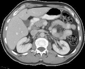


8 CT = density map


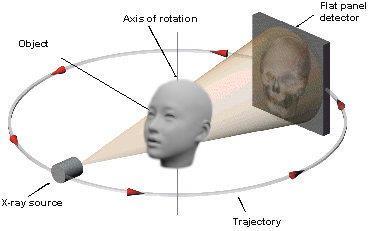
9 Concept of CT Scanner Planar fan beam Type1 Type2
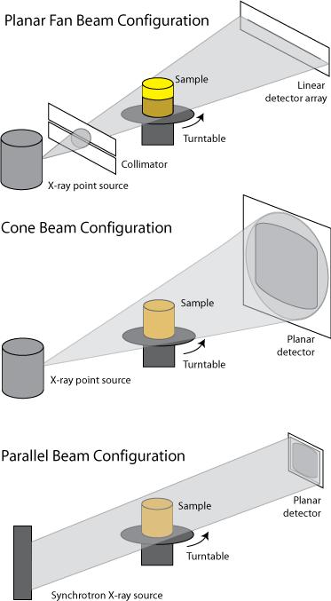
10 CT beam configurations

11 Spiral CT, multi-detector CTs SINGLE SLICE SPIRAL MULTISLICE SPIRAL Spiral path with 4,8,16,32,64, 128, 256, 512 rows of detectors

12 Recent CT scanners (2008)

13 CT gantry inside


14 Modern CT scanners (2018) GE Phillips
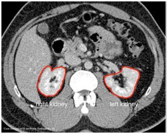
15 CT catered to us as series of slices

16 CT coordinate system pixel Y Field of view X Slice thickness Slice index

17 Navigation between CT slices k -- slice index thk = slice thickness (cm or mm)
18 j pixel (row index) NY number of pixels (typically 512) FOVY size of the body captured (in cm) dy = FOVY / NY (pixel size, in cm) Navigation in a CT Slice i pixel (column index) NX number of pixels (typically 512) FOVX size of the body captured (in cm) dx = FOVX / NX (pixel size, in cm)
 = P(i*dx, j*dy, k*thk) Where: dx = FOVX")

19 Conversion between pixel and metric coordinates in CT imaging Simple scaling: P(x,y,z) = P(i*dx, j*dy, k*thk) Where: dx = FOVX / NX dy = FOVY / NY & FOVX, FOVY, NX, NY, thk are usually printed on the CT image header S = d x d y thk

20 Tilted CT gantry y z Ouch! No longer a Cartesian coordinate system
21 Cone beam CT reconstruction
22 Cone beam CT - truncation

23 Cone beam CT metal artifacts F Edward Boas & Dominik Fleischmann *by

24 Cone beam CT yields isotropic volume


25 Cone beam CT reconstruction examples

26 Organs can be contoured

27 Organs reconstructed from contours

")
28 CT/X-ray fiducial markers (donut)

")
29 CT/X-ray fiducial markers (BB)
30 Next tracked navigation A C B v 3 v 1 v 2
 is known in F Tool F Tool FMar Trac F Trac Tool model of tool Computer F Tool real tool C F A Mar Tool F Tool Tracked")

31 Tracked Navigation F Trac The target anatomy is known in F CT The head markers were found in F CT The head markers are tracked in F trac The tool is tracked in F trac The tool (tip/axis/etc) is known in F Tool F Tool FMar Trac F Trac Tool model of tool Computer F Tool real tool C F A Mar Tool F Tool Tracked navigation = paint the model of the tool over the CT on the computer screen as the real tool is moving over/on/inside the patient in the operating room F CT Mar B v 3 v 1 v 2 F CT F Mar Operating room For this we need to compute the transformation from Tool to CT coordinates F CT Tool = F CT Mar * F Mar Trac * F Trac Tool
Radiology. Marta Anguiano Millán. Departamento de Física Atómica, Molecular y Nuclear Facultad de Ciencias. Universidad de Granada
 Departamento de Física Atómica, Molecular y Nuclear Facultad de Ciencias. Universidad de Granada Overview Introduction Overview Introduction Tecniques of imaging in Overview Introduction Tecniques of imaging
MEDICAL IMAGING 2nd Part Computed Tomography
 MEDICAL IMAGING 2nd Part Computed Tomography Introduction 2 In the last 30 years X-ray Computed Tomography development produced a great change in the role of diagnostic imaging in medicine. In convetional
Ch. 4 Physical Principles of CT
 Ch. 4 Physical Principles of CT CLRS 408: Intro to CT Department of Radiation Sciences Review: Why CT? Solution for radiography/tomography limitations Superimposition of structures Distinguishing between
Shadow casting. What is the problem? Cone Beam Computed Tomography THE OBJECTIVES OF DIAGNOSTIC IMAGING IDEAL DIAGNOSTIC IMAGING STUDY LIMITATIONS
 Cone Beam Computed Tomography THE OBJECTIVES OF DIAGNOSTIC IMAGING Reveal pathology Reveal the anatomic truth Steven R. Singer, DDS srs2@columbia.edu IDEAL DIAGNOSTIC IMAGING STUDY Provides desired diagnostic
[PDR03] RECOMMENDED CT-SCAN PROTOCOLS
![[PDR03] RECOMMENDED CT-SCAN PROTOCOLS](/thumbs/72/66454100.jpg "[PDR03] RECOMMENDED CT-SCAN PROTOCOLS") SURGICAL & PROSTHETIC DESIGN [PDR03] RECOMMENDED CT-SCAN PROTOCOLS WORK-INSTRUCTIONS DOCUMENT (CUSTOMER) RECOMMENDED CT-SCAN PROTOCOLS [PDR03_V1]: LIVE 1 PRESCRIBING SURGEONS Patient-specific implants,
SURGICAL & PROSTHETIC DESIGN [PDR03] RECOMMENDED CT-SCAN PROTOCOLS WORK-INSTRUCTIONS DOCUMENT (CUSTOMER) RECOMMENDED CT-SCAN PROTOCOLS [PDR03_V1]: LIVE 1 PRESCRIBING SURGEONS Patient-specific implants,
Computer-Tomography I: Principles, History, Technology
 Computer-Tomography I: Principles, History, Technology Prof. Dr. U. Oelfke DKFZ Heidelberg Department of Medical Physics (E040) Im Neuenheimer Feld 280 69120 Heidelberg, Germany u.oelfke@dkfz.de History
Computed Tomography. Principles, Design, Artifacts, and Recent Advances. Jiang Hsieh THIRD EDITION. SPIE PRESS Bellingham, Washington USA
 Computed Tomography Principles, Design, Artifacts, and Recent Advances THIRD EDITION Jiang Hsieh SPIE PRESS Bellingham, Washington USA Table of Contents Preface Nomenclature and Abbreviations xi xv 1 Introduction
BME I5000: Biomedical Imaging
 1 Lucas Parra, CCNY BME I5000: Biomedical Imaging Lecture 4 Computed Tomography Lucas C. Parra, parra@ccny.cuny.edu some slides inspired by lecture notes of Andreas H. Hilscher at Columbia University.
Multi-slice CT Image Reconstruction Jiang Hsieh, Ph.D.
 Multi-slice CT Image Reconstruction Jiang Hsieh, Ph.D. Applied Science Laboratory, GE Healthcare Technologies 1 Image Generation Reconstruction of images from projections. textbook reconstruction advanced
Computed tomography - outline
 Computed tomography - outline Computed Tomography Systems Jørgen Arendt Jensen and Mikael Jensen (DTU Nutech) October 6, 216 Center for Fast Ultrasound Imaging, Build 349 Department of Electrical Engineering
Image Reconstruction from Projection
 Image Reconstruction from Projection Reconstruct an image from a series of projections X-ray computed tomography (CT) Computed tomography is a medical imaging method employing tomography where digital
Image Acquisition Systems
 Image Acquisition Systems Goals and Terminology Conventional Radiography Axial Tomography Computer Axial Tomography (CAT) Magnetic Resonance Imaging (MRI) PET, SPECT Ultrasound Microscopy Imaging ITCS
Corso di laurea in Fisica A.A Fisica Medica 4 TC
 Corso di laurea in Fisica A.A. 2007-2008 Fisica Medica 4 TC Computed Tomography Principles 1. Projection measurement 2. Scanner systems 3. Scanning modes Basic Tomographic Principle The internal structure
MEDICAL IMAGING 2nd Part Computed Tomography
 MEDICAL IMAGING 2nd Part Computed Tomography Introduction 2 In the last 30 years X-ray Computed Tomography development produced a great change in the role of diagnostic imaging in medicine. In convetional
Basics of treatment planning II
 Basics of treatment planning II Sastry Vedam PhD DABR Introduction to Medical Physics III: Therapy Spring 2015 Dose calculation algorithms! Correction based! Model based 1 Dose calculation algorithms!
Radon Transform and Filtered Backprojection
 Radon Transform and Filtered Backprojection Jørgen Arendt Jensen October 13, 2016 Center for Fast Ultrasound Imaging, Build 349 Department of Electrical Engineering Center for Fast Ultrasound Imaging Department
CT Reconstruction with Good-Orientation and Layer Separation for Multilayer Objects
 17th World Conference on Nondestructive Testing, 25-28 Oct 2008, Shanghai, China CT Reconstruction with Good-Orientation and Layer Separation for Multilayer Objects Tong LIU 1, Brian Stephan WONG 2, Tai
LAB DEMONSTRATION COMPUTED TOMOGRAPHY USING DESKCAT Lab Manual: 0
 LAB DEMONSTRATION COMPUTED TOMOGRAPHY USING DESKCAT Lab Manual: 0 Introduction This lab demonstration explores the physics and technology of Computed Tomography (CT) and guides the student and instructor
MEDICAL EQUIPMENT: COMPUTED TOMOGRAPHY. Prof. Yasser Mostafa Kadah
 MEDICAL EQUIPMENT: COMPUTED TOMOGRAPHY Prof. Yasser Mostafa Kadah www.k-space.org Recommended Textbook X-Ray Computed Tomography in Biomedical Engineering, by Robert Cierniak, Springer, 211 Computed Tomography
Computational Medical Imaging Analysis Chapter 4: Image Visualization
 Computational Medical Imaging Analysis Chapter 4: Image Visualization Jun Zhang Laboratory for Computational Medical Imaging & Data Analysis Department of Computer Science University of Kentucky Lexington,
Modifications for P551 Fall 2014
 LAB DEMONSTRATION COMPUTED TOMOGRAPHY USING DESKCAT 1 Modifications for P551 Fall 2014 Introduction This lab demonstration explores the physics and technology of Computed Tomography (CT) and guides the
Comparison of Scatter Correction Methods for CBCT. Author(s): Suri, Roland E.; Virshup, Gary; Kaissl, Wolfgang; Zurkirchen, Luis
: Suri, Roland E.; Virshup, Gary; Kaissl, Wolfgang; Zurkirchen, Luis") Research Collection Working Paper Comparison of Scatter Correction Methods for CBCT Author(s): Suri, Roland E.; Virshup, Gary; Kaissl, Wolfgang; Zurkirchen, Luis Publication Date: 2010 Permanent Link:
Research Collection Working Paper Comparison of Scatter Correction Methods for CBCT Author(s): Suri, Roland E.; Virshup, Gary; Kaissl, Wolfgang; Zurkirchen, Luis Publication Date: 2010 Permanent Link:
Computer-Tomography II: Image reconstruction and applications
 Computer-Tomography II: Image reconstruction and applications Prof. Dr. U. Oelfke DKFZ Heidelberg Department of Medical Physics (E040) Im Neuenheimer Feld 280 69120 Heidelberg, Germany u.oelfke@dkfz.de
Digital Image Processing
 Digital Image Processing SPECIAL TOPICS CT IMAGES Hamid R. Rabiee Fall 2015 What is an image? 2 Are images only about visual concepts? We ve already seen that there are other kinds of image. In this lecture
Tomographic Reconstruction
 Tomographic Reconstruction 3D Image Processing Torsten Möller Reading Gonzales + Woods, Chapter 5.11 2 Overview Physics History Reconstruction basic idea Radon transform Fourier-Slice theorem (Parallel-beam)
Joint ICTP-TWAS Workshop on Portable X-ray Analytical Instruments for Cultural Heritage. 29 April - 3 May, 2013
 2455-5 Joint ICTP-TWAS Workshop on Portable X-ray Analytical Instruments for Cultural Heritage 29 April - 3 May, 2013 Lecture NoteBasic principles of X-ray Computed Tomography Diego Dreossi Elettra, Trieste
icatvision Quick Reference
 icatvision Quick Reference Navigating the i-cat Interface This guide shows how to: View reconstructed images Use main features and tools to optimize an image. REMINDER Images are displayed as if you are
Méthodes d imagerie pour les écoulements et le CND
 Méthodes d imagerie pour les écoulements et le CND Journée scientifique FED3G CEA LIST/Lab Imagerie Tomographie et Traitement Samuel Legoupil 15 juin 2012 2D/3D imaging tomography Example Petrochemical
Loma Linda University Medical Center Dept. of Radiation Medicine
 Loma Linda University Medical Center Dept. of Radiation Medicine and Northern Illinois University Dept. of Physics and Dept. of Computer Science Presented by George Coutrakon, PhD NIU Physics Dept. Collaborators
Some reference material
 Some reference material Physics reference book on medical imaging: A good one is The Essential Physics of Medical Imaging, 3 rd Ed. by Bushberg et al. ($170! new). However, there are several similar books
RADIOLOGY AND DIAGNOSTIC IMAGING
 Day 2 part 2 RADIOLOGY AND DIAGNOSTIC IMAGING Dr hab. Zbigniew Serafin, MD, PhD serafin@cm.umk.pl 2 3 4 5 CT technique CT technique 6 CT system Kanal K: RSNA/AAPM web module: CT Systems & CT Image Quality
Alignment and Other Challenges in Reconstructing Cryotomograms with IMOD
 Alignment and Other Challenges in Reconstructing Cryotomograms with IMOD Challenges in Cryotomography Alignment, alignment, alignment It can be hard to get fiducials onto/in the sample The low SNR makes
Development of a multi-axis X-ray CT for highly accurate inspection of electronic devices
 Development of a multi-axis X-ray CT for highly accurate inspection of electronic devices Toru Kano 1, Michihiko Koseki 2 More info about this article: http://www.ndt.net/?id=20843 1 Tokyo University of
Tomography. Forward projectionsp θ (r) are known as a Radon transform. Objective: reverse this process to form the original image
 are known as a Radon transform. Objective: reverse this process to form the original image") C. A. Bouman: Digital Image Processing - January 9, 217 1 Tomography Many medical imaging systems can only measure projections through an object with density f(x,y). Projections must be collected at every
C. A. Bouman: Digital Image Processing - January 9, 217 1 Tomography Many medical imaging systems can only measure projections through an object with density f(x,y). Projections must be collected at every
Introduction to Biomedical Imaging
 Alejandro Frangi, PhD Computational Imaging Lab Department of Information & Communication Technology Pompeu Fabra University www.cilab.upf.edu X-ray Projection Imaging Computed Tomography Digital X-ray
Digital Image Processing
 Digital Image Processing Image Restoration and Reconstruction (Image Reconstruction from Projections) Christophoros Nikou cnikou@cs.uoi.gr University of Ioannina - Department of Computer Science and Engineering
Improvement of Efficiency and Flexibility in Multi-slice Helical CT
 J. Shanghai Jiaotong Univ. (Sci.), 2008, 13(4): 408 412 DOI: 10.1007/s12204-008-0408-x Improvement of Efficiency and Flexibility in Multi-slice Helical CT SUN Wen-wu 1 ( ), CHEN Si-ping 2 ( ), ZHUANG Tian-ge
Scaling Calibration in the ATRACT Algorithm
 Scaling Calibration in the ATRACT Algorithm Yan Xia 1, Andreas Maier 1, Frank Dennerlein 2, Hannes G. Hofmann 1, Joachim Hornegger 1,3 1 Pattern Recognition Lab (LME), Friedrich-Alexander-University Erlangen-Nuremberg,
8/7/2017. Disclosures. MECT Systems Overview and Quantitative Opportunities. Overview. Computed Tomography (CT) CT Numbers. Polyenergetic Acquisition
 CT Numbers. Polyenergetic Acquisition") Quantitative Multi-Energy Computed Tomography: Imaging and Therapy Advancements Disclosures MECT Systems Overview and Quantitative Opportunities The speaker receives research funding from GE Healthcare
Quantitative Multi-Energy Computed Tomography: Imaging and Therapy Advancements Disclosures MECT Systems Overview and Quantitative Opportunities The speaker receives research funding from GE Healthcare
Philips SPECT/CT Systems
 Philips SPECT/CT Systems Ling Shao, PhD Director, Imaging Physics & System Analysis Nuclear Medicine, Philips Healthcare June 14, 2008 *Presented SNM08 Categorical Seminar - Quantitative SPECT and PET
Brilliance CT Big Bore.
 1 2 2 There are two methods of RCCT acquisition in widespread clinical use: cine axial and helical. In RCCT with cine axial acquisition, repeat CT images are taken each couch position while recording respiration.
An approximate cone beam reconstruction algorithm for gantry-tilted CT
 An approximate cone beam reconstruction algorithm for gantry-tilted CT Ming Yan a, Cishen Zhang ab, Hongzhu Liang a a School of Electrical & Electronic Engineering, Nanyang Technological University, Singapore;
Biomedical Imaging. Computed Tomography. Patrícia Figueiredo IST
 Biomedical Imaging Computed Tomography Patrícia Figueiredo IST 2013-2014 Overview Basic principles X ray attenuation projection Slice selection and line projections Projection reconstruction Instrumentation
COMPARATIVE STUDIES OF DIFFERENT SYSTEM MODELS FOR ITERATIVE CT IMAGE RECONSTRUCTION
 COMPARATIVE STUDIES OF DIFFERENT SYSTEM MODELS FOR ITERATIVE CT IMAGE RECONSTRUCTION BY CHUANG MIAO A Thesis Submitted to the Graduate Faculty of WAKE FOREST UNIVERSITY GRADUATE SCHOOL OF ARTS AND SCIENCES
GE s Revolution CT MATLAB III: CT. Kathleen Chen March 20, 2018
 GE s Revolution CT MATLAB III: CT Kathleen Chen chens18@rpi.edu March 20, 2018 https://www.zmescience.com/medicine/inside-human-body-real-time-gifs-demo-power-ct-scan/ Reminders Make sure you have MATLAB
Central Slice Theorem
 Central Slice Theorem Incident X-rays y f(x,y) R x r x Detected p(, x ) The thick line is described by xcos +ysin =R Properties of Fourier Transform F [ f ( x a)] F [ f ( x)] e j 2 a Spatial Domain Spatial
A Fast GPU-Based Approach to Branchless Distance-Driven Projection and Back-Projection in Cone Beam CT
 A Fast GPU-Based Approach to Branchless Distance-Driven Projection and Back-Projection in Cone Beam CT Daniel Schlifske ab and Henry Medeiros a a Marquette University, 1250 W Wisconsin Ave, Milwaukee,
Carestream s 2 nd Generation Metal Artifact Reduction Software (CMAR 2)
") Carestream s 2 nd Generation Metal Artifact Reduction Software (CMAR 2) Author: Levon Vogelsang Introduction Cone beam computed tomography (CBCT), or cone beam CT technology, offers considerable promise
Carestream s 2 nd Generation Metal Artifact Reduction Software (CMAR 2) Author: Levon Vogelsang Introduction Cone beam computed tomography (CBCT), or cone beam CT technology, offers considerable promise
Optimisation of Toshiba Aquilion ONE Volume Imaging
 Optimisation of Toshiba Aquilion ONE Volume Imaging Jane Edwards, RPRSG Royal Free London NHS Foundation Trust Dr Mufudzi Maviki, Plymouth Hospitals NHS Trust Background In 2011/12 Radiology at RFH was
Medical Image Reconstruction Term II 2012 Topic 6: Tomography
 Medical Image Reconstruction Term II 2012 Topic 6: Tomography Professor Yasser Mostafa Kadah Tomography The Greek word tomos means a section, a slice, or a cut. Tomography is the process of imaging a cross
Translational Computed Tomography: A New Data Acquisition Scheme
 2nd International Symposium on NDT in Aerospace 2010 - We.1.A.3 Translational Computed Tomography: A New Data Acquisition Scheme Theobald FUCHS 1, Tobias SCHÖN 2, Randolf HANKE 3 1 Fraunhofer Development
DUE to beam polychromacity in CT and the energy dependence
 1 Empirical Water Precorrection for Cone-Beam Computed Tomography Katia Sourbelle, Marc Kachelrieß, Member, IEEE, and Willi A. Kalender Abstract We propose an algorithm to correct for the cupping artifact
System Optimization and Patient Translational Motion Correction for Reduction of Artifacts in a Fan-Beam CT Scanner
 Wright State University CORE Scholar Browse all Theses and Dissertations Theses and Dissertations 2012 System Optimization and Patient Translational Motion Correction for Reduction of Artifacts in a Fan-Beam
Continuous and Discrete Image Reconstruction
 25 th SSIP Summer School on Image Processing 17 July 2017, Novi Sad, Serbia Continuous and Discrete Image Reconstruction Péter Balázs Department of Image Processing and Computer Graphics University of
Imaging protocols for navigated procedures
 9732379 G02 Rev. 1 2015-11 Imaging protocols for navigated procedures How to use this document This document contains imaging protocols for navigated cranial, DBS and stereotactic, ENT, and spine procedures
Spiral CT. Protocol Optimization & Quality Assurance. Ge Wang, Ph.D. Department of Radiology University of Iowa Iowa City, Iowa 52242, USA
 Spiral CT Protocol Optimization & Quality Assurance Ge Wang, Ph.D. Department of Radiology University of Iowa Iowa City, Iowa 52242, USA Spiral CT Protocol Optimization & Quality Assurance Protocol optimization
Metal Artifact Reduction for Multi-Material Objects
 Metal Artifact Reduction for Multi-Material Objects Bärbel Kratz 1,#, Frank Herold 1, Malte Kurfiß 1, Thorsten M. Buzug 2 1 YXLON International GmbH, Essener Bogen 15, 22419 Hamburg, Germany, e-mail: baerbel.kratz@hbg.yxlon.com,
VXRE Reconstruction Software Manual
 VXRE Reconstruction Software Manual Version 1.7.8 3D INDUSTRIAL IMAGING 3D Industrial Imaging Co.,Ltd. Address :#413 Institute of Computer Technology, Seoul National University, Daehak-dong, Gwanak-gu,
Topics. Projections. Review Filtered Backprojection Fan Beam Spiral CT Applications. Bioengineering 280A Principles of Biomedical Imaging
 Bioengineering 28A Principles of Biomedical Imaging Fall Quarter 24 X-Rays/CT Lecture 2 Topics Review Filtered Backprojection Fan Beam Spiral CT Applications s I θ (r) = I exp µ(x, y)ds Lr,θ = I exp µ(rcosθ
X-ray tomographic study of wetting bentonite
 UNIVERSITY OF JYVÄSKYLÄ X-ray tomographic study of wetting bentonite Tero Harjupatana Department of Physics, University of Jyväskylä BOA seminar 19.8.2014 Goal To develop and apply experimental techniques
Spiral ASSR Std p = 1.0. Spiral EPBP Std. 256 slices (0/300) Kachelrieß et al., Med. Phys. 31(6): , 2004
 Kachelrieß et al., Med. Phys. 31(6): , 2004") Spiral ASSR Std p = 1.0 Spiral EPBP Std p = 1.0 Kachelrieß et al., Med. Phys. 31(6): 1623-1641, 2004 256 slices (0/300) Advantages of Cone-Beam Spiral CT Image quality nearly independent of pitch Increase
Spiral ASSR Std p = 1.0 Spiral EPBP Std p = 1.0 Kachelrieß et al., Med. Phys. 31(6): 1623-1641, 2004 256 slices (0/300) Advantages of Cone-Beam Spiral CT Image quality nearly independent of pitch Increase
Optimization of CT Simulation Imaging. Ingrid Reiser Dept. of Radiology The University of Chicago
 Optimization of CT Simulation Imaging Ingrid Reiser Dept. of Radiology The University of Chicago Optimization of CT imaging Goal: Achieve image quality that allows to perform the task at hand (diagnostic
Spectral analysis of non-stationary CT noise
 Spectral analysis of non-stationary CT noise Kenneth M. Hanson Los Alamos Scientific Laboratory Int. Symposium and Course on Computed Tomography, Las Vegas, April 7-11, 1980 This presentation available
F3-A5: Model-Based Iterative Reconstruction for Advanced Baggage Screening
 F3-A5: Model-Based Iterative Reconstruction for Advanced Baggage Screening Abstract While traditional direct reconstruction algorithms such as filtered back projection (FBP) and direct Fourier method (DFM)
Medical Image Processing: Image Reconstruction and 3D Renderings
 Medical Image Processing: Image Reconstruction and 3D Renderings 김보형 서울대학교컴퓨터공학부 Computer Graphics and Image Processing Lab. 2011. 3. 23 1 Computer Graphics & Image Processing Computer Graphics : Create,
CT Basics Principles of Spiral CT Dose. Always Thinking Ahead.
 1 CT Basics Principles of Spiral CT Dose 2 Who invented CT? 1963 - Alan Cormack developed a mathematical method of reconstructing images from x-ray projections Sir Godfrey Hounsfield worked for the Central
Topics. Review Filtered Backprojection Fan Beam Spiral CT Applications. Bioengineering 280A Principles of Biomedical Imaging
 Bioengineering 28A Principles of Biomedical Imaging Fall Quarter 24 X-Rays/CT Lecture 2 Topics Review Filtered Backprojection Fan Beam Spiral CT Applications Projections I θ (r) = I exp Lr,θ µ(x,y)ds =
Projection and Reconstruction-Based Noise Filtering Methods in Cone Beam CT
 Projection and Reconstruction-Based Noise Filtering Methods in Cone Beam CT Benedikt Lorch 1, Martin Berger 1,2, Joachim Hornegger 1,2, Andreas Maier 1,2 1 Pattern Recognition Lab, FAU Erlangen-Nürnberg
CLASS HOURS: 4 CREDIT HOURS: 4 LABORATORY HOURS: 0
 Revised 10/10 COURSE SYLLABUS TM 220 COMPUTED TOMOGRAPHY PHYSICS CLASS HOURS: 4 CREDIT HOURS: 4 LABORATORY HOURS: 0 CATALOG COURSE DESCRIPTION: This course is one of a three course set in whole body Computed
Reduction of Metal Artifacts in Computed Tomographies for the Planning and Simulation of Radiation Therapy
 Reduction of Metal Artifacts in Computed Tomographies for the Planning and Simulation of Radiation Therapy T. Rohlfing a, D. Zerfowski b, J. Beier a, P. Wust a, N. Hosten a, R. Felix a a Department of
Fundamentals of CT imaging
 SECTION 1 Fundamentals of CT imaging I History In the early 1970s Sir Godfrey Hounsfield s research produced the first clinically useful CT scans. Original scanners took approximately 6 minutes to perform
NON-COLLIMATED SCATTERED RADIATION TOMOGRAPHY
 NON-COLLIMATED SCATTERED RADIATION TOMOGRAPHY Gorshkov V.A., Space Research Institute, Moscow, Russia Yumashev V.M., State corporation "Rosatom", Centre "Atom-innovation", Moscow, Russia Kirilenko K.V.,
Feldkamp-type image reconstruction from equiangular data
 Journal of X-Ray Science and Technology 9 (2001) 113 120 113 IOS Press Feldkamp-type image reconstruction from equiangular data Ben Wang a, Hong Liu b, Shiying Zhao c and Ge Wang d a Department of Elec.
Metal Artifact Reduction CT Techniques. Tobias Dietrich University Hospital Balgrist University of Zurich Switzerland
 Metal Artifact Reduction CT Techniques R S S S Tobias Dietrich University Hospital Balgrist University of Zurich Switzerland N. 1 v o 4 1 0 2. Postoperative CT Metal Implants CT is accurate for assessment
First CT Scanner. How it Works. Contemporary CT. Before and After CT. Computer Tomography: How It Works. Medical Imaging and Pattern Recognition
 Computer Tomography: How t Works Medical maging and Pattern Recognition Lecture 7 Computed Tomography Oleh Tretiak Only one plane is illuminated. Source-subject motion provides added information. 2 How
Cardiac Dual Energy CT: Technique
 RSNA 2013, VSCA51-01, Chicago, Dec. 5, 2013 Cardiac Radiology Series Cardiac Dual Energy CT: Technique Willi A. Kalender, Ph.D. Institute of Medical Physics University of Erlangen www.imp.uni-erlangen.de
(12) Patent Application Publication (10) Pub. No.: US 2011/ A1
 Patent Application Publication (10) Pub. No.: US 2011/ A1") (19) United States US 2011 0255765A1 (12) Patent Application Publication (10) Pub. No.: US 2011/0255765 A1 Carlson et al. (43) Pub. Date: Oct. 20, 2011 (54) REDUCTION AND REMOVAL OF ARTIFACTS FROMA THREE-DIMIENSIONAL
(19) United States US 2011 0255765A1 (12) Patent Application Publication (10) Pub. No.: US 2011/0255765 A1 Carlson et al. (43) Pub. Date: Oct. 20, 2011 (54) REDUCTION AND REMOVAL OF ARTIFACTS FROMA THREE-DIMIENSIONAL
Information page for written examinations at Linköping University TER2
 Information page for written examinations at Linköping University Examination date 2016-08-19 Room (1) TER2 Time 8-12 Course code Exam code Course name Exam name Department Number of questions in the examination
TESTING OF THE CIRCLE AND LINE ALGORITHM IN THE SETTING OF MICRO-CT
 SCA2016-080 1/7 TESTING OF THE CIRCLE AND LINE ALGORITHM IN THE SETTING OF MICRO-CT Alexander Katsevich 1, 2 and Michael Frenkel 1 1 itomography Corp., 2 University of Central Florida (UCF) This paper
3/27/2012 WHY SPECT / CT? SPECT / CT Basic Principles. Advantages of SPECT. Advantages of CT. Dr John C. Dickson, Principal Physicist UCLH
 3/27/212 Advantages of SPECT SPECT / CT Basic Principles Dr John C. Dickson, Principal Physicist UCLH Institute of Nuclear Medicine, University College London Hospitals and University College London john.dickson@uclh.nhs.uk
Advanced single slice rebinning in cone beam spiral CT: Theoretical considerations and medical applications
 Advanced single slice rebinning in cone beam spiral CT: Theoretical considerations and medical applications Marc Kachelrieß a, Stefan Schaller b and Willi A. Kalender a a Institute of Medical Physics,
Phase problem and the Radon transform
 Phase problem and the Radon transform Andrei V. Bronnikov Bronnikov Algorithms The Netherlands The Radon transform and applications Inverse problem of phase-contrast CT Fundamental theorem Image reconstruction
C a t p h a n / T h e P h a n t o m L a b o r a t o r y
 C a t p h a n 5 0 0 / 6 0 0 T h e P h a n t o m L a b o r a t o r y C a t p h a n 5 0 0 / 6 0 0 Internationally recognized for measuring the maximum obtainable performance of axial, spiral and multi-slice
Annexure XII SPECIFICATIONS FOR A NEW STATE OF ART 16 SLICE ALL PURPOSE C. T. SCANNER
 Annexure XII SPECIFICATIONS FOR A NEW STATE OF ART 16 SLICE ALL PURPOSE C. T. SCANNER A) Scanner Design X-Ray generator and tube: 1. Scanner: Whole body spiral CT scanner (16 slices) of latest technology.
Empirical cupping correction: A first-order raw data precorrection for cone-beam computed tomography
 Empirical cupping correction: A first-order raw data precorrection for cone-beam computed tomography Marc Kachelrieß, a Katia Sourbelle, and Willi A. Kalender Institute of Medical Physics, University of
PURE. ViSION Edition PET/CT. Patient Comfort Put First.
 PURE ViSION Edition PET/CT Patient Comfort Put First. 2 System features that put patient comfort and safety first. Oncology patients deserve the highest levels of safety and comfort during scans. Our Celesteion
The theory and practical aspects of proton imaging proton radiography (prad) and proton tomography (pct)
 and proton tomography (pct)") The theory and practical aspects of proton imaging proton radiography (prad) and proton tomography (pct) Fritz DeJongh, ProtonVDA Inc August 30 2018 Stay away from negative people. They have a problem
The theory and practical aspects of proton imaging proton radiography (prad) and proton tomography (pct) Fritz DeJongh, ProtonVDA Inc August 30 2018 Stay away from negative people. They have a problem
A new concept for high-speed atline and inline CT for up to 100% mass production process control allowing both 3D metrology and failure analysis
 A new concept for high-speed atline and inline CT for up to 100% mass production process control allowing both 3D metrology and failure analysis Oliver Brunke 1, Ferdinand Hansen 2, Ingo Stuke 3, Friedrich
The n-pi-method for Helical Cone-Beam CT
 848 IEEE TRANSACTIONS ON MEDICAL IMAGING, VOL. 19, NO. 9, SEPTEMBER 2000 The n-pi-method for Helical Cone-Beam CT R. Proksa*, Th. Köhler, M. Grass, and J. Timmer Abstract A new class of acquisition schemes
Diagnostic imaging techniques. Krasznai Zoltán. University of Debrecen Medical and Health Science Centre Department of Biophysics and Cell Biology
 Diagnostic imaging techniques Krasznai Zoltán University of Debrecen Medical and Health Science Centre Department of Biophysics and Cell Biology 1. Computer tomography (CT) 2. Gamma camera 3. Single Photon
Enhancement Image Quality of CT Using Single Slice Spiral Technique
 Enhancement Image Quality of CT Using Single Slice Spiral Technique Doaa. N. Al Sheack 1 and Dr.Mohammed H. Ali Al Hayani 2 1 2 Electronic and Communications Engineering Department College of Engineering,
Tomography at all Scales. Uccle, 7 April 2014
 Tomography at all Scales Uccle, 7 April 2014 Outline The Vision Lab ASTRA: All Scale Tomographic Reconstruction Antwerp Tomography Discrete Tomography In situ CT Superresolution Dynamic imaging The ASTRA
Interior Reconstruction Using the Truncated Hilbert Transform via Singular Value Decomposition
 Interior Reconstruction Using the Truncated Hilbert Transform via Singular Value Decomposition Hengyong Yu 1, Yangbo Ye 2 and Ge Wang 1 1 CT Laboratory, Biomedical Imaging Division, VT-WFU School of Biomedical
Tracked surgical drill calibration
 Tracked surgical drill calibration An acetabular fracture is a break in the socket portion of the "ball-and-socket" hip joint. The majority of acetabular fractures are caused by some type of highenergy
Radiography and Tomography Lab 2: Computing Radiographs
 Radiography and Tomography Lab 2: Computing Radiographs An Abbreviated View of Radiography Transmission Radiography is the process of measuring and recording changes in a high-energy particle beam (X-rays,
Digital Imaging and Communications in Medicine (DICOM) Supplement 116: 3D X-Ray Storage SOP Class
 Supplement 116: 3D X-Ray Storage SOP Class") 2 4 6 Digital Imaging and Communications in Medicine (DICOM) 8 Supplement 116: 3D X-Ray Storage SOP Class 10 12 14 16 18 20 22 Prepared by: 24 DICOM Standards Committee, Working Groups 02, Projection Imaging
2 4 6 Digital Imaging and Communications in Medicine (DICOM) 8 Supplement 116: 3D X-Ray Storage SOP Class 10 12 14 16 18 20 22 Prepared by: 24 DICOM Standards Committee, Working Groups 02, Projection Imaging
X-ray Target Reconstruction for Cyber Knife Radiosurgery Assignment for CISC/CMPE 330
 X-ray Target Reconstruction for Cyber Knife Radiosurgery Assignment for CISC/CMPE 330 We will perform radiosurgery of multipole liver tumors under X-ray guidance with the Cyber Knife (CK) system. The patient
Principles of Computerized Tomographic Imaging
 Principles of Computerized Tomographic Imaging Parallel CT, Fanbeam CT, Helical CT and Multislice CT Marjolein van der Glas August 29, 2000 Abstract The total attenuation suffered by one beam of x-rays
EECS490: Digital Image Processing. Lecture #16
 Lecture #16 Wiener Filters Constrained Least Squares Filter Computed Tomography Basics Reconstruction and the Radon Transform Fourier Slice Theorem Filtered Backprojections Fan Beams Motion Blurring Model
2D Fan Beam Reconstruction 3D Cone Beam Reconstruction. Mario Koerner
 2D Fan Beam Reconstruction 3D Cone Beam Reconstruction Mario Koerner Moscow-Bavarian Joint Advanced Student School 2006 March 19 2006 to March 29 2006 Overview 2D Fan Beam Reconstruction Shortscan Reconstruction