MEG and EEG analysis using
|
|
|
- Rhoda Parks
- 5 years ago
- Views:
Transcription


1 MEG and EEG analysis using François Tadel & Sylvain Baillet May 2014


2 Foretaste Graphic interface 2

3 Foretaste Scripting environment Rapid selection of files and processes to apply Automatic generation of Matlab scripts Plug-in structure: easy to add custom processes 3
 Matlab & Java:")


4 Brainstorm is A free and open-source application (GPL) Matlab & Java: Platform-independent Designed for Matlab environment Stand-alone version also available Interface-based: click, drag, drop No Matlab experience required Daily updates of the software 4
5 A bit of history 14 years of research and development Collaboration between multiple groups: University of Southern California, Los Angeles, USA La Salpetriere Hospital / CNRS, Paris, France Neurospin / Inserm / CEA, Paris, France Los Alamos National Lab, USA Medical College of Wisconsin, Milwaukee, USA Cleveland Clinic, USA Martinos Center / MGH, USA McGovern Institute / MIT, USA Montreal Neurological Institute / McGill, Canada Over 9000 user accounts / 70 countries 5


6 Workflow Anatomy Sensors EEG/MEG Analysis Co-registration Source estimation 6


7 Database Three levels: Protocol Subject Condition Popup menus All files saved in Matlab.mat Same architecture on the disk 7


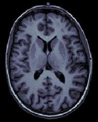
8 Anatomy T1-MRI volume Surfaces extracted with a dedicated software: BrainVISA, FreeSurfer, BrainSuite 8

9 Atlases Support for the surface-based atlases generated automatically by FreeSurfer 9


10 Co-registration MEG / MRI (1) Basic estimation based on three points (NAS,LPA,RPA) MRI: Marked in the volume with the MRI Viewer MEG: Obtained with a tracking system (Polhemus) 10
 with the head surface (from the MRI) Final registration must be checked")
11 Co-registration MEG / MRI (2) Automatic adjustment based on head shape Trying to fit the head points (Polhemus) with the head surface (from the MRI) Final registration must be checked manually 11


12 Continuous recordings Manual inspection of the recordings Identify the noise sources Mark bad channels and bad segments Check stimulus markers, add custom events 12


13 Pre-processing Filtering Sinusoid removal: Remove 50Hz or 60Hz power line contamination Band-pass filter: Remove slow drifts and high frequencies 13


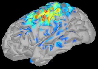
14 Pre-processing Cleaning Artifact detection and removal: heartbeats, eye blinks, movements, ECG EOG ECG EOG 14
15 Pre-processing Cleaning Two categories of artifacts: Well defined, reproducible, short, frequent: Heartbeats, eye blinks, some stimulators Unavoidable and frequent: we cannot just ignore them Can be modeled and removed from the signal efficiently All the other events that can alter the recordings: Movements, building vibrations, metro nearby Too complex or not repeated enough to be modeled Safer to mark them as bad segments, and ignore them 15
 Detect artifacts Concatenate")

16 Pre-processing Cleaning Signal-Space Projection (SSP) Detect artifacts Concatenate epochs Spatial components PCA Select components Compute projectors (linear operator) Apply to EEG/MEG 16


17 Pre-processing Cleaning Example: Cardiac artifact Original SSP 17

18 Pre-processing Cleaning 18
")
19 Pre-processing Averaging Epoching: extraction of small blocks of recordings around an event of interest (stimulus, spike ) 19


20 Pre-processing Averaging Averaging all the trials: Reveals the features of the signals that are locked in time to a given event => Evoked-response field (or potential) 20
 Forward model = head")

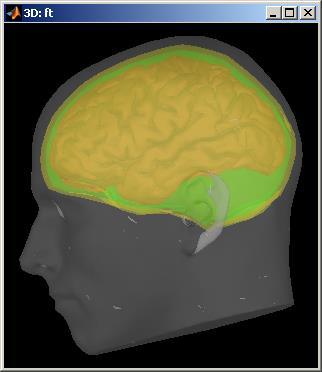
21 Source estimation Source space: cortex surface (or full head volume) Forward model = head model Sources => Sensors Inverse model: Minimum norm estimates Sensors => Sources Forward Inverse 21

22 Minimum Norm Noise covariance matrix Inverse model (minimum norm estimates) requires an estimation of the level of noise on the sensors Noise covariance matrix = covariance of the segments that do not contain any meaningful data Typically: empty room measures, or pre-stim baseline T 22

23 Source activity 23


24 Regions of interest Regions of interest at cortical level (scouts) = Subset of a few dipoles in the brain = Group of vertices of the cortex surface 24
25 Source estimation: MEG Recommended in MEG analysis: The subject head can move in the helmet One sensor is not corresponding to one brain region Different types of sensors (magneto / gradiometers) Difficult to read, reproduce or compare Converting to source space helps solving those issues 25
26 Multi-modal imaging Easy integration of: MEG EEG ECoG SEEG In a near future: NIRS 26


27 Spectral analysis Fast Fourier transform (FFT) Power spectrum density (PSD) 27


28 Time-frequency Morlet wavelets Hilbert transform + band-pass filter 28


29 Time-frequency 29

30 Time-frequency 30
31 Group analysis Normalization Registration of individual brains on a template Subject 1 Subject 2 Group analysis Average Contrast Individual anatomy Standard anatomy 31

32 Group analysis Subject Default anatomy FreeSurfer registration Subject anatomy Right hemisphere Shepard interpolation FreeSurfer registration Template Right hemisphere 32

33 Group analysis Statistics Contrasts between subjects or conditions Statistical analysis: z-score, t-test Quick extraction of measures from complex paradigms => Export to: SPM, R, Excel, Statistica, SPSS, Matlab 33

34 Connectivity Objectives: Describe the interaction between two brain regions, identify the brain networks Non-directed: Functional connectivity Correlation / Coherence Phase locking value Directed: Effective connectivity Granger causality Both at sensor and source levels 34


35 Connectivity 35


36 User support Online tutorials: 20-hour self-teaching program Active user forum: 150 posts/month Daily updates: 500 downloads/month Contact us for specific questions and requests: We will help you adding the features you need 36
37 Sample data Protocol Median nerve stimulation (Nov 2011, Montreal Neurological Institute, McGill) Random electric stimulation of both arms ~ 100 trials per arm Acquisition at 1200 Hz Recorded on CTF 275 MEG sensors + 26 reference sensors + EOG + ECG + STIM + = 302 channels 6 minutes of recordings, 500 Mb 37


38 Sample data Acquisition setup Polhemus Stim 3D points Stimulation computer MEG Acquisition computer 38
39 Morning To-do list Creation of a new protocol, with one subject Preparation of the anatomy (MRI, surfaces) Anatomical atlases Co-registration MRI / MEG Reviewing the continuous file Correcting for eye blinks with SSP Epoching and averaging Source estimation 39
40 Afternoon To-do list Regions of interest (scouts) Frequency analysis: FFT, time-frequency, Hilbert Functional connectivity Scripting interface Group analysis and statistics Registration on default anatomy 40


41 France Investigators Contributors USA McGill Key collaborators Sylvain Baillet MNI Elizabeth Bock MNI Alexandre Gramfort MGH / INRIA Richard Leahy USC Esther Florin MNI Dimitrios Pantazis MIT John Mosher Cleveland Clinic Francois Tadel MNI Rey Ramirez UW Ghislaine Dehaene Claude Delpuech Antoine Ducorps Line Garnero Etienne Labyt Karim N'Diaye Lauri Parkkonen Denis Schwartz Felix Darvas Belma Dogdas Guillaume Dumas John Ermer Matti Hamalainen Sheraz Khan Esen Kucukaltun-Yildirim Alexei Ossadtchi Darren Weber Sergül Aydore USC Syed Ashrafulla USC Sebastien Dery MNI 41
MEG and EEG analysis using
 MEG and EEG analysis using François Tadel Dalhousie University, Halifax February 16, 2013 Foretaste Graphic interface 2 Foretaste Scripting environment Rapid selection of files and processes to apply Automatic
MEG and EEG analysis using
 MEG and EEG analysis using François Tadel Biomag 2012 Paris August 31, 2012 Foretaste Graphic interface 2 Foretaste Scripting environment Rapid selection of files and processes to apply Automatic generation
Update ASA 4.8. Expand your research potential with ASA 4.8. Highly advanced 3D display of single channel coherence
 Update ASA 4.8 Expand your research potential with ASA 4.8. The ASA 4.8 software has everything needed for a complete analysis of EEG / ERP and MEG data. From features like (pre)processing of data, co-registration
Theory: modeling, localization and imaging
 Electromagnetic Brain Mapping with MEG/EEG Theory: modeling, localization and imaging Sylvain Baillet Imaging Group Cognitive Neuroscience & Brain Imaging Lab. Hôpital de la Salpêtrière CNRS UPR640 - LENA
- Graphical editing of user montages for convenient data review - Import of user-defined file formats using generic reader
 Data review and processing Source montages and 3D whole-head mapping Onset of epileptic seizure with 3D whole-head maps and hemispheric comparison of density spectral arrays (DSA) Graphical display of
BESA Research. CE certified software package for comprehensive, fast, and user-friendly analysis of EEG and MEG
 BESA Research CE certified software package for comprehensive, fast, and user-friendly analysis of EEG and MEG BESA Research choose the best analysis tool for your EEG and MEG data BESA Research is the
OHBA M/EEG Analysis Workshop. Mark Woolrich Diego Vidaurre Andrew Quinn Romesh Abeysuriya Robert Becker
 OHBA M/EEG Analysis Workshop Mark Woolrich Diego Vidaurre Andrew Quinn Romesh Abeysuriya Robert Becker Workshop Schedule Tuesday Session 1: Preprocessing, manual and automatic pipelines Session 2: Task
Source Reconstruction in MEG & EEG
 Source Reconstruction in MEG & EEG ~ From Brain-Waves to Neural Sources ~ Workshop Karolinska Institutet June 16 th 2017 Program for today Intro Overview of a source reconstruction pipeline Overview of
M/EEG pre-processing 22/04/2014. GUI Script Batch. Clarification of terms SPM speak. What do we need? Why batch?
 22/04/2014 Clarification of terms SPM speak GUI Script Batch M/EEG pre-processing Vladimir Litvak Wellcome Trust Centre for Neuroimaging UCL Institute of Neurology Why batch? What do we need? As opposed
ASA Getting Started. ANT BV, Enschede, Netherlands Advanced Neuro Technology
 ASA Getting Started ANT BV, Enschede, Netherlands Advanced Neuro Technology www.ant-neuro.com asa@ant-neuro.com TABLE OF CONTENTS DISCLAIMER... 3 NOTICE... 3 INTRODUCTION... 5 ASA-LAB... 6 ASA-LAB RECORDING
Beamformer Source Analysis in MEG
 Beamformer Source Analysis in MEG Douglas Cheyne, PhD Program in Neurosciences and Mental Health Hospital for Sick Children Research Institute & Department of Medical Imaging University of Toronto CCD
Managing custom montage files Quick montages How custom montage files are applied Markers Adding markers...
 AnyWave Contents What is AnyWave?... 3 AnyWave home directories... 3 Opening a file in AnyWave... 4 Quick re-open a recent file... 4 Viewing the content of a file... 5 Choose what you want to view and
ADJUST: An Automatic EEG artifact Detector based on the Joint Use of Spatial and Temporal features
 ADJUST: An Automatic EEG artifact Detector based on the Joint Use of Spatial and Temporal features A Tutorial. Marco Buiatti 1 and Andrea Mognon 2 1 INSERM U992 Cognitive Neuroimaging Unit, Gif sur Yvette,
ERPEEG Tutorial. Version 1.0. This tutorial was written by: Sravya Atluri, Matthew Frehlich and Dr. Faranak Farzan.
 ERPEEG Tutorial Version 1.0 This tutorial was written by: Sravya Atluri, Matthew Frehlich and Dr. Faranak Farzan. Contact: faranak.farzan@sfu.ca Temerty Centre for Therapeutic Brain Stimulation Centre
A comparison of minimum norm and MUSIC for a combined MEG/EEG sensor array
 Adv. Radio Sci., 10, 99 104, 2012 doi:10.5194/ars-10-99-2012 Author(s) 2012. CC Attribution 3.0 License. Advances in Radio Science A comparison of minimum norm and MUSIC for a combined MEG/EEG sensor array
Statistical Analysis of Neuroimaging Data. Phebe Kemmer BIOS 516 Sept 24, 2015
 Statistical Analysis of Neuroimaging Data Phebe Kemmer BIOS 516 Sept 24, 2015 Review from last time Structural Imaging modalities MRI, CAT, DTI (diffusion tensor imaging) Functional Imaging modalities
PREPROCESSING FOR ADVANCED DATA ANALYSIS
 PREPROCESSING FOR ADVANCED DATA ANALYSIS PSYC696B CHAP 7 JL SANGUINETTI Preprocessing Reorganization Transformation Data analysis Collecting EEG Organize Extracting epochs Adjusting event codes Removing
TMSEEG Tutorial. Version 4.0. This tutorial was written by: Sravya Atluri and Matthew Frehlich. Contact:
 TMSEEG Tutorial Version 4.0 This tutorial was written by: Sravya Atluri and Matthew Frehlich Contact: faranak.farzan@sfu.ca For more detail, please see the Method article describing the TMSEEG Toolbox:
This Time. fmri Data analysis
 This Time Reslice example Spatial Normalization Noise in fmri Methods for estimating and correcting for physiologic noise SPM Example Spatial Normalization: Remind ourselves what a typical functional image
New Approaches for EEG Source Localization and Dipole Moment Estimation. Shun Chi Wu, Yuchen Yao, A. Lee Swindlehurst University of California Irvine
 New Approaches for EEG Source Localization and Dipole Moment Estimation Shun Chi Wu, Yuchen Yao, A. Lee Swindlehurst University of California Irvine Outline Motivation why EEG? Mathematical Model equivalent
Introduction to fmri. Pre-processing
 Introduction to fmri Pre-processing Tibor Auer Department of Psychology Research Fellow in MRI Data Types Anatomical data: T 1 -weighted, 3D, 1/subject or session - (ME)MPRAGE/FLASH sequence, undistorted
Towards joint morphometry of white matter tracts and gray matter surfaces
 Towards joint morphometry of white matter tracts and gray matter surfaces P.Gori 1,2, O.Colliot 1,2, Y.Worbe 2, L.Marrakchi 1,2,3, S.Lecomte 1,2,3, C.Poupon 3, A.Hartmann 2, N.Ayache 4, and S.Durrleman
Modeling versus Accuracy in EEG and MEG Data
 Modeling versus Accuracy in EEG and MEG Data John C. Mosher, Richard M. Leahy, Ming-xiong Huang, Michael E. Spencer Los Alamos National Laboratory Group P- MS D Los Alamos, NM 7 mosher@lanl.gov, () -7
Automated MR Image Analysis Pipelines
 Automated MR Image Analysis Pipelines Andy Simmons Centre for Neuroimaging Sciences, Kings College London Institute of Psychiatry. NIHR Biomedical Research Centre for Mental Health at IoP & SLAM. Neuroimaging
CS/NEUR125 Brains, Minds, and Machines. Due: Wednesday, April 5
 CS/NEUR125 Brains, Minds, and Machines Lab 8: Using fmri to Discover Language Areas in the Brain Due: Wednesday, April 5 In this lab, you will analyze fmri data from an experiment that was designed to
NeuroBase: An Information System for Managing Distributed Knowledge and Data Bases in Neuroimaging. Christian BARILLOT DR CNRS
 NeuroBase: An Information System for Managing Distributed Knowledge and Data Bases in Neuroimaging Christian BARILLOT DR CNRS IRISA UMR 6074 CNRS, UR INRIA-Rennes Rennes Vista project Campus de Beaulieu
biomedical engineering program BIOSIGNAL ANALYSIS GUGER TECHNOLOGIES
 www.gtec.at R biomedical engineering program GUGER TECHNOLOGIES Multi-modal Off-line Biosignal Analysis under MATLAB Highlights intuitive Windows instruments for EEG, EOG, EMG, ECG data analysis and documentation
The BERGEN Plug-in for EEGLAB
 The BERGEN Plug-in for EEGLAB July 2009, Version 1.0 What is the Bergen Plug-in for EEGLAB? The Bergen plug-in is a set of Matlab tools developed at the fmri group, University of Bergen, Norway, which
i-eeg: A Software Tool for EEG Feature Extraction, Feature Selection and Classification
 i-eeg: A Software Tool for EEG Feature Extraction, Feature Selection and Classification Baha ŞEN Computer Engineering Department, Yıldırım Beyazıt University, Ulus, Ankara, TURKEY Musa PEKER Computer Engineering
MEG Laboratory Reference Manual for research use
 MEG Laboratory Reference Manual for research use for Ver. 1R007B Manual version 20040224 Index 1. File... 11 1.1 New... 11 1.2 Open... 11 1.3 Transfer...... 11 1.4 Suspended File List... 12 1.5 Save...
Spatial Filtering Methods in MEG. Part 3: Template Normalization and Group Analysis"
 Spatial Filtering Methods in MEG Part 3: Template Normalization and Group Analysis" Douglas Cheyne, PhD" Program in Neurosciences and Mental Health" Hospital for Sick Children Research Institute " &" Department
The NeuroLOG Platform Federating multi-centric neuroscience resources
 Software technologies for integration of process and data in medical imaging The Platform Federating multi-centric neuroscience resources Johan MONTAGNAT Franck MICHEL Vilnius, Apr. 13 th 2011 ANR-06-TLOG-024
Topographic Mapping with fmri
 Topographic Mapping with fmri Retinotopy in visual cortex Tonotopy in auditory cortex signal processing + neuroimaging = beauty! Topographic Mapping with fmri Retinotopy in visual cortex Tonotopy in auditory
AFNI Preprocessing: Outline, Recommendations, and New(ish) Stuff. Robert W Cox SSCC / NIMH & NINDS / NIH / DHHS / USA / EARTH
 Stuff. Robert W Cox SSCC / NIMH & NINDS / NIH / DHHS / USA / EARTH") AFNI Preprocessing: Outline, Recommendations, and New(ish) Stuff Robert W Cox SSCC / NIMH & NINDS / NIH / DHHS / USA / EARTH HBM 2016 As a work of a US Government official, this presentation is not copyrighted
AFNI Preprocessing: Outline, Recommendations, and New(ish) Stuff Robert W Cox SSCC / NIMH & NINDS / NIH / DHHS / USA / EARTH HBM 2016 As a work of a US Government official, this presentation is not copyrighted
MEG & PLS PIPELINE: SOFTWARE FOR MEG DATA ANALYSIS AND PLS STATISTICS
 MEG & PLS PIPELINE: SOFTWARE FOR MEG DATA ANALYSIS AND PLS STATISTICS USER DOCUMENTATION VERSION: 2.00 SEPT. 16, 2014 MEG & PLS Pipeline ([MEG]PLS) Copyright 2013-2014, Michael J. Cheung & Natasa Kovacevic
Journal of Articles in Support of The Null Hypothesis
 Data Preprocessing Martin M. Monti, PhD UCLA Psychology NITP 2016 Typical (task-based) fmri analysis sequence Image Pre-processing Single Subject Analysis Group Analysis Journal of Articles in Support
Tutorial BOLD Module
 m a k i n g f u n c t i o n a l M R I e a s y n o r d i c B r a i n E x Tutorial BOLD Module Please note that this tutorial is for the latest released nordicbrainex. If you are using an older version please
Research Article EEG and MEG Data Analysis in SPM8
 Hindawi Publishing Corporation Computational Intelligence and Neuroscience Volume 211, Article ID 852961, 32 pages doi:1.1155/211/852961 Research Article EEG and MEG Data Analysis in SPM8 Vladimir Litvak,
Digital EEG System Series
 NEURON-SPECTRUM Digital System Series rom 8 to 21 channels up to 4 channels to record EMG, ECG, EOG, etc. Acquisition and review of in arbitrary monopolar and bipolar montages, EP, PSG acquisition Impedance/acquisition
Performing real-time BCI experiments
 Performing real-time BCI experiments g.tec medical engineering GmbH Herbersteinstr. 60 8020 Graz, Austria www.gtec.at guger@gtec.at g.usbamp is a biosignal acquisition system for EEG, ECG, EMG, EOG and
Functional MRI data preprocessing. Cyril Pernet, PhD
 Functional MRI data preprocessing Cyril Pernet, PhD Data have been acquired, what s s next? time No matter the design, multiple volumes (made from multiple slices) have been acquired in time. Before getting
fmri Preprocessing & Noise Modeling
 Translational Neuromodeling Unit fmri Preprocessing & Noise Modeling Lars Kasper September 25 th / October 17 th, 2015 MR-Technology Group & Translational Neuromodeling Unit An SPM Tutorial Institute for
Classification of Mental Task for Brain Computer Interface Using Artificial Neural Network
 Classification of Mental Task for Brain Computer Interface Using Artificial Neural Network Mohammad Naushad 1, Mohammad Waseem Khanooni 2, Nicky Ballani 3 1,2,3 Department of Electronics and Telecommunication
Supplementary methods
 Supplementary methods This section provides additional technical details on the sample, the applied imaging and analysis steps and methods. Structural imaging Trained radiographers placed all participants
NIH Public Access Author Manuscript Neuroimage. Author manuscript; available in PMC 2010 February 1.
 NIH Public Access Author Manuscript Published in final edited form as: Neuroimage. 2009 February 1; 44(3): 932 946. doi:10.1016/j.neuroimage.2008.05.063. A Distributed Spatio-Temporal EEG/MEG Inverse Solver
Estimating Noise and Dimensionality in BCI Data Sets: Towards Illiteracy Comprehension
 Estimating Noise and Dimensionality in BCI Data Sets: Towards Illiteracy Comprehension Claudia Sannelli, Mikio Braun, Michael Tangermann, Klaus-Robert Müller, Machine Learning Laboratory, Dept. Computer
Diffusion-MRI processing for group analysis
 Diffusion-MRI processing for group analysis Felix Renard IRMaGe: Inserm US 17 / CNRS UMS 3552 University Hospital of Grenoble - France 25/09/2015 felixrenard@gmail.com 1 Diffusion-MRI processing for group
SPM Introduction. SPM : Overview. SPM: Preprocessing SPM! SPM: Preprocessing. Scott Peltier. FMRI Laboratory University of Michigan
 SPM Introduction Scott Peltier FMRI Laboratory University of Michigan! Slides adapted from T. Nichols SPM! SPM : Overview Library of MATLAB and C functions Graphical user interface Four main components:
Automatic Generation of Training Data for Brain Tissue Classification from MRI
 Automatic Generation of Training Data for Brain Tissue Classification from MRI Chris A. COCOSCO, Alex P. ZIJDENBOS, and Alan C. EVANS http://www.bic.mni.mcgill.ca/users/crisco/ McConnell Brain Imaging
Basic Introduction to Data Analysis. Block Design Demonstration. Robert Savoy
 Basic Introduction to Data Analysis Block Design Demonstration Robert Savoy Sample Block Design Experiment Demonstration Use of Visual and Motor Task Separability of Responses Combined Visual and Motor
SPM Introduction SPM! Scott Peltier. FMRI Laboratory University of Michigan. Software to perform computation, manipulation and display of imaging data
 SPM Introduction Scott Peltier FMRI Laboratory University of Michigan Slides adapted from T. Nichols SPM! Software to perform computation, manipulation and display of imaging data 1 1 SPM : Overview Library
USER S DOCUMENTATION v.3.4
 USER S DOCUMENTATION v.3.4 BrainWave 2018 Hospital for Sick Children, Toronto, Ontario http://cheynelab.utoronto.ca Table of Contents VERSION UPDATES... 4 GETTING STARTED... 5 INTRODUCTION... 5 SYSTEM
Analysis of fmri data within Brainvisa Example with the Saccades database
 Analysis of fmri data within Brainvisa Example with the Saccades database 18/11/2009 Note : All the sentences in italic correspond to informations relative to the specific dataset under study TP participants
Task Level Hierarchical System for BCI-enabled Shared Autonomy Iretiayo Akinola, Boyuan Chen, Jonathan Koss, Aalhad Patankar, Jake Varley & Peter
 Task Level Hierarchical System for BCI-enabled Shared Autonomy Iretiayo Akinola, Boyuan Chen, Jonathan Koss, Aalhad Patankar, Jake Varley & Peter Allen Columbia University Shared Autonomy Agent 1 Agent
USER S DOCUMENTATION v.3.1
 USER S DOCUMENTATION v.3.1 BrainWave 2015 Hospital for Sick Children, Toronto, Ontario http://cheynelab.utoronto.ca Table of Contents GETTING STARTED... 4 INTRODUCTION... 4 SYSTEM REQUIREMENTS... 5 DISCLAIMER
Multi-atlas labeling with population-specific template and non-local patch-based label fusion
 Multi-atlas labeling with population-specific template and non-local patch-based label fusion Vladimir Fonov, Pierrick Coupé, Simon Eskildsen, Jose Manjon, Louis Collins To cite this version: Vladimir
BioEST ver. 1.5beta User Manual Chang-Hwan Im (Ph.D.)
") BioEST ver. 1.5beta User Manual Chang-Hwan Im (Ph.D.) http://www.bioest.com E-mail: ichism@elecmech.snu.ac.kr imxxx010@umn.edu 1. What is BioEST? BioEST is a special edition of SNUEEG(http://www.bioinverse.com),
BioEST ver. 1.5beta User Manual Chang-Hwan Im (Ph.D.) http://www.bioest.com E-mail: ichism@elecmech.snu.ac.kr imxxx010@umn.edu 1. What is BioEST? BioEST is a special edition of SNUEEG(http://www.bioinverse.com),
Granger Processing Stream
 MASSACHUSETTS GENERAL HOSPITAL Granger Processing Stream Manual for version 1.9 A. Conrad Nied, Bruna Olson, David Gow 2/12/2014 This document outlines the purpose and operation of the Graphical Processing
MNE software version 2.6. Matti Hämäläinen. MGH/MIT/HMS Athinoula A. Martinos Center for Biomedical Imaging Charlestown, MA USA
 MNE software version 2.6 Matti Hämäläinen MGH/MIT/HMS Athinoula A. Martinos Center for Biomedical Imaging Charlestown, MA USA Overall changes Manual: description of the MEG/EEG and MRI coordinate systems
Medical Imaging Introduction
 Medical Imaging Introduction Jan Kybic February 16, 2010 Medical imaging: a collaborative paradigm picture from Atam P. Dhawan: Medical Imaging From physiology to information processing (what we should
User s Documentation 2.3. BrainWave v User s Documentation -- Toronto, Canada
 User s Documentation 2.3 BrainWave v2.2 2013 -- User s Documentation -- Toronto, Canada http://cheynelab.utoronto.ca Table of Contents Getting Started... 5 Introduction... 5 System Requirements... 6 Disclaimer
Section 9. Human Anatomy and Physiology
 Section 9. Human Anatomy and Physiology 9.1 MR Neuroimaging 9.2 Electroencephalography Overview As stated throughout, electrophysiology is the key tool in current systems neuroscience. However, single-
USER S DOCUMENTATION v.3.0 (beta)
") USER S DOCUMENTATION v.3.0 (beta) BrainWave 2015 Toronto, Ontario http://cheynelab.utoronto.ca Table of Contents GETTING STARTED... 4 INTRODUCTION... 4 SYSTEM REQUIREMENTS... 5 DISCLAIMER & LICENSE...
USER S DOCUMENTATION v.3.0 (beta) BrainWave 2015 Toronto, Ontario http://cheynelab.utoronto.ca Table of Contents GETTING STARTED... 4 INTRODUCTION... 4 SYSTEM REQUIREMENTS... 5 DISCLAIMER & LICENSE...
ME scope Application Note 42 Multi-Input Multi-Output (MIMO) FRFs
 FRFs") ME scope Application Note 42 Multi-Input Multi-Output (MIMO) FRFs NOTE: This Application Note uses commands in the VES-3600 Advanced Signal Processing Check Help About to verify authorization of these
ME scope Application Note 42 Multi-Input Multi-Output (MIMO) FRFs NOTE: This Application Note uses commands in the VES-3600 Advanced Signal Processing Check Help About to verify authorization of these
arxiv: v1 [q-bio.nc] 17 Apr 2017
![arxiv: v1 [q-bio.nc] 17 Apr 2017](/thumbs/92/107994755.jpg "arxiv: v1 [q-bio.nc] 17 Apr 2017") EEG SOURCE IMAGING ASSISTS DECODING IN A FACE RECOGNITION TASK arxiv:1704.05748v1 [q-bio.nc] 17 Apr 2017 Rasmus S. Andersen, Anders U. Eliasen, Nicolai Pedersen, Michael Riis Andersen, Sofie Therese Hansen,
EEG SOURCE IMAGING ASSISTS DECODING IN A FACE RECOGNITION TASK arxiv:1704.05748v1 [q-bio.nc] 17 Apr 2017 Rasmus S. Andersen, Anders U. Eliasen, Nicolai Pedersen, Michael Riis Andersen, Sofie Therese Hansen,
Basic principles of MR image analysis. Basic principles of MR image analysis. Basic principles of MR image analysis
 Basic principles of MR image analysis Basic principles of MR image analysis Julien Milles Leiden University Medical Center Terminology of fmri Brain extraction Registration Linear registration Non-linear
White Pixel Artifact. Caused by a noise spike during acquisition Spike in K-space <--> sinusoid in image space
 White Pixel Artifact Caused by a noise spike during acquisition Spike in K-space sinusoid in image space Susceptibility Artifacts Off-resonance artifacts caused by adjacent regions with different
PC based EEG mapping system
 PC based EEG mapping system Piotr Walerjan and Remigiusz Tarnecki Department of Neurophysiology, Nencki Institute of Experimental Biology, 3 Pasteur St., 02-093 Warsaw, Poland, email: piotrwa@nencki.gov.pl.
Clinical Importance. Aortic Stenosis. Aortic Regurgitation. Ultrasound vs. MRI. Carotid Artery Stenosis
 Clinical Importance Rapid cardiovascular flow quantitation using sliceselective Fourier velocity encoding with spiral readouts Valve disease affects 10% of patients with heart disease in the U.S. Most
BioTrace+ What s new V2013
 BioTrace+ What s new V2013 An overview of the new functionality of BioTrace+ V2013 Contents About this document... 2 1. Improved HRV functionality... 3 Resonant frequency calculation... 3 Load and save
SPM8 for Basic and Clinical Investigators. Preprocessing. fmri Preprocessing
 SPM8 for Basic and Clinical Investigators Preprocessing fmri Preprocessing Slice timing correction Geometric distortion correction Head motion correction Temporal filtering Intensity normalization Spatial
SENSING A WORLD OF POTENTIAL TM. Dry Sensor Interface DSI 10/20. Simple Ambulatory No-prep Dry EEG System
 SENSING A WORLD OF POTENTIAL TM Dry Sensor Interface DSI 10/20 Simple Ambulatory No-prep Dry EEG System SENSING A WORLD OF POTENTIAL TM The Dry Sensor Interface s SANDTechnology Simple: Headset is typically
Deformetrica: a software for statistical analysis of anatomical shapes
 Deformetrica: a software for statistical analysis of anatomical shapes Alexandre Routier, Marcel Prastawa, Benjamin Charlier, Cédric Doucet, Joan Alexis Glaunès, Stanley Durrleman To cite this version:
-1- Accepted manuscript
 -1- Accepted manuscript Toda A, Imamizu H, Kawato M, Sato MA. (2011). Reconstruction of two-dimensional movement trajectories from selected magnetoencephalography cortical currents by combined sparse Bayesian
Rapidly recomputable EEG forward models for realistic head shapes
 INSTITUTE OF PHYSICS PUBLISHING PHYSICS IN MEDICINE AND BIOLOGY Phys. Med. Biol. 46 (2001) 1265 1281 www.iop.org/journals/pb PII: S0031-9155(01)19141-4 Rapidly recomputable EEG forward models for realistic
Adapting to Non-Stationarity in EEG using a Mixture of Multiple ICA Models
 Adapting to Non-Stationarity in EEG using a Mixture of Multiple ICA Models Jason A. Palmer 1 Scott Makeig 1 Julie Onton 1 Zeynep Akalin-Acar 1 Ken Kreutz-Delgado 2 Bhaskar D. Rao 2 1 Swartz Center for
2012 IEEE. Personal use of this material is permitted. Permission from IEEE must be obtained for all other uses, in any current or future media,
 2012 IEEE. Personal use of this material is permitted. Permission from IEEE must be obtained for all other uses, in any current or future media, including reprinting/republishing this material for advertising
BrainNet Viewer Manual. Mingrui Xia National Key Laboratory of Cognitive Neuroscience and Learning, Beijing Normal University 7/11/2011
 2011 BrainNet Viewer Manual Mingrui Xia National Key Laboratory of Cognitive Neuroscience and Learning, Beijing Normal University 7/11/2011 Contents 1 Introduction... 2 2 Installation... 3 2.1 Run BrainNet
Neuroimaging and mathematical modelling Lesson 2: Voxel Based Morphometry
 Neuroimaging and mathematical modelling Lesson 2: Voxel Based Morphometry Nivedita Agarwal, MD Nivedita.agarwal@apss.tn.it Nivedita.agarwal@unitn.it Volume and surface morphometry Brain volume White matter
NA-MIC National Alliance for Medical Image Computing fmri Data Analysis
 NA-MIC fmri Data Analysis Sonia Pujol, Ph.D. Wendy Plesniak, Ph.D. Randy Gollub, M.D., Ph.D. Acknowledgments NIH U54EB005149 Neuroimage Analysis Center NIH P41RR013218 FIRST Biomedical Informatics Research
The organization of the human cerebral cortex estimated by intrinsic functional connectivity
 1 The organization of the human cerebral cortex estimated by intrinsic functional connectivity Journal: Journal of Neurophysiology Author: B. T. Thomas Yeo, et al Link: https://www.ncbi.nlm.nih.gov/pubmed/21653723
A P300-speller based on event-related spectral perturbation (ERSP) Ming, D; An, X; Wan, B; Qi, H; Zhang, Z; Hu, Y
 Ming, D; An, X; Wan, B; Qi, H; Zhang, Z; Hu, Y") Title A P300-speller based on event-related spectral perturbation (ERSP) Author(s) Ming, D; An, X; Wan, B; Qi, H; Zhang, Z; Hu, Y Citation The 01 IEEE International Conference on Signal Processing, Communication
Title A P300-speller based on event-related spectral perturbation (ERSP) Author(s) Ming, D; An, X; Wan, B; Qi, H; Zhang, Z; Hu, Y Citation The 01 IEEE International Conference on Signal Processing, Communication
Structural MRI analysis
 Structural MRI analysis volumetry and voxel-based morphometry cortical thickness measurements structural covariance network mapping Boris Bernhardt, PhD Department of Social Neuroscience, MPI-CBS bernhardt@cbs.mpg.de
A Low-Rank + Sparse Decomposition (LR+SD) Method for Automatic EEG Artifact Removal
 Method for Automatic EEG Artifact Removal") A Low-Rank + Sparse Decomposition (LR+SD) Method for Automatic EEG Artifact Removal J. Gilles 1, T.Meyer 1, and P.K. Douglas, 1 Department of Mathematics, University of California, Los Angeles,CA 90095,
A Low-Rank + Sparse Decomposition (LR+SD) Method for Automatic EEG Artifact Removal J. Gilles 1, T.Meyer 1, and P.K. Douglas, 1 Department of Mathematics, University of California, Los Angeles,CA 90095,
Chapter 3 Set Redundancy in Magnetic Resonance Brain Images
 16 Chapter 3 Set Redundancy in Magnetic Resonance Brain Images 3.1 MRI (magnetic resonance imaging) MRI is a technique of measuring physical structure within the human anatomy. Our proposed research focuses
Function-Structure Integration in FreeSurfer
 Function-Structure Integration in FreeSurfer Outline Function-Structure Integration Function-Structure Registration in FreeSurfer fmri Analysis Preprocessing First-Level Analysis Higher-Level (Group) Analysis
Improved MEG/EEG source localization with reweighted mixed-norms
 Improved MEG/EEG source localization with reweighted mixed-norms Daniel Strohmeier, Jens Haueisen, Alexandre Gramfort To cite this version: Daniel Strohmeier, Jens Haueisen, Alexandre Gramfort. Improved
RS SigEdit A module of RS LabSite Advanced Graphical Display and Editing
 RS SigEdit A module of RS LabSite Advanced Graphical Display and Editing Expanding your Signal Editing Capabilities The RS LabSite suite of software offers two applications for data viewing and editing,
The Artifact Subspace Reconstruction method. Christian A Kothe SCCN / INC / UCSD January 2013
 The Artifact Subspace Reconstruction method Christian A Kothe SCCN / INC / UCSD January 2013 Artifact Subspace Reconstruction New algorithm to remove non-stationary highvariance signals from EEG Reconstructs
Users Guide: (Hemodynamic Evoked Response)
") HomER @ Users Guide: (Hemodynamic Evoked Response) Developed by T. Huppert and D. Boas A.A. Martinos Center for Neurological Imaging Massachusetts General Hospital. Charlestown, MA Copyright 2003 MGH Questions
HomER @ Users Guide: (Hemodynamic Evoked Response) Developed by T. Huppert and D. Boas A.A. Martinos Center for Neurological Imaging Massachusetts General Hospital. Charlestown, MA Copyright 2003 MGH Questions
ThE ultimate, INTuITIVE Mr INTErFAcE
 ThE ultimate, INTuITIVE Mr INTErFAcE Empowering you to do more The revolutionary Toshiba M-power user interface takes Mr performance and flexibility to levels higher than ever before. M-power is able to
The iworx 214 and LabScribe V2.0 Tutorial. Overview
 The iworx 214 and LabScribe V2.0 Overview Figure T-1-1: The front and rear panels of IWX/214. The data acquisition unit used in the iworx teaching kits is the IWX/214 (Figure T-1-1 on page T-1-1). The
Functional MRI in Clinical Research and Practice Preprocessing
 Functional MRI in Clinical Research and Practice Preprocessing fmri Preprocessing Slice timing correction Geometric distortion correction Head motion correction Temporal filtering Intensity normalization
Preprocessing II: Between Subjects John Ashburner
 Preprocessing II: Between Subjects John Ashburner Pre-processing Overview Statistics or whatever fmri time-series Anatomical MRI Template Smoothed Estimate Spatial Norm Motion Correct Smooth Coregister
Accelerated MRI Techniques: Basics of Parallel Imaging and Compressed Sensing
 Accelerated MRI Techniques: Basics of Parallel Imaging and Compressed Sensing Peng Hu, Ph.D. Associate Professor Department of Radiological Sciences PengHu@mednet.ucla.edu 310-267-6838 MRI... MRI has low
Identification of the Language Dominant Hemisphere using MEG Cortical Imaging and Permutation Tests
 Identification of the Language Dominant Hemisphere using MEG Cortical Imaging and Permutation Tests DIMITRIO PANTAZI 1 WARREN MERRIFIELD 2 FELIX DARVA 1 WILLIAM UTHERLING 2 RICHARD M. LEAHY 1 1 ignal and
Copyright Notice. Do not remove this notice. COMMONWEALTH OF AUSTRALIA Copyright Regulations 1969 WARNING
 Copyright Notice University of South Australia (UniSA) Do not remove this notice. COMMONWEALTH OF AUSTRALIA Copyright Regulations 969 WARNING This material has been produced and communicated to you by
MEDICAL IMAGE ANALYSIS
 SECOND EDITION MEDICAL IMAGE ANALYSIS ATAM P. DHAWAN g, A B IEEE Engineering in Medicine and Biology Society, Sponsor IEEE Press Series in Biomedical Engineering Metin Akay, Series Editor +IEEE IEEE PRESS
Single Subject Demo Data Instructions 1) click "New" and answer "No" to the "spatially preprocess" question.
 click New and answer No to the spatially preprocess question.") (1) conn - Functional connectivity toolbox v1.0 Single Subject Demo Data Instructions 1) click "New" and answer "No" to the "spatially preprocess" question. 2) in "Basic" enter "1" subject, "6" seconds
(1) conn - Functional connectivity toolbox v1.0 Single Subject Demo Data Instructions 1) click "New" and answer "No" to the "spatially preprocess" question. 2) in "Basic" enter "1" subject, "6" seconds
Last Time. This Time. Thru-plane dephasing: worse at long TE. Local susceptibility gradients: thru-plane dephasing
 Motion Correction Last Time Mutual Information Optimiation Decoupling Translation & Rotation Interpolation SPM Example (Least Squares & MI) A Simple Derivation This Time Reslice example SPM Example : Remind