4/19/2016. Deborah Thames R.T. (R)(M)(QM) Theory & Technology and advancement in 3D imaging DBT
|
|
|
- Bruno Shaw
- 6 years ago
- Views:
Transcription

1 Deborah Thames R.T. (R)(M)(QM) Theory & Technology and advancement in 3D imaging DBT 1


2 Three manufacturers approved for Tomo Hologic and GE, and Siemens Why 2D Digital Mammography 2D FFDM it appears to be slightly more sensitive than digital breast tomosynthesis for the detection of calcification. Diagnostic performance as measured by area under the curve using BI-RADS was not significantly different. With improvements in processing algorithms and display, digital breast tomosynthesis could potentially be improved for this purpose. Mammography Mammography, in particular digital mammography, is the gold standard in breast cancer screening. 2


3 Mammography Screening Requirements for the United States Systems must be capable of: Imaging the whole breast Image all types of breasts Image all lesion types, mass, calcification, distortion Fast and reasonable cost Low radiation dose Hologic Proprietary Information for Training Purposes Only MED Potential Benefits and why we need 3D imaging Reduce recall rate of patients by reducing confusion which arises from tissue overlap. Biopsy rate decreased as there is improvement in separation and visualization of parenchymal structures. Time will possibly show improvement in cancer detection particularly in patients with dense breast tissue. Fewer images required for diagnosis equals a reduction in dose. Compression is a must at this time. NO changes yet but in future may be possible. Why Breast Tomosynthesis? Breast tomosynthesis provides a 3D imaging capability that allows the more accurate evaluation of lesions by enabling better differentiation between overlapping tissues. A lower recall rate, higher positive predictive value for a biopsy recommendation. Higher cancer detection rates, fewer recalls, fewer biopsies, and improved radiologist confidence are expected to result from the use of this technology. Breast tomosynthesis should be valuable in both screening mammography and diagnostic mammography. 3


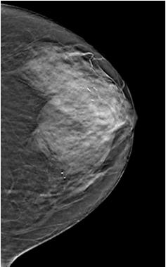
4 Why is There a Need for 3D Tomosynthesis In 2D FFDM: Tissue superimposition hides pathologies in 2D Tissue superimposition mimics pathologies in 2D Why Breast Tomosynthesis? Tomosynthesis should resolve many of the tissue overlap reading problems that are a major source of the need for recalls and additional imaging in 2D mammography exams. The biopsy rate might also decrease through improved visualization of suspect objects. Projections are the basis of the displayed Slices. While stabilizing the breast, images are acquired at a number of different x-ray source angles. Objects at different heights in the breast display differently in the all projections. Some pathologies that are mammographically occult will be discernible through the elimination of structure noise and tomosynthesis may therefore allow improved detection of cancers. Tomosynthesis 3D Breast Tomosynthesis Arc of motion of x-ray tube, showing individual exposures Digital mammography provides images with improved dynamic range and SNR, as well as the ability to adjust image brightness and contrast after acquisition. Despite these improvements it is limited in the same manner as film/screen due to superimposition in a 2D image. A 3D screening modality that preserves the very high resolution of 2D FFDM Multiple images of the breast are acquired at different angles during a sweep of the x-ray tube Allows radiologists to see around overlapping structures Compression Paddle Reconstructed Slices { Compressed Breast Detector Housing 4


5 Why Digital Breast Tomosynthesis (3D)? 3D improves visibility by reducing tissue superimposition Two objects (a spiculated lesion and ellipse) superimpose when the x-rays are at 0º, but the off-axis acquisitions shift the objects shadows relative to one another in the images. Note that additional acquisitions are not required to enhance the visibility of objects at any given height one set of acquired data can be reprocessed to generate the entire 3D volume set. Think of it as Raisin Breast The final step in the tomosynthesis procedure is reconstructing the data to generate images that enhance objects from a given height by appropriate shifting of the projections relative to one another. 5


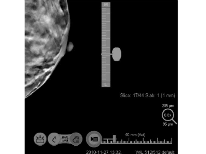
6 Calcifications Example case with Calcs Slabbing May not perceive calcifications as a cluster Radiologist have ability to slab information Look at a 10mm slab vs a 1mm slice 6


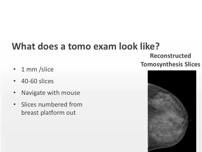
7 2D Imaging vs. 3D Imaging 2D Either molybdenum or tungsten x-ray tube 20 to 39 kvp Moly or rhodium or silver filters 100 ma HTC grid 3D Tungsten x-ray tube 20 to 49 kvp Aluminum filter 200 ma No grid No GRID-when the tube is off axis you would see grid lines) Breast Tomothynthesis The 3D image quality and depth resolution directly depend on the number of projections, angle size and reconstruction algorithm. The Solution is 3D Breast Tomosynthesis How Does Hologic s 3D Breast Tomosynthesis Work? Tomosynthesis is a three-dimensional mammographic examination that can minimize the effects of structure overlap within the breast Tube moves in a 15 º arc 15 low dose images are acquired 1 image at each degree Four second sweep Images are reconstructed into 1 mm slices In combo-mode imaging, the 2D and 3D are taken in the same compression, with no additional positioning for the patient. 7
 Arc of motion of x-ray tube, showing individual exposures Projection image acquired during sweep Images are reconstructed into 1 mm")
8 How Does GE Senoclaire Tomosynthesis work Tube moves in a 25 º arc 9 low dose images are acquired 1 image at 2.8 degree 3D Tomosynthesis Dataset: 2D + 3D (Combo Acquisition) Arc of motion of x-ray tube, showing individual exposures Projection image acquired during sweep Images are reconstructed into 1 mm slices In combo-mode imaging, the 2D and 3D are taken in the same compression, with no additional positioning for the patient. How Does Siemens Inspiration Tomosynthesis work Tube moves in a 50 º arc 25 low dose images are acquired 1 image at 2.0 degree Images are reconstructed into 1 mm slices In combo-mode imaging, the 2D and 3D are taken in the same compression, with no additional positioning for the patient. Projection Image Must have projection image to create slices Reconstruction images are born of projection image Projection image is checked for motion by technologist Slices are from detector to paddle in all views. Example CC-foot to head MLO-lateral to medial Performing the Acquisition The breast is compressed in a standard way. While holding the breast stationary, the x-ray tube is rotated over a limited angular range. A series of low dose exposures are made every degree, creating a series of digital images. Tomosynthesis Dataset: 2D/3D (Combo Acquisition) Finishing with the 2D exposure 8

 Projection Images 15 images/15 0 arc Reconstructed Two Dimensional Tomosynthesis Slices Mammogram Projection Image Angle of")
9 Angular Range 1mm slices: Number of slices dependent upon compressed breast thickness that are reconstructed from projection images 5cm compressed breast 50 1mm slices + 5 Always adds 5 to clear the paddle Larger angular range gives increased reconstructed slice separation Smaller angular ranges keep more structures in focus in a given slice It might be desired for resolving closely lying structures but could impair the appreciation of a cluster of microcalcifications because the individual calcifications would appear in different slices or the appearance of spiculations lying in more than one narrow plane Tomosynthesis Dataset: 2D + 3D (Combo Acquisition) Projection Images 15 images/15 0 arc Reconstructed Two Dimensional Tomosynthesis Slices Mammogram Projection Image Angle of View October 2011 The x-ray tube can move in a continuous or step-and-shoot motion. With continuous motion x-ray exposures must be short enough to avoid image blurring due to focal spot motion If step-and-shoot motion is employed, the gantry must come to a complete stop at each angular location before turning on the x-rays, otherwise vibration will blur the image Hologic has continuous motion Siemens Tomosynthesis With True 3D Breast Tomosynthesis, Siemens has opened up a new chapter in mammography diagnostics. High spatial resolution and the largest acquisition angle allow for excellent resolution depth and superior reconstruction results. This leads to fewer artifacts and greater image detail, thereby improving diagnostic capabilities immensely 9


10 Modes of Acquisition Display The unit must perform existing 2D digital mammography images Tomosynthesis images must be able to be taken in all standard projections not just the CC and MLO Take a normal 2D mammogram and tomosynthesis image in the same compression Similar to CT reconstructed slices View one at a time or display as a cine loop 2D images can also be viewed 2D and 3D acquired in the same compression are completely co-registered Image Reconstruction Image reconstruction is computing high-resolution images whose planes are parallel to the breast support plates Reconstructed with slice separation of 1mm A 5cm compressed breast tomosynthesis study will have 55 reconstructed slices Reconstruction time must be 10 seconds or less Co-Registration The positioning and compression are exactly the same on the 2D and 3D allows the radiologist to view the 2D image and the tomosynthesis slices with perfect co-registration on top of each other or side by side. Image Reconstruction Co-registered: same positioning 2D and 3D 2D image 10
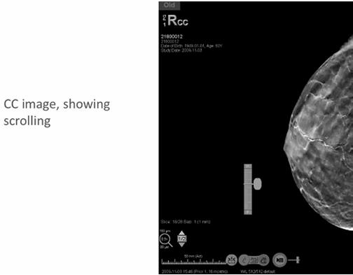
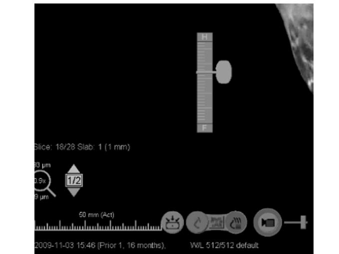

11 Co-registered Tomosynthesis image Scrolling 11


12 Co-registration of 2D & 3D breast images For Technologists 2D and 3D scan acquired with a single positioning/view Workflow is same as FFDM; no learning curve For Patients The clinical trials proved a reduction in recall rates. Reduced recalls can lead to reduced anxiety & reduced inconvenience No noticeable difference in breast screening experience RADIOLOGIST READ TIME Equated to going from film screen to digital Now digital 2D to 3D-same learning curve CAD is applied to 2D images and then compared to slices what was marked on CAD Co-registration of 2D & 3D breast images Visibility of low contrast objects are reduced Even at 4x a conventional dose the digital mammogram (middle) shows inferior low contrast visibility to a tomosynthesis (right) using ¼ the dose For Radiologists Facilitates comparison to priors and images from other facilities Single compression allows co-registration of 2D and 3D images For Administration Reimbursement is available for 2D image and 2D CAD, allowing the facility to continue to generate revenue while offering latest technology Patient throughput is not impacted ACR PHANTOM WITH CADAVER BREAST 12


13 3D Breast Tomosynthesis System X-ray tube swings in arc during 3D scan Preview images displayed on AWS Stationary breast platform The Future of Breast Imaging X-ray tube swings in arc during 3D scan Preview images displayed on AWS What is co-registration? Stationary breast platform Positioning and compression are exactly the same on the 2D and 3D Allows the radiologist to view the 2D image and the tomosynthesis slices with perfect co-registration on top of each other or side by side. X-ray tube swings in arc during 3D scan Stationary breast platform What is the projection image? The images acquired during the tomo sweep These images are used to create the individual slices The tomo slices are born of the projection image Preview images displayed on AWS 13


 plus 2D 1mm slices")
 plus 2D")


14 How many tomo slices are there in a routine tomo acquisition? Depends on the compressed breast thickness Eliminating Superimposition by Reading the Slice(s) plus 2D 1mm slices plus 5 to clear the paddle Eliminating Superimposition by Reading the Slice(s) plus 2D What is the dual acquisition mode called? Combo-mode Tomosynthesis DBT Normal mammogram Normal mammogram 14


15 Tomosynthesis Dataset: 2D/3D (Combo Acquisition) Finishing with the 2D exposure LCC A suspicious area in a 2D Mammography Image 2D October Clinical Image Review The 2D Mammography Image next to one slice of a 3D Image Set Why is tomosynthesis going to revolutionize breast imaging? Images and data courtesy of: Hôpital Privé d Antony, Paris France Massachusetts General Hospital, Boston MA USA Netherlands Cancer Institute Antoni Van Leeuwenhoek Hospital, Amsterdam Holland Centre de Radiologie et d Echographie du Docteur Joussier, Paris France Dartmouth Hitchcock Medical Center, Lebanon NH USA Magee Women s Hospital, Pittsburgh PA USA 2D 3D LCC A 2D Mammography Image Example 2 2D 15


16 A 2D Mammography Image with RMLO a suspicious area identified and blown up A 2D Mammography Image with a suspicious area identified next to a 3D image set RCC RCC 2D 2D 3D The 2D Mammography Image next to one slice of a 3D Image Set Slice D 3D As you go thru the image set, you see that the suspicious area is nothing more than normal breast structures overlapping More Examples Example 3 Superimposed Tissue Examples 16


17 Tomosynthesis: Concepts and Rationale Online November 2011 University of Pittsburg Medical Center Dr. Jules Sumkin Dr. Margarita Zuley FFDM vs F/S So why is digital better Dynamic range Contrast resolution Can change image appearance and size at soft copy workstation Why digital is not always better. Processing algorithms may play a role Reduced spatial resolution lp numbers are lower Learning curve - interpretation 17

18 Screening with tomo Reduce recall rate Up to 2 times the dose if do combo Calcifications and Tomo Confident improvement will be made and will be able to see calcifications as well as on FFDM Screening with Tomo Diagnostic Because of the demonstrated improvement in the ROC area using 2D plus tomo imaging, researchers predicted that the expected sensitivity gain from using tomosynthesis in combo-mode would be considerable. The actual gain will likely vary by site based on individual radiologist s thresholds on detection and recall rate. The exact improvement in cancer detection will not be known until the technology is more widely implemented in screening practices. Useful in lesion characterization Technical Considerations Tomosynthesis Compared to Ultrasound Dose per view Angle of arc Processing algorithms No studies have been published directly comparing the performance of tomosynthesis to ultrasound in breast cancer screening. Nonetheless, several observations may be made about this. Tomosynthesis, like ultrasound, has a superior performance in dense breasts relative to mammography. However, unlike ultrasound, where the recall rate of 2D and ultrasound was 4 times that of 2D mammography alone as was seen in the ACRIN 6666 trial, tomosynthesis improves sensitivity without increasing the recall rate. Further clinical research will be needed to identify the respective roles of tomosynthesis and ultrasound, particularly in screening women with dense breasts. 2D Tomo 2D Tomo 18


19 But will be finished one day Bottom line Tomosynthesis is under construction 19
10/9/2018. Deborah Thames BSRS RT (R)(M)(QM) Theory & Technology and advancement in 3D imaging DBT
(M)(QM) Theory & Technology and advancement in 3D imaging DBT") Deborah Thames BSRS RT (R)(M)(QM) Theory & Technology and advancement in 3D imaging DBT 1 Mammography Five FFDM approved for Tomo Hologic, Ge Senoclair and GE Pristina, Siemens, and Fujifilm 2 Why 2D Digital
Deborah Thames BSRS RT (R)(M)(QM) Theory & Technology and advancement in 3D imaging DBT 1 Mammography Five FFDM approved for Tomo Hologic, Ge Senoclair and GE Pristina, Siemens, and Fujifilm 2 Why 2D Digital
Design Considerations in Optimizing a Breast Tomosynthesis System
 Design Considerations in Optimizing a Breast Tomosynthesis System Andrew Smith, Ph.D., Vice President - Imaging Science, Hologic Introduction Breast tomosynthesis, also referred to as three-dimensional
Unique Features of the GE Senoclaire Tomosynthesis System. Tyler Fisher, M.S., DABR Therapy Physics, Inc.
 Unique Features of the GE Senoclaire Tomosynthesis System Tyler Fisher, M.S., DABR Therapy Physics, Inc. Conflict of Interest Disclosure I have no conflicts to disclose. Learning Objectives Overview of
Limitations of Projection Radiography. Stereoscopic Breast Imaging. Limitations of Projection Radiography. 3-D Breast Imaging Methods
 Stereoscopic Breast Imaging Andrew D. A. Maidment, Ph.D. Chief, Physics Section Department of Radiology University of Pennsylvania Limitations of Projection Radiography Mammography is a projection imaging
Acknowledgments and financial disclosure
 AAPM 2012 Annual Meeting Digital breast tomosynthesis: basic understanding of physics principles James T. Dobbins III, Ph.D., FAAPM Director, Medical Physics Graduate Program Ravin Advanced Imaging Laboratories
Certificate Extension II 8/1/2016. Certification Extension Process for Digital Breast Tomosynthesis and Medical Physicists Role
 Certification Extension Process for Digital Breast Tomosynthesis and Medical Physicists Role Kish Chakrabarti, Ph.D., FAAPM Division of Mammography Quality Standards Center for Devices and Radiological
ENGLISH. Digital Breast Tomosynthesis
 ENGLISH Digital Breast Tomosynthesis Medical imaging excellence Planmed is strongly committed to improving the early detection of breast cancer. Our mission is to provide top quality imaging systems for
Synthesized 2D Mammographic Imaging
 Synthesized 2D Mammographic Imaging Theory and Clinical Performance Synthesized 2D Mammographic Imaging Theory and Clinical Performance Andrew Smith, Ph.D., Vice President, Image Research Hologic, Inc.,
RECENTLY APPROVED GE FFDM DBT TESTING PROCEDURES.
 RECENTLY APPROVED GE FFDM DBT TESTING PROCEDURES. S. G U R U P R A S A D, P H. D., D A B R, E M E R I T U S, M AT H E W H A L L M. S. N O RT H S H O R E U N I V E R S I T Y H E A LT H S Y S T E M A N D
Digital Breast Tomosynthesis: The Fundamentals
 Digital Breast Tomosynthesis: The Fundamentals 8:00 am Registration and Coffee 8:30 am Introduction What is DBT? Why do we need DBT? How does DBT work? Clinical issues Clinical advantages 10:10 am Informal
Simulation of Mammograms & Tomosynthesis imaging with Cone Beam Breast CT images
 Simulation of Mammograms & Tomosynthesis imaging with Cone Beam Breast CT images Tao Han, Chris C. Shaw, Lingyun Chen, Chao-jen Lai, Xinming Liu, Tianpeng Wang Digital Imaging Research Laboratory (DIRL),
Refraction Corrected Transmission Ultrasound Computed Tomography for Application in Breast Imaging
 Refraction Corrected Transmission Ultrasound Computed Tomography for Application in Breast Imaging Joint Research With Trond Varslot Marcel Jackowski Shengying Li and Klaus Mueller Ultrasound Detection
Digital breast tomosynthesis: comparison of different methods to calculate patient doses
 Digital breast tomosynthesis: comparison of different methods to calculate patient doses Poster No.: C-2220 Congress: ECR 2011 Type: Scientific Paper Authors: A. Jacobs 1, L. Cockmartin 1, D. R. Dance
8/2/2016. Hands-on GE SenoClaire DBT: technical characteristics & quality control. Disclosures and Acknowledgements
 Hands-on GE SenoClaire DBT: technical characteristics & quality control Razvan Iordache, Ph.D. GE Healthcare Imagination at work Disclosures and Acknowledgements Razvan Iordache Employee of GE Healthcare
Breast Tomosynthesis: Impact on IT Systems. dondennison.com
 Breast Tomosynthesis: Impact on IT Systems MIIT 2016, HAMILTON Don K Dennison Industry consultant with 14+ years experience in imaging IT Society for Imaging Informatics in Medicine (SIIM) Board of Directors
Digital Scatter Removal in Mammography to enable Patient Dose Reduction
 Digital Scatter Removal in Mammography to enable Patient Dose Reduction Mary Cocker Radiation Physics and Protection Oxford University Hospitals NHS Trust Chris Tromans, Mike Brady University of Oxford
Lorad FFDM QC Procedures for Medical Physicist. Tao Wu, Ph.D. Hologic, Inc.
 Lorad FFDM QC Procedures for Medical Physicist Tao Wu, Ph.D. Hologic, Inc. Lorad Selenia FFDM Tests Following Lorad QC Manual Collimation Assessment Artifact Evaluation System Resolution Phantom Image
Background. Outline. Radiographic Tomosynthesis: Image Quality and Artifacts Reduction 1 / GE /
 Radiographic Tomosynthesis: Image Quality and Artifacts Reduction Baojun Li, Ph.D Department of Radiology Boston University Medical Center 2012 AAPM Annual Meeting Background Linear Trajectory Tomosynthesis
Detection of microcalcification clusters by 2D-mammography and narrow and wide angle digital breast tomosynthesis
 Detection of microcalcification clusters by 2D-mammography and narrow and wide angle digital breast tomosynthesis Andria Hadjipanteli a, Premkumar Elangovan b, Padraig T Looney a, Alistair Mackenzie a,
Planmed Sophie Classic Mobile. English. dedicated for mobile mammography
 Sophie Classic Mobile English at its best Sophie Classic Mobile proven mammography on wheels The Sophie Classic Mobile system takes transportable mammography to a new level. Generously equipped with integrated
Background 8/2/2011. Development of Breast Models for Use in Simulation of Breast Tomosynthesis and CT Breast Imaging. Stephen J.
 Development of Breast Models for Use in Simulation of Breast Tomosynthesis and CT Breast Imaging Stephen J. Glick* J. Michael O Connor**, Clay Didier**, Mini Das*, * University of Massachusetts Medical
Image Acquisition Systems
 Image Acquisition Systems Goals and Terminology Conventional Radiography Axial Tomography Computer Axial Tomography (CAT) Magnetic Resonance Imaging (MRI) PET, SPECT Ultrasound Microscopy Imaging ITCS
Assessment of 3D performance metrics. X-ray based Volumetric imaging systems: Fourier-based imaging metrics. The MTF in CT
 Assessment of 3D performance metrics D and 3D Metrics of Performance Towards Quality Index: Volumetric imaging systems X-ray based Volumetric imaging systems: CBCT/CT Tomosynthesis Samuel Richard and Ehsan
3/27/2012 WHY SPECT / CT? SPECT / CT Basic Principles. Advantages of SPECT. Advantages of CT. Dr John C. Dickson, Principal Physicist UCLH
 3/27/212 Advantages of SPECT SPECT / CT Basic Principles Dr John C. Dickson, Principal Physicist UCLH Institute of Nuclear Medicine, University College London Hospitals and University College London john.dickson@uclh.nhs.uk
Shadow casting. What is the problem? Cone Beam Computed Tomography THE OBJECTIVES OF DIAGNOSTIC IMAGING IDEAL DIAGNOSTIC IMAGING STUDY LIMITATIONS
 Cone Beam Computed Tomography THE OBJECTIVES OF DIAGNOSTIC IMAGING Reveal pathology Reveal the anatomic truth Steven R. Singer, DDS srs2@columbia.edu IDEAL DIAGNOSTIC IMAGING STUDY Provides desired diagnostic
Noise power spectrum and modulation transfer function analysis of breast tomosynthesis imaging
 Noise power spectrum and modulation transfer function analysis of breast tomosynthesis imaging Weihua Zhou a, Linlin Cong b, Xin Qian c, Yueh Z. Lee d, Jianping Lu c,e, Otto Zhou c,e, *Ying Chen a,b a
Medical Image Processing: Image Reconstruction and 3D Renderings
 Medical Image Processing: Image Reconstruction and 3D Renderings 김보형 서울대학교컴퓨터공학부 Computer Graphics and Image Processing Lab. 2011. 3. 23 1 Computer Graphics & Image Processing Computer Graphics : Create,
Improving Image Reconstruction for Digital Breast Tomosynthesis
 Improving Image Reconstruction for Digital Breast Tomosynthesis by Jiabei Zheng A dissertation submitted in partial fulfillment of the requirements for the degree of Doctor of Philosophy (Electrical Engineering:
AAPM Standard of Practice: CT Protocol Review Physicist
 AAPM Standard of Practice: CT Protocol Review Physicist Dianna Cody, Ph.D., DABR, FAAPM U.T.M.D. Anderson Cancer Center September 11, 2014 2014 Texas Radiation Regulatory Conference Goals Understand purpose
CT Protocol Review: Practical Tips for the Imaging Physicist Physicist
 CT Protocol Review: Practical Tips for the Imaging Physicist Physicist Dianna Cody, Ph.D., DABR, FAAPM U.T.M.D. Anderson Cancer Center August 8, 2013 AAPM Annual Meeting Goals Understand purpose and importance
Power Spectrum Analysis of an Anthropomorphic Breast Phantom Compared to Patient Data in 2D Digital Mammography and Breast Tomosynthesis
 Power Spectrum Analysis of an Anthropomorphic Breast Phantom Compared to Patient Data in 2D Digital Mammography and Breast Tomosynthesis Lesley Cockmartin 1,*, Predrag R. Bakic 2, Hilde Bosmans 1, Andrew
CT NOISE POWER SPECTRUM FOR FILTERED BACKPROJECTION AND ITERATIVE RECONSTRUCTION
 CT NOISE POWER SPECTRUM FOR FILTERED BACKPROJECTION AND ITERATIVE RECONSTRUCTION Frank Dong, PhD, DABR Diagnostic Physicist, Imaging Institute Cleveland Clinic Foundation and Associate Professor of Radiology
DIRECT RADIOGRAPHY. flexible. Delivering. imaging excellence. DX-D Direct Radiography from Agfa HealthCare
 DIRECT RADIOGRAPHY flexible Delivering imaging excellence DX-D 300 - Direct Radiography from Agfa HealthCare versatile a and compact solution for all General Radiographic exams Maximum flexibility. The
A Method for Producing Simulated Mammograms: Observer Study
 A Method for Producing Simulated Mammograms: Observer Study Payam Seifi M.Sc. Michael R. Chinander Ph.D. Robert M. Nishikawa Ph.D., FAAPM Carl J. Vyborny Translational Laboratory for Breast Imaging Research
A comparative study of limited-angle cone-beam reconstruction methods for breast tomosynthesis
 A comparative study of limited-angle cone-beam reconstruction methods for breast tomosynthesis Yiheng Zhang, a Heang-Ping Chan, Berkman Sahiner, Jun Wei, Mitchell M. Goodsitt, Lubomir M. Hadjiiski, Jun
Optimization of CT Simulation Imaging. Ingrid Reiser Dept. of Radiology The University of Chicago
 Optimization of CT Simulation Imaging Ingrid Reiser Dept. of Radiology The University of Chicago Optimization of CT imaging Goal: Achieve image quality that allows to perform the task at hand (diagnostic
Introduction to Biomedical Imaging
 Alejandro Frangi, PhD Computational Imaging Lab Department of Information & Communication Technology Pompeu Fabra University www.cilab.upf.edu X-ray Projection Imaging Computed Tomography Digital X-ray
WorkstationOne. - a diagnostic breast imaging workstation. User Training Presentation. doc #24 (v2.0) Let MammoOne Assist You in Digital Mammography
 Let MammoOne Assist You in Digital Mammography") WorkstationOne - a diagnostic breast imaging workstation User Training Presentation WorkstationOne Unique streamlined workflow for efficient digital mammography reading Let MammoOne Let MammoOne Assist
WorkstationOne - a diagnostic breast imaging workstation User Training Presentation WorkstationOne Unique streamlined workflow for efficient digital mammography reading Let MammoOne Let MammoOne Assist
Ch. 4 Physical Principles of CT
 Ch. 4 Physical Principles of CT CLRS 408: Intro to CT Department of Radiation Sciences Review: Why CT? Solution for radiography/tomography limitations Superimposition of structures Distinguishing between
SCENARIA 64-Slice CT Putting Advanced CT Within Reach.
 SCENARIA 64-Slice CT Putting Advanced CT Within Reach. Advancing the technology of Computed Tomography for over 30 years, Hitachi is a recognized innovator of lower dose*, high diagnostic value CT solutions.
Fundamentals of CT imaging
 SECTION 1 Fundamentals of CT imaging I History In the early 1970s Sir Godfrey Hounsfield s research produced the first clinically useful CT scans. Original scanners took approximately 6 minutes to perform
Image quality of microcalcifications in digital breast tomosynthesis: Effects of projection-view distributions
 Image quality of microcalcifications in digital breast tomosynthesis: Effects of projection-view distributions Yao Lu, a) Heang-Ping Chan, Jun Wei, Mitch Goodsitt, Paul L. Carson, and Lubomir Hadjiiski
Computer-Tomography II: Image reconstruction and applications
 Computer-Tomography II: Image reconstruction and applications Prof. Dr. U. Oelfke DKFZ Heidelberg Department of Medical Physics (E040) Im Neuenheimer Feld 280 69120 Heidelberg, Germany u.oelfke@dkfz.de
DX-D 100 with WITH ITS EXCELLENT IMAGE QUALITY AND
 M O B I L E D R S O L U T I O N DX-D 100 with wireless detector Patients who most need imaging exams may lack the mobility necessary to move to the X-ray room or to position themselves properly for optimum
AIDR 3D Iterative Reconstruction:
 Iterative Reconstruction: Integrated, Automated and Adaptive Dose Reduction Erin Angel, PhD Manager, Clinical Sciences, CT Canon Medical Systems USA Iterative Reconstruction 1 Since the introduction of
TomoTherapy Related Projects. An image guidance alternative on Tomo Low dose MVCT reconstruction Patient Quality Assurance using Sinogram
 TomoTherapy Related Projects An image guidance alternative on Tomo Low dose MVCT reconstruction Patient Quality Assurance using Sinogram Development of A Novel Image Guidance Alternative for Patient Localization
Optimisation of Toshiba Aquilion ONE Volume Imaging
 Optimisation of Toshiba Aquilion ONE Volume Imaging Jane Edwards, RPRSG Royal Free London NHS Foundation Trust Dr Mufudzi Maviki, Plymouth Hospitals NHS Trust Background In 2011/12 Radiology at RFH was
CT Basics Principles of Spiral CT Dose. Always Thinking Ahead.
 1 CT Basics Principles of Spiral CT Dose 2 Who invented CT? 1963 - Alan Cormack developed a mathematical method of reconstructing images from x-ray projections Sir Godfrey Hounsfield worked for the Central
Superior Imaging on the Go
 Superior Imaging on the Go About Samsung Samsung Electronics Co., Ltd. is a global leader in technology, opening new possibilities for people everywhere. Through relentless innovation and discovery, we
Fujifilm DR Solution. FDR AcSelerate. The new pinnacle in diagnostic imaging from Fujifilm ISS. CsI. Dynamic Visualization. Technology.
 Fujifilm DR Solution FDR AcSelerate The new pinnacle in diagnostic imaging from Fujifilm CsI Scintillator ISS Technology Dynamic Visualization Welcome to the X-ray room of the future! A streamlined solution
Experience Boundless Performance
 Experience Boundless Performance About Samsung Samsung Electronics Co., Ltd. inspires the world and shapes the future with transformative ideas and technologies, redefining the worlds of TVs, smartphones,
MIXTURE MODELING FOR DIGITAL MAMMOGRAM DISPLAY AND ANALYSIS 1
 MIXTURE MODELING FOR DIGITAL MAMMOGRAM DISPLAY AND ANALYSIS 1 Stephen R. Aylward, Bradley M. Hemminger, and Etta D. Pisano Department of Radiology Medical Image Display and Analysis Group University of
Generalized Filtered Backprojection for Digital Breast Tomosynthesis Reconstruction
 Generalized Filtered Backprojection for Digital Breast Tomosynthesis Reconstruction Klaus Erhard a, Michael Grass a, Sebastian Hitziger b, Armin Iske b and Tim Nielsen a a Philips Research Europe Hamburg,
Digital Imaging and Communications in Medicine (DICOM) Supplement 116: 3D X-Ray Storage SOP Class
 Supplement 116: 3D X-Ray Storage SOP Class") 2 4 6 Digital Imaging and Communications in Medicine (DICOM) 8 Supplement 116: 3D X-Ray Storage SOP Class 10 12 14 16 18 20 22 Prepared by: 24 DICOM Standards Committee, Working Groups 02, Projection Imaging
2 4 6 Digital Imaging and Communications in Medicine (DICOM) 8 Supplement 116: 3D X-Ray Storage SOP Class 10 12 14 16 18 20 22 Prepared by: 24 DICOM Standards Committee, Working Groups 02, Projection Imaging
ACMP 25th Annual Meeting
 Surveying and QC of Stereotactic Breast Biopsy Units for ACR Accreditation ACMP 25th Annual Meeting Seattle, WA May 3, 2008 Melissa C. Martin, M.S., FACR Therapy Physics, Inc. 879 West 190th St., Ste 419,
3-D Compounding of B-Scan Ultrasound Images
 3-D Compounding of B-Scan Ultrasound Images Jochen F. Krücker, Charles R. Meyer, Theresa A. Tuthill, Gerald L. LeCarpentier, J. Brian Fowlkes, Paul L. Carson University of Michigan, Dept. of Radiology,
[PDR03] RECOMMENDED CT-SCAN PROTOCOLS
![[PDR03] RECOMMENDED CT-SCAN PROTOCOLS](/thumbs/72/66454100.jpg "[PDR03] RECOMMENDED CT-SCAN PROTOCOLS") SURGICAL & PROSTHETIC DESIGN [PDR03] RECOMMENDED CT-SCAN PROTOCOLS WORK-INSTRUCTIONS DOCUMENT (CUSTOMER) RECOMMENDED CT-SCAN PROTOCOLS [PDR03_V1]: LIVE 1 PRESCRIBING SURGEONS Patient-specific implants,
SURGICAL & PROSTHETIC DESIGN [PDR03] RECOMMENDED CT-SCAN PROTOCOLS WORK-INSTRUCTIONS DOCUMENT (CUSTOMER) RECOMMENDED CT-SCAN PROTOCOLS [PDR03_V1]: LIVE 1 PRESCRIBING SURGEONS Patient-specific implants,
digital imaging A world of dedicated to animal healthcare Fast, affordable, easy-to-use solutions that offer high-quality digital imaging
 Digital radiography small animals A world of digital imaging dedicated to animal healthcare Fast, affordable, easy-to-use solutions that offer high-quality digital imaging 2 Digital radiography small animals
Enhancement Image Quality of CT Using Single Slice Spiral Technique
 Enhancement Image Quality of CT Using Single Slice Spiral Technique Doaa. N. Al Sheack 1 and Dr.Mohammed H. Ali Al Hayani 2 1 2 Electronic and Communications Engineering Department College of Engineering,
Computed Tomography. Principles, Design, Artifacts, and Recent Advances. Jiang Hsieh THIRD EDITION. SPIE PRESS Bellingham, Washington USA
 Computed Tomography Principles, Design, Artifacts, and Recent Advances THIRD EDITION Jiang Hsieh SPIE PRESS Bellingham, Washington USA Table of Contents Preface Nomenclature and Abbreviations xi xv 1 Introduction
(12) Patent Application Publication (10) Pub. No.: US 2007/ A1
 Patent Application Publication (10) Pub. No.: US 2007/ A1") (19) United States (12) Patent Application Publication (10) Pub. No.: US 2007/0268999 A1 Ullberg et al. US 20070268.999A1 (43) Pub. Date: (54) (75) (73) (21) (22) (30) APPARATUS AND METHOD FOR CREATING
(19) United States (12) Patent Application Publication (10) Pub. No.: US 2007/0268999 A1 Ullberg et al. US 20070268.999A1 (43) Pub. Date: (54) (75) (73) (21) (22) (30) APPARATUS AND METHOD FOR CREATING
INSPIRED BY YOUR PATIENTS.
 Hitachi healthcare americas HHA INSPIRED BY YOUR PATIENTS. Inspired by your patients 1 HHA Hitachi healthcare americas Hitachi healthcare americas HHA WHAT WE ARE about... Since our founding in 1910, Hitachi
DX-D 100 with wireless detector
 M O B I L E D R S O L U T I O N DX-D 100 with wireless detector Patients who most need imaging exams may lack the mobility necessary to move to the X-ray room or to position themselves properly for optimum
Micro-CT Methodology Hasan Alsaid, PhD
 Micro-CT Methodology Hasan Alsaid, PhD Preclinical & Translational Imaging LAS, PTS, GlaxoSmithKline 20 April 2015 Provide basic understanding of technical aspects of the micro-ct Statement: All procedures
Technical Publications
 GE Medical Systems Technical Publications Direction 2188003-100 Revision 0 Tissue Volume Analysis DICOM for DICOM V3.0 Copyright 1997 By General Electric Co. Do not duplicate REVISION HISTORY REV DATE
FLEXIBLE DIRECT RADIOGRAPHY SYSTEM DX-D 300
 FLEXIBLE DIRECT RADIOGRAPHY SYSTEM DX-D 300 The DX-D 300 DR system unites excellent image quality with complete convenience. It offers top-of-the-line technology, a single detector and a fully-motorized
Image Quality Assessment and Quality Assurance of Advanced Imaging Systems for IGRT. AAPM Penn-Ohio Chapter Sep 25, 2015 Soyoung Lee, PhD
 Image Quality Assessment and Quality Assurance of Advanced Imaging Systems for IGRT AAPM Penn-Ohio Chapter Sep 25, 2015 Soyoung Lee, PhD 1 Outline q Introduction q Imaging performances in 4D-CBCT Image
7/13/2015 EVALUATION OF NONLINEAR RECONSTRUCTION METHODS. Outline. This is a decades-old challenge
 EVALUATION OF NONLINEAR RECONSTRUCTION METHODS Kyle J. Myers, Ph.D. Director, Division of Imaging, Diagnostics, and Software Reliability Office of Science and Engineering Laboratories, CDRH, FDA 2 Outline
Slide 1. Technical Aspects of Quality Control in Magnetic Resonance Imaging. Slide 2. Annual Compliance Testing. of MRI Systems.
 Slide 1 Technical Aspects of Quality Control in Magnetic Resonance Imaging Slide 2 Compliance Testing of MRI Systems, Ph.D. Department of Radiology Henry Ford Hospital, Detroit, MI Slide 3 Compliance Testing
CHAPTER 6 DETECTION OF MASS USING NOVEL SEGMENTATION, GLCM AND NEURAL NETWORKS
 130 CHAPTER 6 DETECTION OF MASS USING NOVEL SEGMENTATION, GLCM AND NEURAL NETWORKS A mass is defined as a space-occupying lesion seen in more than one projection and it is described by its shapes and margin
Design and performance characteristics of a Cone Beam CT system for Leksell Gamma Knife Icon
 Design and performance characteristics of a Cone Beam CT system for Leksell Gamma Knife Icon WHITE PAPER Introduction Introducing an image guidance system based on Cone Beam CT (CBCT) and a mask immobilization
Whole Body Submillimeter Scan
 128 Whole Body Submillimeter Scan The real value of 64ch/128slice CT systems does not come from the ability to perfom cardiac scans but the capability to scan all parts of the body in high definition submillimeter
A ranklet-based CAD for digital mammography
 A ranklet-based CAD for digital mammography Enrico Angelini 1, Renato Campanini 1, Emiro Iampieri 1, Nico Lanconelli 1, Matteo Masotti 1, Todor Petkov 1, and Matteo Roffilli 2 1 Physics Department, University
Superior Imaging on the Go
 Superior Imaging on the Go About Samsung Samsung Electronics Co., Ltd. is a global leader in technology, opening new possibilities for people everywhere. Through relentless innovation and discovery, we
LOGIQ. V2 Ultrasound. Part of LOGIQ Vision Series. Imagination at work LOGIQ is a trademark of General Electric Company.
 TM LOGIQ V2 Ultrasound Part of LOGIQ Vision Series Imagination at work The brilliance of color. The simplicity of GE. Now you can add the advanced capabilities of color Doppler to patient care with the
DX-D 300. Flexible Direct Radiography System
 DX-D 300 Flexible Direct Radiography System MUSICA 2 processing provides superior contrast detail and exam-independent, consistent image quality Cesium Iodide DR detector technology offers potential for
COMPUTED RADIOGRAPHY. Mixed to. DX-M - Computed Radiography from Agfa HealthCare. For Digital Mammography and General Radiography
 COMPUTED RADIOGRAPHY per Mixed to fection DX-M - Computed Radiography from Agfa HealthCare For Digital Mammography and General Radiography Mammog quality images Needle-based technology delivers A CR solution
Tomographic Reconstruction
 Tomographic Reconstruction 3D Image Processing Torsten Möller Reading Gonzales + Woods, Chapter 5.11 2 Overview Physics History Reconstruction basic idea Radon transform Fourier-Slice theorem (Parallel-beam)
Iterative CT Reconstruction Using Curvelet-Based Regularization
 Iterative CT Reconstruction Using Curvelet-Based Regularization Haibo Wu 1,2, Andreas Maier 1, Joachim Hornegger 1,2 1 Pattern Recognition Lab (LME), Department of Computer Science, 2 Graduate School in
Abbie M. Diak, PhD Loyola University Medical Center Dept. of Radiation Oncology
 Abbie M. Diak, PhD Loyola University Medical Center Dept. of Radiation Oncology Outline High Spectral and Spatial Resolution MR Imaging (HiSS) What it is How to do it Ways to use it HiSS for Radiation
Financial disclosure. Onboard imaging modality for IGRT
 Tetrahedron Beam Computed Tomography Based On Multi-Pixel X- Ray Source and Its Application in Image Guided Radiotherapy Tiezhi Zhang, Ph.D. Advanced X-ray imaging Lab Financial disclosure Patent royalty
Contrast Enhancement with Dual Energy CT for the Assessment of Atherosclerosis
 Contrast Enhancement with Dual Energy CT for the Assessment of Atherosclerosis Stefan C. Saur 1, Hatem Alkadhi 2, Luca Regazzoni 1, Simon Eugster 1, Gábor Székely 1, Philippe Cattin 1,3 1 Computer Vision
Digital Image Processing
 Digital Image Processing SPECIAL TOPICS CT IMAGES Hamid R. Rabiee Fall 2015 What is an image? 2 Are images only about visual concepts? We ve already seen that there are other kinds of image. In this lecture
Virtual Phantoms for IGRT QA
 TM Virtual Phantoms for IGRT QA Why ImSimQA? ImSimQA was developed to overcome the limitations of physical phantoms for testing modern medical imaging and radiation therapy software systems, when there
BME I5000: Biomedical Imaging
 1 Lucas Parra, CCNY BME I5000: Biomedical Imaging Lecture 4 Computed Tomography Lucas C. Parra, parra@ccny.cuny.edu some slides inspired by lecture notes of Andreas H. Hilscher at Columbia University.
Technical evaluation of EIZO RadiForce RX850 8MP monitor
 Technical evaluation of EIZO RadiForce RX850 8MP monitor NCCPM Technical Report 1801 June 2018 Cecilia J Strudley, Lucy M Warren and Kenneth C Young 2 Acknowledgements The authors are grateful to the staff
3D breast tomosynthesis intelligent technology for clear clinical benefits. White Paper.
 3D breast tomosynthesis intelligent technology for clear clinical benefits White Paper www.siemens.com/healthcare 3D breast tomosynthesis intelligent technology for clear clinical benefits by Dr. Thomas
Z-MOTION. Universal Digital Radiographic System Z-MOTION. Control-X Medical CONTROL-X MEDICAL
 Control-X Medical Z-MOTION Compact design, low ceiling height requirement Motorized and manual movement capability Wide motion / SID range Best-in-class image quality Flexible connectivity to PACS systems
Solid Capabilities Are Built Into the Supria Plus. Putting You On The Path of High Quality, Cost-Effective CT Scanning
 Specification Data Putting You On The Path of High Quality, Cost-Effective CT Scanning Solid Capabilities Are Built Into the Supria Plus Addressing the challenges of controlling healthcare organization
Advanced Image Reconstruction Methods for Photoacoustic Tomography
 Advanced Image Reconstruction Methods for Photoacoustic Tomography Mark A. Anastasio, Kun Wang, and Robert Schoonover Department of Biomedical Engineering Washington University in St. Louis 1 Outline Photoacoustic/thermoacoustic
RADIOLOGY AND DIAGNOSTIC IMAGING
 Day 2 part 2 RADIOLOGY AND DIAGNOSTIC IMAGING Dr hab. Zbigniew Serafin, MD, PhD serafin@cm.umk.pl 2 3 4 5 CT technique CT technique 6 CT system Kanal K: RSNA/AAPM web module: CT Systems & CT Image Quality
Multi-slice CT Image Reconstruction Jiang Hsieh, Ph.D.
 Multi-slice CT Image Reconstruction Jiang Hsieh, Ph.D. Applied Science Laboratory, GE Healthcare Technologies 1 Image Generation Reconstruction of images from projections. textbook reconstruction advanced
ImPACT. Information Leaflet No. 1: CT Scanner Acceptance Testing
 ImPACT Information Leaflet No. 1: CT Scanner Acceptance Testing Version 1.02, 18/05/01 CONTENTS: 1. SCOPE OF LEAFLET 2. GENERAL PRINCIPLES OF ACCEPTANCE AND COMMISSIONING 2.1 PHANTOMS 2.2 EXPOSURE AND
If it matters to you, it matters to us
 If it matters to you, it matters to us Philips clinical innovations in nuclear medicine Innovation with insight We understand that clinical innovations are only as valuable as the day-to-day difference
UNIVERSITY OF SOUTHAMPTON
 UNIVERSITY OF SOUTHAMPTON PHYS2007W1 SEMESTER 2 EXAMINATION 2014-2015 MEDICAL PHYSICS Duration: 120 MINS (2 hours) This paper contains 10 questions. Answer all questions in Section A and only two questions
연구용유방초음파질관리 원광대학병원김혜원
 연구용유방초음파질관리 원광대학병원김혜원 Why US QC? Quality control (QC) testing of ultrasound scanners is important to verify the proper and consistent operation of these devices. main goal ; quality improvement Guidelines
Technical Publications
 g GE Medical Systems Technical Publications Direction 2275362-100 Revision 0 DICOM for DICOM V3.0 Copyright 2000 By General Electric Co. Do not duplicate REVISION HISTORY REV DATE REASON FOR CHANGE 0 May
Lesion Ground Truth Estimation for a Physical Breast Phantom
 Lesion Ground Truth Estimation for a Physical Breast Phantom Suneeza Hanif 1, Frank Schebesch 1, Anna Jerebko 2, Ludwig Ritschl 2, Thomas Mertelmeier 2, Andreas Maier 1 1 Pattern Recognition Lab, FAU Erlangen-Nürnberg,
MUSICA makes the difference
 MUSICA makes the difference At busy Logan Hospital, MUSICA and the DR 600 are keeping the imaging workflow smooth and efficient, while meeting the quality and dose reduction needs for patients and staff
Deviceless respiratory motion correction in PET imaging exploring the potential of novel data driven strategies
 g Deviceless respiratory motion correction in PET imaging exploring the potential of novel data driven strategies Presented by Adam Kesner, Ph.D., DABR Assistant Professor, Division of Radiological Sciences,