Novel methods and tools for MR Commissioning and Quality Control
|
|
|
- Randolf Jennings
- 6 years ago
- Views:
Transcription

1 Novel methods and tools for MR Commissioning and Quality Control T. Stanescu, PhD, MCCPM Medical Physicist, RMP, Princess Margaret Cancer Centre Assistant Professor, Radiation Oncology, University of Toronto Affiliated Faculty, Guided Therapeutics, Techna Institute, UHN
2 Disclaimer License agreement with Modus Medical Devices Inc. to develop a phantom for the quantification of MR image system-related distortions.
3 Topics for MR-guided RT system Commissioning & QC MR data for RT planning and in-room guidance - MR image distortion: system/scanner-related - MR image distortion: susceptibility-induced - Quantification of motion MR-guided systems: design specific - RF noise - Magnetic field coupling - MR-radiation source system: iso-to-iso registration System performance monitoring & Reporting - Open-source software for semi/auto-qc monitoring - Data base record: in-house, commercial, cloud solutions

4 MRI Guidance MR data for RT planning MR images suffer of intrinsic distortions affect geometric accuracy > System scanner - related: - B0 field inhomogeneities - Imaging gradients non-linearity > Patient object - induced: - Tissue magnetic susceptibility - Chemical shift - The distortions can be treated as separate problems - Organ motion present 4D composite distortion field


5 MRI Guidance MR data for RT planning System-related distortions MR images suffer of intrinsic distortions affect geometric accuracy 1. B0 field inhomogeneities - High field homogeneity required for the static magnetic field - Typical value: a few ppm in a cm spherical volume Siemens Espree 1.5T - B0 field homogeneity specs - 70 cm bore cm long

6 MRI Guidance MR data for RT planning System-related distortions MR images suffer of intrinsic distortions affect geometric accuracy 2. Gradient non-linearity - Most significant source of geometric distortions Distortion field magnitude with d-iso B Gx B Gx non-linear profile x 1 x 2 ideal profile


7 System-related distortions MRI Guidance 1.5T Multiple MR scanner quantification 3T 1.5T 3T
8 MRI Guidance MR data for RT planning System-related distortions MR images suffer of intrinsic distortions affect geometric accuracy Methods for quantifying the 3D distortion field a. Measurements using phantoms or linearity objects b. Theoretical evaluation using spherical harmonics c. Hybrid approach


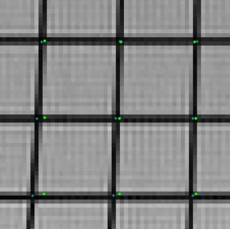
9 System-related distortions MRI Guidance image processing algorithm acquire data identify control point locations determine 3D distortion correct MR images validate method


10 System-related distortions MRI Guidance acquire data identify control point locations determine 3D distortion correct MR images validate method MR CT - unsharp mask and Gaussian blur - adaptive thresholding - 3D Gaussian blurring in x and y - watershed: identify and analyze each dot - center of mass: control points coordinates


11 System-related distortions MRI Guidance acquire data identify control point locations determine 3D distortion correct MR images validate method - register CT+MR control points - clean data - 3D polynomial fit - determine 3D distortion field
")

12 3D distortion field (vectorial) System-related distortions MRI Guidance x y z r


13 difference corrected uncorrected System-related distortions MRI Guidance uncorrected corrected MR CT (true location) MR CT (true location)

14 System-related distortions MRI Guidance Spherical harmonics analysis - Anm, Bnm are the spherical harmonic coefficients - Provided by the manufacturer for a certain region of interest - Example: 29 coeff for Gx and Gy 7 coeff for Gz
15 System-related distortions MRI Guidance Hybrid technique: harmonics analysis + phantom measurements Boundary measurements Make use of the harmonic nature of the 3D distortion vector field Distortions measured on a volume boundary Laplace s equation is solved to reconstruct the full 3D distortion field within the entire VOI (volume of interest)


16 System-related distortions MRI Guidance Hybrid technique: harmonics analysis + phantom measurements Boundary measurements Collaboration with Modus Medical Devices - Large field 3D distortions - Harmonic analysis - 38 diameter, 32 long - Light weight, hollow, <17 Kg - Option for inserts

17 System-related distortions MRI Guidance Harmonics analysis can be extended to arbitrary geometries
18 System-related distortions MRI Guidance Summary - Manufacturers provide a 1 st order correction (2D/3D) - Detailed quantification depends on clinical applications - MR used for diagnostic - MR-only planning - Limited standardization and lack of user friendly solutions


19 Patient-induced distortions Tissue magnetic susceptibility Cylinder geometry: interior and exterior mapped diff Interior: const field offset, no shape distortion along Gr Exterior: ( ) inhomogeneous dipole field \ shape distortions \ arrows indicate magnitude & direction of warp
 inhomogeneous dipole field \ shape distortions \ arrows indicate magnitude & direction")
20 Patient-induced distortions Tissue magnetic susceptibility image of the interior image of ext boundary original cross-section Cylinder geometry: interior and exterior mapped diff Interior: const field offset, no shape distortion along Gr Exterior: ( ) inhomogeneous dipole field \ shape distortions \ arrows indicate magnitude & direction of warp
21 Patient-induced distortions Methods for quantifying the distortion field: 1. Measurement of B0 field distortion map - double-echo GE sequence phase diff of the 2 echoes 2. Correlating at least 2 images of the same sample - without calculating or measuring the field 3. Numerical computations of the magnetic field on datasets converted into tissue susceptibility maps


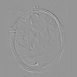
22 Patient-induced distortions Tissue magnetic susceptibility (~ mm) validation workflow CT raw image image mask magnetic field geometric distortion


23 Patient-induced distortions Tissue magnetic susceptibility (~ mm)


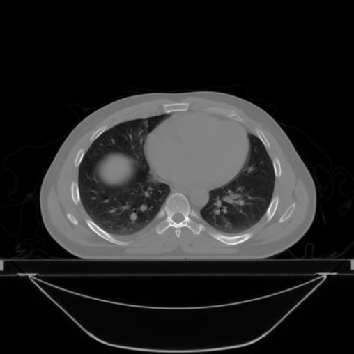
24 Patient-induced distortions Tissue magnetic susceptibility (~ mm) lung


25 Organ / target motion: lung MRI Guidance Case study: lung patient, 10 bins 4D CT 4D distortion field associated with organ motion: - 2 independent steps 1. System distortions - register anatomy to 3D field - track dist as local target/organs move - static field - measured with phantom 2. Magnetic susceptibility - numerical methods - anatomy specific - dynamic distortion field Total: combine contributions from 1 & cm exhale inhale


26 Organ / target motion: lung MRI Guidance

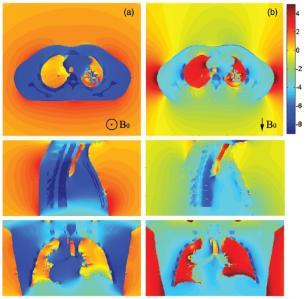
27 Organ / target motion: lung MRI Guidance B0 z-axis B0 y-axis
28 MRI Guidance MR data for RT planning Issue: MR images suffer of intrinsic distortions affect geometric accuracy Strategies: Several methods proposed claiming adequate accuracy Limitations and Challenges: Vendor and application specific Large FOVs still posing practical issues for distortion field mapping Susceptibility-induced distortions minimized via protocol optimization Real-time correction limited Streamlining and clinical integration
29 MRI Guidance MR data for Treatment Delivery - Patient setup verification & Tracking/Gating Aim: Reliable quantification and validation of methods used for organ motion assessment (real-time or retrospective data availability) - Real-time imaging: - 1D / 2D readily available, platform specific - 3D 4D (3D+time): most techniques under development - Retrospective 4D image data binning and image reconstruction - Available, implementation is vendor specific - Growing literature: 2D 4D, 3D 4D
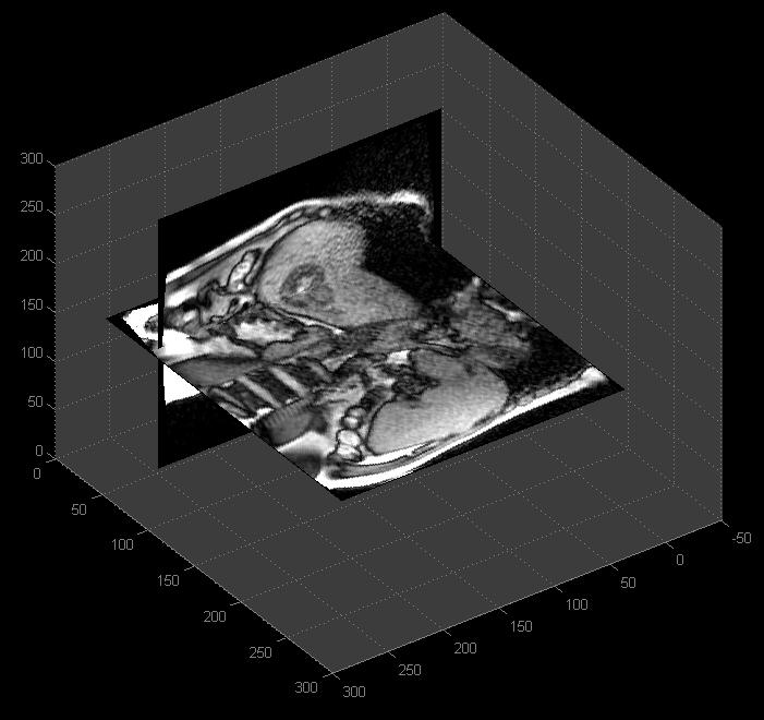


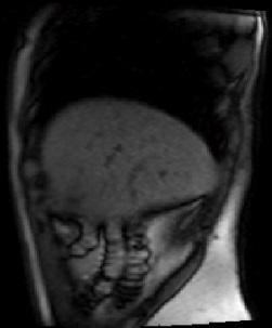
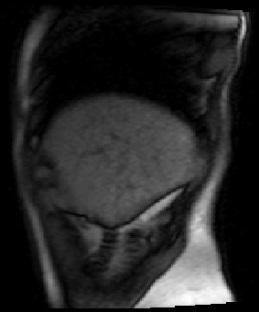
30 4D MRI Retrospective - 2D 4D MRI Guidance 2-plane sync: motion info dome of diaphragm tumor Sagittal


31 4D MRI Retrospective - 2D 4D Data acquisition 2D cine sagittal acquisition Multiple subsequent slice locations Cover volume of interest Additional coronal scan MRI Guidance Sagittal: scans 1 to N scan N N scan 3 Coronal: scan N N+1 scan 2 scan 1
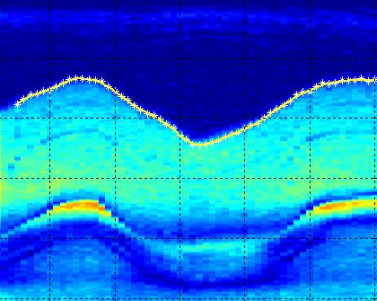


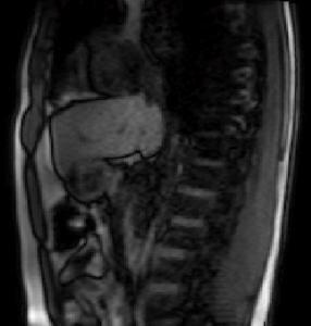


32 4D MRI Retrospective - 2D 4D MRI Guidance Organ motion curves & 4D data bining scan 1 scan 14 Exhale phase 3D volume, slice-by-slice
33 4D MRI Retrospective - 3D 4D MRI Guidance 3D fast acquisition with 4D image data sorting and reconstruction Similarity with 4D CBCT potential solution for motion quantification Strategies: Breathhold + multiple 3D acquisitions (< 15s) at diff respiratory phases Free breathing - Continuous acquisition (radial sampling) + post processing
34 4D MRI Retrospective - 3D 4D MRI Guidance 3D fast acquisition with 4D image data sorting and reconstruction Similarity with 4D CBCT potential solution for motion quantification Strategies: Breathhold + multiple 3D acquisitions (< 15s) at diff respiratory phases Free breathing - Continuous acquisition (radial sampling) + post processing 4D MRI Dynamic Strategies: Free breathing - Multiple 3D scans with ~s time sampling, low image resolution Sparse/parallel imaging 3D acquisitions, good temporal sampling (?)


35 QC of motion sequences: 2D/4D MRI Guidance


36 Phantom data analysis MRI Guidance Prototype provided by Modus Medical Devices

37 Phantom data analysis MRI Guidance BW (Hz/p) SNR (%) Time (s) Increased BW Decreased SNR Decreased Susceptibility effects better geometric accuracy Decreased acquisition time/frame faster imaging TurboFLASH - 1.9x 1.9 x 8 mm FOV 300 min TE/TR


38 Phantom data analysis RadialVIBE: FOV: 310x310 Voxel resolution 1.3x.1.3x3.0 Mid/High BW View sharing mode: golden angle, total acquisition time < 1 min
39 MRI Guidance MR data for Treatment Delivery - Patient setup verification & Tracking/Gating Aim: Reliable quantification and validation of methods used for organ motion assessment (real-time or retrospective data availability) Strategies: 1D / 2D available, several proposed 4D techniques Limitations and Challenges: Vendor implementation and application specific 4D motion - quantification of distortions still to be investigated Motion phantoms & QA methods still to be developed Motion data integration in clinical workflows
40 6% 8% 32% 27% Which is the main contributor to the MR image distortion field for RT applications? 27% 1. MR main field (B0) inhomogeneity 2. Chemical shift 3. Tissue susceptibility 4. Imaging gradient non-linearities 5. Motion
41 Which is the main contributor to the MR image distortion field for RT applications? Answer: Imaging gradient non-linearities Ref: Doran et al, Phys Med Biol 50, , 2005
42 Topics for MR-guided RT system Commissioning & QC System performance monitoring - Open-source software for semi/auto-qc monitoring - ACR guidelines, AAPM, NEMA, etc. MR data for RT planning and in-room guidance - MR image distortion: system/scanner-related - MR image distortion: susceptibility-induced - Quantification of motion MR-guided systems: design specific - MR-radiation source system: iso-to-iso registration - RF noise - Magnetic field coupling Reporting - Data base record: in-house, commercial, cloud solutions


43 MR-to-Radiation source isocenter registration Cylindrical phantom filled with water Scribe lines for alignment to lasers Circular film between two halves of phantom Wrap-around film strip Once MLC accuracy is established, imaging this phantom provides information about MR-RT isocenter alignment Once RT isocenter is established, MR isocenter coordinate shift is implemented in software Courtesy of Olga Green, Washington University, St. Louis


44 MR-to-Radiation source isocenter registration Designed for Elekta s Atlantic system MR-to-MV alignment Ceramic, non-conductive markers for MV 3D analysis to locate markers Automatic co-registration MR/MV Testing done at UMC, Utrecht MR image res: 1x1x1 mm3 MV image res: 0.5x0.5x0.5 mm3 Analysis mean error: ~0.3 mm MV CBCT MR Images & info courtesy of M. Sell, M. Luzzaro (Elekta/Philips)


45 MR-to-Radiation source isocenter registration Designed for IMRIS MR-linac system In collaboration with Modus Medical Devices MR-to-kV and MV alignment Daily QA 3D analysis to locate markers Automatic co-registration Ongoing testing

46 MRI Guidance MR-linac systems Radiofrequency (RF) interference MR needs to be isolated Collects weak signal from patient Linac is a significant source of RF MR Linac RF shield

47 MRI Guidance MR-linac systems Radiofrequency (RF) interference MR needs to be isolated Collects weak signal from patient Linac is a significant source of RF MR Linac RF

48 MRI Guidance MR-linac systems Radiofrequency (RF) interference MR needs to be isolated Collects weak signal from patient Linac is a significant source of RF Solutions: Relocate linac main RF sources in adjacent rooms Enclose linac head or MR in a Faraday cage QC monitoring: MR scanner RF noise tests RF sniffer kit for troubleshooting

 e - in")
49 MRI Guidance MR-linac systems Magnetic field mutual interaction: MR magnet Linac B0 fringe field of MR scanner reaching the Linac structure Linac performance affected Beam output = f(fringe B-field) Linac waveguide MR e - magnetic field (on all the time) e - in magnetic field

50 non-uniform MRI Guidance MR-linac systems Magnetic field mutual interaction: MR magnet Linac Linac is a large metallic structure, ferromagnetic components» MR imaging field homogeneity affected MR magnetic field uniform

51 MRI Guidance MR-linac systems Magnetic field mutual interaction: MR magnet Linac Linac is a large metallic structure, ferromagnetic components» MR imaging field homogeneity affected Solutions: Passive and/or active shielding Physical separation QC monitoring: Simulation environment: baseline, monitor perturbations MR: B0 mapping & Shimming Linac: rad beam, imaging


52 Princess Margaret MRgRT Project QC monitoring: Simulation environment: baseline for B0 fringe field mapping Establish margins of tolerance for sub-components - MR scanner: active imaging field homogeneity - Linac: beam optimal specs - Couch: safety margins on pull forces, upgradability impact on MR


53 Princess Margaret MRgRT Project QC monitoring: Simulation environment: baseline for B0 fringe field mapping Establish margins of tolerance for sub-components - MR scanner: active imaging field homogeneity - Linac: beam optimal specs - Couch: safety margins on pull forces, upgradability impact on MR


54 Princess Margaret MRgRT Project QC monitoring: B0 mapping for testing system performance Direct measurements to ensure B-field decoupling - MR should stay within specs over time, all intended configurations - Negligible impact from hysteresis/residual B-field related effects - Measurements more often than for a standalone MR implementation 1 st order harmonics tune-up


55 MR testing & commissioning MRgRT: MR Shimming Study Methods: - Siemens service procedures: Phantom Shim & Phantom Shim Check - B0 mapping technique: dual-echo GRE field mapping sequence - Metrics: Brms, Bpp, FWHM water spectral peak Results: - Transient effects due to B-field priming of the environment - The effects are reproducible - MR shim stays within the specs outlined by Siemens/IMRIS B0 mapping technique: Magnitude Phantom Unwrapped phase Analysis Brms, Bpp Phase


56 Princess Margaret MRgRT Project QC monitoring: Linac beam: Flatness & Symmetry v. Gantry angle rotation Direct measurements to ensure B-field decoupling - Beam stirring servos turned on/off - IC Profiler mounted on linac head via custom built accessory - Look for remnant magnetization and transient effects
57 Princess Margaret MRgRT Project QC monitoring: Linac beam: Flatness & Symmetry v. Gantry angle rotation Direct measurements to ensure B-field decoupling - Beam stirring servos turned on/off - IC Profiler mounted on linac head via custom built accessory - Look for remnant magnetization and transient effects flatness symmetry
58 For MR-guided RT systems, which MR-related test is new and has to be added to the QC routine? 10% 1. Magnetic field drift 81% 4% 3% 3% 2. Imaging-to-treatment isocenter co-registration 3. Center frequency 4. Image uniformity 5. Ghosting
59 For MR-guided RT systems, which MR-related test is new and has to be added to the QC routine? Answer: Imaging-to-treatment isocenter co-registration Ref: Lagendijk et al, Phys Med Biol 59, R349-R369, 2014
60 Topics for MR-guided RT system Commissioning & QC MR data for RT planning and in-room guidance - MR image distortion: system/scanner-related - MR image distortion: susceptibility-induced - Quantification of motion MR-guided systems: design specific - RF noise - Magnetic field coupling - MR-radiation source system: iso-to-iso registration System performance monitoring & Reporting - Open-source software for semi/auto-qc monitoring - Data base record: in-house, commercial, cloud solutions


61 Topics for MR-guided RT system Commissioning & QC System performance monitoring - Open-source software for semi/auto QC analysis - Developed by J. Sun at el - Calvary Mater Hospital, NSW - Supports ACR, MagPhan and MagIQ phantoms - Matlab code - Can be configured for broader purpose
62 Topics for MR-guided RT system Commissioning & QC Data record and Reporting In-house: - AAPM 2015 presentation: TU-G-CAMPUS-I-15 - Developed by J. Yung et al at MD Anderson - Semi-automatic QC program - Analyze and record measurements - Built on open-source software (Linux, Apache, MySQL, Python) - Analysis performed on 27 MR scanner: 1.5/3T, GE/Siemens - Tests: geometric accuracy/linearity, position accuracy, image uniformity, signal, noise, ghosting, transmit gain, center frequency, magnetic field drift
63 Topics for MR-guided RT system Commissioning & QC Data record and Reporting In-house / Commercial: - AQUA - Developed at Princess Margaret (Toronto) - Initially aimed for linac QC - Can be configured to include MRI tests - Analysis is semi-automatic - Data record is manual - Allows for data trending, control charts - The software is currently developed by Acumyn (
64 Topics for MR-guided RT system Commissioning & QC Data record and Reporting Commercial / Cloud: - QUMULATE - Developed by Varian for linac QA - Store, visualize, manage QC data - Arbitrary tests can be configured - Potential platform for MRI - Monthly/annual fee for service
65 MR-guided RT system Commissioning & QC Summary - Quantify and mitigate for system-related and patientinduced image distortions - QC of motion sequences may be required, especially for new techniques - MR-guided RT systems new tests may be required - RF noise - Magnetic field coupling - MR iso to radiation source iso co-registration - Establishing a QC program including data reporting
8/3/2016. MR-guided RT: Commissioning and Quality Control. Topics for MR-guided RT system commissioning & QC
 MR-guided RT: Commissioning and Quality Control T. Stanescu, PhD, MCCPM Medical Physicist, Princess Margaret Cancer Centre, UHN Assistant Professor, Radiation Oncology, University of Toronto Affiliated
Image Guidance and Beam Level Imaging in Digital Linacs
 Image Guidance and Beam Level Imaging in Digital Linacs Ruijiang Li, Ph.D. Department of Radiation Oncology Stanford University School of Medicine 2014 AAPM Therapy Educational Course Disclosure Research
7/31/2011. Learning Objective. Video Positioning. 3D Surface Imaging by VisionRT
 CLINICAL COMMISSIONING AND ACCEPTANCE TESTING OF A 3D SURFACE MATCHING SYSTEM Hania Al-Hallaq, Ph.D. Assistant Professor Radiation Oncology The University of Chicago Learning Objective Describe acceptance
Slide 1. Technical Aspects of Quality Control in Magnetic Resonance Imaging. Slide 2. Annual Compliance Testing. of MRI Systems.
 Slide 1 Technical Aspects of Quality Control in Magnetic Resonance Imaging Slide 2 Compliance Testing of MRI Systems, Ph.D. Department of Radiology Henry Ford Hospital, Detroit, MI Slide 3 Compliance Testing
Lucy Phantom MR Grid Evaluation
 Lucy Phantom MR Grid Evaluation Anil Sethi, PhD Loyola University Medical Center, Maywood, IL 60153 November 2015 I. Introduction: The MR distortion grid, used as an insert with Lucy 3D QA phantom, is
Image Quality Assessment and Quality Assurance of Advanced Imaging Systems for IGRT. AAPM Penn-Ohio Chapter Sep 25, 2015 Soyoung Lee, PhD
 Image Quality Assessment and Quality Assurance of Advanced Imaging Systems for IGRT AAPM Penn-Ohio Chapter Sep 25, 2015 Soyoung Lee, PhD 1 Outline q Introduction q Imaging performances in 4D-CBCT Image
Brilliance CT Big Bore.
 1 2 2 There are two methods of RCCT acquisition in widespread clinical use: cine axial and helical. In RCCT with cine axial acquisition, repeat CT images are taken each couch position while recording respiration.
MR-guided radiotherapy: Vision, status and research at the UMC Utrecht. Dipl. Ing. Dr. Markus Glitzner
 MR-guided radiotherapy: Vision, status and research at the UMC Utrecht Dipl. Ing. Dr. Markus Glitzner About myself Training Medizintechnik TU Graz PhD UMC Utrecht Clinical work Software implementation
Initial Clinical Experience with 3D Surface Image Guidance
 Initial Clinical Experience with 3D Surface Image Guidance Amanda Havnen-Smith, Ph.D. Minneapolis Radiation Oncology Ridges Radiation Therapy Center Burnsville, MN April 20 th, 2012 Non-funded research
Use of MRI in Radiotherapy: Technical Consideration
 Use of MRI in Radiotherapy: Technical Consideration Yanle Hu, PhD Department of Radiation Oncology, Mayo Clinic Arizona 04/07/2018 2015 MFMER slide-1 Conflict of Interest: None 2015 MFMER slide-2 Objectives
Thank-You Members of TG147 TG 147: QA for nonradiographic
 Thank-You Members of TG147 TG 147: QA for nonradiographic localization and positioning systems Twyla Willoughby, M.S. Medical Physicist Clinical AAPM Meeting March 2013 Department of Radiation Oncology
Respiratory Motion Compensation for Simultaneous PET/MR Based on Strongly Undersampled Radial MR Data
 Respiratory Motion Compensation for Simultaneous PET/MR Based on Strongly Undersampled Radial MR Data Christopher M Rank 1, Thorsten Heußer 1, Andreas Wetscherek 1, and Marc Kachelrieß 1 1 German Cancer
Overview of Proposed TG-132 Recommendations
 Overview of Proposed TG-132 Recommendations Kristy K Brock, Ph.D., DABR Associate Professor Department of Radiation Oncology, University of Michigan Chair, AAPM TG 132: Image Registration and Fusion Conflict
SPECT QA and QC. Bruce McBride St. Vincent s Hospital Sydney.
 SPECT QA and QC Bruce McBride St. Vincent s Hospital Sydney. SPECT QA and QC What is needed? Why? How often? Who says? QA and QC in Nuclear Medicine QA - collective term for all the efforts made to produce
Deviceless respiratory motion correction in PET imaging exploring the potential of novel data driven strategies
 g Deviceless respiratory motion correction in PET imaging exploring the potential of novel data driven strategies Presented by Adam Kesner, Ph.D., DABR Assistant Professor, Division of Radiological Sciences,
Image Co-Registration II: TG132 Quality Assurance for Image Registration. Image Co-Registration II: TG132 Quality Assurance for Image Registration
 Image Co-Registration II: TG132 Quality Assurance for Image Registration Preliminary Recommendations from TG 132* Kristy Brock, Sasa Mutic, Todd McNutt, Hua Li, and Marc Kessler *Recommendations are NOT
State-of-the-Art IGRT
 in partnership with State-of-the-Art IGRT Exploring the Potential of High-Precision Dose Delivery and Real-Time Knowledge of the Target Volume Location Antje-Christin Knopf IOP Medical Physics Group Scientific
DAILY LINAC QA BEAM QA
 BEAM QA DAILY LINAC QA The QA BeamChecker Plus allows for fast, reliable, and uncomplicated daily QA of Varian, Elekta, Siemens, and Accuray Treatment Machines. The QA BeamChecker Plus is specifically
REAL-TIME ADAPTIVITY IN HEAD-AND-NECK AND LUNG CANCER RADIOTHERAPY IN A GPU ENVIRONMENT
 REAL-TIME ADAPTIVITY IN HEAD-AND-NECK AND LUNG CANCER RADIOTHERAPY IN A GPU ENVIRONMENT Anand P Santhanam Assistant Professor, Department of Radiation Oncology OUTLINE Adaptive radiotherapy for head and
Virtual Phantoms for IGRT QA
 TM Virtual Phantoms for IGRT QA Why ImSimQA? ImSimQA was developed to overcome the limitations of physical phantoms for testing modern medical imaging and radiation therapy software systems, when there
Ini$al Performance Test Results on an Elekta MR-Linac
 Ini$al Performance Test Results on an Elekta MR-Linac Eric Paulson, PhD DABR Radia$on Oncology, Radiology, and Biophysics Froedtert and Medical College of Wisconsin Milwaukee, Wisconsin, United States
IMRT and VMAT Patient Specific QA Using 2D and 3D Detector Arrays
 IMRT and VMAT Patient Specific QA Using 2D and 3D Detector Arrays Sotiri Stathakis Outline Why IMRT/VMAT QA AAPM TG218 UPDATE Tolerance Limits and Methodologies for IMRT Verification QA Common sources
ICARO Vienna April Implementing 3D conformal radiotherapy and IMRT in clinical practice: Recommendations of IAEA- TECDOC-1588
 ICARO Vienna April 27-29 2009 Implementing 3D conformal radiotherapy and IMRT in clinical practice: Recommendations of IAEA- TECDOC-1588 M. Saiful Huq, Ph.D., Professor and Director, Dept. of Radiation
Estimating 3D Respiratory Motion from Orbiting Views
 Estimating 3D Respiratory Motion from Orbiting Views Rongping Zeng, Jeffrey A. Fessler, James M. Balter The University of Michigan Oct. 2005 Funding provided by NIH Grant P01 CA59827 Motivation Free-breathing
Optical Guidance. Sanford L. Meeks. July 22, 2010
 Optical Guidance Sanford L. Meeks July 22, 2010 Optical Tracking Optical tracking is a means of determining in real-time the position of a patient relative to the treatment unit. Markerbased systems track
Raising the Bar in IMRT QA
 MapCHECK 2TM Raising the Bar in IMRT QA The leader in quick and precise measurement of modulated radiotherapy beams Benefits Proven solution for film-less rotational delivery and IMRT QA - More than 1500
Chapter 9 Field Shaping: Scanning Beam
 Chapter 9 Field Shaping: Scanning Beam X. Ronald Zhu, Ph.D. Department of Radiation Physics M. D. Anderson Cancer Center Houston, TX June 14-18, 2015 AAPM - Summer School 2015, Colorado Spring Acknowledgement
URGENT IMPORTANT FIELD SAFETY NOTIFICATION
 Subject: Incorrect Movement of the Treatment Table Product: MOSAIQ Scope: Sites affected will be those: 1. Running MOSAIQ and, 2. Treating on linear accelerators with the RATM license Notification Released:
Artefakt-resistente Bewegungsschätzung für die bewegungskompensierte CT
 Artefakt-resistente Bewegungsschätzung für die bewegungskompensierte CT Marcus Brehm 1,2, Thorsten Heußer 1, Pascal Paysan 3, Markus Oehlhafen 3, and Marc Kachelrieß 1,2 1 German Cancer Research Center
TG 132: Use of Image Registration and Fusion in RT
 TG 132: Use of Image Registration and Fusion in RT Kristy K Brock, PhD, DABR, FAAPM Associate Professor Department of Radiation Oncology, University of Michigan Chair, AAPM TG 132: Image Registration and
Tomotherapy Physics. Machine Twinning and Quality Assurance. Emilie Soisson, MS
 Tomotherapy Physics Machine Twinning and Quality Assurance Emilie Soisson, MS Tomotherapy at UW- Madison Treating for nearly 5 years Up to ~45 patients a day on 2 tomo units Units twinned to facilitate
TomoTherapy Related Projects. An image guidance alternative on Tomo Low dose MVCT reconstruction Patient Quality Assurance using Sinogram
 TomoTherapy Related Projects An image guidance alternative on Tomo Low dose MVCT reconstruction Patient Quality Assurance using Sinogram Development of A Novel Image Guidance Alternative for Patient Localization
1. Learn to incorporate QA for surface imaging
 Hania Al-Hallaq, Ph.D. Assistant Professor Radiation Oncology The University of Chicago ***No disclosures*** 1. Learn to incorporate QA for surface imaging into current QA procedures for IGRT. 2. Understand
Clinical implementation of photon beam flatness measurements to verify beam quality
 JOURNAL OF APPLIED CLINICAL MEDICAL PHYSICS, VOLUME 16, NUMBER 6, 2015 Clinical implementation of photon beam flatness measurements to verify beam quality Simon Goodall, a Nicholas Harding, Jake Simpson,
Automated Quality Assurance for Image-Guided Radiation Therapy
 JOURNAL OF APPLIED CLINICAL MEDICAL PHYSICS, VOLUME 10, NUMBER 1, WINTER 2009 Automated Quality Assurance for Image-Guided Radiation Therapy Eduard Schreibmann, a Eric Elder, Tim Fox Department of Radiation
UvA-DARE (Digital Academic Repository) Motion compensation for 4D PET/CT Kruis, M.F. Link to publication
 Motion compensation for 4D PET/CT Kruis, M.F. Link to publication") UvA-DARE (Digital Academic Repository) Motion compensation for 4D PET/CT Kruis, M.F. Link to publication Citation for published version (APA): Kruis, M. F. (2014). Motion compensation for 4D PET/CT General
UvA-DARE (Digital Academic Repository) Motion compensation for 4D PET/CT Kruis, M.F. Link to publication Citation for published version (APA): Kruis, M. F. (2014). Motion compensation for 4D PET/CT General
Volumetric Modulated Arc Therapy - Clinical Implementation. Outline. Acknowledgement. History of VMAT. IMAT Basics of IMAT
 Volumetric Modulated Arc Therapy - Clinical Implementation Daliang Cao, PhD, DABR Swedish Cancer Institute, Seattle, WA Acknowledgement David M. Shepard, Ph.D. Muhammad K. N. Afghan, Ph.D. Fan Chen, Ph.D.
Automated Image Analysis Software for Quality Assurance of a Radiotherapy CT Simulator
 Automated Image Analysis Software for Quality Assurance of a Radiotherapy CT Simulator Andrew J Reilly Imaging Physicist Oncology Physics Edinburgh Cancer Centre Western General Hospital EDINBURGH EH4
CT Protocol Review: Practical Tips for the Imaging Physicist Physicist
 CT Protocol Review: Practical Tips for the Imaging Physicist Physicist Dianna Cody, Ph.D., DABR, FAAPM U.T.M.D. Anderson Cancer Center August 8, 2013 AAPM Annual Meeting Goals Understand purpose and importance
CT NOISE POWER SPECTRUM FOR FILTERED BACKPROJECTION AND ITERATIVE RECONSTRUCTION
 CT NOISE POWER SPECTRUM FOR FILTERED BACKPROJECTION AND ITERATIVE RECONSTRUCTION Frank Dong, PhD, DABR Diagnostic Physicist, Imaging Institute Cleveland Clinic Foundation and Associate Professor of Radiology
Evaluation of AutoQA Lite TM Image Quality Measurement Software
 Evaluation of AutoQA Lite TM Image Quality Measurement Software Andrew J Reilly Imaging Physicist Oncology Physics Edinburgh Cancer Centre Western General Hospital EDINBURGH EH4 2XU Phone: 0131 537 1161
8/3/2016. Image Guidance Technologies. Introduction. Outline
 8/3/26 Session: Image Guidance Technologies and Management Strategies Image Guidance Technologies Jenghwa Chang, Ph.D.,2 Department of Radiation Medicine, Northwell Health 2 Hofstra Northwell School of
Using a research real-time control interface to go beyond dynamic MLC tracking
 in partnership with Using a research real-time control interface to go beyond dynamic MLC tracking Dr. Simeon Nill Joint Department of Physics at The Institute of Cancer Research and the Royal Marsden
VALIDATION OF DIR. Raj Varadhan, PhD, DABMP Minneapolis Radiation Oncology
 VALIDATION OF DIR Raj Varadhan, PhD, DABMP Minneapolis Radiation Oncology Overview Basics: Registration Framework, Theory Discuss Validation techniques Using Synthetic CT data & Phantoms What metrics to
8/4/2016. Emerging Linac based SRS/SBRT Technologies with Modulated Arc Delivery. Disclosure. Introduction: Treatment delivery techniques
 Emerging Linac based SRS/SBRT Technologies with Modulated Arc Delivery Lei Ren, Ph.D. Duke University Medical Center 2016 AAPM 58 th annual meeting, Educational Course, Therapy Track Disclosure I have
Facility Questionnaire PART I (General Information for 3DCRT and IMRT)
") Facility Questionnaire PART I (General Information for 3DCRT and IMRT) The following items are required before you can enter cases on any RTOG protocol that requires data submission to the Image-Guided
Facility Questionnaire PART I (General Information for 3DCRT and IMRT) The following items are required before you can enter cases on any RTOG protocol that requires data submission to the Image-Guided
An investigation of temporal resolution parameters in cine-mode four-dimensional computed tomography acquisition
 JOURNAL OF APPLIED CLINICAL MEDICAL PHYSICS, VOLUME 9, NUMBER 4, FALL 2008 An investigation of temporal resolution parameters in cine-mode four-dimensional computed tomography acquisition Yildirim D. Mutaf
ViewRay System Commissioning
 ViewRay System Commissioning Kyle R. Padgett, PhD, DABR Assistant Professor University of Miami Viewray Team: Matt Studenski, PhD Chet Ford, PhD Yidong Yang, PhD Nesrin Dogan, PhD Medical Physics Residents:
AAPM Standard of Practice: CT Protocol Review Physicist
 AAPM Standard of Practice: CT Protocol Review Physicist Dianna Cody, Ph.D., DABR, FAAPM U.T.M.D. Anderson Cancer Center September 11, 2014 2014 Texas Radiation Regulatory Conference Goals Understand purpose
C a t p h a n / T h e P h a n t o m L a b o r a t o r y
 C a t p h a n 5 0 0 / 6 0 0 T h e P h a n t o m L a b o r a t o r y C a t p h a n 5 0 0 / 6 0 0 Internationally recognized for measuring the maximum obtainable performance of axial, spiral and multi-slice
PURE. ViSION Edition PET/CT. Patient Comfort Put First.
 PURE ViSION Edition PET/CT Patient Comfort Put First. 2 System features that put patient comfort and safety first. Oncology patients deserve the highest levels of safety and comfort during scans. Our Celesteion
Acknowledgments. Ping Xia, Ph.D., UCSF. Pam Akazawa, CMD, UCSF. Cynthia Chuang, Ph.D., UCSF
 Page 1 Quality Assurance of IMRT Delivery Systems - Siemens Lynn J. Verhey, Ph.D. Professor and Vice-Chair UCSF Dept. of Radiation Oncology AAPM 22 Annual Meeting, Montreal Acknowledgments Ping Xia, Ph.D.,
New Technology in Radiation Oncology. James E. Gaiser, Ph.D. DABR Physics and Computer Planning Charlotte, NC
 New Technology in Radiation Oncology James E. Gaiser, Ph.D. DABR Physics and Computer Planning Charlotte, NC Technology s s everywhere From the imaging chain To the planning system To the linac To QA..it..it
IAEA-TECDOC-1583 Commissioning of Radiotherapy Treatment Planning Systems: Testing for Typical External Beam Treatment Techniques
 IAEA-TECDOC-1583 Commissioning of Radiotherapy Treatment Planning Systems: Testing for Typical External Beam Treatment Techniques Report of the Coordinated Research Project (CRP) on Development of Procedures
Digital Tomosynthesis for Target Localization
 Digital Tomosynthesis for Target Localization Fang-Fang Yin, Devon Godfrey, Lei Ren Jacqueline Maurer, Jackie Q-L Wu Duke University Medical Center Acknowledgements Duke Radiation Oncology faculty and
Optimization of CT Simulation Imaging. Ingrid Reiser Dept. of Radiology The University of Chicago
 Optimization of CT Simulation Imaging Ingrid Reiser Dept. of Radiology The University of Chicago Optimization of CT imaging Goal: Achieve image quality that allows to perform the task at hand (diagnostic
Position accuracy analysis of the stereotactic reference defined by the CBCT on Leksell Gamma Knife Icon
 Position accuracy analysis of the stereotactic reference defined by the CBCT on Leksell Gamma Knife Icon WHITE PAPER Introduction An image guidance system based on Cone Beam CT (CBCT) is included in Leksell
Design and performance characteristics of a Cone Beam CT system for Leksell Gamma Knife Icon
 Design and performance characteristics of a Cone Beam CT system for Leksell Gamma Knife Icon WHITE PAPER Introduction Introducing an image guidance system based on Cone Beam CT (CBCT) and a mask immobilization
S. Guru Prasad, Ph.D., DABR
 PURPOSE S. Guru Prasad, Ph.D., DABR Director of Medical Physics IAEA Consultant NorthShore University Health System and University of Chicago, Pritzker School of Medicine Current TPS utilize more information
Moving Metal Artifact Reduction for Cone-Beam CT (CBCT) Scans of the Thorax Region
 Scans of the Thorax Region") Moving Metal Artifact Reduction for Cone-Beam CT (CBCT) Scans of the Thorax Region Andreas Hahn 1,2, Sebastian Sauppe 1,2, Michael Knaup 1, and Marc Kachelrieß 1,2 1 German Cancer Research Center (DKFZ),
Moving Metal Artifact Reduction for Cone-Beam CT (CBCT) Scans of the Thorax Region Andreas Hahn 1,2, Sebastian Sauppe 1,2, Michael Knaup 1, and Marc Kachelrieß 1,2 1 German Cancer Research Center (DKFZ),
Computational Algorithm for Estimating the Gradient Isocenter of an MRI Scanner
 Computational Algorithm for Estimating the Gradient Isocenter of an MRI Scanner R Cai 1, KE Schubert 1, R Schulte 2 ritchiecai@r2labsorg, keith@r2labsorg, rschulte@dominionllumcedu 1 School of Computer
Version 5.6. Quick Start Guide
 WWW..COM Version 5.6 Quick Start Guide STANDARD IMAGING, INC. 3120 Deming Way Middleton, WI 53562-1461 Jul / 2018 2018 Standard Imaging, Inc. TEL 800.261.4446 TEL 608.831.0025 FAX 608.831.2202 DOC # 80714-05
3DVH : SUN NUCLEAR On The Accuracy Of The corporation Planned Dose Perturbation Algorithm Your Most Valuable QA and Dosimetry Tools *Patent Pending
 3DVH : On The Accuracy Of The Planned Dose Perturbation Algorithm SUN NUCLEAR corporation Your Most Valuable QA and Dosimetry Tools *Patent Pending introduction State-of-the-art IMRT QA of static gantry
8/2/2017. PerFRACTION from Sun Nuclear Automated Transit Dosimetry for In-Vivo QA
 PerFRACTION from Sun Nuclear Automated Transit Dosimetry for In-Vivo QA Jennifer R. Clark, M.S., DABR Conflict of Interest Statement: I am an employee of Sun Nuclear Corporation Outline: Platform overview
Tumor motion during liver SBRT
 Tumor motion during liver SBRT - projects at Aarhus University Hospital - Per Poulsen, Esben Worm, Walther Fledelius, Morten Høyer Aarhus University Hospital, Denmark SBRT: Stereotactic Body Radiation
ImPACT. Information Leaflet No. 1: CT Scanner Acceptance Testing
 ImPACT Information Leaflet No. 1: CT Scanner Acceptance Testing Version 1.02, 18/05/01 CONTENTS: 1. SCOPE OF LEAFLET 2. GENERAL PRINCIPLES OF ACCEPTANCE AND COMMISSIONING 2.1 PHANTOMS 2.2 EXPOSURE AND
An Automated Image-based Method for Multi-Leaf Collimator Positioning Verification in Intensity Modulated Radiation Therapy
 An Automated Image-based Method for Multi-Leaf Collimator Positioning Verification in Intensity Modulated Radiation Therapy Chenyang Xu 1, Siemens Corporate Research, Inc., Princeton, NJ, USA Xiaolei Huang,
HST.583 Functional Magnetic Resonance Imaging: Data Acquisition and Analysis Fall 2008
 MIT OpenCourseWare http://ocw.mit.edu HST.583 Functional Magnetic Resonance Imaging: Data Acquisition and Analysis Fall 2008 For information about citing these materials or our Terms of Use, visit: http://ocw.mit.edu/terms.
Technological Advances and Challenges: Experience with Time-Of-Flight PET Combined with 3T MRI. Floris Jansen, GE Healthcare July, 2015
 Technological Advances and Challenges: Experience with Time-Of-Flight PET Combined with 3T MRI Floris Jansen, GE Healthcare July, 2015 PET/MR 101 : challenges Thermal Workflow & Apps RF interactions?!!
IMSURE QA SOFTWARE FAST, PRECISE QA SOFTWARE
 QA SOFTWARE FAST, PRECISE Software for accurate and independent verification of monitor units, dose, and overall validity of standard, IMRT, VMAT, SRS and brachytherapy plans no film, no phantoms, no linac
ph fax Deming Way Middleton WI USA
 www.standardimaging.com 800-261-4446. ph 608-831-0025. fax 608-831-2202 3120 Deming Way Middleton WI 53562-1461 USA 2007 Standard Imaging, Inc. 1239-21 (03.07) beam qa LINAC AND TOMOTHERAPY QA The NEW
Development and Optimization of Fourdimensional Magnetic Resonance Imaging (4D-MRI) for Radiation Therapy
 for Radiation Therapy") Development and Optimization of Fourdimensional Magnetic Resonance Imaging (4D-MRI) for Radiation Therapy by Yilin Liu Medical Physics Graduate Program Duke University Date: Approved: Jing Cai, co-advisor
Development and Optimization of Fourdimensional Magnetic Resonance Imaging (4D-MRI) for Radiation Therapy by Yilin Liu Medical Physics Graduate Program Duke University Date: Approved: Jing Cai, co-advisor
TG-148 overview. Introduction. System Overview. System Overview. QA for Helical Tomotherapy: Report of the AAPM Task Group 148. Conflict of Interest:
 QA or Helical Tomotherapy: Report o the AAPM Tas Group 148 Members: Conlict o Interest: r. John Balog owns TomoTherapy stoc. Katja Langen (Co-chair) Nio Papaniolaou (Co-chair) Walter Grant Richard Crilly
Monte Carlo methods in proton beam radiation therapy. Harald Paganetti
 Monte Carlo methods in proton beam radiation therapy Harald Paganetti Introduction: Proton Physics Electromagnetic energy loss of protons Distal distribution Dose [%] 120 100 80 60 40 p e p Ionization
Table of Contents. About AQUA
 User Manual About AQUA Table of Contents 1 About AQUA... 6 1.1 Radiation Therapy... 6 1.1.1 Workflow manager... 6 1.1.2 Reports... 7 1.1.3 Tests... 7 1.1.4 Many Ways to Work... 7 1.2 Technology... 7 1.3
THE WIRELESS PHANTOM PERFORM ACCURATE PATIENT QA IN LESS TIME THAN EVER!
 THE WIRELESS PHANTOM PERFORM ACCURATE PATIENT QA IN LESS TIME THAN EVER! Confidence in complex treatments Modern radiation therapy uses complex plans with techniques such as IMRT, VMAT and Tomotherapy.
A fluence convolution method to account for respiratory motion in three-dimensional dose calculations of the liver: A Monte Carlo study
 A fluence convolution method to account for respiratory motion in three-dimensional dose calculations of the liver: A Monte Carlo study Indrin J. Chetty, a) Mihaela Rosu, Neelam Tyagi, Lon H. Marsh, Daniel
Patient Set-ups and Tumor Localizations
 Patient Set-ups and Tumor Localizations Amy S. Harrison Patient Positioning Prior to starting any localization or simulation procedure patients need to be positioned and immobilized Patients disease location
Quick Reference Datasheet For All RIT113 Packages
 Quick Reference Datasheet For All RIT113 Packages For Rotational Therapies, IMRT & TG142 Highlights and selected product information only. A complete TG142 brochure is available. For more information on
ADVANCING CANCER TREATMENT
 The RayPlan treatment planning system makes proven, innovative RayStation technology accessible to clinics that need a cost-effective and streamlined solution. Fast, efficient and straightforward to use,
PET-CT in Radiation Treatment Planning
 PET-CT in Radiation Treatment Planning TA van de Water, Radiotherapeutic Institute Friesland, Leeuwarden JA van Dalen, Isala, Zwolle ACKNOWLEDGEMENTS A. van der Schaaf (University of Groningen, University
Laser Systems for Patient Alignment
 Laser Systems for Patient Alignment GALAXY Patient TOPOGRAPHY Laser System Page: 2 Date: 28 March 2009 The GALAXY System NEW METHOD FOR HIGHER ALIGNMENT ACCURACY. Patient Positioning System for Linac Simulator
Real Time Tumor Motion Tracking with CyberKnife
 Real Time Tumor Motion Tracking with CyberKnife Martina Descovich, Ph.D University of California San Francisco July 16, 2015 Learning objectives Review the principles of real-time tumor motion tracking
Helical 4D CT pitch management for the Brilliance CT Big Bore in clinical practice
 JOURNAL OF APPLIED CLINICAL MEDICAL PHYSICS, VOLUME 16, NUMBER 3, 2015 Helical 4D CT pitch management for the Brilliance CT Big Bore in clinical practice Guido Hilgers, a Tonnis Nuver, André Minken Department
MapCHECK 2 & 3DVH. The Gold Standard for 2D Arrays
 MapCHECK 2 & 3DVH The Gold Standard for 2D Arrays Your Most Valuable QA and Dosimetry Tools THE GOLD STANDARD FOR 2D ARRAYS The MapCHECK 2 is the world s most selected independent 2D measurement array.
3/27/2012 WHY SPECT / CT? SPECT / CT Basic Principles. Advantages of SPECT. Advantages of CT. Dr John C. Dickson, Principal Physicist UCLH
 3/27/212 Advantages of SPECT SPECT / CT Basic Principles Dr John C. Dickson, Principal Physicist UCLH Institute of Nuclear Medicine, University College London Hospitals and University College London john.dickson@uclh.nhs.uk
Choosing and Commissioning a Video Based Motion Management System
 Choosing and Commissioning a Video Based Motion Management System David Shepard Swedish Cancer Institute 2 1 Acknowledgments Daliang Cao, PhD, SCI Mohammed Jermoumi, PhD, SCI Roger Xie, PhD, SCI Malin
Multi-Purpose Body Phantom
 Multi-Purpose Body Phantom Legal & Copyright Notice Information in this manual is subject to change without notice. Permission is granted to Owners of the to reproduce the Worksheets included in this manual
7/31/ D Cone-Beam CT: Developments and Applications. Disclosure. Outline. I have received research funding from NIH and Varian Medical System.
 4D Cone-Beam CT: Developments and Applications Lei Ren, PhD, DABR Department of Radiation Oncology Duke University Medical Center Disclosure I have received research funding from NIH and Varian Medical
MapCHECK 2 & 3DVH The Gold Standard for 2D Arrays
 MapCHECK 2 & 3DVH The Gold Standard for 2D Arrays Your Most Valuable QA and Dosimetry Tools THE GOLD STANDARD FOR 2D ARRAYS The MapCHECK 2 is the world s most selected independent 2D measurement array.
RADIOMICS: potential role in the clinics and challenges
 27 giugno 2018 Dipartimento di Fisica Università degli Studi di Milano RADIOMICS: potential role in the clinics and challenges Dr. Francesca Botta Medical Physicist Istituto Europeo di Oncologia (Milano)
Good Morning! Thank you for joining us
 Good Morning! Thank you for joining us Deformable Registration, Contour Propagation and Dose Mapping: 101 and 201 Marc Kessler, PhD, FAAPM The University of Michigan Conflict of Interest I receive direct
Abbie M. Diak, PhD Loyola University Medical Center Dept. of Radiation Oncology
 Abbie M. Diak, PhD Loyola University Medical Center Dept. of Radiation Oncology Outline High Spectral and Spatial Resolution MR Imaging (HiSS) What it is How to do it Ways to use it HiSS for Radiation
Comparison of Two Phantoms for End-to-End SRS/SBRT Testing. Vikren Sarkar, Ph.D. Associate Professor University of Utah Huntsman Cancer Center
 Comparison of Two Phantoms for End-to-End SRS/SBRT Testing Vikren Sarkar, Ph.D. Associate Professor University of Utah Huntsman Cancer Center Disclosure Travel and Speaker Honorarium from Sun Nuclear Corp.
Implementation and evaluation of a fully 3D OS-MLEM reconstruction algorithm accounting for the PSF of the PET imaging system
 Implementation and evaluation of a fully 3D OS-MLEM reconstruction algorithm accounting for the PSF of the PET imaging system 3 rd October 2008 11 th Topical Seminar on Innovative Particle and Radiation
MRI-LINAC Dynamic Phantom
 MRI-LINAC Dynamic Phantom Model 008M IMAGE ACQUISITION TREATMENT PLANNING DOSE DELIVERY Strict QA procedures for the imaging, planning and delivery of radiotherapy using respiratory management devices
ADVANCING CANCER TREATMENT
 3 ADVANCING CANCER TREATMENT SUPPORTING CLINICS WORLDWIDE RaySearch is advancing cancer treatment through pioneering software. We believe software has un limited potential, and that it is now the driving
Future is Effortless
 AQUATM Radiology is a machine Quality Assurance management solution that standardizes, streamlines and automates QA procedures throughout rediology department and across multiple sites. AQUATM Radiology
Lab Location: MRI, B2, Cardinal Carter Wing, St. Michael s Hospital, 30 Bond Street
 Lab Location: MRI, B2, Cardinal Carter Wing, St. Michael s Hospital, 30 Bond Street MRI is located in the sub basement of CC wing. From Queen or Victoria, follow the baby blue arrows and ride the CC south
2005 IEEE Nuclear Science Symposium Conference Record M10-2
 25 IEEE Nuclear Science Symposium Conference Record M1-2 Estimating 3D Respiratory Motion from Orbiting Views Rongping Zeng 1, Jeffrey A. Fessler 1, and James M. Balter 2 rzeng@eecs.umich.edu, fessler@eecs.umich.edu,