From DQE to TRE: Image Science, Image Guidance, and Cone-Beam CT
|
|
|
- Milo Mosley
- 6 years ago
- Views:
Transcription


1 W task = F task C task Magnification Focal Spot Size Scatter-to-Primary Ratio kvp, Dose Scintillator, Detector Pixel Aperture, 2D Sampling Reconstruction Filter Backprojection Voxel Size 3D Sampling PW, PWE NPW, NPWE f J. H. Siewerdsen (Johns Hopkins University) From DQE to TRE: Image Science, Image Guidance, and Cone-Beam CT Jeff Siewerdsen, PhD Department of Biomedical Engineering Johns Hopkins University Johns Hopkins University Schools of Medicine and Engineering Overview Cone-Beam CT Principles and applications Task-based models for imaging performance 3D NPS 3D NEQ Detectability ROC Imaging Task System Geometry Detectability Index Observer Model NEQ Anatomical Background Quantum Noise k b System Design and Optimization Dedicated MSK scanner Physics-driven design Performance / advanced applications Clinical Translation and System Integration A mobile C-arm for intraop CBCT Spine, thoracic, skull base surgery Integration of Imaging + Registration + Tracking + Augmentation 1


2 Cone-Beam CT Projection data Multiple projections (~ ) over ~180 o -360 o Volume reconstruction Sub-mm spatial resolution + soft tissue visibility Cone-Beam CT 2

 X-ray Tube 50 150 kvp,")


3 Cone-Beam CT Imaging Bench Flat-Panel Detector a-si:h PD + CsI:Tl (1536 x 2048 pixels, 1-30 fps) X-ray Tube kvp, Pulsed fluoro f cone ~5 o -15 o Optical Bench + Motion Control System 8-axis translation / rotation, 180 o +fan 360 o Repeat for ~360 o Projection Data Pre-Processing CBCT Reconstruction 3D Filtered Backprojection 3


4 Modeling the Imaging Chain Stage Physical Process 0 Incident quanta 1 Interacting quanta 2 Conversion to secondary quanta A: Complete absorption B: K x-ray escape C: K x-ray reabsorption 3 Spread of secondary quanta 4 Coupling of secondary quanta 5 Integration by pixel aperture 6 Sampling of pixel matrix 7 Readout with additive noise A B C Modeling the Imaging Chain Stage Physical Process 0 Incident quanta 1 Interacting quanta 2 Conversion to secondary quanta A: Complete absorption B: K x-ray escape C: K x-ray reabsorption 3 Spread of secondary quanta 4 Coupling of secondary quanta 5 Integration by pixel aperture 6 Sampling of pixel matrix 7 Readout with additive noise 4


 Broken Sagittal Axial")
5 Extension to Cone-Beam CT Stage Physical Process 0 Incident quanta 7 Projection readout Stage Mathematical Process 8 Ramp Filter 9 Apodization Filter 10 Interpolation 11 Backprojection 12 Sampling Projection T 8 T 9 T III 12 3D Noise-Power Spectrum S(f x, f y, f z ) Broken Sagittal Axial Symmetry NPS S(f x, f y z ) 5
 NPS (m 2 mm 3 ) Axial NPS S(f")


 0.")
6 3D Noise-Power Spectrum Axial Plane (x,y) NPS (m 2 mm 3 ) Axial NPS S(f x, f y ) 0.4 mas 1 mas 2 mas 4 mas Spatial Frequency, f x (mm -1 ) 3D Noise-Power Spectrum Sagittal Plane (x,z) NPS (m 2 mm 3 ) Sagittal NPS S(f x, f z ) 0.4 mas 1 mas 2 mas 4 mas Spatial Frequency, f z (mm -1 ) 6


 DQE Fraction of quanta used at each")
 ~ Projection DQE(0) 3D")
7 Image Noise CT image noise depends on Dose Detector efficiency Voxel size Axial, a xy Slice thickness, a z Reconstruction filter 2 k K E 1 D a o xy 3 xy az D o a xy 1 a z Barrett, Gordon, and Hershel (1976) The 3-D NEQ and DQE NEQ Effective number of quanta used at each spatial frequency (Efficiency x Fluence) DQE Fraction of quanta used at each each frequency. Observations: 3D DQE(0) ~ Projection DQE(0) 3D DQE(f) dependent on reconstruction parameters 7


8 A Task-Based Model Detectability Index Imaging Task W task = F task C task Observer Model PW, PWE NPW, NPWE System Geometry Magnification Focal Spot Size Scatter-to-Primary Ratio NEQ Anatomical Background k b f NPS Q Quantum Noise kvp, Dose Scintillator, Detector Pixel Aperture, 2D Sampling Reconstruction Filter Backprojection Voxel Size 3D Sampling A Task-Based Model Detectability Index Imaging Task W task = F task C task System Geometry Magnification Focal Spot Size Scatter-to-Primary Ratio NEQ Observer Model PW, PWE NPW, NPWE Anatomical Background k b f NPS Q 2 W task ( f ) Quantum Noise kvp, Dose Scintillator, Detector Pixel Aperture, 2D Sampling Reconstruction Filter Backprojection Voxel Size 3D Sampling 8


9 A Task-Based Model Detectability Index Imaging Task Observer Model W task = F task C task PW, PWE NPW, NPWE NEQ NPS Q NPS B System Geometry Magnification Focal Spot Size Scatter-to-Primary Ratio Anatomical Background k b f Quantum Noise kvp, Dose Scintillator, Detector Pixel Aperture, 2D Sampling Reconstruction Filter Backprojection Voxel Size 3D Sampling A Task-Based Model Detectability Index Imaging Task Observer Model W task = F task C task PW, PWE NPW, NPWE System Geometry Magnification Focal Spot Size Scatter-to-Primary Ratio NEQ Anatomical Background k b f NPS Q 2 2 MTF spot MTF scatter primary +NPS Q scatter Quantum Noise kvp, Dose Scintillator, Detector Pixel Aperture, 2D Sampling Reconstruction Filter Backprojection Voxel Size 3D Sampling 1 1+SPR 2 MTF spot tot NPS Q 9

 Quantum Noise Limited q tot = 180 o 150 o 90 o")

10 A Task-Based Model Detectability Index Imaging Task Observer Model W task = F task C task PW, PWE NPW, NPWE NEQ 1 1+SPR 2 MTF spot NPS + MTF 2 Q NPS B System Geometry Magnification Focal Spot Size Scatter-to-Primary Ratio Anatomical Background k b f Quantum Noise kvp, Dose Scintillator, Detector Pixel Aperture, 2D Sampling Reconstruction Filter Backprojection Voxel Size 3D Sampling Application: Dose Reduction and Tomosynthesis Detectability Index (d ) Quantum Noise Limited q tot = 180 o 150 o 90 o 40 o Anatomical Noise Limited 10 o Anatomical noise typically dominates over quantum noise. Increasing dose Improved detectability! Including anatomical noise is essential to meaningful performance modeling Dose (mgy) 10
11 Human Observer Study 9AFC Tests Darkened reading room Diagnostic-quality display Fixed win / level [90%min, 110%max] 6 observers (physicists) Training set distinct from test set 5 repeats (distinct stimuli) Randomized reading order ~100 minutes for each observer Human Observer Study Uniform Background: Sphere detection Cluttered Background: Large sphere Small sphere Cube / sphere discrimination Encapsulated sphree 11
12 Human Observer Study z x Total Orbit (Tomo Angle) q: 360 o 90 o 40 o 10 o Human Observer Study Theoretical calculation (cascaded systems + task + model observer) 1 d ' A Z (1 erf ( )) 2 2 P corr A Z d' 2 1 ( x d) M 1 Pcorr ( d ', M ) exp f( x) dx 2 2 Measured directly from human observer MAFC tests 12
13 0.8 A z 0.7 B A z 0.7 B PW, PWEi, NPW, NPWE 1.0 NPWEi q tot 0.6 Uniform Background Sphere Detection Human Observer Cube vs. Sphere PW, PWEi, NPW, NPWE NPWEi Human Observer q tot and it Works! 0.8 A z NPW 0.7 NPWEi 0.6 Human Observer q tot B A z NPW 0.7 NPWEi 0.6 Human Observer q tot B Cluttered Background Small Sphere PW, PWEi, NPWE Encapsulated Sphere PW, PWEi, NPWE NPW A z 0.7 NPWEi 0.6 Human Observer q tot B Cluttered Background Large Sphere PW, PWEi NPWE Application to CBCT System Design 13


 Scope of Applications Trauma (bone, joint, soft tissue); Osteoporosis Inflammation")

14 Clinical Motivation Extremities Imaging Musculoskeletal (MSK) / Orthopaedics CT high-resolution bone, ~soft tissue MR exquisite soft tissue; limited resolution CT ( Limitations and Challenges Imaging under load or tension Total knee replacement Longitudinal monitoring Cost, space, and workflow Impingement Subsidence Bone disintegration Fracture healing Therapy response MRI ( Scope of Applications Trauma (bone, joint, soft tissue); Osteoporosis Inflammation (arthritis, gout, lupus) Neoplasm / soft tissue (tendon, cartilage, vascular) A Dedicated Extremities Scanner Compact Self-shielded Upper / lower extremities Allows side entry Standing configuration (weight-bearing) Sitting configuration Standing Configuration Sitting Configuration 14


15 A Dedicated Extremities Scanner FPD Side Entry X-ray Tube Gantry: SDD 55 cm SAD 42 cm Up to 240 o rotation Flat-Panel Detector PaxScan FPD mm pixels 1-30 fps Dual/Dynamic gain X-ray source: Fixed anode 0.5 mm focal spot kvp ma (0.7 kw@100 kvp) Theoretical Basis for System Design Detectability Index 60 kvp Imaging Task Observer Model W task = F task C task PW, PWE NPW, NPWE NEQ d (Mag,kVp) System Geometry Magnification Focal Spot Size Scatter-to-Primary Ratio Anatomical Background k b f Quantum Noise kvp, Dose Scintillator, Detector Pixel Aperture, 2D Sampling Reconstruction Filter Backprojection Voxel Size 3D Sampling High-Frequency Task 3 (High Detection Frequency) Task kvp Magnification Beam Energy (kvp) 15
 0.4 0.35 0.3 0.25 0.2 0.15 0.1 0.")
 Low-Frequency Detection Task 1.")

 1.7 1.6 1.5 1.")


16 Theoretical Basis for System Design Pixel size (mm) High-Frequency Detection Task Beam Energy (kvp) Low-Frequency Detection Task x1 Binning a pix = mm 2x2 Binning a pix = mm Pixel size (mm) Beam Energy (kvp) CBCT for MSK Radiology and Orthopaedics Compact Self-shielded Upper / lower extremities Allows side entry Standing configuration (weight-bearing) Sitting configuration Standing Configuration Sitting Configuration 16


17 Cadaver Studies Hand Knee Qualitative Study Fresh cadavers Dose = ~4 mgy (~0.06 msv) (compare msv)* zoom Half resolution (0.388 mm) 0.25 mm isotropic voxels W/ and w/o scatter grid Simple scatter correction Elbow Ankle *Knee CT = 0.16 msv Biswas et al. J. Bone Joint Surg. Am. 91 (2009) Cadaver Studies Hand Knee Qualitative Study Fresh cadavers Dose = ~4 mgy (~0.06 msv) (compare msv)* zoom Half resolution (0.388 mm) 0.25 mm isotropic voxels W/ and w/o scatter grid Simple scatter correction Elbow Ankle *Knee CT = 0.16 msv Biswas et al. J. Bone Joint Surg. Am. 91 (2009) 17
 Dual-Energy Cone-Beam CT Exogenous Materials Iodine")
")
")
18 Advanced Imaging Modes Multi-Mode Operation Radiography / Fluoroscopy Cone-Beam CT Single-Energy Dual-Energy (Ca-I) Dual-Energy Cone-Beam CT Exogenous Materials Iodine arthroplasty Vessels adjacent to bone Devices and implants Endogenous Tissue Contrasts Tendon / ligament collagen density Cartilage degeneration (Gd-proteoglycan) Arthritis (gout) Iodine basis Calcium basis Iterative Reconstruction Improved soft-tissue image quality Artifact correction Model-based reconstruction (implants) Applications in IGI 18

19 A Mobile C-Arm for Intraoperative Cone-Beam CT Multiple projection images acquired over ~180 o Image acquisition - Nominal: 60 s - High-speed: 20 s Image quality - Sub-mm spatial resolution - Soft-tissue visibility Radiation dose - ~1/10 th that of Dx CT 19


20 J. H. Siewerdsen (Johns Hopkins University) Applications in IG Surgery Orthopedic Surgery Spine Surgery Brachytherapy Ear Surgery Interventional Radiology Urology Lung Surgery Breast Surgery Head and Neck Surgery Maximal target ablation and critical structure avoidance Image-Guided Spine Surgery 20


21 100 kvp mas 1-2 mgy ( msv) Spine Protocols Task-Specific Imaging Techniques Bony Detail Soft-Tissue kvp mas 5-10 mgy ( msv) Example Intra-op Protocol Thoracic Scan 0 3D Fast 1 3D Fast 1 3D Hi-Q 5 3D Fast 1 3D Hi-Q 5 Total Fluoro 1 Lumbar 5 mgy Scan 0 10 mgy 3D Fast 2 3D Fast 2 3D Hi-Q 10 3D Fast 2 3D Hi-Q 10 Total Fluoro 1 TOTAL Typical Diagnostic CT Dose: 56 mgy >60 mgy Iterative Reconstruction Known-Component Reconstruction (KCR) Simultaneous Registration and PL Reconstruction of a Known Component (e.g., spine screw) in an Unknown Background T3 Iteration #0 #1 #2 #3 Comments R Awl Perforation 2.6mm X 2.6 mm square pyramid hole TRUTH FBP PL KCR 21


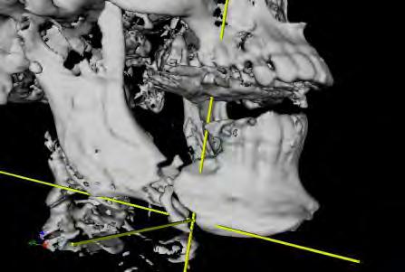


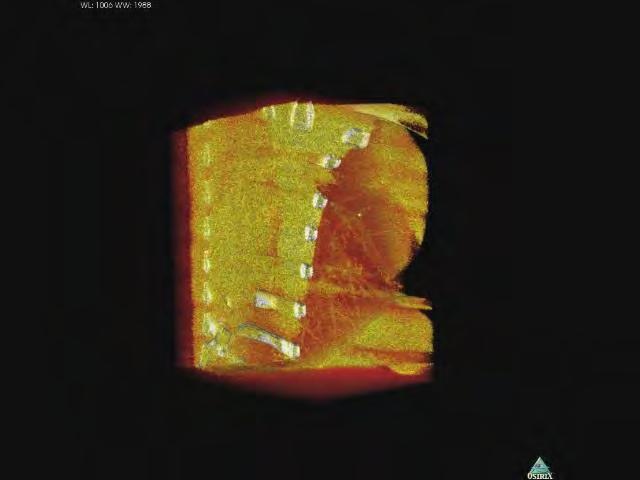
22 J. H. Siewerdsen (Johns Hopkins University) Image-Guided Head and Neck Surgery C-Spine Facial Nerve Cochlea Stapes Crura C-Arm Trials: Mandibulectomy Scan 4 Scan 3 Scan 2 Scan 1 Fibula Reconstruction Target (Radionecrosis) Resection Plates 22


23 C-Arm Trials: Skull Base Scan 4 Scan 3 Scan 2 Scan 1 Craniotomy Tumor margins Tumor Packing resection Chondrosarcoma Closure CBCT Guidance: Endoscopic Skull Base 23
 Real-time video augmentation Motion imaging Image-Guided")
24 Image-Guided Thoracic Surgery Low-dose CT screening Early detection Stage Ia tumors Reduced mortality Video-assisted thoracic surgery (VATS): a growing challenge Localization and resection of subpalpable lung tumors Intraoperative CBCT Direct localization of tumors and critical structures Deformable registration (inflated deflated) Real-time video augmentation Motion imaging Image-Guided Thoracic Surgery Goal: Direct visualization / localization of nodules in the deflated lung Inflated Deflated Porcine Study: Implanted Lung Nodules 24


25 Image-Guided Thoracic Surgery Goal: Direct visualization / localization of nodules in the deflated lung (B) 0.9 mm 1.5 mm Inflated Deflated 2.95 msv 1.63 msv 0.41 msv Inflated 1.63 msv Collapsed Image-Guided Thoracic Surgery Goal: Direct visualization / localization of nodules in the deflated lung Inflated Deflated 25
26 Segmentation and Registration Goal: Deformable registration from inhale to deflated lung Segmentation Inhaled Deflated Semi-Automatic Seeding + Region Growing Segmentation and Registration Goal: Deformable registration from inhale to deflated lung Segmentation Inhaled Deflated Semi-Automatic Seeding + Region Growing 26


27 Medial Line and Bifurcations Inhaled Deflated Segmentation Medial Line Junctions Corresponding Points Goal: Deformable registration from inhale to deflated lung Inhaled Deflated REG 27


")

 Thoracoscopic")
28 Corresponding Points + Surfaces Goal: Deformable registration from inhale to deflated lung Inhaled Deflated REG Preliminary Results: Segmentation Surface Mesh Bifurcations Inflated-Deflated (nodule in wedge) Correspondences Inflated-Deflated (mm-precision critical structures) Thoracoscopic Video-CBCT Fusion PA Fissure Target Video Overlay Sagittal Axial Coronal 28
 System geometry System configuration Acquisition techniques Reconstruction techniques Accelerates system development, translation, and system integration Improved system")
29 J. H. Siewerdsen (Johns Hopkins University) Summary and Conclusions CBCT entering a broad scope of applications Diagnostic imaging (breast, dental, ENT, MSK, orthopaedics) Image guidance (e.g., MI interventions, IGRT) Image science foundation for CBCT Spatial resolution Noise, NEQ Dose Artifacts (e.g., x-ray scatter) System geometry System configuration Acquisition techniques Reconstruction techniques Accelerates system development, translation, and system integration Improved system configurations (detector, speed, electronics) New applications (e.g. MSK imaging and DE-CBCT) Streamlined system integration (esp. for IGI and IGRT) Advanced reconstruction techniques, artifact reduction Acknowledgments I-STAR Laboratory JW Stayman, W Zbijewski S Schafer, P DeJean, Y Otake J Lee, A Uneri, S Nithiananthan D Mirota, P Prakash, GJ Gang Y Ding, W Liu, H Dang Hopkins Collaborators J Carrino (MSK Radiology) J Prince (Electrical Engineering) R Taylor, G Hager (Comp. Science) D Reh (Head and Neck Surgery) G Gallia (Neurosurgery) J Khanna (Spine Surgery) M Sussman (Thoracic Surgery) Funding Support / Disclosures National Institutes of Health Siemens Healthcare (SP, Erlangen) Carestream Health Elekta Oncology Systems 29
Acknowledgments. Nesterov s Method for Accelerated Penalized-Likelihood Statistical Reconstruction for C-arm Cone-Beam CT.
 June 5, Nesterov s Method for Accelerated Penalized-Likelihood Statistical Reconstruction for C-arm Cone-Beam CT Adam S. Wang, J. Webster Stayman, Yoshito Otake, Gerhard Kleinszig, Sebastian Vogt, Jeffrey
Acknowledgments. High Performance Cone-Beam CT of Acute Traumatic Brain Injury
 A. Sisniega et al. (presented at RSNA 214) High Performance Cone-Beam CT of Acute Traumatic Brain Injury A. Sisniega 1 W. Zbijewski 1, H. Dang 1, J. Xu 1 J. W. Stayman 1, J. Yorkston 2, N. Aygun 3 V. Koliatsos
Incorporation of Prior Knowledge for Region of Change Imaging from Sparse Scan Data in Image-Guided Surgery
 Incorporation of Prior Knowledge for Region of Change Imaging from Sparse Scan Data in Image-Guided Surgery J. Lee a, J. W. Stayman b, Y. Otake c, S. Schafer b, W. Zbijewski b, A. J. Khanna b,d, J. L.
Acknowledgements. Atlas-based automatic measurements of the morphology of the tibiofemoral joint
 Atlas-based automatic measurements of the morphology of the tibiofemoral joint M Brehler 1, G Thawait 2, W Shyr 1, J Ramsay 3, JH Siewerdsen 1,2, W Zbijewski 1 1 Dept. of Biomedical Engineering, Johns
Hot Topics in Imaging Physics A Key Interdisciplinary Science for 21 st Century Medicine
 Hot Topics in Imaging Physics A Key Interdisciplinary Science for 21 st Century Medicine Jeff Siewerdsen, PhD FAAPM Department of Biomedical Engineering Department of Computer Science Russell H. Morgan
Design and performance characteristics of a Cone Beam CT system for Leksell Gamma Knife Icon
 Design and performance characteristics of a Cone Beam CT system for Leksell Gamma Knife Icon WHITE PAPER Introduction Introducing an image guidance system based on Cone Beam CT (CBCT) and a mask immobilization
Self-Calibration of Cone-Beam CT Using 3D-2D Image Registration
 Self-Calibration of Cone-Beam CT Using 3D-2D Image Registration Sarah Ouadah 1 J. Webster Stayman 1 Grace Jianan Gang 1 Ali Uneri 2 Tina Ehtiati 3 Jeffrey H. Siewerdsen 1,2 1. Department of Biomedical
Philips SPECT/CT Systems
 Philips SPECT/CT Systems Ling Shao, PhD Director, Imaging Physics & System Analysis Nuclear Medicine, Philips Healthcare June 14, 2008 *Presented SNM08 Categorical Seminar - Quantitative SPECT and PET
Assessment of 3D performance metrics. X-ray based Volumetric imaging systems: Fourier-based imaging metrics. The MTF in CT
 Assessment of 3D performance metrics D and 3D Metrics of Performance Towards Quality Index: Volumetric imaging systems X-ray based Volumetric imaging systems: CBCT/CT Tomosynthesis Samuel Richard and Ehsan
Carestream s 2 nd Generation Metal Artifact Reduction Software (CMAR 2)
") Carestream s 2 nd Generation Metal Artifact Reduction Software (CMAR 2) Author: Levon Vogelsang Introduction Cone beam computed tomography (CBCT), or cone beam CT technology, offers considerable promise
Carestream s 2 nd Generation Metal Artifact Reduction Software (CMAR 2) Author: Levon Vogelsang Introduction Cone beam computed tomography (CBCT), or cone beam CT technology, offers considerable promise
Shadow casting. What is the problem? Cone Beam Computed Tomography THE OBJECTIVES OF DIAGNOSTIC IMAGING IDEAL DIAGNOSTIC IMAGING STUDY LIMITATIONS
 Cone Beam Computed Tomography THE OBJECTIVES OF DIAGNOSTIC IMAGING Reveal pathology Reveal the anatomic truth Steven R. Singer, DDS srs2@columbia.edu IDEAL DIAGNOSTIC IMAGING STUDY Provides desired diagnostic
CT Reconstruction Using Spectral and Morphological Prior Knowledge: Application to Imaging the Prosthetic Knee
 CT Reconstruction Using Spectral and Morphological Prior Knowledge: Application to Imaging the Prosthetic Knee Wojciech Zbijewski, J. Webster Stayman, Abdullah Muhit, John Yorkston, John A. Carrino and
Spiral CT. Protocol Optimization & Quality Assurance. Ge Wang, Ph.D. Department of Radiology University of Iowa Iowa City, Iowa 52242, USA
 Spiral CT Protocol Optimization & Quality Assurance Ge Wang, Ph.D. Department of Radiology University of Iowa Iowa City, Iowa 52242, USA Spiral CT Protocol Optimization & Quality Assurance Protocol optimization
CT NOISE POWER SPECTRUM FOR FILTERED BACKPROJECTION AND ITERATIVE RECONSTRUCTION
 CT NOISE POWER SPECTRUM FOR FILTERED BACKPROJECTION AND ITERATIVE RECONSTRUCTION Frank Dong, PhD, DABR Diagnostic Physicist, Imaging Institute Cleveland Clinic Foundation and Associate Professor of Radiology
3/27/2012 WHY SPECT / CT? SPECT / CT Basic Principles. Advantages of SPECT. Advantages of CT. Dr John C. Dickson, Principal Physicist UCLH
 3/27/212 Advantages of SPECT SPECT / CT Basic Principles Dr John C. Dickson, Principal Physicist UCLH Institute of Nuclear Medicine, University College London Hospitals and University College London john.dickson@uclh.nhs.uk
Background. Outline. Radiographic Tomosynthesis: Image Quality and Artifacts Reduction 1 / GE /
 Radiographic Tomosynthesis: Image Quality and Artifacts Reduction Baojun Li, Ph.D Department of Radiology Boston University Medical Center 2012 AAPM Annual Meeting Background Linear Trajectory Tomosynthesis
Acknowledgments and financial disclosure
 AAPM 2012 Annual Meeting Digital breast tomosynthesis: basic understanding of physics principles James T. Dobbins III, Ph.D., FAAPM Director, Medical Physics Graduate Program Ravin Advanced Imaging Laboratories
Image Quality Assessment and Quality Assurance of Advanced Imaging Systems for IGRT. AAPM Penn-Ohio Chapter Sep 25, 2015 Soyoung Lee, PhD
 Image Quality Assessment and Quality Assurance of Advanced Imaging Systems for IGRT AAPM Penn-Ohio Chapter Sep 25, 2015 Soyoung Lee, PhD 1 Outline q Introduction q Imaging performances in 4D-CBCT Image
ML reconstruction for CT
 ML reconstruction for CT derivation of MLTR rigid motion correction resolution modeling polychromatic ML model dual energy ML model Bruno De Man, Katrien Van Slambrouck, Maarten Depypere, Frederik Maes,
Optimization of CT Simulation Imaging. Ingrid Reiser Dept. of Radiology The University of Chicago
 Optimization of CT Simulation Imaging Ingrid Reiser Dept. of Radiology The University of Chicago Optimization of CT imaging Goal: Achieve image quality that allows to perform the task at hand (diagnostic
CT vs. VolumeScope: image quality and dose comparison
 CT vs. VolumeScope: image quality and dose comparison V.N. Vasiliev *a, A.F. Gamaliy **b, M.Yu. Zaytsev b, K.V. Zaytseva ***b a Russian Sci. Center of Roentgenology & Radiology, 86, Profsoyuznaya, Moscow,
Financial disclosure. Onboard imaging modality for IGRT
 Tetrahedron Beam Computed Tomography Based On Multi-Pixel X- Ray Source and Its Application in Image Guided Radiotherapy Tiezhi Zhang, Ph.D. Advanced X-ray imaging Lab Financial disclosure Patent royalty
Image Acquisition Systems
 Image Acquisition Systems Goals and Terminology Conventional Radiography Axial Tomography Computer Axial Tomography (CAT) Magnetic Resonance Imaging (MRI) PET, SPECT Ultrasound Microscopy Imaging ITCS
Computer-Tomography II: Image reconstruction and applications
 Computer-Tomography II: Image reconstruction and applications Prof. Dr. U. Oelfke DKFZ Heidelberg Department of Medical Physics (E040) Im Neuenheimer Feld 280 69120 Heidelberg, Germany u.oelfke@dkfz.de
Radiology. Marta Anguiano Millán. Departamento de Física Atómica, Molecular y Nuclear Facultad de Ciencias. Universidad de Granada
 Departamento de Física Atómica, Molecular y Nuclear Facultad de Ciencias. Universidad de Granada Overview Introduction Overview Introduction Tecniques of imaging in Overview Introduction Tecniques of imaging
Cone-beam computed tomography with a flat-panel imager: Initial performance characterization
 Cone-beam computed tomography with a flat-panel imager: Initial performance characterization D. A. Jaffray a) and J. H. Siewerdsen Department of Radiation Oncology, William Beaumont Hospital, Royal Oak,
CT Basics Principles of Spiral CT Dose. Always Thinking Ahead.
 1 CT Basics Principles of Spiral CT Dose 2 Who invented CT? 1963 - Alan Cormack developed a mathematical method of reconstructing images from x-ray projections Sir Godfrey Hounsfield worked for the Central
1. INTRODUCTION ABSTRACT
 Incorporating Tissue Excision in Deformable Image Registration: A Modified Demons Algorithm for Cone-Beam CT-Guided Surgery S.Nithiananthan, a D. Mirota, b A. Uneri, b S. Schafer, a Y. Otake, b J.W. Stayman,
8/7/2017. Disclosures. MECT Systems Overview and Quantitative Opportunities. Overview. Computed Tomography (CT) CT Numbers. Polyenergetic Acquisition
 CT Numbers. Polyenergetic Acquisition") Quantitative Multi-Energy Computed Tomography: Imaging and Therapy Advancements Disclosures MECT Systems Overview and Quantitative Opportunities The speaker receives research funding from GE Healthcare
Quantitative Multi-Energy Computed Tomography: Imaging and Therapy Advancements Disclosures MECT Systems Overview and Quantitative Opportunities The speaker receives research funding from GE Healthcare
PURE. ViSION Edition PET/CT. Patient Comfort Put First.
 PURE ViSION Edition PET/CT Patient Comfort Put First. 2 System features that put patient comfort and safety first. Oncology patients deserve the highest levels of safety and comfort during scans. Our Celesteion
RADIOLOGY AND DIAGNOSTIC IMAGING
 Day 2 part 2 RADIOLOGY AND DIAGNOSTIC IMAGING Dr hab. Zbigniew Serafin, MD, PhD serafin@cm.umk.pl 2 3 4 5 CT technique CT technique 6 CT system Kanal K: RSNA/AAPM web module: CT Systems & CT Image Quality
Image Guidance and Beam Level Imaging in Digital Linacs
 Image Guidance and Beam Level Imaging in Digital Linacs Ruijiang Li, Ph.D. Department of Radiation Oncology Stanford University School of Medicine 2014 AAPM Therapy Educational Course Disclosure Research
Tomographic Reconstruction
 Tomographic Reconstruction 3D Image Processing Torsten Möller Reading Gonzales + Woods, Chapter 5.11 2 Overview Physics History Reconstruction basic idea Radon transform Fourier-Slice theorem (Parallel-beam)
Spatial Resolution Properties in Penalized-Likelihood Reconstruction of Blurred Tomographic Data
 Spatial Resolution Properties in Penalized-Likelihood Reconstruction of Blurred Tomographic Data Wenying Wang, Grace J. Gang and J. Webster Stayman Department of Biomedical Engineering, Johns Hopkins University,
8/3/2016. Outline. The EPID Strikes Back: Future EPID Technology and Applications. Active Matrix Flat-Panel Imagers (AMFPIs)
") 8//6 The EPID Strikes Back: Future EPID Technology and Applications Larry E. Antonuk Department of Radiation Oncology University of Michigan, Ann Arbor Acknowledgements: Youcef El-Mohri, Qihua Zhao (U.
8//6 The EPID Strikes Back: Future EPID Technology and Applications Larry E. Antonuk Department of Radiation Oncology University of Michigan, Ann Arbor Acknowledgements: Youcef El-Mohri, Qihua Zhao (U.
Applications of Medical Imaging
 Applications of Medical Imaging Imaging for Therapy Guidance eff Siewerdsen, Ph.D. Department of Biomedical Engineering ohns Hopkins University Screening Is there an abnormality? At-risk populations Asymptomatic
Spectral analysis of non-stationary CT noise
 Spectral analysis of non-stationary CT noise Kenneth M. Hanson Los Alamos Scientific Laboratory Int. Symposium and Course on Computed Tomography, Las Vegas, April 7-11, 1980 This presentation available
Metal Artifact Reduction CT Techniques. Tobias Dietrich University Hospital Balgrist University of Zurich Switzerland
 Metal Artifact Reduction CT Techniques R S S S Tobias Dietrich University Hospital Balgrist University of Zurich Switzerland N. 1 v o 4 1 0 2. Postoperative CT Metal Implants CT is accurate for assessment
Towards full-body X-ray images
 Towards full-body X-ray images Christoph Luckner 1,2, Thomas Mertelmeier 2, Andreas Maier 1, Ludwig Ritschl 2 1 Pattern Recognition Lab, FAU Erlangen-Nuernberg 2 Siemens Healthcare GmbH, Forchheim christoph.luckner@fau.de
Fujifilm DR Solution. FDR AcSelerate. The new pinnacle in diagnostic imaging from Fujifilm ISS. CsI. Dynamic Visualization. Technology.
 Fujifilm DR Solution FDR AcSelerate The new pinnacle in diagnostic imaging from Fujifilm CsI Scintillator ISS Technology Dynamic Visualization Welcome to the X-ray room of the future! A streamlined solution
Multi-slice CT Image Reconstruction Jiang Hsieh, Ph.D.
 Multi-slice CT Image Reconstruction Jiang Hsieh, Ph.D. Applied Science Laboratory, GE Healthcare Technologies 1 Image Generation Reconstruction of images from projections. textbook reconstruction advanced
8/3/2016. Image Guidance Technologies. Introduction. Outline
 8/3/26 Session: Image Guidance Technologies and Management Strategies Image Guidance Technologies Jenghwa Chang, Ph.D.,2 Department of Radiation Medicine, Northwell Health 2 Hofstra Northwell School of
Scatter Correction for Dual source Cone beam CT Using the Pre patient Grid. Yingxuan Chen. Graduate Program in Medical Physics Duke University
 Scatter Correction for Dual source Cone beam CT Using the Pre patient Grid by Yingxuan Chen Graduate Program in Medical Physics Duke University Date: Approved: Lei Ren, Supervisor Fang Fang Yin, Chair
Lecture 6: Medical imaging and image-guided interventions
 ME 328: Medical Robotics Winter 2019 Lecture 6: Medical imaging and image-guided interventions Allison Okamura Stanford University Updates Assignment 3 Due this Thursday, Jan. 31 Note that this assignment
Integration of CBCT and a Skull Base Drilling Robot
 Integration of CBCT and a Skull Base Drilling Robot Computer Integrated Surgery II, Spring, 2011 Hao Dang and Zihan Chen Mentors: Dr. Jeffrey Siewerdsen and Dr. Peter Kazanzides 2011-5-19 Contents 1. Introduction
Introduction to Biomedical Imaging
 Alejandro Frangi, PhD Computational Imaging Lab Department of Information & Communication Technology Pompeu Fabra University www.cilab.upf.edu X-ray Projection Imaging Computed Tomography Digital X-ray
8/2/2016. Measures the degradation/distortion of the acquired image (relative to an ideal image) using a quantitative figure-of-merit
 using a quantitative figure-of-merit") Ke Li Assistant Professor Department of Medical Physics and Department of Radiology School of Medicine and Public Health, University of Wisconsin-Madison This work is partially supported by an NIH Grant
Ke Li Assistant Professor Department of Medical Physics and Department of Radiology School of Medicine and Public Health, University of Wisconsin-Madison This work is partially supported by an NIH Grant
Artefakt-resistente Bewegungsschätzung für die bewegungskompensierte CT
 Artefakt-resistente Bewegungsschätzung für die bewegungskompensierte CT Marcus Brehm 1,2, Thorsten Heußer 1, Pascal Paysan 3, Markus Oehlhafen 3, and Marc Kachelrieß 1,2 1 German Cancer Research Center
Penalized-Likelihood Reconstruction for Sparse Data Acquisitions with Unregistered Prior Images and Compressed Sensing Penalties
 Penalized-Likelihood Reconstruction for Sparse Data Acquisitions with Unregistered Prior Images and Compressed Sensing Penalties J. W. Stayman* a, W. Zbijewski a, Y. Otake b, A. Uneri b, S. Schafer a,
Fits you like no other
 Fits you like no other BrightView X and XCT specifications The new BrightView X system is a fully featured variableangle camera that is field-upgradeable to BrightView XCT without any increase in room
CT imaging using energy-sensitive photon-counting detectors. Disclosure. Vision 20/20 paper
 CT imaging using energy-sensitive photon-counting detectors Katsuyuki Ken Taguchi, Ph.D. ktaguchi@jhmi.edu Division of Medical Imaging Physics Department of Radiology Johns Hopkins University No financial
Computational Medical Imaging Analysis Chapter 4: Image Visualization
 Computational Medical Imaging Analysis Chapter 4: Image Visualization Jun Zhang Laboratory for Computational Medical Imaging & Data Analysis Department of Computer Science University of Kentucky Lexington,
Task-Based Imaging Performance in 3D X-Ray Tomography: Noise, Detectability, and Implications for System Design
 Task-Based Imaging Performance in 3D X-Ray Tomography: Noise, Detectability, and Implications for System Design by Jianan (Grace) Gang A thesis submitted in conformity with the requirements for the degree
Automated Image Analysis Software for Quality Assurance of a Radiotherapy CT Simulator
 Automated Image Analysis Software for Quality Assurance of a Radiotherapy CT Simulator Andrew J Reilly Imaging Physicist Oncology Physics Edinburgh Cancer Centre Western General Hospital EDINBURGH EH4
Z-MOTION. Universal Digital Radiographic System Z-MOTION. Control-X Medical CONTROL-X MEDICAL
 Control-X Medical Z-MOTION Compact design, low ceiling height requirement Motorized and manual movement capability Wide motion / SID range Best-in-class image quality Flexible connectivity to PACS systems
GPU implementation for rapid iterative image reconstruction algorithm
 GPU implementation for rapid iterative image reconstruction algorithm and its applications in nuclear medicine Jakub Pietrzak Krzysztof Kacperski Department of Medical Physics, Maria Skłodowska-Curie Memorial
Optimisation of Toshiba Aquilion ONE Volume Imaging
 Optimisation of Toshiba Aquilion ONE Volume Imaging Jane Edwards, RPRSG Royal Free London NHS Foundation Trust Dr Mufudzi Maviki, Plymouth Hospitals NHS Trust Background In 2011/12 Radiology at RFH was
DX-D 100 with WITH ITS EXCELLENT IMAGE QUALITY AND
 M O B I L E D R S O L U T I O N DX-D 100 with wireless detector Patients who most need imaging exams may lack the mobility necessary to move to the X-ray room or to position themselves properly for optimum
Limits of Dose Reduction in CT: How low is too low? Acknowledgement. Disclosures 8/2/2012
 8/2/22 Limits of Dose Reduction in CT: How low is too low? Ehsan Samei Duke University Acknowledgement Disclosures Research grant: NIH Research grant: DHS Research grant: RSNA, QIBA Research grant: General
The theory and practical aspects of proton imaging proton radiography (prad) and proton tomography (pct)
 and proton tomography (pct)") The theory and practical aspects of proton imaging proton radiography (prad) and proton tomography (pct) Fritz DeJongh, ProtonVDA Inc August 30 2018 Stay away from negative people. They have a problem
The theory and practical aspects of proton imaging proton radiography (prad) and proton tomography (pct) Fritz DeJongh, ProtonVDA Inc August 30 2018 Stay away from negative people. They have a problem
Estimating 3D Respiratory Motion from Orbiting Views
 Estimating 3D Respiratory Motion from Orbiting Views Rongping Zeng, Jeffrey A. Fessler, James M. Balter The University of Michigan Oct. 2005 Funding provided by NIH Grant P01 CA59827 Motivation Free-breathing
Intraoperative Prostate Tracking with Slice-to-Volume Registration in MR
 Intraoperative Prostate Tracking with Slice-to-Volume Registration in MR Sean Gill a, Purang Abolmaesumi a,b, Siddharth Vikal a, Parvin Mousavi a and Gabor Fichtinger a,b,* (a) School of Computing, Queen
Unique Features of the GE Senoclaire Tomosynthesis System. Tyler Fisher, M.S., DABR Therapy Physics, Inc.
 Unique Features of the GE Senoclaire Tomosynthesis System Tyler Fisher, M.S., DABR Therapy Physics, Inc. Conflict of Interest Disclosure I have no conflicts to disclose. Learning Objectives Overview of
A prototype table-top inverse-geometry volumetric CT system
 A prototype table-top inverse-geometry volumetric CT system Taly Gilat Schmidt a Department of Radiology, Stanford University, Stanford, California 94305 Josh Star-Lack NexRay, Inc., Los Gatos, California
Simulation of Mammograms & Tomosynthesis imaging with Cone Beam Breast CT images
 Simulation of Mammograms & Tomosynthesis imaging with Cone Beam Breast CT images Tao Han, Chris C. Shaw, Lingyun Chen, Chao-jen Lai, Xinming Liu, Tianpeng Wang Digital Imaging Research Laboratory (DIRL),
Cross Scatter in Dual Cone X ray Imaging: Magnitude, Avoidance, Correction, and. Artifact Reduction. William Michael Giles
 Cross Scatter in Dual Cone X ray Imaging: Magnitude, Avoidance, Correction, and Artifact Reduction by William Michael Giles Department of Medical Physics Duke University Date: Approved: Fang Fang Yin,
Digital Image Processing
 Digital Image Processing SPECIAL TOPICS CT IMAGES Hamid R. Rabiee Fall 2015 What is an image? 2 Are images only about visual concepts? We ve already seen that there are other kinds of image. In this lecture
Gradient-Based Differential Approach for Patient Motion Compensation in 2D/3D Overlay
 Gradient-Based Differential Approach for Patient Motion Compensation in 2D/3D Overlay Jian Wang, Anja Borsdorf, Benno Heigl, Thomas Köhler, Joachim Hornegger Pattern Recognition Lab, Friedrich-Alexander-University
A closer look at CT scanning
 Vet Times The website for the veterinary profession https://www.vettimes.co.uk A closer look at CT scanning Author : Charissa Lee, Natalie Webster Categories : General, Vets Date : April 3, 2017 A basic
Fits you like no other
 Fits you like no other Philips BrightView X and XCT specifications The new BrightView X system is a fully featured variableangle camera that is field-upgradeable to BrightView XCT without any increase
BME I5000: Biomedical Imaging
 1 Lucas Parra, CCNY BME I5000: Biomedical Imaging Lecture 4 Computed Tomography Lucas C. Parra, parra@ccny.cuny.edu some slides inspired by lecture notes of Andreas H. Hilscher at Columbia University.
REAL-TIME ADAPTIVITY IN HEAD-AND-NECK AND LUNG CANCER RADIOTHERAPY IN A GPU ENVIRONMENT
 REAL-TIME ADAPTIVITY IN HEAD-AND-NECK AND LUNG CANCER RADIOTHERAPY IN A GPU ENVIRONMENT Anand P Santhanam Assistant Professor, Department of Radiation Oncology OUTLINE Adaptive radiotherapy for head and
First CT Scanner. How it Works. Contemporary CT. Before and After CT. Computer Tomography: How It Works. Medical Imaging and Pattern Recognition
 Computer Tomography: How t Works Medical maging and Pattern Recognition Lecture 7 Computed Tomography Oleh Tretiak Only one plane is illuminated. Source-subject motion provides added information. 2 How
Limitations of Projection Radiography. Stereoscopic Breast Imaging. Limitations of Projection Radiography. 3-D Breast Imaging Methods
 Stereoscopic Breast Imaging Andrew D. A. Maidment, Ph.D. Chief, Physics Section Department of Radiology University of Pennsylvania Limitations of Projection Radiography Mammography is a projection imaging
Computed Tomography. Principles, Design, Artifacts, and Recent Advances. Jiang Hsieh THIRD EDITION. SPIE PRESS Bellingham, Washington USA
 Computed Tomography Principles, Design, Artifacts, and Recent Advances THIRD EDITION Jiang Hsieh SPIE PRESS Bellingham, Washington USA Table of Contents Preface Nomenclature and Abbreviations xi xv 1 Introduction
MEDICAL EQUIPMENT: COMPUTED TOMOGRAPHY. Prof. Yasser Mostafa Kadah
 MEDICAL EQUIPMENT: COMPUTED TOMOGRAPHY Prof. Yasser Mostafa Kadah www.k-space.org Recommended Textbook X-Ray Computed Tomography in Biomedical Engineering, by Robert Cierniak, Springer, 211 Computed Tomography
Developments in Dimensional Metrology in X-ray Computed Tomography at NPL
 Developments in Dimensional Metrology in X-ray Computed Tomography at NPL Wenjuan Sun and Stephen Brown 10 th May 2016 1 Possible factors influencing XCT measurements Components Influencing variables Possible
Joint ICTP-TWAS Workshop on Portable X-ray Analytical Instruments for Cultural Heritage. 29 April - 3 May, 2013
 2455-5 Joint ICTP-TWAS Workshop on Portable X-ray Analytical Instruments for Cultural Heritage 29 April - 3 May, 2013 Lecture NoteBasic principles of X-ray Computed Tomography Diego Dreossi Elettra, Trieste
DICOM. Supplement 188 Multi-Energy CT Imaging. DICOM Working Group 21 Computed Tomography
 DICOM Supplement 188 Multi-Energy CT Imaging DICOM Working Group 21 Computed Tomography Rationale Short introduction of Multi Energy (ME) s Overview: Imaging techniques, including scanning, reconstruction,
Background 8/2/2011. Development of Breast Models for Use in Simulation of Breast Tomosynthesis and CT Breast Imaging. Stephen J.
 Development of Breast Models for Use in Simulation of Breast Tomosynthesis and CT Breast Imaging Stephen J. Glick* J. Michael O Connor**, Clay Didier**, Mini Das*, * University of Massachusetts Medical
Ch. 4 Physical Principles of CT
 Ch. 4 Physical Principles of CT CLRS 408: Intro to CT Department of Radiation Sciences Review: Why CT? Solution for radiography/tomography limitations Superimposition of structures Distinguishing between
O-ARM IMAGING SYSTEM TECHNICAL SPECIFICATION GUIDE
 IMAGING SYSTEM TECHNICAL SPECIFICATION GUIDE SYSTEM FEATURES CATEGORY PHYSICAL DIMENSIONS IMAGING MODALITY PERFORMANCE SPECIFICATION Length Width Height Weight Gantry Opening Bore Diameter Single Plane
Slide 1. Technical Aspects of Quality Control in Magnetic Resonance Imaging. Slide 2. Annual Compliance Testing. of MRI Systems.
 Slide 1 Technical Aspects of Quality Control in Magnetic Resonance Imaging Slide 2 Compliance Testing of MRI Systems, Ph.D. Department of Radiology Henry Ford Hospital, Detroit, MI Slide 3 Compliance Testing
Implementation of Advanced Image Guided Radiation Therapy
 Image Acquisition Course Outline Principles, characteristics& applications of the available modalities Image Processing in the T x room Image guided treatment delivery What can / can t we do in the room
New Technology in Radiation Oncology. James E. Gaiser, Ph.D. DABR Physics and Computer Planning Charlotte, NC
 New Technology in Radiation Oncology James E. Gaiser, Ph.D. DABR Physics and Computer Planning Charlotte, NC Technology s s everywhere From the imaging chain To the planning system To the linac To QA..it..it
CBCT: Past, Present and Future. Disclosures. Computed Tomography. ATec Education Course Feb 28-Mar3, Douglas Moseley PhD, DABR
 CBCT: Past, Present and Future Douglas Moseley PhD, DABR Disclosures License Agreement Modus Medical Educational Consultant Elekta Oncology Systems Computed Tomography First CT Scanner Third Generation
Evaluation of Spectrum Mismatching using Spectrum Binning Approach for Statistical Polychromatic Reconstruction in CT
 Evaluation of Spectrum Mismatching using Spectrum Binning Approach for Statistical Polychromatic Reconstruction in CT Qiao Yang 1,4, Meng Wu 2, Andreas Maier 1,3,4, Joachim Hornegger 1,3,4, Rebecca Fahrig
The protocols used to scan the musculoskeletal system are tailored to each patient and
 Chapter 22. Musculoskeletal Protocols The protocols used to scan the musculoskeletal system are tailored to each patient and region being examined. The clinical indication for the examination will also
Micro-CT Methodology Hasan Alsaid, PhD
 Micro-CT Methodology Hasan Alsaid, PhD Preclinical & Translational Imaging LAS, PTS, GlaxoSmithKline 20 April 2015 Provide basic understanding of technical aspects of the micro-ct Statement: All procedures
Refraction Corrected Transmission Ultrasound Computed Tomography for Application in Breast Imaging
 Refraction Corrected Transmission Ultrasound Computed Tomography for Application in Breast Imaging Joint Research With Trond Varslot Marcel Jackowski Shengying Li and Klaus Mueller Ultrasound Detection
AIDR 3D Iterative Reconstruction:
 Iterative Reconstruction: Integrated, Automated and Adaptive Dose Reduction Erin Angel, PhD Manager, Clinical Sciences, CT Canon Medical Systems USA Iterative Reconstruction 1 Since the introduction of
Some reference material
 Some reference material Physics reference book on medical imaging: A good one is The Essential Physics of Medical Imaging, 3 rd Ed. by Bushberg et al. ($170! new). However, there are several similar books
Biomedical Imaging. Computed Tomography. Patrícia Figueiredo IST
 Biomedical Imaging Computed Tomography Patrícia Figueiredo IST 2013-2014 Overview Basic principles X ray attenuation projection Slice selection and line projections Projection reconstruction Instrumentation
Constructing System Matrices for SPECT Simulations and Reconstructions
 Constructing System Matrices for SPECT Simulations and Reconstructions Nirantha Balagopal April 28th, 2017 M.S. Report The University of Arizona College of Optical Sciences 1 Acknowledgement I would like
TomoTherapy Related Projects. An image guidance alternative on Tomo Low dose MVCT reconstruction Patient Quality Assurance using Sinogram
 TomoTherapy Related Projects An image guidance alternative on Tomo Low dose MVCT reconstruction Patient Quality Assurance using Sinogram Development of A Novel Image Guidance Alternative for Patient Localization
Cascaded Systems Analysis of the 3D NEQ for Cone-Beam CT and Tomosynthesis
 Cascaded Systems Analysis of the 3D NEQ for Cone-Beam CT and Tomosynthesis D. J. Tward, a J. H. Siewerdsen, a,b R. A. Fahrig, c and A. R. Pineda d a Ontario Cancer nstitute, Princess Margaret Hospital,
Position accuracy analysis of the stereotactic reference defined by the CBCT on Leksell Gamma Knife Icon
 Position accuracy analysis of the stereotactic reference defined by the CBCT on Leksell Gamma Knife Icon WHITE PAPER Introduction An image guidance system based on Cone Beam CT (CBCT) is included in Leksell
Digital Tomosynthesis for Target Localization
 Digital Tomosynthesis for Target Localization Fang-Fang Yin, Devon Godfrey, Lei Ren Jacqueline Maurer, Jackie Q-L Wu Duke University Medical Center Acknowledgements Duke Radiation Oncology faculty and
Imaging protocols for navigated procedures
 9732379 G02 Rev. 1 2015-11 Imaging protocols for navigated procedures How to use this document This document contains imaging protocols for navigated cranial, DBS and stereotactic, ENT, and spine procedures
Whole Body Submillimeter Scan
 128 Whole Body Submillimeter Scan The real value of 64ch/128slice CT systems does not come from the ability to perfom cardiac scans but the capability to scan all parts of the body in high definition submillimeter