BDP: BrainSuite Diffusion Pipeline. Chitresh Bhushan
|
|
|
- Cynthia Matthews
- 5 years ago
- Views:
Transcription
1 BDP: BrainSuite Diffusion Pipeline Chitresh Bhushan


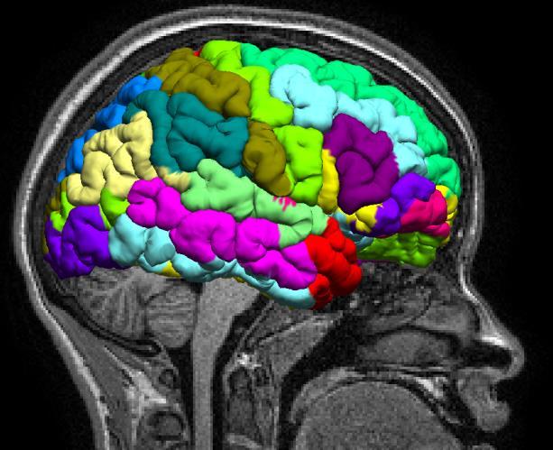
2 Why diffusion MRI? T 2 weighted MPRAGE FA map Fiber track Quantify microstructural tissue characteristics Structural connectivity Connectome Clinical Abnormalities in white matter stroke etc. Multimodal image analysis Sporns et al. 2005; Wedeen et al. 2008; Hagmann et al. 2007; Jones et al. 2011; Johansen-Berg et al. 2009


3 BDP MPRAGE Diffusion Tensor ODF ROI Connectivity ROI Statistics
4 Diffusion Pipeline Dicom to NIfTI Bias-field corrected MPRAGE Co-registration SVReg ROIs Diffusion Modeling ROI-wise Statistics Tractography Custom ROIs Connectivity analysis
5 Diffusion Pipeline Dicom to NIfTI Bias-field corrected MPRAGE Co-registration bdp13.exe bdp13.sh SVReg ROIs Diffusion Modeling bdp13.exe bdp13.sh ROI-wise Statistics Custom ROIs Tractography Connectivity analysis BrainSuite13 GUI


6 bdp.exe overview Co-register diffusion and MPRAGE scan Distortion correction multiple methods Fit diffusion model Multiple models Tensor, ODFs Compute basic ROI-wise statistics Custom ROIs, track based ROIs etc.
7 Co-registration { & distortion correction
")

8 Co-register MPRAGE Coord (Surfaces, Labels) Diffusion Coord Diffusion MRI - Rigid registration is not enough!
 Susceptibility differences Magnetic field (B 0 )")
 Field")
9 EPI distortion Diffusion MRI uses fast acquisition Echo planar Imaging (EPI) Susceptibility differences Magnetic field (B 0 ) inhomogeneity EPI is sensitive to B 0 inhomogeneity Localized geometric distortion MPRAGE image b=0 image (EPI) Field inhomogeneity map
")


10 EPI distortion b=0 image (EPI) MPRAGE Overlay with edges Misalignment with structural scans by several millimeters Limits the accuracy of multi-modal analysis


11 Distortion correction framework MPRAGE φ Distortion/deformation map b=0 image (EPI) Somehow estimate/find the deformation map φ
12 Distortion correction in BDP 1. Registration based distortion correction Uses structural image to estimate distortion field Does not require any field inhomogeneity map 2. Fieldmap based distortion correction Requires field inhomogeneity map Lower computational requirement 3. No distortion correction Only Rigid registration to MPRAGE
13 Fieldmap based correction (Indirectly) Acquire the deformation map φ Computed from field inhomogeneity map (fieldmap) B 0 (x, y) Deformation map Fieldmap Echo spacing Corrected Image Distorted Image Phase encoding gradient duration Field inhomogeneity map Jezzard 1995, 2011, Bhushan et al. 2012


14 Fieldmap based correction Accurate correction in most regions Drawbacks: Distorted image Fieldmap based correction MPRAGE Additional Data acquisition (fieldmap) Can not be used on data already acquired without field map Sensitive to errors in fieldmap acquisition/estimation
15 Registration based correction No extra data (fieldmap) is required Estimate a deformation map φ which best aligns MPRAGE and b=0 image Uses anatomical information Mutual-information based non-rigid registration MPRAGE Corrected Image Regularization Estimated Deformation map Normalized MI Bhushan et al. 2012

16 Registration based correction Similar performance to fieldmap method Before After Before After Bhushan et al. 2012


17 Comparison Registration based correction Fieldmap based correction Reversed Gradient / Interlaced sampling* MPRAGE Be aware of limitation of your dataset and/or correction method * To be included in future version of BDP. Bhushan et al., ISMRM 2013, p55
18 Summary: Distortion correction 1. Registration based distortion correction Uses structural image to estimate distortion field Does not require any field inhomogeneity map 2. Fieldmap based distortion correction Requires field inhomogeneity map Lower computational requirement 3. No distortion correction Only Rigid registration to MPRAGE --no-distortion-correction


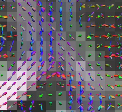
19 Diffusion models Estimates diffusion tensors FA, MD, color-fa Axial, Radial diffusivity ODFs using FRT and FRACT Haldar et al. 2013

 and diffusion")
20 Tractography & connectivity Combine labels (from MPRAGE space) and diffusion information

21 Tractography & connectivity Fiber tracking in MPRAGE and diffusion space ROI-wise connectivity analysis

22 Syntax NIfTI input (.nii or.nii.gz) bdp13.exe <BFC File> [Optional Flags] --nii <4D DWI NIfTI> --bvec <Gradient file> --bval <b-value file> BDP expects diffusion gradient direction in voxel coordinates BDP uses NIfTI header matrix extensively for registration


23 BDP: Flexible flags.t1_coord.d_coord ~40 optional flags Refer documentation for full descriptions and usage

24 Example multimodal study Choi et al., A Multimodal Investigation of Neuronal/Axonal Integrity Using Structural T1-weighted Imaging, Diffusion Tensor Imaging, and H1 MR Spectroscopy, ISMRM 2013, Salt Lake City, p. 1951
25 References C Bhushan, JP Haldar, AA Joshi, RM Leahy, Correcting susceptibilityinduced distortion in diffusion-weighted MRI using constrained nonrigid registration, APSIPA ASC, Hollywood, 3-6 Dec 2012 DW Shattuck, AA Joshi, JP Haldar, C Bhushan, S Choi, AC Krause, JL Wisnowski, H Damasio, AW Toga, RM Leahy, New BrainSuite13 Tools for Image Segmentation, Registration, Connectivity Analysis and Visualization, OHBM, Seattle, 2013, p DW Shattuck, AA Joshi, JP Haldar, C Bhushan, S Choi, AC Krause, JL Wisnowski, AW Toga and RM Leahy, Tools for Brain Image Segmentation, Registration, and Connectivity, ISMRM, Salt Lake City, 2013, p C Bhushan, AA Joshi, RM Leahy, JP Haldar, Accelerating Data Acquisition for Reversed-Gradient Distortion Correction in Diffusion MRI: A Constrained Reconstruction Approach, ISMRM, Salt Lake City, 2013, p. 55
26 Download hands-on dataset
27 Running BDP { or bdp13.exe
28 Diffusion Pipeline Dicom to NIfTI Bias-field corrected MPRAGE Co-registration bdp13.exe bdp13.sh SVReg ROIs Diffusion Modeling bdp13.exe bdp13.sh ROI-wise Statistics Custom ROIs Tractography Connectivity analysis BrainSuite13 GUI
29 bdp13.exe / bdp13.sh Command line tool Highly extensible using your batch/shell scripts Flexible numerous flags for custom processing Requires Matlab 2012a MCR Visual C++ runtime package (windows only) Documentation Detailed flag description
30 Syntax DICOM bdp13.exe <BFC File> [Optional Flags] -d <DICOM path> [DICOM path...] Limited support BDP extracts (most) relevant diffusion scan parameters NIfTI (.nii or.nii.gz) bdp13.exe <BFC File> [Optional Flags] --nii <4D DWI NIfTI> --bvec <Gradient file> --bval <b-value file> BDP expects diffusion gradient direction in voxel coordinates BDP uses NIfTI header matrix extensively for registration Linux and Macintosh Replace bdp13.exe by bdp13.sh
31 Example C:\bdp13p17_win64\bdp13.exe C:\5934\5934.bfc.nii.gz - nii C:\5934\5934.dwi.nii.gz --bvec C:\5934\5934.dwi.bvec --bval C:\5934\5934.dwi.bval Flags are separated by space If required file are not in current working directory, then specify full path to files Any number of flags can be added Output files: Many many files. (see documentation for all details) <fileprefix>.bdpsummary.txt Summary of all the processing with references The command used for future reference
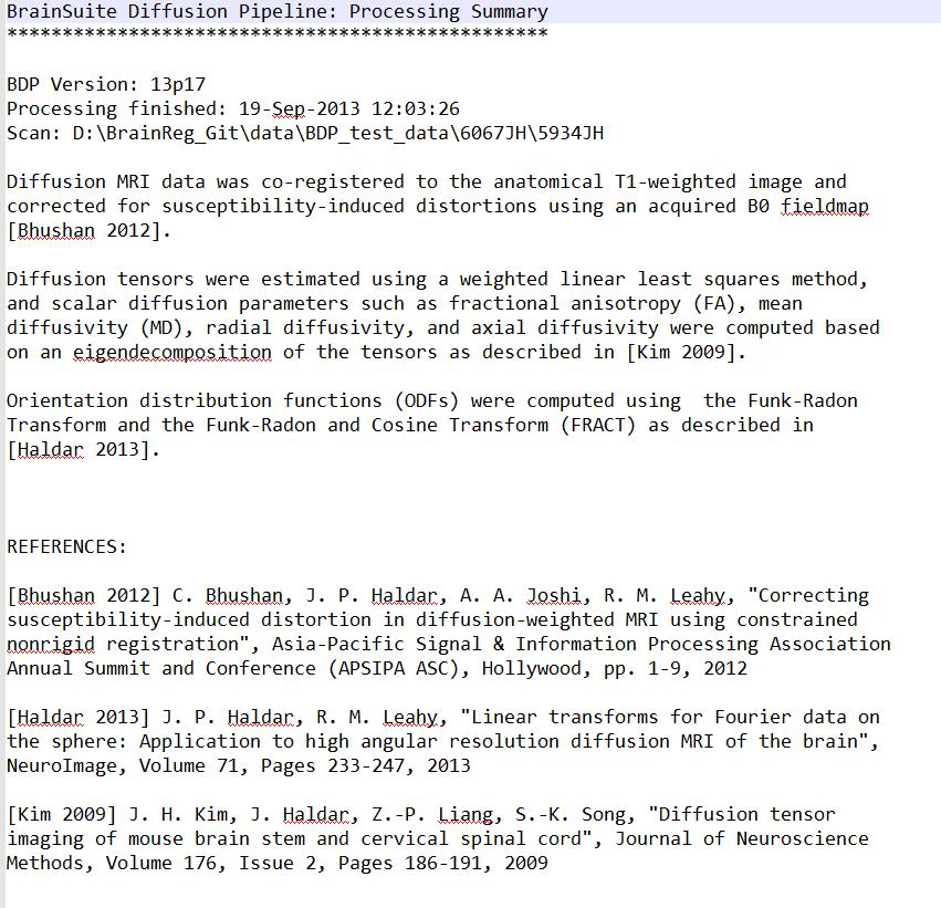

32 <fileprefix>.bdpsummary.txt
33 Command line output Command line output Always verbose with relevant important information

34 Default flags When no optional flag is defined: --tensor --dir=y Registration based distortion correction Only T1-coordinate outputs Outputs are saved in same directory as bfc file --threads=4 C:\bdp13p17_win64\bdp13.exe C:\5934\5934.bfc.nii.gz - nii C:\5934\5934.dwi.nii.gz --bvec C:\5934\5934.dwi.bvec --bval C:\5934\5934.dwi.bval
35 Help! --help or h Prints out description of all BDP flags Also reports the version of BDP executable being run --check-for-updates Connects to BrainSuite server to check if a new version of BDP is available All other flags and options are ignored and BDP terminates after printing help or checking for updates. Of course online documentation:
36 Diffusion models Multiple model flags can be used at once: Diffusion Tensor --tensor <name>.eig.nii.gz saves all eigen value/vectors FA, colorfa, axial, radial, L2, L3, MD ODFs --FRT --FRACT <name>.odf Load saved Spherical harmonic coefficients Coordinate filename suffix.t1_coord : In T1/MPRAGE coordinates.d_coord : In diffusion coordinates
37 Distortion direction --dir=<direction> Define phase encoding direction x : increases along the Right side of the subject x- : increases along the left side of the subject. y : increases along the Anterior direction of the subject y- : increases along the posterior direction of the subject z : increases along the Superior direction z- : increases along the inferior direction Example --dir=y- -ve sign is important only for fieldmap based correction
 --ignore-fieldmap-fov BDP checks for overlap of field of view (FOV) of diffusion scan and fieldmap scan Overrides FOV check Example:")
38 Fieldmap based correction Required --fieldmap-correction <fname.nii.gz> (in rad/sec) --echo-spacing=<t> (in sec) Example --fieldmap-correction fieldmap.radians.nii.gz --echo-spacing= Optional --fieldmap-smooth3=<s> (in mm) --ignore-fieldmap-fov BDP checks for overlap of field of view (FOV) of diffusion scan and fieldmap scan Overrides FOV check Example: --fieldmap-smooth3=0.75
39 Some (more) useful flags --output-subdir <directory_name> allows to specify a sub-directory name in which output files would be written Example: --output-subdir BDPv17 --output-diffusion-coordinate Enables estimation of diffusion tensors and/or ODFs in the native diffusion coordinate All native diffusion coordinate files are saved in a seperate folder named diffusion_coord_outputs Outputs in MPRAGE coordinates are always saved
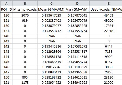
40 Statistics flags --generate-stats Requires extraction (& SVReg) output files Writes statistics for white matter(wm), grey matter(gm), and both WM and GM combined Outputs in.csv format

41 Statistics flags Default information: WM/GM from <name>.cortex.dewisp.mask.nii.gz SVReg labels from <name>.svreg.corr.label.nii.gz ROI_ID from brainsuite_labeldescription.xml --custom-label-xml <filename.xml>

42 Statistics FOV issues BDP detects overlay of field of view (FOV) of MPRAGE and diffusion scan Computes missing voxels in each ROI By default does not compute stats for ROI missing any voxel --force-partial-roi-stats Force stats computation in all ROIs
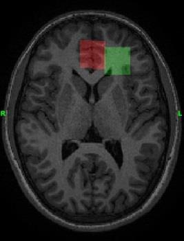
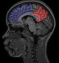
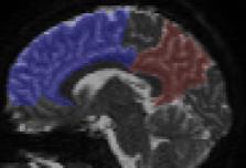
43 Custom labels --custom-diffusion-label <name> --custom-t1-label <name> Define custom labels in either coordinates <name> can be either NIfTI filename or directory name Custom labels can be painted in BrainSuite13 --custom-label-xml <filename.xml> Example: --custom-diffusion-label ROI26.nii.gz --custom-t1-label T1_labels When --custom-label-xml is not used: BDP generates 5-digit ROI IDs for each label found Saves ROI ID maps (to labels found) in an.xml file <fileprefix>.bdp_roi_map.xml
44 Re-compute statistics --only-generate-stats --generate-stats-only Refined/manually corrected labels re-run BDP to only compute statistics Skip all of the processing (co-registration, distortion correction and tensor/odf estimation) All of the other flags MUST be used in the same way as they were in the initial BDP run (<fileprefix>.bdpsummary.txt)
45 Transform image volumes --transform-diffusion-volume <name> --transform-t1-volume <name> To-and-fro from diffusion and T1-coordinates <name> can be either NIfTI filename or directory name This does not perform any distortion correction --transform-interpolation <method> Define interpolation method linear, nearest, cubic or spline --transform-data-only Skip all of the processing (co-registration, distortion correction and tensor/odf estimation) All of the other flags MUST be used in the same way as they were in the initial BDP run (<fileprefix>.bdpsummary.txt)
46 Error!

47 BDP Documentation: Detailed flag description:
BDP: BrainSuite Diffusion Pipeline. Chitresh Bhushan
 BDP: BrainSuite Diffusion Pipeline Chitresh Bhushan Why diffusion MRI? T 2 weighted MPRAGE FA map Fiber track Quantify microstructural tissue characteristics Structural connectivity Connectome Clinical
UCLA Advanced Neuroimaging Summer Program David W. Shattuck, PhD
 BrainSuite UCLA Advanced Neuroimaging Summer Program Presented 18 July 2013 David W. Shattuck, PhD Associate Professor Department of Neurology David Geffen School of Medicine at UCLA http://www.loni.ucla.edu/~shattuck/
BrainSuite. presented at the UCLA/NITP Advanced Neuroimaging Summer Program 29 July 2014
 BrainSuite presented at the UCLA/NITP Advanced Neuroimaging Summer Program 29 July 2014 David Shattuck Ahmanson-Lovelace Brain Mapping Center Department of Neurology David Geffen School of Medicine at
Playing with data from lab
 Playing with data from lab Getting data off the scanner From the Patient Browser, select the folder for the study you want (or within that study, the set of images you want), and then from the Transfer
HST.583 Functional Magnetic Resonance Imaging: Data Acquisition and Analysis Fall 2008
 MIT OpenCourseWare http://ocw.mit.edu HST.583 Functional Magnetic Resonance Imaging: Data Acquisition and Analysis Fall 2008 For information about citing these materials or our Terms of Use, visit: http://ocw.mit.edu/terms.
FROM IMAGE RECONSTRUCTION TO CONNECTIVITY ANALYSIS: A JOURNEY THROUGH THE BRAIN'S WIRING. Francesca Pizzorni Ferrarese
 FROM IMAGE RECONSTRUCTION TO CONNECTIVITY ANALYSIS: A JOURNEY THROUGH THE BRAIN'S WIRING Francesca Pizzorni Ferrarese Pipeline overview WM and GM Segmentation Registration Data reconstruction Tractography
BrainSuite Lab Exercises. presented at the UCLA/NITP Advanced Neuroimaging Summer Program 29 July 2014
 BrainSuite Lab Exercises presented at the UCLA/NITP Advanced Neuroimaging Summer Program 29 July 2014 1. Opening and Displaying an MRI Start BrainSuite Drag and drop the T1 image from the native space
Supplementary methods
 Supplementary methods This section provides additional technical details on the sample, the applied imaging and analysis steps and methods. Structural imaging Trained radiographers placed all participants
Brain Extraction, Registration & EPI Distortion Correction
 Brain Extraction, Registration & EPI Distortion Correction What use is Registration? Some common uses of registration: Combining across individuals in group studies: including fmri & diffusion Quantifying
SPM8 for Basic and Clinical Investigators. Preprocessing. fmri Preprocessing
 SPM8 for Basic and Clinical Investigators Preprocessing fmri Preprocessing Slice timing correction Geometric distortion correction Head motion correction Temporal filtering Intensity normalization Spatial
Diffusion-MRI processing for group analysis
 Diffusion-MRI processing for group analysis Felix Renard IRMaGe: Inserm US 17 / CNRS UMS 3552 University Hospital of Grenoble - France 25/09/2015 felixrenard@gmail.com 1 Diffusion-MRI processing for group
PANDA Manual Zaixu Cui & Suyu Zhong & Gaolang Gong
 PANDA Manual Zaixu Cui & Suyu Zhong & Gaolang Gong National Key Laboratory of Cognitive Neuroscience and Learning Beijing Normal University, China Contents Overview Setup Files/Directories selection Preparing
HST.583 Functional Magnetic Resonance Imaging: Data Acquisition and Analysis Fall 2008
 MIT OpenCourseWare http://ocw.mit.edu HST.583 Functional Magnetic Resonance Imaging: Data Acquisition and Analysis Fall 2008 For information about citing these materials or our Terms of Use, visit: http://ocw.mit.edu/terms.
Diffusion MRI Acquisition. Karla Miller FMRIB Centre, University of Oxford
 Diffusion MRI Acquisition Karla Miller FMRIB Centre, University of Oxford karla@fmrib.ox.ac.uk Diffusion Imaging How is diffusion weighting achieved? How is the image acquired? What are the limitations,
Basic fmri Design and Analysis. Preprocessing
 Basic fmri Design and Analysis Preprocessing fmri Preprocessing Slice timing correction Geometric distortion correction Head motion correction Temporal filtering Intensity normalization Spatial filtering
Quantitative MRI of the Brain: Investigation of Cerebral Gray and White Matter Diseases
 Quantities Measured by MR - Quantitative MRI of the Brain: Investigation of Cerebral Gray and White Matter Diseases Static parameters (influenced by molecular environment): T, T* (transverse relaxation)
ASAP_2.0 (Automatic Software for ASL Processing) USER S MANUAL
 USER S MANUAL") ASAP_2.0 (Automatic Software for ASL Processing) USER S MANUAL ASAP was developed as part of the COST Action "Arterial Spin Labelling Initiative in Dementia (AID)" by: Department of Neuroimaging, Institute
ASAP_2.0 (Automatic Software for ASL Processing) USER S MANUAL ASAP was developed as part of the COST Action "Arterial Spin Labelling Initiative in Dementia (AID)" by: Department of Neuroimaging, Institute
User s Guide Neuroimage Processing ToolKit (NPTK) Version 2.0 fmri Registration Software Pipeline for Functional Localization
 Version 2.0 fmri Registration Software Pipeline for Functional Localization") User s Guide Neuroimage Processing ToolKit (NPTK) Version 2.0 fmri Registration Software Pipeline for Functional Localization Software Written by Ali Gholipour SIP Lab, UTD, 2005-2010 Revision 2.0 February
User s Guide Neuroimage Processing ToolKit (NPTK) Version 2.0 fmri Registration Software Pipeline for Functional Localization Software Written by Ali Gholipour SIP Lab, UTD, 2005-2010 Revision 2.0 February
2. Creating Field Maps Using the Field Map GUI (Version 2.0) in SPM5
 in SPM5") 1. Introduction This manual describes how to use the Field Map Toolbox Version 2.0 for creating unwrapped field maps that can be used to do geometric distortion correction of EPI images in SPM5. 1. 1.
1. Introduction This manual describes how to use the Field Map Toolbox Version 2.0 for creating unwrapped field maps that can be used to do geometric distortion correction of EPI images in SPM5. 1. 1.
Super-Resolution Reconstruction of Diffusion-Weighted Images from Distortion Compensated Orthogonal Anisotropic Acquisitions.
 Super-Resolution Reconstruction of Diffusion-Weighted Images from Distortion Compensated Orthogonal Anisotropic Acquisitions. Benoit Scherrer Ali Gholipour Simon K. Warfield Children s Hospital Boston,
The WU-Minn HCP Open Access Initial Data Release: User Guide. Appendix 2 File Names and Directory Structure for Minimally Pre-Processed Data
 The WU-Minn HCP Open Access Initial Data Release: User Guide Appendix 2 File Names and Directory Structure for Minimally Pre-Processed Data Version 2 release: 21 December 2012 Table of Contents File Names
Multimodal Imaging Brain Connectivity Analysis (MIBCA)
") Multimodal Imaging Brain Connectivity Analysis (MIBCA) Andre Santos Ribeiro, Luis Miguel Lacerda, Hugo Ferreira April 23, 2015 Abstract In recent years, connectivity studies using neuroimaging data have
Multimodal Imaging Brain Connectivity Analysis (MIBCA) Andre Santos Ribeiro, Luis Miguel Lacerda, Hugo Ferreira April 23, 2015 Abstract In recent years, connectivity studies using neuroimaging data have
Introduction to fmri. Pre-processing
 Introduction to fmri Pre-processing Tibor Auer Department of Psychology Research Fellow in MRI Data Types Anatomical data: T 1 -weighted, 3D, 1/subject or session - (ME)MPRAGE/FLASH sequence, undistorted
Functional MRI in Clinical Research and Practice Preprocessing
 Functional MRI in Clinical Research and Practice Preprocessing fmri Preprocessing Slice timing correction Geometric distortion correction Head motion correction Temporal filtering Intensity normalization
High Fidelity Brain Connectivity Imaging
 CNI Inauguration Workshop Stanford, March 22 nd, 2012 High Fidelity Brain Connectivity Imaging -Recent Progress on Diffusion Weighted MRI for High Resolution and Low Distortion Allen W. Song, PhD Brain
Lilla Zöllei A.A. Martinos Center, MGH; Boston, MA
 Lilla Zöllei lzollei@nmr.mgh.harvard.edu A.A. Martinos Center, MGH; Boston, MA Bruce Fischl Gheorghe Postelnicu Jean Augustinack Anastasia Yendiki Allison Stevens Kristen Huber Sita Kakonoori + the FreeSurfer
Diffusion model fitting and tractography: A primer
 Diffusion model fitting and tractography: A primer Anastasia Yendiki HMS/MGH/MIT Athinoula A. Martinos Center for Biomedical Imaging 03/18/10 Why n how Diffusion model fitting and tractography 0/18 Why
SPM8 for Basic and Clinical Investigators. Preprocessing
 SPM8 for Basic and Clinical Investigators Preprocessing fmri Preprocessing Slice timing correction Geometric distortion correction Head motion correction Temporal filtering Intensity normalization Spatial
Susceptibility Distortion Correction for Echo Planar Images with Non-uniform B-Spline Grid Sampling: A Diffusion Tensor Image Study
 Susceptibility Distortion Correction for Echo Planar Images with Non-uniform B-Spline Grid Sampling: A Diffusion Tensor Image Study M.O. Irfanoglu 1,L.Walker 3,S.Sammet 2, C. Pierpaoli 3,andR.Machiraju
EPI Data Are Acquired Serially. EPI Data Are Acquired Serially 10/23/2011. Functional Connectivity Preprocessing. fmri Preprocessing
 Functional Connectivity Preprocessing Geometric distortion Head motion Geometric distortion Head motion EPI Data Are Acquired Serially EPI Data Are Acquired Serially descending 1 EPI Data Are Acquired
How to create a head model
 How to create a head model This document describes the command line tools: mri2mesh: Central tool to reconstruct a head model from T1w and T2w data dwi2cond: Reconstruct conductivity tensors for brain
Atelier 2 : Calcul Haute Performance et Sciences du Vivant Forum er juillet, Paris, France
 From Diffusion MR Image Analysis to Whole Brain Connectivity Simulation Jean-Philippe Thiran EPFL Lausanne, Switzerland EPFL - Lausanne HPC in life sciences at EPFL The Blue Brain project: create a biologically
Fmri Spatial Processing
 Educational Course: Fmri Spatial Processing Ray Razlighi Jun. 8, 2014 Spatial Processing Spatial Re-alignment Geometric distortion correction Spatial Normalization Smoothing Why, When, How, Which Why is
Image Registration + Other Stuff
 Image Registration + Other Stuff John Ashburner Pre-processing Overview fmri time-series Motion Correct Anatomical MRI Coregister m11 m 21 m 31 m12 m13 m14 m 22 m 23 m 24 m 32 m 33 m 34 1 Template Estimate
ECSE 626 Project Report Multimodality Image Registration by Maximization of Mutual Information
 ECSE 626 Project Report Multimodality Image Registration by Maximization of Mutual Information Emmanuel Piuze McGill University Montreal, Qc, Canada. epiuze@cim.mcgill.ca Abstract In 1997, Maes et al.
DIFFUSION TENSOR IMAGING ANALYSIS. Using Analyze
 DIFFUSION TENSOR IMAGING ANALYSIS Using Analyze 2 Table of Contents 1. Introduction page 3 2. Loading DTI Data page 4 3. Computing DTI Maps page 5 4. Defining ROIs for Fiber Tracking page 6 5. Visualizing
GLM for fmri data analysis Lab Exercise 1
 GLM for fmri data analysis Lab Exercise 1 March 15, 2013 Medical Image Processing Lab Medical Image Processing Lab GLM for fmri data analysis Outline 1 Getting Started 2 AUDIO 1 st level Preprocessing
EMSegmenter Tutorial (Advanced Mode)
") EMSegmenter Tutorial (Advanced Mode) Dominique Belhachemi Section of Biomedical Image Analysis Department of Radiology University of Pennsylvania 1/65 Overview The goal of this tutorial is to apply the
EMSegmenter Tutorial (Advanced Mode) Dominique Belhachemi Section of Biomedical Image Analysis Department of Radiology University of Pennsylvania 1/65 Overview The goal of this tutorial is to apply the
C. Leon Partain JOURNAL OF MAGNETIC RESONANCE IMAGING EDITOR-IN-CHIEF
 JOURNAL OF MAGNETIC RESONANCE IMAGING ri m j / m o c. E ning M C ear l lth a he li ey w w. w w VOLUME 37 NUMBER 6 JUNE 2013 VOLUME 37 NUMBER 6 JUNE 2013 PAGES 1257 1504 CORRECTION OF EDDY CURRENT DISTORTIONS
Saturn User Manual. Rubén Cárdenes. 29th January 2010 Image Processing Laboratory, University of Valladolid. Abstract
 Saturn User Manual Rubén Cárdenes 29th January 2010 Image Processing Laboratory, University of Valladolid Abstract Saturn is a software package for DTI processing and visualization, provided with a graphic
fmri Basics: Spatial Pre-processing Workshop
 fmri Basics: Spatial Pre-processing Workshop Starting a VNC session: Most of your fmri analysis will be done on the central Linux machines accessed via a VNC (Virtual Network Computing) server. This is
ECE1778 Final Report MRI Visualizer
 ECE1778 Final Report MRI Visualizer David Qixiang Chen Alex Rodionov Word Count: 2408 Introduction We aim to develop a mobile phone/tablet based neurosurgical MRI visualization application with the goal
TRACULA: Troubleshooting, visualization, and group analysis
 TRACULA: Troubleshooting, visualization, and group analysis Anastasia Yendiki HMS/MGH/MIT Athinoula A. Martinos Center for Biomedical Imaging 18/11/13 TRACULA: troubleshooting, visualization, group analysis
User s Guide Neuroimage Processing ToolKit (NPTK) Version.1.7 (beta) fmri Registration Software Pipeline for Functional Localization
 Version.1.7 (beta) fmri Registration Software Pipeline for Functional Localization") User s Guide Neuroimage Processing ToolKit (NPTK) Version.1.7 (beta) fmri Registration Software Pipeline for Functional Localization Software Written by Ali Gholipour SIP Lab, UTD, 2005-2007 Revision 1.7
User s Guide Neuroimage Processing ToolKit (NPTK) Version.1.7 (beta) fmri Registration Software Pipeline for Functional Localization Software Written by Ali Gholipour SIP Lab, UTD, 2005-2007 Revision 1.7
Automatic Subthalamic Nucleus Targeting for Deep Brain Stimulation. A Validation Study
 Automatic Subthalamic Nucleus Targeting for Deep Brain Stimulation. A Validation Study F. Javier Sánchez Castro a, Claudio Pollo a,b, Jean-Guy Villemure b, Jean-Philippe Thiran a a École Polytechnique
Motion Correction in fmri by Mapping Slice-to-Volume with Concurrent Field-Inhomogeneity Correction
 Motion Correction in fmri by Mapping Slice-to-Volume with Concurrent Field-Inhomogeneity Correction Desmond T.B. Yeo 1,2, Jeffery A. Fessler 2, and Boklye Kim 1 1 Department of Radiology, University of
CATNAP User Manual v1.0
 CATNAP User Manual v1.0 July 6, 2006 Nera-Lee Patel and Bennett Landman Note: Items in green text are only sample value. Your values may be different. I. CreateParManifest -- Creates a spreadsheet to show
Computational Neuroanatomy
 Computational Neuroanatomy John Ashburner john@fil.ion.ucl.ac.uk Smoothing Motion Correction Between Modality Co-registration Spatial Normalisation Segmentation Morphometry Overview fmri time-series kernel
MriCloud DTI Processing Pipeline. DTI processing can be initiated by choosing DTI Processing in the top menu bar.
 MriCloud DTI Processing Pipeline 1: Data Upload 1-1: Web interface DTI processing can be initiated by choosing DTI Processing in the top menu bar. To avoid any HIPPA issues, data need to be first converted
Fiber Selection from Diffusion Tensor Data based on Boolean Operators
 Fiber Selection from Diffusion Tensor Data based on Boolean Operators D. Merhof 1, G. Greiner 2, M. Buchfelder 3, C. Nimsky 4 1 Visual Computing, University of Konstanz, Konstanz, Germany 2 Computer Graphics
Methods for data preprocessing
 Methods for data preprocessing John Ashburner Wellcome Trust Centre for Neuroimaging, 12 Queen Square, London, UK. Overview Voxel-Based Morphometry Morphometry in general Volumetrics VBM preprocessing
Neuroimaging and mathematical modelling Lesson 2: Voxel Based Morphometry
 Neuroimaging and mathematical modelling Lesson 2: Voxel Based Morphometry Nivedita Agarwal, MD Nivedita.agarwal@apss.tn.it Nivedita.agarwal@unitn.it Volume and surface morphometry Brain volume White matter
Correction of susceptibility artifacts in diffusion tensor data using non-linear registration
 Correction of susceptibility artifacts in diffusion tensor data using non-linear registration D. Merhof a,b,, G. Soza c, A. Stadlbauer b, G. Greiner a, C. Nimsky b a Computer Graphics Group, University
NEURO M203 & BIOMED M263 WINTER 2014
 NEURO M203 & BIOMED M263 WINTER 2014 MRI Lab 2: Neuroimaging Connectivity Lab In today s lab we will work with sample diffusion imaging data and the group averaged fmri data collected during your scanning
Anatomic parcellation based on DTI data with FSL taking the example of SMA/preSMA
 Groupe de Travail IRMf/MEG 02/2014 Anatomic parcellation based on DTI data with FSL taking the example of SMA/preSMA Magdalena Wutte, Lucile Brun & Boris Burle SMA/preSMA Parcellation - what for? - connectivity
Advanced Visual Medicine: Techniques for Visual Exploration & Analysis
 Advanced Visual Medicine: Techniques for Visual Exploration & Analysis Interactive Visualization of Multimodal Volume Data for Neurosurgical Planning Felix Ritter, MeVis Research Bremen Multimodal Neurosurgical
syngo.mr Neuro 3D: Your All-In-One Post Processing, Visualization and Reporting Engine for BOLD Functional and Diffusion Tensor MR Imaging Datasets
 syngo.mr Neuro 3D: Your All-In-One Post Processing, Visualization and Reporting Engine for BOLD Functional and Diffusion Tensor MR Imaging Datasets Julien Gervais; Lisa Chuah Siemens Healthcare, Magnetic
FSL Pre-Processing Pipeline
 The Art and Pitfalls of fmri Preprocessing FSL Pre-Processing Pipeline Mark Jenkinson FMRIB Centre, University of Oxford FSL Pre-Processing Pipeline Standard pre-processing: Task fmri Resting-state fmri
Slide 1. Technical Aspects of Quality Control in Magnetic Resonance Imaging. Slide 2. Annual Compliance Testing. of MRI Systems.
 Slide 1 Technical Aspects of Quality Control in Magnetic Resonance Imaging Slide 2 Compliance Testing of MRI Systems, Ph.D. Department of Radiology Henry Ford Hospital, Detroit, MI Slide 3 Compliance Testing
A Novel Contrast for DTI Visualization for Thalamus Delineation
 A Novel Contrast for DTI Visualization for Thalamus Delineation Xian Fan a, Meredith Thompson a,b, John A. Bogovic a, Pierre-Louis Bazin c, Jerry L. Prince a,c a Johns Hopkins University, Baltimore, MD,
Characterizing brain connectivity using ɛ-radial nodes: application to autism classification
 Characterizing brain connectivity using ɛ-radial nodes: application to autism classification Nagesh Adluru, Moo K. Chung, Kim M. Dalton Andrew L. Alexander, and Richard J. Davidson University of Wisconsin,
Diffusion Imaging Models 1: from DTI to HARDI models
 Diffusion Imaging Models 1: from DTI to HARDI models Flavio Dell Acqua, PhD. www.natbrainlab.com flavio.dellacqua@kcl.ac.uk @flaviodellacqua Diffusion Tensor Imaging (DTI) z λ 1 λ 2 The profile of the
Basic principles of MR image analysis. Basic principles of MR image analysis. Basic principles of MR image analysis
 Basic principles of MR image analysis Basic principles of MR image analysis Julien Milles Leiden University Medical Center Terminology of fmri Brain extraction Registration Linear registration Non-linear
MITK-DI. A new Diffusion Imaging Component for MITK. Klaus Fritzsche, Hans-Peter Meinzer
 MITK-DI A new Diffusion Imaging Component for MITK Klaus Fritzsche, Hans-Peter Meinzer Division of Medical and Biological Informatics, DKFZ Heidelberg k.fritzsche@dkfz-heidelberg.de Abstract. Diffusion-MRI
Introduction to MRI data processing with FSL. Anna Blazejewska
 Introduction to MRI data processing with FSL Anna Blazejewska FSL = FMRIB Software Library FMRIB = Functional Magnetic Resonance Imaging of the Brain @ Oxford since 2000, last stable FSL 5.0, free! for
Free-Form Fibers: A Whole Brain Fiber-to-DTI Registration Method
 Free-Form Fibers: A Whole Brain Fiber-to-DTI Registration Method Chao Li 1,2, Xiaotian He 2,3, Vincent Mok 4, Winnie Chu 5, Jing Yuan 5, Ying Sun 1, and Xiaogang Wang 2,3 1 Department of Electrical and
TractoR and Other Software
 TractoR and Other Software Jon Clayden DIBS Teaching Seminar, 11 Dec 2015 Photo by José Martín Ramírez Carrasco https://www.behance.net/martini_rc TractoR A set of R packages Additional
Joint Reconstruction of Multi-contrast MR Images for Multiple Sclerosis Lesion Segmentation
 Joint Reconstruction of Multi-contrast MR Images for Multiple Sclerosis Lesion Segmentation Pedro A Gómez 1,2,3, Jonathan I Sperl 3, Tim Sprenger 2,3, Claudia Metzler-Baddeley 4, Derek K Jones 4, Philipp
Advanced MRI Techniques (and Applications)
") Advanced MRI Techniques (and Applications) Jeffry R. Alger, PhD Department of Neurology Ahmanson-Lovelace Brain Mapping Center Brain Research Institute Jonsson Comprehensive Cancer Center University of
Advanced MRI Techniques (and Applications) Jeffry R. Alger, PhD Department of Neurology Ahmanson-Lovelace Brain Mapping Center Brain Research Institute Jonsson Comprehensive Cancer Center University of
MIDAS Processing of Segmentation Data for Brain Lesions
 MIDAS Processing of Segmentation Data for Brain Lesions A. A. Maudsley 4/6/2012 For studies that contain lesions, the MIDAS processing has two steps that benefit from a modified processing pipeline to
Comprehensive Approach for Correction of Motion and Distortion in Diffusion-Weighted MRI
 Magnetic Resonance in Medicine 51:103 114 (2004) Comprehensive Approach for Correction of Motion and Distortion in Diffusion-Weighted MRI G.K. Rohde, 1,3 * A.S. Barnett, 2 P.J. Basser, 1 S. Marenco, 2
MRI Physics II: Gradients, Imaging
 MRI Physics II: Gradients, Imaging Douglas C., Ph.D. Dept. of Biomedical Engineering University of Michigan, Ann Arbor Magnetic Fields in MRI B 0 The main magnetic field. Always on (0.5-7 T) Magnetizes
Where are we now? Structural MRI processing and analysis
 Where are we now? Structural MRI processing and analysis Pierre-Louis Bazin bazin@cbs.mpg.de Leipzig, Germany Structural MRI processing: why bother? Just use the standards? SPM FreeSurfer FSL However:
fmri pre-processing Juergen Dukart
 fmri pre-processing Juergen Dukart Outline Why do we need pre-processing? fmri pre-processing Slice time correction Realignment Unwarping Coregistration Spatial normalisation Smoothing Overview fmri time-series
Good Morning! Thank you for joining us
 Good Morning! Thank you for joining us Deformable Registration, Contour Propagation and Dose Mapping: 101 and 201 Marc Kessler, PhD, FAAPM The University of Michigan Conflict of Interest I receive direct
Automated MR Image Analysis Pipelines
 Automated MR Image Analysis Pipelines Andy Simmons Centre for Neuroimaging Sciences, Kings College London Institute of Psychiatry. NIHR Biomedical Research Centre for Mental Health at IoP & SLAM. Neuroimaging
ANALYSIS OF FUNCTIONAL MAGNETIC RESONANCE IMAGING DATA USING SPM99: VOXEL-BASED MORPHOMETRY DONNA ROSE ADDIS
 Donna Rose Addis, TWRI, May 2004 1 ANALYSIS OF FUNCTIONAL MAGNETIC RESONANCE IMAGING DATA USING SPM99: VOXEL-BASED MORPHOMETRY DONNA ROSE ADDIS DEPT. OF PSYCHOLOGY, UNIVERSITY OF TORONTO TORONTO WESTERN
MITK-DI. A new Diffusion Imaging Component for MITK. Klaus Fritzsche, Hans-Peter Meinzer
 MITK-DI A new Diffusion Imaging Component for MITK Klaus Fritzsche, Hans-Peter Meinzer Division of Medical and Biological Informatics, DKFZ Heidelberg k.fritzsche@dkfz-heidelberg.de Abstract. Diffusion-MRI
Evaluation of Local Filter Approaches for Diffusion Tensor based Fiber Tracking
 Evaluation of Local Filter Approaches for Diffusion Tensor based Fiber Tracking D. Merhof 1, M. Buchfelder 2, C. Nimsky 3 1 Visual Computing, University of Konstanz, Konstanz 2 Department of Neurosurgery,
Table of Contents. IntroLab < SPMLabs < Dynevor TWiki
 Table of Contents Lab 1: Introduction to SPM and data checking...1 Goals of this Lab...1 Prerequisites...1 An SPM Installation...1 SPM Defaults...2 L/R Brain Orientation...2 Memory Use for Data Processing...2
Correction of susceptibility artifacts in diffusion tensor data using non-linear registration
 Available online at www.sciencedirect.com Medical Image Analysis 11 (2007) 588 603 www.elsevier.com/locate/media Correction of susceptibility artifacts in diffusion tensor data using non-linear registration
Normalization for clinical data
 Normalization for clinical data Christopher Rorden, Leonardo Bonilha, Julius Fridriksson, Benjamin Bender, Hans-Otto Karnath (2012) Agespecific CT and MRI templates for spatial normalization. NeuroImage
Cardiac diffusion tensor imaging based on compressed sensing using joint sparsity and low-rank approximation
 Technology and Health Care 24 2016) S593 S599 DOI 10.3233/THC-161186 IOS Press S593 Cardiac diffusion tensor imaging based on compressed sensing using joint sparsity and low-rank approximation Jianping
MRI Segmentation MIDAS, 2007, 2010
 MRI Segmentation MIDAS, 2007, 2010 Lawrence O. Hall, Dmitry Goldgof, Yuhua Gu, Prodip Hore Dept. of Computer Science & Engineering University of South Florida CONTENTS: 1. Introduction... 1 2. Installing
n o r d i c B r a i n E x Tutorial DTI Module
 m a k i n g f u n c t i o n a l M R I e a s y n o r d i c B r a i n E x Tutorial DTI Module Please note that this tutorial is for the latest released nordicbrainex. If you are using an older version please
Preprocessing II: Between Subjects John Ashburner
 Preprocessing II: Between Subjects John Ashburner Pre-processing Overview Statistics or whatever fmri time-series Anatomical MRI Template Smoothed Estimate Spatial Norm Motion Correct Smooth Coregister
Diffusion Tensor Processing and Visualization
 NA-MIC National Alliance for Medical Image Computing http://na-mic.org Diffusion Tensor Processing and Visualization Guido Gerig University of Utah Martin Styner, UNC NAMIC: National Alliance for Medical
M R I Physics Course
 M R I Physics Course Multichannel Technology & Parallel Imaging Nathan Yanasak, Ph.D. Jerry Allison Ph.D. Tom Lavin, B.S. Department of Radiology Medical College of Georgia References: 1) The Physics of
Appendix E1. Supplementary Methods. MR Image Acquisition. MR Image Analysis
 RSNA, 2015 10.1148/radiol.2015150532 Appendix E1 Supplementary Methods MR Image Acquisition By using a 1.5-T system (Avanto, Siemens Medical, Erlangen, Germany) under a program of regular maintenance (no
BrainSpace V2.1: Geometric mapping and diffusion-based software for cross-subject multimodality brain imaging informatics
 Brief Introduction to BrainSpace V2.1 BrainSpace V2.1: Geometric mapping and diffusion-based software for cross-subject multimodality brain imaging informatics Graphics and Imaging Laboratory Wayne State
Brain Explorer for Connectomic Analysis (BECA) Software Manual
 Software Manual") Brain Explorer for Connectomic Analysis (BECA) Software Manual Table of Contents 1. Launch the application... 2 2. Tractography Visualization... 3 3. Gray Matter Visualization... 5 4. Fiber classification
Brain Explorer for Connectomic Analysis (BECA) Software Manual Table of Contents 1. Launch the application... 2 2. Tractography Visualization... 3 3. Gray Matter Visualization... 5 4. Fiber classification
Nonrigid Registration using Free-Form Deformations
 Nonrigid Registration using Free-Form Deformations Hongchang Peng April 20th Paper Presented: Rueckert et al., TMI 1999: Nonrigid registration using freeform deformations: Application to breast MR images
Head motion in diffusion MRI
 Head motion in diffusion MRI Anastasia Yendiki HMS/MGH/MIT Athinoula A. Martinos Center for Biomedical Imaging 11/06/13 Head motion in diffusion MRI 0/33 Diffusion contrast Basic principle of diffusion
A Method for Registering Diffusion Weighted Magnetic Resonance Images
 A Method for Registering Diffusion Weighted Magnetic Resonance Images Xiaodong Tao and James V. Miller GE Research, Niskayuna, New York, USA Abstract. Diffusion weighted magnetic resonance (DWMR or DW)
Correction of Partial Volume Effects in Arterial Spin Labeling MRI
 Correction of Partial Volume Effects in Arterial Spin Labeling MRI By: Tracy Ssali Supervisors: Dr. Keith St. Lawrence and Udunna Anazodo Medical Biophysics 3970Z Six Week Project April 13 th 2012 Introduction
A Model-Independent, Multi-Image Approach to MR Inhomogeneity Correction
 Tina Memo No. 2007-003 Published in Proc. MIUA 2007 A Model-Independent, Multi-Image Approach to MR Inhomogeneity Correction P. A. Bromiley and N.A. Thacker Last updated 13 / 4 / 2007 Imaging Science and
Dual Tensor Atlas Generation Based on a Cohort of Coregistered non-hardi Datasets
 Dual Tensor Atlas Generation Based on a Cohort of Coregistered non-hardi Datasets Matthan Caan 1,2, Caroline Sage 3, Maaike van der Graaf 1, Cornelis Grimbergen 1, Stefan Sunaert 3, Lucas van Vliet 2,
Non-rigid Image Registration using Electric Current Flow
 Non-rigid Image Registration using Electric Current Flow Shu Liao, Max W. K. Law and Albert C. S. Chung Lo Kwee-Seong Medical Image Analysis Laboratory, Department of Computer Science and Engineering,
Network connectivity via inference over curvature-regularizing line graphs
 Network connectivity via inference over curvature-regularizing line graphs Asian Conference on Computer Vision Maxwell D. Collins 1,2, Vikas Singh 2,1, Andrew L. Alexander 3 1 Department of Computer Sciences
3D Steady-State Diffusion-Weighted Imaging with Trajectory Using Radially Batched Internal Navigator Echoes (TURBINE)
") 3D Steady-State Diffusion-Weighted Imaging with Trajectory Using Radially Batched Internal Navigator Echoes (TURBINE) Jennifer A. McNab, Daniel Gallichan, Karla L. Miller Oxford Centre for Functional Magnetic
3D Steady-State Diffusion-Weighted Imaging with Trajectory Using Radially Batched Internal Navigator Echoes (TURBINE) Jennifer A. McNab, Daniel Gallichan, Karla L. Miller Oxford Centre for Functional Magnetic