Biomedical Imaging and Image Analysis
|
|
|
- Alison Franklin
- 5 years ago
- Views:
Transcription

1 Biomedical Imaging and Image Analysis Lecture in Medical Informatics Course Ewert Bengtsson Professor of computerized image analysis Centrum för bildanalys
2 The theme Images are of central importance in medical diagnosis There has been a dramatic development in medical imaging during the last few decades In this lecture we will briefly describe different ways of creating and interpreting medical images

3 Medical imaging Using different parts of the electromagnetic spectrum PET hard gamma rays, 511keV X-ray images, CT Visible light Heat images, thermography Radio waves from nuclear spinn, MRT The electric activity of the body, EEG Sound waves, ultrasound

4 Medical imaging modalities


5 Classical X-ray projection, gives a 2D shadow image

6 X-rays: Röntgen the inventor

7 X-ray technology trends Since about 100 years X- ray imaging through analogue electronic technology and photography Since about 30 years with digital technology Digital technology is rapidly taking over in this field as in most other
8 Fluoroscopy vs radiography Fluoroscopy transillumination, Creates a live image of the patient Can support real time diagnosis Shows dynamics Can control certain invasive diagnostic procedures Gives a relative high dose also to the medical doctor Radiography X-ray photography Creates a frozen permanent image Can be interpreted without rush Gives medical and legal documentation
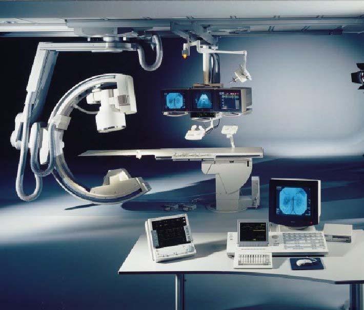
9 Fluoroscopy Fluoroscope, originally zinkcadmiumsulphide screen, 7% efficiency Electro-optical image amplifiers with fluorescent screen (> x amplification) Image amplifier with TV-camera (tube or CCD) Digitally registering the image from the TVcamera Digital fluoroscopy Digital subtraction angiography

10 Blood vessels - Angiography

11 Digital fluoroscopy
12 Radiography Original direct film exposure, gives the sharpest images but low efficiency, only used in special cases such as dental imaging Amplification screens converts X-rays to light, gain x Can use secondary aperture, a grid to decrease scattered light and increase contrast The film can be replaced by image plates, gives a greater dynamic range and possibilities of directly digitizing and improving the image through image processing

13 Muscles and bones

14 Conventional vs digital, high-frequency amplified X-ray image
15 Digital radiography, advantages Greater contrast range gives fewer retakes because of poor exposure Digital image handling gives fewer lost films and simplified archiving More enviromentally friendly through less use of film and chemicals Easier to consult other experts over the network

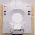
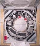
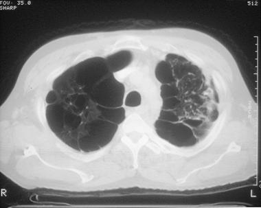
16 Computed Tomography (CT) Creates images of slices through the body

17 How the tomograph functions

18 How the tomograph functions
19 CT-functional principles In a large number of projection rays though the body the X-ray absorption is measured, this yields many density profiles. These can be reprojected into the slice through Radons formula or through filtered back projection CT gives good contrast resolution and very good geometric accuracy

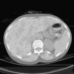
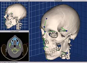
20 Computed tomography CT gives anatomical information
21
22 CT image properties CT measures X-ray density in absolute units according to the Hounsfields scale for air 0 for water for bone Through different contrast windows in the display different tissues can be displayed optimally

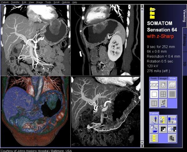
23 A modern CT can have 64 parallel channels Typical specifications; 64 x 0.625mm acquisition 0.34mm x 0.34mm x 0.34mm isotropic resolution 0.4 second rotation time Up to 24 Lp/cm ultra-high spatial resolution High resolution 768 and 1024 reconstruction matrices Reconstruction up to 40 images per second

24 CT examples
 Based")


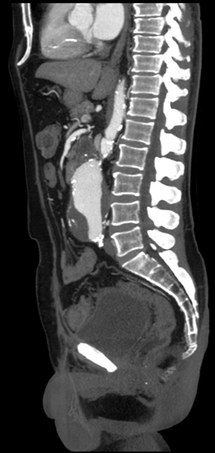
25 Magnetic Resonance Tomography (MRT) Based on magnetic pulse sequences in a strong magnetic field Different pulse sequences gives different contrast The orientation of the slices can be chosen freely through manipulation of the magnetic fields

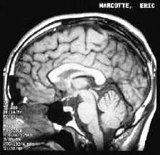
26 Magnetic Resonance Imaging MRI gives anatomical information


27 How MRT works Nuclei with odd number of protons/neutrons has spin The spin vector can be aligned to a (very) strong magnetic field Can be disturbed by a radio signal in resonance with the spin frequency, the so called Larmorfrequency When the atoms returns to rest position they become radio transmitters which can be detected by sensitive receivers Through conrol of field gradients and pulse sequences one can determine which atoms are activated and listened to respectively and thus images can be created in 2D and 3D
28 Some fundamental MR-concepts MR-images can be weighted to show two time constants giving different contrast: T1 is the time constant that determines how fast the spin M Z returns to equilibrium, it is called spin lattice relaxation time M z = M o ( 1 - e -t/t1 ) T2 is the time constant that determines the return to equilibrium for the transversal magnetisation M XY, it is called spin-spin relaxation time M XY =M XYo ( e -t/t2 )
29 MRT image properties Very good contrast resolution for soft tissue Very flexible, different pulse sequences gives different contrast Not possible to determine the signal levels in absolute terms Poor geometric precision No known harmful effects Still under strong development

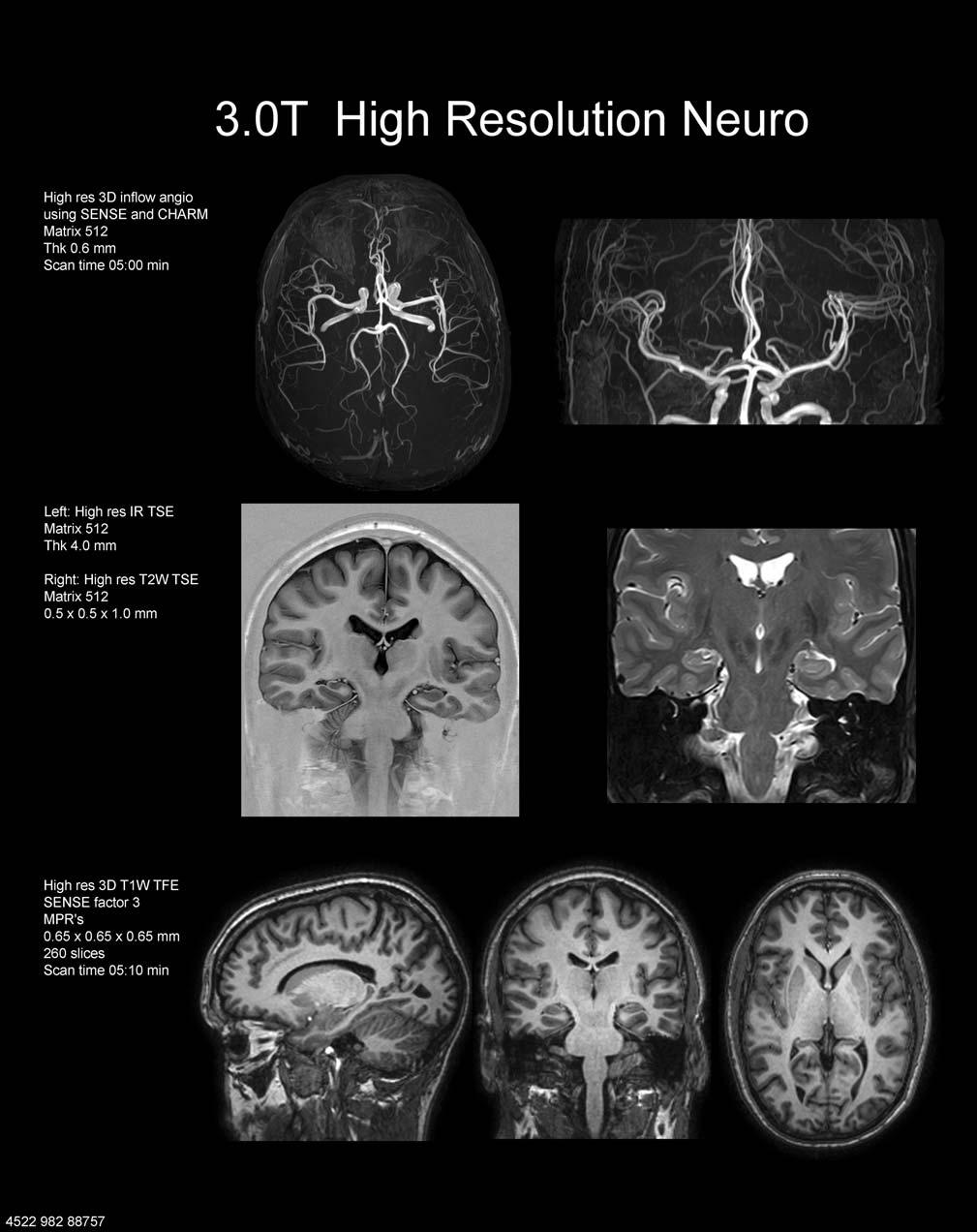
30 MR Neuro
 05-10-10")
31 Muscles and bones (joints)
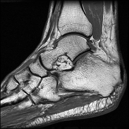
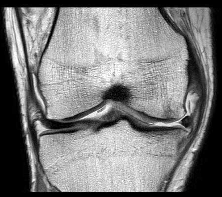
32 Impressive skeletal details

33 Microscopic resolution for orthopedics mm in-plane resolution of wrist Observe clear delineation of fine structures such as the vessel walls Technical details: T1 FLASH TR 591 ms, TE 7.5 ms, TA 6:09 min, SL 3 mm, slices 19, matrix 1024, FoV 80 mm.


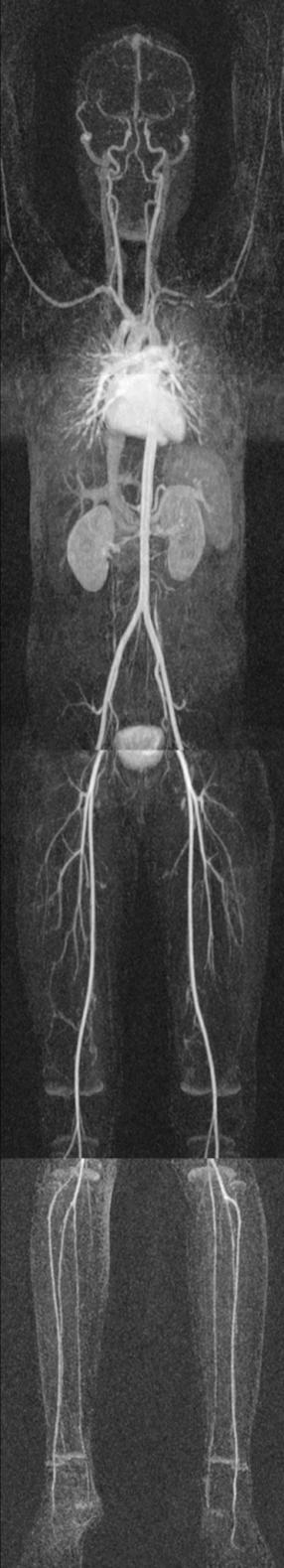
34 Whole body MR imaging
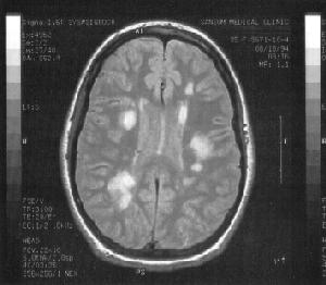
35 Neurological Multiple sclerosis
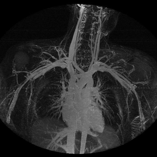
36 Angiography

37 The heart

38 Open MR designs allows intervention while imaging
39 MRT technologies The image properties are influenced by many factors: Radio antenna coils can be adapted to anatomy and pathology Closer coil gives better image Different pulse sequences gives different contrast, resolution, signal noise and registration times Triggering by heart beat, blood motion and breading can increase the resolution Contrast media can enhance certain structures With functional MR, fmri activity in the brain can be registered and imaged

40 Functional imaging


41 MR diffusion tensor imaging Showing the connections of fibers in the brain
42 For further studies about MRT A good description of the MRI technology at: A good popular description at:

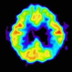
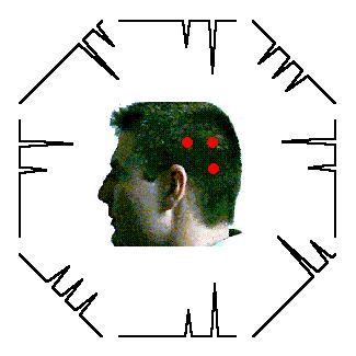
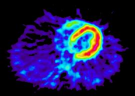
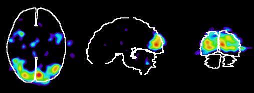
43 Positron Emission Tomograpy (PET) PET shows the concentration and distribution of positron emitting tracer substances in the patient. These images are functional, not anatomical, i.e. they show physiological parameters

44 PET functional principle 44
45 PET functional principles A positron emitting compound is injected into the body (must be produced in an accelerator) The positrons will, within a couple of mm, collide with an electron and create two co-linear 511keV gamma rays These are detected by two detectors located in opposite locations in rings around the person and based on this one can figure out where the event took place Re-projection based on the tomographic principle

46 Positron Emission Tomography PET gives functional information 46

47 Positron Emission Tomografi : accelerator for creating the radioactive tracer substances 47
48 The properties of PET images Gives functional images with rather good resolution at least 1 cm Glucose can be labelled with C11 and this makes it possible to see where in the brain fuel is needed i.e. where the brain is working Very specific substances can be labelled so PET has many applications in pharmaceutical research The need for an accelerator and a chemical lab which can handle high speed synthesis of radioactive compounds makes the technology very expensive
49 PET in Uppsala The PET-research in Uppsala is in the international front-line In 2001 the university PET-centre was sold to Amersham-Biosciences and Imanet AB was created Amersham-Biosciences was bought by GE Medical a few years ago The research co-operation with the university continues

50 Typical result from PCA image enhancement of PET images HV NK1-receptor tracer GLD Pasha Razifar PhD thesis work at IMANET AB and CBA

51 Single Photon Emission Computed Tomography (SPECT) SPECT is similar to PET and shows the concentration and distribution of a radioactive tracer in the patient. The images are functional, not anatomical.
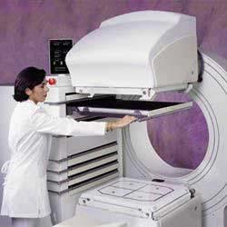
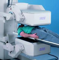
52 Scintigraphy - SPECT camera 52
53 SPECT functional principles A radioactive tracer is injected into the body With a matrix of detectors arranged above the body the location of the radioactive disintegrations is approximately determined The detector can be moved into different positions, which makes tomographic reconstruction possible Alternatively a collimator with slanted holes can be used - ectomography

54 Single Photon Emission Tomography SPECT gives functional information 54
55 The SPECT image properties SPECT gives a functional image with relatively low resolution, some cm The images are intrinsically 3D The radioactive compounds can be obtained from long lived mother isotopes which is much cheaper than accelerators Dynamic processes can be studied through long registrations
56 Ultrasound, US Based on the sonar, acoustic echo principle. Sound with high frequency, typically a few MHz is sent into the body and the echoes are studied. Can with a small, compact equipment give dynamic images in 2D or 3D. The images has problems with coherent noise, specle, and with non-linearities in the sound propagation.
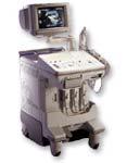
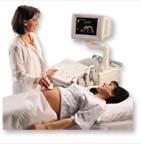
57 Ultrasound equipment 57

58 Ultrasound, best at showing soft tissue

59 Heart 59

60 Ultrasound images of a heart Sharp images of structures in a moving heart
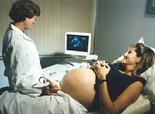
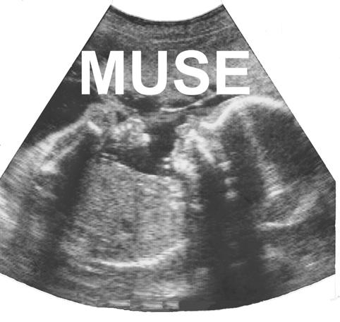
61 Ultrasound for fetal examinations

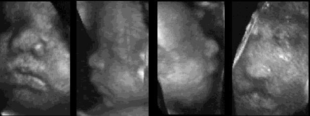
62 3D rendering of dynamic Ultrasound

63 Ultrasound can show flow through Doppler technology
64 Advantages of digital technology Can create images with greater contrast range with less radiation Can handle the images more efficiently through PACS Picture Archiving and Communication Systems Can create completely new types of images Slice images, computer tomography Three dimensional volume images Images of new physiological aspects e.g. oxygen consumption or flow Can visualize the images in new ways, 3D Can extract quantitative information from the images
65 Man vs computer Man is superior when it comes to recognising and interpreting patterns The computer is superior when it comes to Store Transport Present Count and measure The computer can make the images better for human visual analysis
66 PACS the computer as an administrative tool Large amounts of images are registered dayly at a modern hospital. Administration and storage of these requires great resources A Picture Archiving and Communication System, PACS, can make this more rational Requires high capacity storage units and networks. Typically several TB needs to be handled and stored. Sectra-Imtec in Linköping is a leading company in this field
67 Digital image enhancement When the images are available in digital format the computer can be used to help presenting them optimally In order to enhance the images they are filtered point-wise through neighbourhood filters or in the spectral domain
68 Point-wise greyscale transforms

69 Example of simple greyscale transforms: Contrast inverted mammograms

70 Contrastenhancement with nonlinear greyscaletransform

71 Image subtraction image with contrast image without

72 Spatial filtering

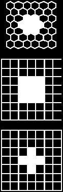
73 Mean filtering Linear quadratic mean filter with increasing size 3,5,9,15,35


74 Noise reducing filtering Original image 3x3 mean filter 3x3 medianfilter
75 Laplace filter 3x3

76 Edge sharpening filter
 Laplace filtered c) Sum a and")
 c*e g) a+f h) Greyscale transf.")
77 Image filtering example a) Whole body image b) Laplace filtered c) Sum a and b d) Sobel filtered a e) 5x5 mean of a f) c*e g) a+f h) Greyscale transf. of g
")
78 Image enhancement with the Context Vision method (adaptive neighboorhood filtering)


79 Context Vision filtering of MR
80 Medical image analysis: CAD - Computer Aided Diagnosis To filter an image so that it becomes significantly better for visual analysis is difficult, the visual system is very adaptive and can handle rather poor images To automatically find abnormalities in images is even harder, requires advanced image analysis The techology is about to mature in this area
81 Typical Mammography image

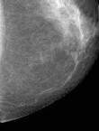
82 Typical Mammography image
83 Typical Mammography image Automated detection of suspicious cancerous lesions


84 3D MRI An MR camera gives a 3D image. Classical X- ray image handling works with 2D film. 3D images gives a whole stack of 2D images to be interpreted jointly

85 Volume rendering An imaginative light ray is sent through each pixel in the image plane. The colour and intensity is determined through the interaction between the ray and the volume elements in the volume in combination with different light sources.
86 Volume rendering methods Single modalities Greylevel gradient shading Maximum intensity projection (MIP) Integrated projection Multiple modalities Combined rendering Implicit segmentation Surface projection of cortical activity 86


87 Greylevel gradient shading A greylevel threshold is set and rays are sent into the volume until a volume element with a value greater than the threshold is encountered The intensity gradients at these positions are combined with the light sources to render the the image Cutting planes can be used to remove parts of the volume to make other parts more visible MRI PET

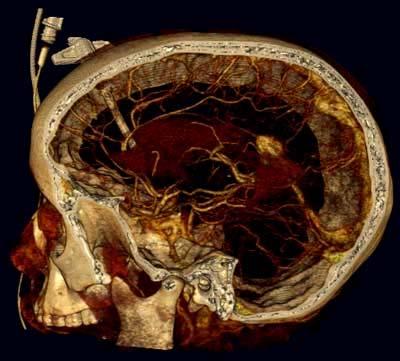
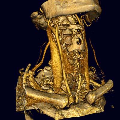
88 3D volume rendering used for CT Much easier than for MR because of fixed Hounsfield units
 the different vessel")

89 With special image analysis (based on greyscale connectivity) the different vessel can be separated MIP projections of a contrast enhanced MRA volume. Original MIP Arteries Veins
90 Maximum intensity projection (MIP) Along each ray the maximal density/intensity value is determined This is particularly useful for small intense structures such as the vessels in angiography Can become complex if several vessels are crossing and overlapping each other
91 Image Fusion Different modalities give complimentary information, anatomy and physiology respectively. There are therefore needs to fuse data from different modalities Image fusion includes spatial registration combined visualisation combined analysis 91
92 Reference Study Choose starting par Transform Study Evaluate similarity (cost function) Choose new set of parameters Yes Converged? No

93 PET-MRI 93

94 SPECT-MRI


95 Multimodal registration can also be combined with 3D visualization - =
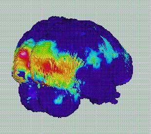
96 Surface projection of cortical activity

97

98

99
100 3D visualisation requires segmentation Small differences in the properties of different tissue types makes advanced segmentation methods necessary High demands of correct reproduction of small details in the anatomy Need for rapid interaction between man and the system Greate needs for research
101 Summary Humans are good at recognising patterns Computers are good at counting and measuring The 3D reality is hard to represent accurately in 2D images Computers can significantly improve and facilitate medical diagnostics So far mainly by producing new types of images In the future 3D visualisation and CAD will probably also have great importance
102 THE END That's all, thanks for your attention!
Medical Images Analysis and Processing
 Medical Images Analysis and Processing - 25642 Emad Course Introduction Course Information: Type: Graduated Credits: 3 Prerequisites: Digital Image Processing Course Introduction Reference(s): Insight
Medical Image Analysis
 Computer assisted Image Analysis VT04 29 april 2004 Medical Image Analysis Lecture 10 (part 1) Xavier Tizon Medical Image Processing Medical imaging modalities XRay,, CT Ultrasound MRI PET, SPECT Generic
BME I5000: Biomedical Imaging
 1 Lucas Parra, CCNY BME I5000: Biomedical Imaging Lecture 4 Computed Tomography Lucas C. Parra, parra@ccny.cuny.edu some slides inspired by lecture notes of Andreas H. Hilscher at Columbia University.
Computational Medical Imaging Analysis
 Computational Medical Imaging Analysis Chapter 1: Introduction to Imaging Science Jun Zhang Laboratory for Computational Medical Imaging & Data Analysis Department of Computer Science University of Kentucky
Lecture 6: Medical imaging and image-guided interventions
 ME 328: Medical Robotics Winter 2019 Lecture 6: Medical imaging and image-guided interventions Allison Okamura Stanford University Updates Assignment 3 Due this Thursday, Jan. 31 Note that this assignment
Image Acquisition Systems
 Image Acquisition Systems Goals and Terminology Conventional Radiography Axial Tomography Computer Axial Tomography (CAT) Magnetic Resonance Imaging (MRI) PET, SPECT Ultrasound Microscopy Imaging ITCS
MEDICAL IMAGE ANALYSIS
 SECOND EDITION MEDICAL IMAGE ANALYSIS ATAM P. DHAWAN g, A B IEEE Engineering in Medicine and Biology Society, Sponsor IEEE Press Series in Biomedical Engineering Metin Akay, Series Editor +IEEE IEEE PRESS
Introduction to Biomedical Imaging
 Alejandro Frangi, PhD Computational Imaging Lab Department of Information & Communication Technology Pompeu Fabra University www.cilab.upf.edu X-ray Projection Imaging Computed Tomography Digital X-ray
Tomographic Reconstruction
 Tomographic Reconstruction 3D Image Processing Torsten Möller Reading Gonzales + Woods, Chapter 5.11 2 Overview Physics History Reconstruction basic idea Radon transform Fourier-Slice theorem (Parallel-beam)
Digital Image Processing
 Digital Image Processing SPECIAL TOPICS CT IMAGES Hamid R. Rabiee Fall 2015 What is an image? 2 Are images only about visual concepts? We ve already seen that there are other kinds of image. In this lecture
Diagnostic imaging techniques. Krasznai Zoltán. University of Debrecen Medical and Health Science Centre Department of Biophysics and Cell Biology
 Diagnostic imaging techniques Krasznai Zoltán University of Debrecen Medical and Health Science Centre Department of Biophysics and Cell Biology 1. Computer tomography (CT) 2. Gamma camera 3. Single Photon
UNIVERSITY OF SOUTHAMPTON
 UNIVERSITY OF SOUTHAMPTON PHYS2007W1 SEMESTER 2 EXAMINATION 2014-2015 MEDICAL PHYSICS Duration: 120 MINS (2 hours) This paper contains 10 questions. Answer all questions in Section A and only two questions
Computational Medical Imaging Analysis
 Computational Medical Imaging Analysis Chapter 2: Image Acquisition Systems Jun Zhang Laboratory for Computational Medical Imaging & Data Analysis Department of Computer Science University of Kentucky
Medical Imaging Introduction
 Medical Imaging Introduction Jan Kybic February 16, 2010 Medical imaging: a collaborative paradigm picture from Atam P. Dhawan: Medical Imaging From physiology to information processing (what we should
Medical Imaging BMEN Spring 2016
 Name Medical Imaging BMEN 420-501 Spring 2016 Homework #4 and Nuclear Medicine Notes All questions are from the introductory Powerpoint (based on Chapter 7) and text Medical Imaging Signals and Systems,
Visualisation : Lecture 1. So what is visualisation? Visualisation
 So what is visualisation? UG4 / M.Sc. Course 2006 toby.breckon@ed.ac.uk Computer Vision Lab. Institute for Perception, Action & Behaviour Introducing 1 Application of interactive 3D computer graphics to
Physical bases of X-ray diagnostics
 Physical bases of X-ray diagnostics Dr. István Voszka Possibilities of X-ray production (X-ray is produced, when charged particles of high velocity are stopped) X-ray tube: Relatively low accelerating
Radiology. Marta Anguiano Millán. Departamento de Física Atómica, Molecular y Nuclear Facultad de Ciencias. Universidad de Granada
 Departamento de Física Atómica, Molecular y Nuclear Facultad de Ciencias. Universidad de Granada Overview Introduction Overview Introduction Tecniques of imaging in Overview Introduction Tecniques of imaging
Advanced Visual Medicine: Techniques for Visual Exploration & Analysis
 Advanced Visual Medicine: Techniques for Visual Exploration & Analysis Interactive Visualization of Multimodal Volume Data for Neurosurgical Planning Felix Ritter, MeVis Research Bremen Multimodal Neurosurgical
The University of Chicago. Center for EPR Imaging in Vivo Physiology. Image Registration. Boris Epel
 The University of Chicago Center for EPR Imaging in Vivo Physiology Image Registration Boris Epel Imaging Methods are Complimentary CT MRI EPRI High resolution anatomic images Quantitative Poor soft tissue
Constructing System Matrices for SPECT Simulations and Reconstructions
 Constructing System Matrices for SPECT Simulations and Reconstructions Nirantha Balagopal April 28th, 2017 M.S. Report The University of Arizona College of Optical Sciences 1 Acknowledgement I would like
Ch. 4 Physical Principles of CT
 Ch. 4 Physical Principles of CT CLRS 408: Intro to CT Department of Radiation Sciences Review: Why CT? Solution for radiography/tomography limitations Superimposition of structures Distinguishing between
Medical Imaging Modalities
 Image Science Introduction Medical Imaging Modalities Ho Kyung Kim hokyung@pusan.ac.kr Pusan National University Projection Radiography Routine diagnostic radiography Chest x rays, fluoroscopy, mammography,
FIELD PARADIGM FOR 3D MEDICAL IMAGING: Safer, More Accurate, and Faster SPECT/PET, MRI, and MEG
 FIELD PARADIGM FOR 3D MEDICAL IMAGING: Safer, More Accurate, and Faster SPECT/PET, MRI, and MEG July 1, 2011 First, do no harm. --Medical Ethics (Hippocrates) Dr. Murali Subbarao, Ph. D. murali@fieldparadigm.com,
Emission Computed Tomography Notes
 Noll (24) ECT Notes: Page 1 Emission Computed Tomography Notes Introduction Emission computed tomography (ECT) is the CT applied to nuclear medicine. There are two varieties of ECT: 1. SPECT single-photon
Reconstruction in CT and relation to other imaging modalities
 Reconstruction in CT and relation to other imaging modalities Jørgen Arendt Jensen November 1, 2017 Center for Fast Ultrasound Imaging, Build 349 Department of Electrical Engineering Center for Fast Ultrasound
Medical Image Processing: Image Reconstruction and 3D Renderings
 Medical Image Processing: Image Reconstruction and 3D Renderings 김보형 서울대학교컴퓨터공학부 Computer Graphics and Image Processing Lab. 2011. 3. 23 1 Computer Graphics & Image Processing Computer Graphics : Create,
Joint CI-JAI advanced accelerator lecture series Imaging and detectors for medical physics Lecture 1: Medical imaging
 Joint CI-JAI advanced accelerator lecture series Imaging and detectors for medical physics Lecture 1: Medical imaging Dr Barbara Camanzi barbara.camanzi@stfc.ac.uk Course layout Day AM 09.30 11.00 PM 15.30
Introduction to Medical Image Processing
 Introduction to Medical Image Processing Δ Essential environments of a medical imaging system Subject Image Analysis Energy Imaging System Images Image Processing Feature Images Image processing may be
CP467 Image Processing and Pattern Recognition
 CP467 Image Processing and Pattern Recognition Instructor: Hongbing Fan Introduction About DIP & PR About this course Lecture 1: an overview of DIP DIP&PR show What is Digital Image? We use digital image
Corso di laurea in Fisica A.A Fisica Medica 5 SPECT, PET
 Corso di laurea in Fisica A.A. 2007-2008 Fisica Medica 5 SPECT, PET Step 1: Inject Patient with Radioactive Drug Drug is labeled with positron (β + ) emitting radionuclide. Drug localizes
Index. aliasing artifacts and noise in CT images, 200 measurement of projection data, nondiffracting
 Index Algebraic equations solution by Kaczmarz method, 278 Algebraic reconstruction techniques, 283-84 sequential, 289, 293 simultaneous, 285-92 Algebraic techniques reconstruction algorithms, 275-96 Algorithms
ThE ultimate, INTuITIVE Mr INTErFAcE
 ThE ultimate, INTuITIVE Mr INTErFAcE Empowering you to do more The revolutionary Toshiba M-power user interface takes Mr performance and flexibility to levels higher than ever before. M-power is able to
MRI Physics II: Gradients, Imaging
 MRI Physics II: Gradients, Imaging Douglas C., Ph.D. Dept. of Biomedical Engineering University of Michigan, Ann Arbor Magnetic Fields in MRI B 0 The main magnetic field. Always on (0.5-7 T) Magnetizes
Introduction to Positron Emission Tomography
 Planar and SPECT Cameras Summary Introduction to Positron Emission Tomography, Ph.D. Nuclear Medicine Basic Science Lectures srbowen@uw.edu System components: Collimator Detector Electronics Collimator
Automated Image Analysis Software for Quality Assurance of a Radiotherapy CT Simulator
 Automated Image Analysis Software for Quality Assurance of a Radiotherapy CT Simulator Andrew J Reilly Imaging Physicist Oncology Physics Edinburgh Cancer Centre Western General Hospital EDINBURGH EH4
New Technology Allows Multiple Image Contrasts in a Single Scan
 These images were acquired with an investigational device. PD T2 T2 FLAIR T1 MAP T1 FLAIR PSIR T1 New Technology Allows Multiple Image Contrasts in a Single Scan MR exams can be time consuming. A typical
Computer Graphics. - Volume Rendering - Philipp Slusallek
 Computer Graphics - Volume Rendering - Philipp Slusallek Overview Motivation Volume Representation Indirect Volume Rendering Volume Classification Direct Volume Rendering Applications: Bioinformatics Image
Technical aspects of SPECT and SPECT-CT. John Buscombe
 Technical aspects of SPECT and SPECT-CT John Buscombe What does the clinician need to know? For SPECT What factors affect SPECT How those factors should be sought Looking for artefacts For SPECT-CT Issues
Computed tomography (Item No.: P )
") Computed tomography (Item No.: P2550100) Curricular Relevance Area of Expertise: Biology Education Level: University Topic: Modern Imaging Methods Subtopic: X-ray Imaging Experiment: Computed tomography
Computed tomography (Item No.: P2550100) Curricular Relevance Area of Expertise: Biology Education Level: University Topic: Modern Imaging Methods Subtopic: X-ray Imaging Experiment: Computed tomography
Spiral CT. Protocol Optimization & Quality Assurance. Ge Wang, Ph.D. Department of Radiology University of Iowa Iowa City, Iowa 52242, USA
 Spiral CT Protocol Optimization & Quality Assurance Ge Wang, Ph.D. Department of Radiology University of Iowa Iowa City, Iowa 52242, USA Spiral CT Protocol Optimization & Quality Assurance Protocol optimization
Introduction to Medical Image Analysis
 Introduction to Medical Image Analysis Rasmus R. Paulsen DTU Compute rapa@dtu.dk http://courses.compute.dtu.dk/02511 http://courses.compute.dtu.dk/02511 Plenty of slides adapted from Thomas Moeslunds lectures
COMPREHENSIVE QUALITY CONTROL OF NMR TOMOGRAPHY USING 3D PRINTED PHANTOM
 COMPREHENSIVE QUALITY CONTROL OF NMR TOMOGRAPHY USING 3D PRINTED PHANTOM Mažena MACIUSOVIČ *, Marius BURKANAS *, Jonas VENIUS *, ** * Medical Physics Department, National Cancer Institute, Vilnius, Lithuania
A Generic Lie Group Model for Computer Vision
 A Generic Lie Group Model for Computer Vision Within this research track we follow a generic Lie group approach to computer vision based on recent physiological research on how the primary visual cortex
Medical Imaging and Beyond
 Medical Imaging and Beyond Jesus J. Caban Schedule! Today:! Lecture: Medical Imaging and Beyond! Wednesday:! No Class (Thanksgiving Eve)! Final presentations:! Nov 29 th : W. Griffin, F. Zafar! Dec 1 st
Slide 1. Technical Aspects of Quality Control in Magnetic Resonance Imaging. Slide 2. Annual Compliance Testing. of MRI Systems.
 Slide 1 Technical Aspects of Quality Control in Magnetic Resonance Imaging Slide 2 Compliance Testing of MRI Systems, Ph.D. Department of Radiology Henry Ford Hospital, Detroit, MI Slide 3 Compliance Testing
CS 5630/6630 Scientific Visualization. Volume Rendering I: Overview
 CS 5630/6630 Scientific Visualization Volume Rendering I: Overview Motivation Isosurfacing is limited It is binary A hard, distinct boundary is not always appropriate Slice Isosurface Volume Rendering
Positron Emission Tomography
 Physics 656 Seminar on Physical Fundamentals of Medical Imaging Positron Emission Tomography Ahmed Qamesh Outline What is PET? PET mechanism Radionuclide and its synthesis Detection concept and Development
An Introduction to Images
 An Introduction to Images CS6640/BIOENG6640/ECE6532 Ross Whitaker, Tolga Tasdizen SCI Institute, School of Computing, Electrical and Computer Engineering University of Utah 1 What Is An Digital Image?
Computed tomography of simple objects. Related topics. Principle. Equipment TEP Beam hardening, artefacts, and algorithms
 Related topics Beam hardening, artefacts, and algorithms Principle The CT principle is demonstrated with the aid of simple objects. In the case of very simple targets, only a few images need to be taken
Medical Image Registration
 Medical Image Registration Submitted by NAREN BALRAJ SINGH SB ID# 105299299 Introduction Medical images are increasingly being used within healthcare for diagnosis, planning treatment, guiding treatment
FOREWORD TO THE SPECIAL ISSUE ON MOTION DETECTION AND COMPENSATION
 Philips J. Res. 51 (1998) 197-201 FOREWORD TO THE SPECIAL ISSUE ON MOTION DETECTION AND COMPENSATION This special issue of Philips Journalof Research includes a number of papers presented at a Philips
LOGIQ. V2 Ultrasound. Part of LOGIQ Vision Series. Imagination at work LOGIQ is a trademark of General Electric Company.
 TM LOGIQ V2 Ultrasound Part of LOGIQ Vision Series Imagination at work The brilliance of color. The simplicity of GE. Now you can add the advanced capabilities of color Doppler to patient care with the
Computer-Tomography I: Principles, History, Technology
 Computer-Tomography I: Principles, History, Technology Prof. Dr. U. Oelfke DKFZ Heidelberg Department of Medical Physics (E040) Im Neuenheimer Feld 280 69120 Heidelberg, Germany u.oelfke@dkfz.de History
Methodological progress in image registration for ventilation estimation, segmentation propagation and multi-modal fusion
 Methodological progress in image registration for ventilation estimation, segmentation propagation and multi-modal fusion Mattias P. Heinrich Julia A. Schnabel, Mark Jenkinson, Sir Michael Brady 2 Clinical
Limitations of Projection Radiography. Stereoscopic Breast Imaging. Limitations of Projection Radiography. 3-D Breast Imaging Methods
 Stereoscopic Breast Imaging Andrew D. A. Maidment, Ph.D. Chief, Physics Section Department of Radiology University of Pennsylvania Limitations of Projection Radiography Mammography is a projection imaging
Clinical Importance. Aortic Stenosis. Aortic Regurgitation. Ultrasound vs. MRI. Carotid Artery Stenosis
 Clinical Importance Rapid cardiovascular flow quantitation using sliceselective Fourier velocity encoding with spiral readouts Valve disease affects 10% of patients with heart disease in the U.S. Most
MR Advance Techniques. Vascular Imaging. Class III
 MR Advance Techniques Vascular Imaging Class III 1 Vascular Imaging There are several methods that can be used to evaluate the cardiovascular systems with the use of MRI. MRI will aloud to evaluate morphology
SPECT QA and QC. Bruce McBride St. Vincent s Hospital Sydney.
 SPECT QA and QC Bruce McBride St. Vincent s Hospital Sydney. SPECT QA and QC What is needed? Why? How often? Who says? QA and QC in Nuclear Medicine QA - collective term for all the efforts made to produce
CP Generalize Concepts in Abstract Multi-dimensional Image Model Component Semantics. David Clunie.
 CP-1390 - Generalize Concepts in Abstract Multi-dimensional Image Model Semantics Page 1 STATUS Date of Last Update Person Assigned Submitter Name Submission Date Assigned 2014/06/09 David Clunie mailto:dclunie@dclunie.com
Advanced MRI Techniques (and Applications)
") Advanced MRI Techniques (and Applications) Jeffry R. Alger, PhD Department of Neurology Ahmanson-Lovelace Brain Mapping Center Brain Research Institute Jonsson Comprehensive Cancer Center University of
Advanced MRI Techniques (and Applications) Jeffry R. Alger, PhD Department of Neurology Ahmanson-Lovelace Brain Mapping Center Brain Research Institute Jonsson Comprehensive Cancer Center University of
SIGMI Meeting ~Image Fusion~ Computer Graphics and Visualization Lab Image System Lab
 SIGMI Meeting ~Image Fusion~ Computer Graphics and Visualization Lab Image System Lab Introduction Medical Imaging and Application CGV 3D Organ Modeling Model-based Simulation Model-based Quantification
VISUALIZATION AND EXPLORATION OF TIME-VARYING AND DIFFUSION TENSOR MEDICAL IMAGE DATA SETS
 VISUALIZATION AND EXPLORATION OF TIME-VARYING AND DIFFUSION TENSOR MEDICAL IMAGE DATA SETS by Zhe Fang B.Sc. with Honours, Acadia University, 2003 a thesis submitted in partial fulfillment of the requirements
Corso di laurea in Fisica A.A Fisica Medica 4 TC
 Corso di laurea in Fisica A.A. 2007-2008 Fisica Medica 4 TC Computed Tomography Principles 1. Projection measurement 2. Scanner systems 3. Scanning modes Basic Tomographic Principle The internal structure
THE DICOM 2013 INTERNATIONAL CONFERENCE & SEMINAR. DICOM Fields of Use. Klaus Neuner. Brainlab AG. Software Project Manager Feldkirchen, Germany
 THE DICOM 2013 INTERNATIONAL CONFERENCE & SEMINAR March 14-16 Bangalore, India DICOM Fields of Use Klaus Neuner Brainlab AG Software Project Manager Feldkirchen, Germany Introduction This presentation
3/27/2012 WHY SPECT / CT? SPECT / CT Basic Principles. Advantages of SPECT. Advantages of CT. Dr John C. Dickson, Principal Physicist UCLH
 3/27/212 Advantages of SPECT SPECT / CT Basic Principles Dr John C. Dickson, Principal Physicist UCLH Institute of Nuclear Medicine, University College London Hospitals and University College London john.dickson@uclh.nhs.uk
Introduction to Emission Tomography
 Introduction to Emission Tomography Gamma Camera Planar Imaging Robert Miyaoka, PhD University of Washington Department of Radiology rmiyaoka@u.washington.edu Gamma Camera: - collimator - detector (crystal
Data Fusion Virtual Surgery Medical Virtual Reality Team. Endo-Robot. Database Functional. Database
 2017 29 6 16 GITI 3D From 3D to 4D imaging Data Fusion Virtual Surgery Medical Virtual Reality Team Morphological Database Functional Database Endo-Robot High Dimensional Database Team Tele-surgery Robotic
Shadow casting. What is the problem? Cone Beam Computed Tomography THE OBJECTIVES OF DIAGNOSTIC IMAGING IDEAL DIAGNOSTIC IMAGING STUDY LIMITATIONS
 Cone Beam Computed Tomography THE OBJECTIVES OF DIAGNOSTIC IMAGING Reveal pathology Reveal the anatomic truth Steven R. Singer, DDS srs2@columbia.edu IDEAL DIAGNOSTIC IMAGING STUDY Provides desired diagnostic
Continuation Format Page
 C.1 PET with submillimeter spatial resolution Figure 2 shows two views of the high resolution PET experimental setup used to acquire preliminary data [92]. The mechanics of the proposed system are similar
Introduction to Neuroimaging Janaina Mourao-Miranda
 Introduction to Neuroimaging Janaina Mourao-Miranda Neuroimaging techniques have changed the way neuroscientists address questions about functional anatomy, especially in relation to behavior and clinical
A NEURAL NETWORK BASED IMAGING SYSTEM FOR fmri ANALYSIS IMPLEMENTING WAVELET METHOD
 6th WSEAS International Conference on CIRCUITS, SYSTEMS, ELECTRONICS,CONTROL & SIGNAL PROCESSING, Cairo, Egypt, Dec 29-31, 2007 454 A NEURAL NETWORK BASED IMAGING SYSTEM FOR fmri ANALYSIS IMPLEMENTING
A Study of Medical Image Analysis System
 Indian Journal of Science and Technology, Vol 8(25), DOI: 10.17485/ijst/2015/v8i25/80492, October 2015 ISSN (Print) : 0974-6846 ISSN (Online) : 0974-5645 A Study of Medical Image Analysis System Kim Tae-Eun
MRI. When to use What sequences. Outline 2012/09/19. Sequence: Definition. Basic Principles: Step 2. Basic Principles: Step 1. Govind Chavhan, MD
 MRI When to use What sequences Govind Chavhan, MD Assistant Professor and Staff Radiologist The Hospital For Sick Children, Toronto Planning Acquisition Post processing Interpretation Patient history and
Biomedical Image Processing for Human Elbow
 Biomedical Image Processing for Human Elbow Akshay Vishnoi, Sharad Mehta, Arpan Gupta Department of Mechanical Engineering Graphic Era University Dehradun, India akshaygeu001@gmail.com, sharadm158@gmail.com
Annales UMCS Informatica AI 1 (2003) UMCS. Registration of CT and MRI brain images. Karol Kuczyński, Paweł Mikołajczak
 UMCS. Registration of CT and MRI brain images. Karol Kuczyński, Paweł Mikołajczak") Annales Informatica AI 1 (2003) 149-156 Registration of CT and MRI brain images Karol Kuczyński, Paweł Mikołajczak Annales Informatica Lublin-Polonia Sectio AI http://www.annales.umcs.lublin.pl/ Laboratory
Annales Informatica AI 1 (2003) 149-156 Registration of CT and MRI brain images Karol Kuczyński, Paweł Mikołajczak Annales Informatica Lublin-Polonia Sectio AI http://www.annales.umcs.lublin.pl/ Laboratory
4DM Packages. 4DM Packages & License Types. Information to help you order the appropriate licenses for your site.
 4DM Packages 4DM Packages & License Types. Information to help you order the appropriate licenses for your site. Nuclear Cardiac Quantification, Review, and Reporting Select Your 4DM Package and corresponding
Volume Illumination and Segmentation
 Volume Illumination and Segmentation Computer Animation and Visualisation Lecture 13 Institute for Perception, Action & Behaviour School of Informatics Overview Volume illumination Segmentation Volume
Medical Imaging Projects
 NSF REU MedIX Summer 2006 Medical Imaging Projects Daniela Stan Raicu, PhD http://facweb.cs.depaul.edu/research draicu@cs.depaul.edu Outline Medical Informatics Imaging Modalities Computed Tomography Medical
CT Basics Principles of Spiral CT Dose. Always Thinking Ahead.
 1 CT Basics Principles of Spiral CT Dose 2 Who invented CT? 1963 - Alan Cormack developed a mathematical method of reconstructing images from x-ray projections Sir Godfrey Hounsfield worked for the Central
Cardiac Dual Energy CT: Technique
 RSNA 2013, VSCA51-01, Chicago, Dec. 5, 2013 Cardiac Radiology Series Cardiac Dual Energy CT: Technique Willi A. Kalender, Ph.D. Institute of Medical Physics University of Erlangen www.imp.uni-erlangen.de
Chapter 3. Image Processing Methods. (c) 2008 Prof. Dr. Michael M. Richter, Universität Kaiserslautern
 2008 Prof. Dr. Michael M. Richter, Universität Kaiserslautern") Chapter 3 Image Processing Methods The Role of Image Processing Methods (1) An image is an nxn matrix of gray or color values An image processing method is algorithm transforming such matrices or assigning
Chapter 3 Image Processing Methods The Role of Image Processing Methods (1) An image is an nxn matrix of gray or color values An image processing method is algorithm transforming such matrices or assigning
Whole Body MRI Intensity Standardization
 Whole Body MRI Intensity Standardization Florian Jäger 1, László Nyúl 1, Bernd Frericks 2, Frank Wacker 2 and Joachim Hornegger 1 1 Institute of Pattern Recognition, University of Erlangen, {jaeger,nyul,hornegger}@informatik.uni-erlangen.de
Computed tomography - outline
 Computed tomography - outline Computed Tomography Systems Jørgen Arendt Jensen and Mikael Jensen (DTU Nutech) October 6, 216 Center for Fast Ultrasound Imaging, Build 349 Department of Electrical Engineering
Tomography. Forward projectionsp θ (r) are known as a Radon transform. Objective: reverse this process to form the original image
 are known as a Radon transform. Objective: reverse this process to form the original image") C. A. Bouman: Digital Image Processing - January 9, 217 1 Tomography Many medical imaging systems can only measure projections through an object with density f(x,y). Projections must be collected at every
C. A. Bouman: Digital Image Processing - January 9, 217 1 Tomography Many medical imaging systems can only measure projections through an object with density f(x,y). Projections must be collected at every
Image Restoration. Diffusion Denoising Deconvolution Super-resolution Tomographic Reconstruction
 Image Restoration Image Restoration Diffusion Denoising Deconvolution Super-resolution Tomographic Reconstruction Diffusion Term Consider only the regularization term E-L equation: (Laplace equation) Steepest
Mathematical methods and simulations tools useful in medical radiation physics
 Mathematical methods and simulations tools useful in medical radiation physics Michael Ljungberg, professor Department of Medical Radiation Physics Lund University SE-221 85 Lund, Sweden Major topic 1:
DUAL energy X-ray radiography [1] can be used to separate
![DUAL energy X-ray radiography [1] can be used to separate](/thumbs/91/106229592.jpg "DUAL energy X-ray radiography [1] can be used to separate") IEEE TRANSACTIONS ON NUCLEAR SCIENCE, VOL. 53, NO. 1, FEBRUARY 2006 133 A Scatter Correction Using Thickness Iteration in Dual-Energy Radiography S. K. Ahn, G. Cho, and H. Jeon Abstract In dual-energy
IEEE TRANSACTIONS ON NUCLEAR SCIENCE, VOL. 53, NO. 1, FEBRUARY 2006 133 A Scatter Correction Using Thickness Iteration in Dual-Energy Radiography S. K. Ahn, G. Cho, and H. Jeon Abstract In dual-energy
Chapter 3 Set Redundancy in Magnetic Resonance Brain Images
 16 Chapter 3 Set Redundancy in Magnetic Resonance Brain Images 3.1 MRI (magnetic resonance imaging) MRI is a technique of measuring physical structure within the human anatomy. Our proposed research focuses
Enhanced material contrast by dual-energy microct imaging
 Enhanced material contrast by dual-energy microct imaging Method note Page 1 of 12 2 Method note: Dual-energy microct analysis 1. Introduction 1.1. The basis for dual energy imaging Micro-computed tomography
Computer-Aided Diagnosis in Abdominal and Cardiac Radiology Using Neural Networks
 Computer-Aided Diagnosis in Abdominal and Cardiac Radiology Using Neural Networks Du-Yih Tsai, Masaru Sekiya and Yongbum Lee Department of Radiological Technology, School of Health Sciences, Faculty of
MEDICAL EQUIPMENT: COMPUTED TOMOGRAPHY. Prof. Yasser Mostafa Kadah
 MEDICAL EQUIPMENT: COMPUTED TOMOGRAPHY Prof. Yasser Mostafa Kadah www.k-space.org Recommended Textbook X-Ray Computed Tomography in Biomedical Engineering, by Robert Cierniak, Springer, 211 Computed Tomography
Volume rendering for interactive 3-d segmentation
 Volume rendering for interactive 3-d segmentation Klaus D. Toennies a, Claus Derz b a Dept. Neuroradiology, Inst. Diagn. Radiology, Inselspital Bern, CH-3010 Berne, Switzerland b FG Computer Graphics,
Certificate in Clinician Performed Ultrasound (CCPU)
") Certificate in Clinician Performed Ultrasound (CCPU) Syllabus Physics Tutorial Physics Tutorial Purpose: Training: Assessments: This unit is designed to cover the theoretical and practical curriculum for
Certificate in Clinician Performed Ultrasound (CCPU) Syllabus Physics Tutorial Physics Tutorial Purpose: Training: Assessments: This unit is designed to cover the theoretical and practical curriculum for
Extraction and recognition of the thoracic organs based on 3D CT images and its application
 1 Extraction and recognition of the thoracic organs based on 3D CT images and its application Xiangrong Zhou, PhD a, Takeshi Hara, PhD b, Hiroshi Fujita, PhD b, Yoshihiro Ida, RT c, Kazuhiro Katada, MD
Radiographic Simulator artist: Version 2
 18th World Conference on Nondestructive Testing, 16-20 April 2012, Durban, South Africa Radiographic Simulator artist: Version 2 Carsten Bellon 1, Andreas Deresch 1, Christian Gollwitzer 1, Gerd-Rüdiger
Micro-CT Methodology Hasan Alsaid, PhD
 Micro-CT Methodology Hasan Alsaid, PhD Preclinical & Translational Imaging LAS, PTS, GlaxoSmithKline 20 April 2015 Provide basic understanding of technical aspects of the micro-ct Statement: All procedures
Range Sensors (time of flight) (1)
 (1)") Range Sensors (time of flight) (1) Large range distance measurement -> called range sensors Range information: key element for localization and environment modeling Ultrasonic sensors, infra-red sensors
Range Sensors (time of flight) (1) Large range distance measurement -> called range sensors Range information: key element for localization and environment modeling Ultrasonic sensors, infra-red sensors
Multimodal Image Fusion Of The Human Brain
 Multimodal Image Fusion Of The Human Brain Isis Lázaro(1), Jorge Marquez(1), Juan Ortiz(2), Fernando Barrios(2) isislazaro@gmail.com Centro de Ciencias Aplicadas y Desarrollo Tecnológico, UNAM Circuito
Fundamentals of CT imaging
 SECTION 1 Fundamentals of CT imaging I History In the early 1970s Sir Godfrey Hounsfield s research produced the first clinically useful CT scans. Original scanners took approximately 6 minutes to perform